Gesetz gegen den unlauteren Wettbewerb: § 5 Irreführende geschäftliche Handlungen
Gesetz gegen den unlauteren Wettbewerb: § 5 Irreführende geschäftliche Handlungen
(1) Unlauter handelt, wer eine irreführende geschäftliche Handlung vornimmt, die geeignet ist, den Verbraucher oder sonstigen Marktteilnehmer zu einer geschäftlichen Entscheidung zu veranlassen, die er andernfalls nicht getroffen hätte.
(2) Eine geschäftliche Handlung ist irreführend, wenn sie unwahre Angaben enthält oder sonstige zur Täuschung geeignete Angaben über folgende Umstände enthält:
1.
die wesentlichen Merkmale der Ware oder Dienstleistung wie Verfügbarkeit, Art, Ausführung, Vorteile, Risiken, Zusammensetzung, Zubehör, Verfahren oder Zeitpunkt der Herstellung, Lieferung oder Erbringung, Zwecktauglichkeit, Verwendungsmöglichkeit, Menge, Beschaffenheit, Kundendienst und Beschwerdeverfahren, geographische oder betriebliche Herkunft, von der Verwendung zu erwartende Ergebnisse oder die Ergebnisse oder wesentlichen Bestandteile von Tests der Waren oder Dienstleistungen;
2.
den Anlass des Verkaufs wie das Vorhandensein eines besonderen Preisvorteils, den Preis oder die Art und Weise, in der er berechnet wird, oder die Bedingungen, unter denen die Ware geliefert oder die Dienstleistung erbracht wird;
3.
die Person, Eigenschaften oder Rechte des Unternehmers wie Identität, Vermögen einschließlich der Rechte des geistigen Eigentums, den Umfang von Verpflichtungen, Befähigung, Status, Zulassung, Mitgliedschaften oder Beziehungen, Auszeichnungen oder Ehrungen, Beweggründe für die geschäftliche Handlung oder die Art des Vertriebs;
4.
Aussagen oder Symbole, die im Zusammenhang mit direktem oder indirektem Sponsoring stehen oder sich auf eine Zulassung des Unternehmers oder der Waren oder Dienstleistungen beziehen;
5.
die Notwendigkeit einer Leistung, eines Ersatzteils, eines Austauschs oder einer Reparatur;
6.
die Einhaltung eines Verhaltenskodexes, auf den sich der Unternehmer verbindlich verpflichtet hat, wenn er auf diese Bindung hinweist, oder
7.
Rechte des Verbrauchers, insbesondere solche auf Grund von Garantieversprechen oder Gewährleistungsrechte bei Leistungsstörungen.
(3) Eine geschäftliche Handlung ist auch irreführend, wenn
1.
sie im Zusammenhang mit der Vermarktung von Waren oder Dienstleistungen einschließlich vergleichender Werbung eine Verwechslungsgefahr mit einer anderen Ware oder Dienstleistung oder mit der Marke oder einem anderen Kennzeichen eines Mitbewerbers hervorruft oder
2.
mit ihr eine Ware in einem Mitgliedstaat der Europäischen Union als identisch mit einer in anderen Mitgliedstaaten der Europäischen Union auf dem Markt bereitgestellten Ware vermarktet wird, obwohl sich diese Waren in ihrer Zusammensetzung oder in ihren Merkmalen wesentlich voneinander unterscheiden, sofern dies nicht durch legitime und objektive Faktoren gerechtfertigt ist.
(4) Angaben im Sinne von Absatz 2 sind auch Angaben im Rahmen vergleichender Werbung sowie bildliche Darstellungen und sonstige Veranstaltungen, die darauf zielen und geeignet sind, solche Angaben zu ersetzen.
(5)1Es wird vermutet, dass es irreführend ist, mit der Herabsetzung eines Preises zu werben, sofern der Preis nur für eine unangemessen kurze Zeit gefordert worden ist. 2Ist streitig, ob und in welchem Zeitraum der Preis gefordert worden ist, so trifft die Beweislast denjenigen, der mit der Preisherabsetzung geworben hat.
Fassung aufgrund des Gesetzes zur Umsetzung der Richtlinie (EU) 2020/1828 über Verbandsklagen zum Schutz der Kollektivinteressen der Verbraucher und zur Aufhebung der Richtlinie 2009/22/EG sowie zur Änderung des Kapitalanleger-Musterverfahrensgesetzes (Verbandsklagenrichtlinienumsetzungsgesetz) vom 08.10.2023 (BGBl. I Nr. 272), in Kraft getreten am 13.10.2023 Gesetzesbegründung verfügbar
Nachweis von DNA-Fragmenten in COVID-19-Impfstoffen in Kanada: Eine Analyse der Residual-DNA und deren Auswirkungen auf die Impfstoffsicherheit
Nachweis von DNA-Fragmenten in COVID-19-Impfstoffen in Kanada: Eine Analyse der Residual-DNA und deren Auswirkungen auf die Impfstoffsicherheit
Speicher, D. J. (2023, October 19). DNA fragments detected in monovalent and bivalent Pfizer/BioNTech and Moderna modRNA COVID-19 vaccines from Ontario, Canada: Exploratory dose response relationship with serious adverse events. Retrieved from osf.io/xv3nz
Die vorliegende Studie untersucht das Vorhandensein von DNA-Fragmenten in COVID-19-Impfstoffen, die in Kanada verteilt wurden. Durch den Einsatz von quantitativer PCR (qPCR) und Qubit®-Fluorometrie analysierten die Autoren 27 mRNA-Impfstoffampullen aus 12 einzigartigen Chargen, die verschiedene Pfizer- und Moderna-Produkte umfassen.
Die qPCR-Analyse ergab Rest-DNA-Werte von 0,22-4,27 ng/Dosis für die Pfizer-Spike- und Ori-Sequenzen sowie 0,01-0,78 ng/Dosis für die Moderna-Spike- und Ori-Sequenzen. Die Qubit®-Fluorometrie zeigte jedoch wesentlich höhere gesamte Rest-DNA-Werte, die die behördlichen Richtlinien um das 188- bis 509-fache überschritten. Dieser Unterschied wird auf die Unfähigkeit von qPCR zurückgeführt, kleinere DNA-Fragmente zu erkennen.
Interessanterweise wurde die SV40-Promotor-Enhancer-Ori-Sequenz nur in den Pfizer-Ampullen nachgewiesen. Eine explorative Analyse deutet auf eine potenzielle Dosis-Wirkungs-Beziehung zwischen Rest-DNA-Werten und der Häufigkeit schwerwiegender unerwünschter Ereignisse (SAEs) hin, die dem Vaccine Adverse Event Reporting System (VAERS) gemeldet wurden, wobei Unterschiede zwischen den Pfizer- und Moderna-Produkten beobachtet wurden.
Die Oxford-Nanopore-Sequenzierung einer Pfizer-Impfstoffprobe zeigte DNA-Fragmentgrößen von bis zu 3,5 kb mit einem Mittelwert von 214 Basenpaaren. Weitere Experimente zeigten, dass die Rest-DNA durch die Lipidnanopartikel geschützt ist und gegenüber der DNase-I-Verdauung resistent ist.
Die Autoren argumentieren, dass die aktuellen behördlichen Richtlinien zur Rest-DNA in Impfstoffen, die vor der Einführung einer effizienten DNA-Transfektion mittels Lipidnanopartikeln entwickelt wurden, möglicherweise nicht ausreichen. Sie fordern weitere Untersuchungen und eine Überarbeitung dieser Richtlinien, um die potenziellen Risiken durch hohe Rest-DNA-Werte, ihre effiziente Lieferung und die kumulative Dosierung dieser Impfstoffe angemessen zu berücksichtigen
Berufsordnung für die in Deutschland tätigen Ärzte
§ 1 Aufgaben der Ärztinnen und Ärzte
(1) Ärztinnen und Ärzte dienen der Gesundheit des einzelnen Menschen und der Bevölkerung. Der ärztliche Beruf ist kein Gewerbe. Er ist seiner Natur nach ein freier Beruf.
(2) Aufgabe der Ärztinnen und Ärzte ist es, das Leben zu erhalten, die Gesundheit zu schützen und wiederherzustellen, Leiden zu lindern, Sterbenden Beistand zu leisten und an der Erhaltung der natürlichen Lebensgrundlagen im Hinblick auf ihre Bedeutung für die Gesundheit der Menschen mitzuwirken.
***
(1) Ärztinnen und Ärzte üben ihren Beruf nach ihrem Gewissen, den Geboten der ärztlichen Ethik und der Menschlichkeit aus. Sie dürfen keine Grundsätze anerkennen und keine Vorschriften oder Anweisungen beachten, die mit ihren Aufgaben nicht vereinbar sind oder deren Befolgung sie nicht verantworten können.
(2) Ärztinnen und Ärzte haben ihren Beruf gewissenhaft auszuüben und dem ihnen bei ihrer Berufsausübung entgegengebrachten Vertrauen zu entsprechen. Sie haben dabei ihr ärztliches Handeln am Wohl der Patientinnen und Patienten auszurichten. Insbesondere dürfen sie nicht das Interesse Dritter über das Wohl der Patientinnen und Patienten stellen.
(3) Eine gewissenhafte Ausübung des Berufs erfordert insbesondere die notwendige fachliche Qualifikation und die Beachtung des anerkannten Standes der medizinischen Erkenntnisse.
(4) Ärztinnen und Ärzte dürfen hinsichtlich ihrer ärztlichen Entscheidungen keine Weisungen von Nichtärzten entgegennehmen.
(5) Ärztinnen und Ärzte sind verpflichtet, die für die Berufsausübung geltenden Vorschriften zu beachten.
***
§ 4 Fortbildung
(1) Ärztinnen und Ärzte, die ihren Beruf ausüben, sind verpflichtet, sich in dem Umfange beruflich fortzubilden, wie es zur Erhaltung und Entwicklung der zu ihrer Berufsausübung erforderlichen Fachkenntnisse notwendig ist.
(2) Auf Verlangen müssen Ärztinnen und Ärzte ihre Fortbildung nach Absatz 1 gegenüber der Ärztekammer durch ein Fortbildungszertifikat einer Ärztekammer nachweisen.
Für jede Ärztin und jeden Arzt gilt folgendes Gelöbnis:
Bei meiner Aufnahme in den ärztlichen Berufsstand gelobe ich, mein Leben in den Dienst der Menschlichkeit zu stellen.
Ich werde meinen Beruf mit Gewissenhaftigkeit und Würde ausüben.
Die Erhaltung und Wiederherstellung der Gesundheit meiner Patientinnen und Patienten soll oberstes Gebot meines Handelns sein.
Ich werde alle mir anvertrauten Geheimnisse auch über den Tod der Patientin oder des Patienten hinaus wahren.
Ich werde mit allen meinen Kräften die Ehre und die edle Überlieferung des ärztlichen Berufes aufrechterhalten und bei der Ausübung meiner ärztlichen Pflichten keinen Unterschied machen weder aufgrund einer etwaigen Behinderung noch nach Religion, Nationalität, Rasse noch nach Parteizugehörigkeit oder sozialer Stellung.
Ich werde jedem Menschenleben von der Empfängnis an Ehrfurcht entgegenbringen und selbst unter Bedrohung meine ärztliche Kunst nicht in Widerspruch zu den Geboten der Menschlichkeit anwenden.
Ich werde meinen Lehrerinnen und Lehrern sowie Kolleginnen und Kollegen die schuldige Achtung erweisen. Dies alles verspreche ich auf meine Ehre.
N.B.: Es ist unvorstellbar, wie ein verantwortungsbewusster Arzt seinen Patienten ungetestete und potenziell gefährliche genetische De-novo-Eingriffe verabreichen könnte. Es ist offensichtlich, dass mehr als 95 % der betroffenen Ärzte dabei eindeutig gegen ihre ärztliche Ethik verstoßen haben. Wie kann dies übersehen werden? Die geltenden Gesetze sind in dieser Hinsicht eindeutig formuliert.
Die bloße Behauptung, "Ich habe nur Anweisungen befolgt", ist kein gültiges rechtliches Argument. Ein Blick in die jüngste Vergangenheit, wie zum Beispiel die Nürnberger Prozesse, verdeutlicht dies. In der DDR gab es die sogenannten "Mauerschützen", Beamte, die angewiesen waren, jeden zu erschießen, der die Grenze überschritt. Obwohl sie nach DDR-Recht legal handelten, wurden sie nach der Auflösung der DDR für ihre Taten zur Rechenschaft gezogen. Ähnlich müssen auch die "Doktoren", die sich an dieser äußerst bedenklichen "Impf"-Kampagne beteiligt haben, für ihre Handlungen verantwortlich gemacht werden. Sie haben ihre Patienten durch die Verabreichung ungetesteter genetischer Interventionen nicht nur einer potenziellen Gefahr ausgesetzt, sondern auch gegen grundlegende ärztliche Ethikgrundsätze verstoßen. Es ist für jeden vernünftigen Menschen offensichtlich, dass solches Handeln nicht nur moralisch verwerflich ist, sondern auch den Berufsvorschriften widerspricht. Ärzte, die an solchen Praktiken beteiligt waren, müssen zur Rechenschaft gezogen werden. Es steht außer Frage, dass solche Personen den falschen Beruf gewählt haben, da sie offensichtlich nicht in der Lage sind, zwischen richtig und falsch zu unterscheiden, weder fachlich noch moralisch.
Quelle: https://youtu.be/wnhL1W9dj1w
Transkript:
„Es war das dunkelste Kapitel der deutschen Geschichte, eine Zeit, in der ein aufgestachelter Mob Steine in Schaufenster unschuldiger Ladenbesitzer warf und Frauen sowie Kinder auf grausame Weise und auf offener Straße gedemütigt wurden. In dieser Zeit begann der junge Pfarrer Dietrich Bonhoeffer, sich öffentlich gegen die Grausamkeiten zu äußern. Nachdem er jahrelang versucht hatte, die Menschen zum Umdenken zu bewegen, kam er an einem Abend nach Hause und sein eigener Vater teilte ihm mit, dass zwei Männer in seinem Zimmer warteten, um ihn festzunehmen. Im Gefängnis begann Bonhoeffer darüber nachzudenken, wie sich sein Land der Dichter und Denker in ein Kollektiv von Feiglingen, Gaunern und Verbrechern verwandelt hatte. Er kam letztendlich zu dem Schluss, dass die Wurzel des Problems nicht Bosheit, sondern Dummheit war. In seinen berühmten Briefen aus dem Gefängnis vertrat Bonhoeffer die Ansicht, dass die Dummheit ein gefährlicherer Feind des Guten sei als die Bosheit. Denn während man gegen das Böse protestieren und es durch den Einsatz von Gewalt aufdecken und verhindern kann, sind wir gegen die Dummheit wehrlos. Weder Proteste noch Gewaltanwendung bewirken hier etwas. Argumente stoßen auf taube Ohren.
Fakten, die dem Vorurteil eines dummen Menschen widersprechen, braucht man einfach nicht zu glauben. Und wenn sie unwiderlegbar sind, werden sie einfach als unwichtig, als nebensächlich beiseite geschoben. Bei all dem ist der dumme Mensch selbst zufrieden. Er ist leicht reizbar und wird gefährlich, wenn er auf Angriff geht. Aus diesem Grund ist im Umgang mit einem dummen Menschen größere Vorsicht geboten als mit einem Bösartigen. Wenn wir wissen wollen, wie wir die Dummheit überwinden können, müssen wir versuchen, ihr Wesen zu verstehen. So viel ist sicher: Dummheit ist im Grunde kein intellektueller, sondern ein moralischer Defekt. Es gibt Menschen, die intellektuell bemerkenswert beweglich sind und dennoch dumm erscheinen, und andere, die intellektuell stumpf, aber alles andere als dumm sind. Man hat weniger den Eindruck, dass Dummheit ein angeborener Defekt ist, sondern dass Menschen unter bestimmten Umständen dumm werden oder besser gesagt, dass sie es zulassen, dass dies mit ihnen geschieht. Menschen, die in Einsamkeit leben, zeigen diesen Defekt seltener als Individuen in Gruppen. Es scheint also, dass Dummheit weniger ein psychologisches als ein soziologisches Problem ist. Es zeigt sich, dass jeder starke Machtzuwachs, sei er nun politischer oder religiöser Natur, einen großen Teil der Menschheit mit Dummheit infiziert. Es ist fast so, als sei dies ein soziologisches, psychologisches Gesetz, wonach die Macht des Einen die Dummheit des Anderen braucht.
Der Prozess, der hier am Werk ist, besteht nicht darin, dass bestimmte menschliche Fähigkeiten wie der Intellekt plötzlich versagen. Vielmehr scheint es so zu sein, dass der Mensch unter dem überwältigenden Einfluss der steigenden Macht seiner inneren Unabhängigkeit beraubt wird und mehr oder weniger bewusst seine autonome Position aufgibt. Die Tatsache, dass der dumme Mensch oft stur ist, darf uns nicht von der Tatsache ablenken, dass er nicht selbstbestimmt ist. Wenn man sich mit ihm unterhält, hat man fast das Gefühl, dass man es gar nicht mit ihm als Person zu tun hat, sondern mit Parolen, Schlagworten und dergleichen, die von ihm Besitz ergriffen haben. Er ist wie verhext, verblendet, wird benutzt und in seinem Wesen missbraucht. Der so zum willenlosen Werkzeug gewordene Dumme ist auch zu allen bösen Taten fähig, und er erkennt nicht, dass es böse ist. Nur ein Akt der Befreiung, nicht der Belehrung, kann die Dummheit überwinden. Hier müssen wir uns mit der Tatsache abfinden, dass eine echte innere Befreiung in den meisten Fällen erst dann möglich wird, wenn ihr eine äußere Befreiung vorausgegangen ist. Bis dahin müssen wir alle Versuche aufgeben, den dummen Menschen zu überzeugen. Bonhoeffer wurde wegen seiner Beteiligung an einem Komplott gegen Adolf Hitler im Morgengrauen des neunten April 1945 im Konzentrationslager Flossenbürg hingerichtet, nur zwei Wochen vor der Befreiung des Lagers durch Soldaten aus den Vereinigten Staaten.“* * *
„Von guten Mächten wunderbar geborgen, erwarten wir getrost, was kommen mag.“ – Dieses Gedicht schrieb Dietrich Bonhoeffer 1945 im KZ Flossenbürg, kurz vor seiner Ermordung durch die Nationalsozialisten.
*1 Original Text aus Bonhoeffers Brief: „Dummheit ist ein gefährlicherer Feind des Guten als Bosheit. Gegen das Böse läßt sich protestieren, es läßt sich bloßstellen, es läßt sich notfalls mit Gewalt verhindern, das Böse trägt immer den Keim der Selbstzersetzung in sich, indem es mindestens ein Unbehagen im Menschen zurückläßt. Gegen die Dummheit sind wir wehrlos. Weder mit Protesten noch durch Gewalt läßt sich hier etwas ausrichten; Gründe verfangen I nicht; Tatsachen, die dem eigenen Vorurteil widersprechen, brauchen einfach nicht geglaubt zu werden - in solchen Fällen wird der Dumme sogar kritisch -, und wenn sie unausweichlich sind, können sie ein- fach als nichtssagende Einzelfälle beiseitegeschoben werden. Dabei ist der Dumme im Unterschied zum Bösen restlos mit sich selbst zufrieden; ja, er wird sogar gefährlich, indem er leicht gereizt zum Angriff übergeht. Daher ist dem Dummen gegen- über mehr Vorsicht geboten als gegenüber dem Bösen. Niemals werden wir mehr versuchen, den Dummen durch Gründe zu überzeugen, es ist sinnlos und gefährlich. [...] Um zu wissen, wie wir der Dummheit beikommen können, müssen wir ihr Wesen zu verstehen suchen. Soviel ist sicher, daß sie nicht [wesentlich] ein intellektueller, sondern ein mensch- licher Defekt ist. Es gibt intellektuell außerordentlich bewegli- che Menschen, die dumm sind, und intellektuell sehr Schwer- fällige, die alles andere als dumm sind. Diese Entdeckung ma- chen wir zu unserer Überraschung anläßlich bestimmter Situa- tionen. Dabei gewinnt man weniger den Eindruck, daß die Dummheit ein angeborener Defekt ist, als daß unter bestimm-ten Umständen die Menschen dumm gemacht werden, bzw. sich dumm machen lassen. Wir beobachten weiterhin, daß abgeschlossen und einsam lebende Menschen diesen Defekt seltener zeigen als zur Gesellung neigende oder verurteilte Menschen undMenschengruppen. So scheint die Dummheit vielleicht weniger ein psychologisches als ein soziologisches Problem zu sein. Sie ist eine besondere Form der Einwirkung geschichtlicher Um- stände auf den Menschen, eine psychologische Begleiterschei- nung bestimmter äußerer Verhältnisse. Bei genauerem Zusehen zeigt sich, daß jede starke äußere Machtentfaltung, sei sie politi- scher oder religiöser Art, einen großen Teil der Menschen mit Dummheit schlägt. Ja, es hat den Anschein, als sei das geradezu ein soziologisch-psychologisches Gesetz. Die Macht der einen braucht die Dummheit der anderen. Der Vorgang ist dabei nicht der, daß bestimmte - also etwa intellektuelle - Anlagen des Menschen plötzlich verkümmern oder ausfallen, sondern daß unter dem überwältigenden Eindruck der Machtentfaltung dem Menschen seine innere Selbständigkeit geraubt wird und daß dieser nun- mehr oder weniger unbewußt - darauf verzichtet, zu den sich ergebenden Lebenslagen ein eigenes Verhalten zu finden. Daß der Dumme oft bockig ist, darf nicht darüber hin- wegtäuschen, daß er nicht selbständig ist. Man spürt es gerade- zu im Gespräch mit ihm, daß man es garnicht mit ihm selbst, mit ihm persönlich, sondern mit über ihn mächtig gewordenen Schlagworten, Parolen etc. zu tun hat. Er ist in einem Banne, er ist verblendet, er ist in seinem eigenen Wesen mißbraucht, mißhandelt. So zum willenlosen Instrument geworden, wird der Dumme auch zu allem Bösen fähig sein und zugleich unfähig, dies als Böses zu erkennen. Hier liegt die Gefahr eines diabolischen Mißbrauches, dadurch werden Menschen für immer zugrunde gerichtet werden können. [...] Übrigens haben diese Gedanken über die Dummheit doch dies Tröstliche für sich, daß sie ganz und garnicht zulassen, die Mehrzahl der Menschen unter allen Umständen für dumm zu halten. Es wird wirklich darauf ankommen, ob Machthaber sich mehr von der Dummheit oder von der inneren Selbständigkeit und Klugheit der Menschen versprechen. “
Weiterführende Literatur:
*Brown, P., & Stokes, P. (2020). Bonhoeffer and Løgstrup: the Ethics of Disclosure in a State of Exception. Sophia, 59(2), 229–246. https://doi.org/10.1007/s11841-018-0683-4
*Huber, W. (2018). Dietrich Bonhoeffer. Zeitschrift Für Evangelische Ethik, 62(2), 148–150. https://doi.org/10.14315/zee-2018-0210
*Alston, W., & Welker, M. (1996). Dokumentation: Dietrich Bonhoeffer: Bisher unveröffentlichte Briefe an Paul Lehmann. Evangelische Theologie, 56(5), 465–475. https://doi.org/10.14315/evth-1996-0507
*Green, C. (2021). Dietrich Bonhoeffer’s Letter to Mahatma Gandhi. The Journal of Ecclesiastical History, 72(1), 113–121. https://doi.org/10.1017/S0022046920000093
*Weeks, G., & de Gruchy, J. W. (2001). The Cambridge Companion to Dietrich Bonhoeffer. German Studies Review, 24(1), 204. https://doi.org/10.2307/1433194
*Holder, R. D. (2009). Science and religion in the theorlogy of Dietrich Bonhoeffer. Zygon, 44(1), 115–132. https://doi.org/10.1111/j.1467-9744.2009.00989.x
*Peter-Dass, R. (2021). Bonhoeffer in India: An Embodied Theology of Public Engagement. Journal of Ecumenical Studies, 56(4), 504–520. https://doi.org/10.1353/ecu.2021.0037
*Meiring, P. (2007). Bonhoeffer in South Africa: Role model and prophet. Verbum et Ecclesia, 28(1), 150–165. https://doi.org/10.4102/ve.v28i1.101
Techniken der Propaganda und Manipulation (Wikipedia)
Techniken der Propaganda und Manipulation (Wikipedia)
Techniken der Propaganda und Manipulation werden unter anderem in Logik und Rhetorik, Kognitionspsychologie, Massenpsychologie, Totalitarismusforschung, Medienkritik, Public Relations und Werbung oder Marketing analysiert. Auch im Bereich der hybriden Kriegsführung und der psychologischen Operationen (PSYOPS), der Gehirnwäsche und Umerziehung spielen sie eine entscheidende Rolle.
Derailment (deutsch: „Entgleisung“) oder auch Derailing (deutsch: „Entgleisen“, umgangssprachlich: „aus dem Ruder Laufen“) ist ein Anglizismus, der als transitive Konstruktion – „etwas zum Entgleisen Bringen“ – oder intransitiv als „außer Kontrolle geratendes Verhalten“ verwendet wird. Die Bezeichnung ist nicht im Sinne eines Terminus, sondern metaphorisch zu verstehen. In der Wirtschaftspsychologie bezeichnet Derailing fatale Verhaltensweisen unkontrolliert agierender Führungskräfte. Innerhalb der Argumentationstheorie und diskursiven Strategieforschung hat sich der Begriff für eine Reihe von Taktiken etabliert, mit denen Debatten absichtsvoll zum Entgleisen gebracht werden. In diesem Zusammenhang fand der Begriff zunächst Eingang in einschlägige wissenschaftliche Veröffentlichungen im englischsprachigen Raum, nachfolgend auch in deutsche Fachpublikationen und schließlich in populärwissenschaftliche Literatur.
Als Oberbegriff fasst Derailment eine Reihe rhetorischer Taktiken zusammen, die sich insbesondere in der Netzkommunikation und anderen sozialen Medien verbreitet haben und dazu dienen, Diskussionen zu manipulieren. In diesem Wortsinn beruht Derailment auf der Absicht, ein Gespräch mit Hilfe gezielter Strategien des Argumentierens in eine andere als die ursprünglich gewollte Richtung zu leiten, vom Thema abzulenken oder Kontroversen eskalieren zu lassen. So verstanden läuft Derailment einer guten Streitkultur zuwider. Mit Aufkommen und Verbreitung des Internets werden Algorithmen und virale Effekte zunehmend strategisch genutzt, um Diskussionen – und darüber hinaus gesellschaftliche Vorgänge oder Wahlen – zu manipulieren. Infolgedessen haben sich Begriffe wie Fake News (in manipulativer Absicht verbreitete Falschmeldungen) oder Whataboutism (Ablenkung von Kritik durch Fokussieren auf andere Missstände) als neue Anglizismen bzw. sogenannte Neologismen etabliert. Praktisch reicht ihre Verwendung von einer Nebenbemerkung bis zum Versuch, eine Diskussion zu dominieren und Deutungshoheit über das anstehende Thema zu gewinnen. Besonders populäre, dem Derailing dienliche Sophismen, wie das Totschlag-, Dammbruch- und Strohmann-Argument oder der Nazi-Vergleich (siehe auch reductio ad Hitlerum) haben Eingang in die Umgangssprache gefunden.
Als Filibuster (vom spanischen „Filibustero“) wird die Taktik einer Minderheit in einem Parlament bezeichnet, durch Dauerreden oder die bloße Androhung von Dauerreden eine Beschlussfassung durch die Mehrheit zu verhindern oder zu verzögern. Dabei wird hinter den Kulissen meist zugleich versucht, Überzeugungsarbeit bei einzelnen Abgeordneten der Mehrheitsfraktion gegen den Beschluss zu leisten. Der Filibuster ist kein neues Phänomen, sondern geht auf die römische Tradition der Ermüdungsrede zurück.
Im deutschen Sprachraum ist der verallgemeinerte Begriff der Filibusterei eingesickert, der jede zermürbende Abstimmungstaktik bezeichnet. Auch im englischen Sprachraum gibt es diese Verallgemeinerung („filibustering“). Nahezu alle demokratischen Systeme kennen geschichtlich eine Form der Filibusterei, auch wenn die Redezeit im Parlament begrenzt ist, etwa durch Anträge zur Tagesordnung, Anfragen zur Klärung einzelner Punkte und Nutzung von verlängerten Pausen.
Der Ausdruck Chewbacca-Verteidigung (im Original: Chewbacca defense) ist ein vor allem in den Vereinigten Staaten gebräuchlicher Begriff für die juristische oder politische Verteidigung eines Standpunktes mit unsinnigen Argumenten.
Der Ausdruck Chewbacca-Verteidigung stammt aus der Folge Kohle für den Chefkoch (Originaltitel Chef Aid; Folge 14 der zweiten Staffel) aus der Zeichentrick-Fernsehserie South Park, die am 7. Oktober 1998 erstausgestrahlt wurde.
Die Folge persifliert den bekannten Rechtsanwalt Johnnie Cochran, der O. J. Simpson in dessen Mordprozess verteidigte. Das Wort Chewbacca wurde bei Cochrans Verteidigung nicht erwähnt.
In besagter South-Park-Episode tritt Cochran auf und verteidigt eine Musikgesellschaft. Cochran benutzt hierbei die populäre Figur Chewbacca aus der Star-Wars-Filmreihe zur Verteidigung seines Klienten:
„Meine angeblichen Damen und Herren Geschworenen, ich bitte Sie, eine letzte Sache zu berücksichtigen.“ (Er zeigt ein Bild von Chewbacca) „Dies ist Chewbacca. Chewbacca ist ein Wookiee vom Planeten Kashyyyk, aber Chewbacca lebt auf dem Planeten Endor. Denken Sie darüber nach! Es ergibt keinen Sinn! Warum sollte ein Wookiee – ein zwei Meter großer Wookiee – auf Endor leben wollen, zusammen mit einem Haufen winziger Ewoks? Es ergibt keinen Sinn! Aber was noch wichtiger ist: Sie müssen sich ernsthaft fragen: Was hat das mit diesem Fall zu tun? Gar nichts! Meine Damen und Herren, es hat nichts mit diesem Fall zu tun. Es ergibt keinen Sinn! […] Nichts von alledem ergibt einen Sinn. […] Wenn Chewbacca auf Endor lebt, müssen Sie ihn freisprechen! Das Plädoyer ist abgeschlossen.“
Robert Arp: The Chewbacca Defense: A South Park Logic Lesson. In: Robert Arp (Hrsg.): South Park and Philosophy. You know, I learned something Today. Blackwell, Malden MA u. a. 2007, ISBN 978-1-4051-6160-2, S. 40–53.
m Gish-Galopp (englisch Gish gallop) zu reiten, ist die Bezeichnung für eine Methode des Debattierens, in welcher der Gegner in einer Flut aus Halbwahrheiten und ihm unterstellten falschen oder lächerlichen Annahmen ertränkt werden soll, so dass es ihm unmöglich wird, alle diese Postulate zu widerlegen.
Beim Gish-Galopp wird versucht, das Gegenüber mit einer Flut von Fragen, Halbwahrheiten und Falschaussagen einzudecken und oft von einem Punkt zum nächsten zu springen. Der „Galoppierende“ erscheint dabei als allwissend, während der Diskussionsgegner als unfähig und ständig einen Schritt hinterher erscheint. Durch die Vielzahl an vorgebrachten Argumenten steht der Gegner ständig unter Erklärungszwang und kann unmöglich alle vorgebrachten Punkte auf der Stelle entkräften. Selbst wenn es gelingt, ein Argument zu entkräften, folgt sofort der Wechsel auf eine andere Diskussionsebene oder die Erklärung wird schlicht verneint. Die dann nötige Erläuterung kostet den Gegner viel Diskussionszeit. Im Publikum soll so Zweifel an der Fähigkeit des Gegners erzeugt werden. Die Technik funktioniert besonders live vor ungeschultem Publikum, während bei aufgezeichneten Debatten die fraglichen Punkte im Nachhinein überprüft werden können.
Bei einem bereits bekannten Diskutanten dessen Argumente vorwegnehmen und einzeln widerlegen.
In einer strukturierten Debatte ist es schwerer, den Gish-Galopp einzusetzen, als in einer Freiform-Debatte.
Multifokale nekrotisierende Enzephalitis und Myokarditis nach BNT162b2 mRNA-Impfung gegen COVID-19 (Übersetzt)
Multifokale nekrotisierende Enzephalitis und Myokarditis nach BNT162b2 mRNA-Impfung gegen COVID-19 (Übersetzt)
Lange wurden die Behauptungen, dass das Spike-Protein auch nach der Impfung über einen längeren Zeitraum im Körper verbleibe und Gewebe und Organe schädige, von den Faktenchecker kurzerhand als Fakenews deklariert.
Seit der Veröffentlichung des Fallberichts von Dr. Mörz tut sich endlich was in der Diskurslandschaft. Auch die Nachrichten-Webseite „heise.de“ veröffentlichte daraufhin die Schlagzeile „Corona-Impfungen: "Das Problem des Nicht-Wissen-Wollens".
Dies ist ein Bericht, der das Vorhandensein des Spike-Proteins in den enzephalitischen Läsionen nachweist und es auf eine Impfung und nicht auf eine Infektion zurückführt. Diese Ergebnisse bestätigen eine ursächliche Rolle der genbasierten Covid-19-Impfstoffe, und dieser diagnostische Ansatz ist auch für potenziell impfstoffinduzierte Schäden an anderen Organen relevant.
Nature Publikation: “Die Ergebnisse legen nahe, dass SARS-CoV-2-mRNA-Impfstoffe routinemäßig bis zu 30 Tage nach der Impfung persistieren und im Herzen nachgewiesen werden können.”
Nature Publikation: “Die Ergebnisse legen nahe, dass SARS-CoV-2-mRNA-Impfstoffe routinemäßig bis zu 30 Tage nach der Impfung persistieren und im Herzen nachgewiesen werden können.”
https://www.nature.com/articles/s41541-023-00742-7
Zusammenfassung
Zu Beginn der COVID-19-Pandemie wurden die mRNA-Impfstoffe BNT162b2 (BioNTech-Pfizer) und mRNA-1273 (Moderna) rasch entwickelt und in Massenproduktion hergestellt. Beide Impfstoffe produzieren das SARS-CoV-2-Spike-Protein in voller Länge und haben die Mortalität und Morbidität bei SARS-CoV-2-Infektionen stark reduziert. Die Verteilung und Dauer der Persistenz von SARS-CoV-2-mRNA-Impfstoffen im menschlichen Gewebe ist unklar. Hier haben wir spezifische RT-qPCR-basierte Assays zum Nachweis jedes mRNA-Impfstoffs entwickelt und Lymphknoten, Leber, Milz und Herzmuskel von kürzlich geimpften verstorbenen Patienten untersucht. Bei der Mehrheit der Patienten, die innerhalb von 30 Tagen nach der Impfung starben, wurde der Impfstoff in den axillären Lymphknoten nachgewiesen, nicht jedoch bei Patienten, die mehr als 30 Tage nach der Impfung starben. Der Impfstoff wurde nicht in den mediastinalen Lymphknoten, der Milz oder der Leber nachgewiesen. Bei einer Untergruppe von Patienten, die innerhalb von 30 Tagen vor dem Tod geimpft wurden, wurde der Impfstoff im Herzmuskel nachgewiesen. Die Herzkammern, in denen der Impfstoff nachgewiesen wurde, wiesen zum Zeitpunkt der Impfung eine heilende Myokardschädigung auf und hatten mehr Myokardmakrophagen als die Herzkammern, in denen kein Impfstoff nachgewiesen wurde. Diese Ergebnisse legen nahe, dass SARS-CoV-2-mRNA-Impfstoffe routinemäßig bis zu 30 Tage nach der Impfung persistieren und im Herzen nachgewiesen werden können.
Nobelpreisträger Sir Bertrand Russel: Wissenschaftliches Management der Gesellschaft
Nobelpreisträger Sir Bertrand Russel: Wissenschaftliches Management der Gesellschaft
"Die wissenschaftlichen Gesellschaften stecken noch in den Kinderschuhen... Es ist zu erwarten, dass die Fortschritte in der Physiologie und Psychologie den Regierungen viel mehr Kontrolle über die individuelle Mentalität geben werden, als sie jetzt selbst in totalitären Ländern haben. Fichte legte fest, dass die Erziehung darauf abzielen sollte, den freien Willen zu zerstören, so dass die Schüler, nachdem sie die Schule verlassen haben, für den Rest ihres Lebens unfähig sind, anders zu denken oder zu handeln, als ihre Schulmeister es sich gewünscht haben. [...] Diät, Injektionen und Anordnungen werden von einem sehr frühen Alter an zusammenwirken, um die Art von Charakter und die Art von Überzeugungen hervorzubringen, die die Behörden für wünschenswert halten, und jede ernsthafte Kritik an den Mächten, die da sind, wird psychologisch unmöglich werden..."
- Bertrand Russell, 1953
"Die Erziehung in einer wissenschaftlichen Gesellschaft lässt sich, glaube ich, am besten nach dem Vorbild der von den Jesuiten vermittelten Erziehung konzipieren. Die Jesuiten boten eine Art von Erziehung für die Jungen, die normale Männer der Welt werden sollten, und eine andere für diejenigen, die Mitglieder der Gesellschaft Jesu werden sollten. In gleicher Weise werden die wissenschaftlichen Herrscher eine Art von Erziehung für die gewöhnlichen Männer und Frauen anbieten und eine andere für diejenigen, die Inhaber der wissenschaftlichen Macht werden sollen. Von gewöhnlichen Männern und Frauen wird erwartet, dass sie gefügig, fleißig, pünktlich, gedankenlos und zufrieden sind. Von diesen Eigenschaften wird wahrscheinlich die Zufriedenheit als die wichtigste angesehen werden. Um sie zu erzeugen, werden alle Forschungen der Psychoanalyse, des Behaviorismus und der Biochemie ins Spiel gebracht werden".
- Russel, Erziehung in einer wissenschaftlichen Gesellschaft, S.251
"Die Erziehung sollte darauf abzielen, den freien Willen zu zerstören, so dass die Schüler, nachdem sie auf diese Weise erzogen worden sind, für den Rest ihres Lebens unfähig sind, anders zu denken oder zu handeln, als ihre Schulmeister es sich gewünscht haben ... Der Sozialpsychologe der Zukunft wird eine Reihe von Schulklassen haben, an denen er verschiedene Methoden ausprobieren wird, um eine unerschütterliche Überzeugung zu erzeugen, dass Schnee schwarz ist. Wenn die Technik vervollkommnet ist, wird jede Regierung, die seit mehr als einer Generation für die Erziehung zuständig ist, in der Lage sein, ihre Untertanen sicher zu kontrollieren, ohne Armeen oder Polizisten zu benötigen."
- Russell zitiert Johann Gottlieb Fichte, den Leiter der Philosophie und Psychologie, der Hegel und andere beeinflusste - Preußische Universität in Berlin, 1810
"Man darf nicht annehmen, dass die mit der Erziehung beauftragten Beamten wünschen, dass die Jugend gebildet werde. Im Gegenteil, ihr Problem ist es, Informationen zu vermitteln, ohne Intelligenz zu vermitteln. Die Erziehung soll zweierlei bezwecken: erstens, bestimmte Kenntnisse zu vermitteln - Lesen und Schreiben, Sprachen und Mathematik und so weiter; zweitens, jene geistigen Gewohnheiten zu schaffen, die den Menschen befähigen, sich Wissen anzueignen und für sich selbst vernünftige Urteile zu bilden. Das erste können wir Information nennen, das zweite Intelligenz. Der Nutzen der Information ist sowohl praktisch als auch theoretisch anerkannt; ohne eine gebildete Bevölkerung ist ein moderner Staat unmöglich. Aber der Nutzen der Intelligenz wird nur theoretisch, nicht praktisch anerkannt; es ist nicht erwünscht, dass das gewöhnliche Volk selbständig denkt, weil man der Meinung ist, dass Menschen, die selbständig denken, schwer zu verwalten sind und Schwierigkeiten in der Verwaltung verursachen. Nur die Wächter sollen, in Platons Sprache, denken; der Rest soll gehorchen oder den Führern wie eine Schafherde folgen. Diese Doktrin hat, oft unbewusst, die Einführung der politischen Demokratie überlebt und alle nationalen Bildungssysteme radikal verdorben."
- Russell, 1922 in "Free Thought And Official Propaganda"; Volltext: https://archive.org/stream/freethoughtoffic00russuoft
"Die Menschen fürchten das Denken, wie sie nichts anderes auf der Welt fürchten - mehr als den Ruin, sogar mehr als den Tod. Der Gedanke ist subversiv und revolutionär, zerstörerisch und schrecklich, der Gedanke ist gnadenlos gegenüber Privilegien, etablierten Institutionen und bequemen Gewohnheiten; der Gedanke ist anarchisch und gesetzlos, gleichgültig gegenüber Autoritäten, gleichgültig gegenüber der bewährten Weisheit der Zeitalter. Der Gedanke blickt in den Abgrund der Hölle und fürchtet sich nicht ... Der Gedanke ist groß und schnell und frei, das Licht der Welt und der größte Ruhm des Menschen."
- Russell, 1920 in "Why Men Fight: A Method of Abolishing the International Duel" S. 178-179; Volltext: https://www.gutenberg.org/ebooks/55610
Nobelpreisträger Sir Betrand Russel über wissenschaftliche Methoden zur Bevölkerungsreduktion
Nobelpreisträger Sir Betrand Russel über wissenschaftliche Methoden zur Bevölkerungsreduktion
Auszug aus dem Buch (siehe insbesondere S.27 ff.)
Aus der Originalausgabe von 1953
Translation:
Nehmen wir zunächst die Frage der Ernährung und der Bevölkerung. Gegenwärtig
wächst die Weltbevölkerung mit einer Rate von etwa
20 Millionen pro Jahr. Der größte Teil dieses Anstiegs findet in Russland und
Südostasien. Die Bevölkerung in Westeuropa und den
den Vereinigten Staaten bleibt nahezu unverändert. Unterdessen droht die Nahrungsmittel
die Nahrungsmittelversorgung der Welt als Ganzes zu schrumpfen droht.
durch unkluge Anbaumethoden und die Zerstörung der
Wälder. Dies ist eine explosive Situation. Sich selbst überlassen, muss sie
zu einer Nahrungsmittelknappheit und in der Folge zu einem Weltkrieg führen. Die Technik,
macht jedoch andere Dinge möglich.
Die Lebensstatistiken im Westen werden von der Medizin
und Geburtenkontrolle: die eine verringert die Todesfälle, die andere
die Geburten. Die Folge ist, dass das Durchschnittsalter im Westen
ansteigt: Es gibt einen geringeren Prozentsatz an jungen Menschen und
einen größeren Prozentsatz an alten Menschen. Manche Leute meinen, dass
dies unglückliche Folgen haben muss, aber als alter Mensch
Person bin ich mir da nicht sicher.
Die Gefahr einer weltweiten Nahrungsmittelknappheit kann
eine Zeit lang durch Verbesserungen in der Technik der Landwirtschaft abgewendet werden.
Aber wenn die Bevölkerung weiterhin so schnell wächst wie bisher,
können solche Verbesserungen nicht lange ausreichen. Es wird dann zwei Gruppen geben
zwei Gruppen geben, eine arme mit einer wachsenden Bevölkerung, die
die andere reich mit einer gleichbleibenden Bevölkerung. Eine solche Situation kann
nicht zu einem Weltkrieg führen. Wenn es nicht zu einer
endlose Reihe von Kriegen geben soll, muss die Bevölkerung
Bevölkerung in der ganzen Welt stationär werden, und dies wird wahrscheinlich
in vielen Ländern durch staatliche Maßnahmen erreicht werden
Maßnahmen. Dies wird eine Ausweitung der wissenschaftlichen Technik
nik auf sehr intime Angelegenheiten. Es gibt jedoch zwei
andere Möglichkeiten. Der Krieg kann so zerstörerisch werden, dass zumindest
dass zumindest eine Zeit lang keine Gefahr der Überbevölkerung besteht; oder
die wissenschaftlichen Nationen können besiegt werden und die Anarchie kann die
die wissenschaftliche Technik zerstören.
Die Biologie wird das menschliche Leben durch das Studium der
Vererbung. Ohne Wissenschaft haben die Menschen Haustiere
Nutztiere und Nahrungspflanzen auf vorteilhafte Weise verändert.
Es ist anzunehmen, dass er sie noch viel mehr verändern wird,
und viel schneller verändern wird, wenn er die Wissenschaft der Genetik
zum Tragen kommt. Vielleicht wird es sogar möglich sein, auf künstliche Weise
erwünschte Mutationen in den Genen herbeizuführen. (Bislang sind die einzigen Muta
Mutationen künstlich herbeigeführt werden können, sind neutral oder schädlich.)
In jedem Fall ist es ziemlich sicher, dass die wissenschaftliche Technik
sehr bald große Verbesserungen bei den Tieren und
Pflanzen, die für den Menschen nützlich sind.
Wenn solche Methoden zur Veränderung des angeborenen Charakters
von Tieren und Pflanzen lange genug verfolgt worden sind, um ihren
Erfolg offensichtlich wird, ist es wahrscheinlich, dass es eine
eine starke Bewegung zur Anwendung wissenschaftlicher Methoden auf die menschliche
Fortpflanzung. Es gäbe zunächst starke religiöse und
religiöse und emotionale Hindernisse für die Annahme einer solchen Politik. Aber angenommen
nehmen wir an, Russland wäre in der Lage, diese Hindernisse zu überwinden
und eine Rasse zu züchten, die stärker, intelligenter und
und widerstandsfähiger gegen Krankheiten zu züchten als jede Rasse, die bisher
und angenommen, die anderen Nationen würden erkennen, dass sie, wenn sie
dass sie im Krieg besiegt werden würden, wenn sie nicht nachziehen, dann würden entweder
würden die anderen Nationen ihre Vorurteile freiwillig aufgeben oder,
oder sie würden nach einer Niederlage gezwungen sein, sie aufzugeben. Jede
wissenschaftliche Technik, wie bestialisch sie auch sein mag, muss sich verbreiten, wenn
wenn sie im Krieg nützlich ist - bis zu dem Zeitpunkt, an dem die Menschen beschließen, dass sie
genug vom Krieg haben und von nun an in Frieden leben wollen. Wie
dieser Tag nicht gekommen zu sein scheint, muss die wissenschaftliche Züchtung von
des Menschen zu erwarten. Ich werde
diesem Thema in einem späteren Kapitel zurückkommen.
Die Physiologie und die Psychologie bieten Felder für wissenschaftliche Technik
nik, die noch der Entwicklung harren. Zwei große Männer, Pawlow
und Freud, haben den Grundstein gelegt. Ich akzeptiere nicht die Ansicht
Ich bin nicht der Ansicht, dass sie in einem wesentlichen Konflikt stehen, aber
auf ihren Fundamenten aufgebaut werden soll, ist noch ungewiss.
Ich denke, das Thema, das politisch am wichtigsten sein wird, ist die Massenpsychologie.
ist die Massenpsychologie. Die Massenpsychologie ist, wissenschaftlich
Massenpsychologie ist, wissenschaftlich gesehen, nicht sehr weit fortgeschritten, und
nicht an Universitäten, sondern in der Werbung,
Politiker und vor allem Diktatoren. Diese Studie ist ungemein
nützlich für praktische Menschen, egal ob sie reich werden
oder die Regierung übernehmen wollen. Sie ist natürlich eine Wissenschaft,
auf der Psychologie des Individuums gegründet, aber bisher hat sie
Methoden verwendet, die auf einer Art intuitivem
eine Art intuitiver gesunder Menschenverstand. Ihre Bedeutung hat sich
modernen Methoden der Propaganda enorm an Bedeutung gewonnen.
Propaganda. Die einflussreichste davon ist das, was man als
"Erziehung". Die Religion spielt eine Rolle, wenn auch eine abnehmende;
die Presse, das Kino und das Radio spielen eine immer größere Rolle.
Das Wesentliche in der Massenpsychologie ist die Kunst der Per- sussion.
Überredungskunst. Wenn Sie eine Rede Hitlers mit einer Rede von
(Edmund Burke vergleicht, wird man sehen, welche Fortschritte
welche Fortschritte in dieser Kunst seit dem achtzehnten Jahrhundert gemacht wurden. Was schief
war, dass die Leute in Büchern gelesen hatten, dass der Mensch
ein rationales Tier ist, und ihre Argumente auf diese
Hypothese. Heute wissen wir, dass Rampenlicht und eine Blaskapelle
mehr zu überzeugen vermögen als die eleganteste Aneinanderreihung von
Aneinanderreihung von Syllogismen. Es ist zu hoffen, dass mit der Zeit jeder
in der Lage sein wird, jeden von allem zu überzeugen, wenn er
der Patient jung ist und vom Staat mit Geld und Ausrüstung ausgestattet
und Ausrüstung.
Berichte über Todesfälle sind eine Übertreibung: Gesamtmortalität in Deutschland während der SARS-CoV-2-Ära
Berichte über Todesfälle sind eine Übertreibung: Gesamtmortalität in Deutschland während der SARS-CoV-2-Ära
Rockenfeller R., Günther M. and Mörl F. (2023). Reports of deaths are an exaggeration: all-cause and NAA-test-conditional mortality in Germany during the SARS-CoV-2 eraR. Soc. open sci.10221551221551: https://royalsocietypublishing.org/doi/10.1098/rsos.221551
Die Studie liefert eine detaillierte Analyse der Sterblichkeitsraten in Deutschland für jede Altersgruppe und berechnet daraus die mutmaßliche Übersterblichkeit während der Jahre der Corona-Pandemie. Im ersten Jahr der Pandemie, 2020, gab es eine Untersterblichkeit von etwa 11.500 Menschen. Das bedeutet, dass im sogenannten gefährlichen Jahr der Coronapandemie weniger Menschen gestorben sind als erwartet, und das sogar ohne Impfung. Diese Ergebnisse widersprechen klar der Panik, die in dieser Zeit verbreitet wurde.
***
Konklusion: Die weltweite Medienberichterstattung über die "CoViD-19-Pandemie" ist unübertroffen von allen Ereignissen der letzten Zeit (oder wahrscheinlich aller Zeiten). Dementsprechend wurde eine Fülle von Daten gesammelt und analysiert, um Informationen und statistische Erkenntnisse über die Infektionsdynamik sowie wichtige Leistungsindikatoren für ihre Eindämmung zu gewinnen. Die dramatisch gestiegene Sterblichkeitsrate war eines der wichtigsten Argumente der Politiker für die Verhängung strenger sozioökonomischer Maßnahmen gegen die Bevölkerung. In dieser Arbeit haben wir uns zum Ziel gesetzt, die angeblich hohe EM in den Jahren 2020 und 2021 genauer zu untersuchen. Im krassen Gegensatz zu den WHO-Schätzungen von 101500 bis 195000 überzähligen Sterbefällen in Deutschland in diesen beiden Jahren fanden wir sogar eine Netto-Untersterblichkeit von -11 500, was bedeutet, dass nichts anderes als ein "Sterben wie immer" stattgefunden hat, zumindest wenn man das Nettoverfahren über alle Alterskohorten betrachtet. Unsere Analyse zeigt, dass die WHO-Schätzungen übertrieben sind, was auf (i) eine verkürzte Datenbasis, (ii) fehleranfällige Spline-Extrapolationen und (iii) fehlende alterskohortenspezifische Merkmale der Gesamtmortalität zurückzuführen ist, wodurch ein zugrunde liegendes Simpson-Paradoxon verschleiert wird.
Abbildung 4. Deutsche Daten der gezählten jährlichen Todesfälle (abgeleitet aus täglichen Zählungen in schwarzen Punkten, aus wöchentlichen Zählungen in braunen Quadraten), versus Modellschätzungen oder Prognosen der zu erwartenden Todesfälle (Exponentialmodell in grünen, nach oben zeigenden Dreiecken, konstantes Modell in nach unten zeigenden blauen Dreiecken) einschließlich 90% CIs für die Jahre 2000-2022, im Vergleich zur AMC-Berechnung nach der Methode des Statistischen Bundesamtes (Destatis) und im Vergleich zu den im Mai 2022 (rote Quadrate), Dezember 2022 (rote Kreise) und Mai 2023 (rote Sterne) veröffentlichten AMC-Prognosen der WHO für 2020 und 2021, einschließlich ihrer 95%-KI.
***
Die Königliche Gesellschaft, besser bekannt als die Royal Society, ist eine britische Akademie der Wissenschaften, die 1660 mit dem Ziel der Förderung wissenschaftlicher Forschung gegründet wurde. Sie konzentriert sich auf die Naturwissenschaften und ihre Mitglieder tragen den Titel "Fellow of the Royal Society" (kurz FRS oder F.R.S.). Neben ihrer Funktion als nationale wissenschaftliche Akademie für das Vereinigte Königreich, vergibt die Royal Society auch wissenschaftliche Preise und Ehrungen, darunter namhafte Auszeichnungen wie die Copley-Medaille, die Royal Medal sowie weitere Medaillen, die spezialisierten Forschungsgebieten gewidmet sind.
»Was ist denn die Wissenschaft?«
Sie ist nur des Lebens Kraft.
Ihr erzeuget nicht das Leben,
Leben erst muß Leben geben.
~ Johann Wolfgang von Goethe (1749-1832)
"Der Zauberlehrling" ist eine Ballade von Johann Wolfgang von Goethe, die 1797 veröffentlicht wurde. Die Geschichte handelt von einem jungen Lehrling, der, während sein Meister abwesend ist, dessen Magie benutzt, um einen Besen zum Leben zu erwecken und ihn arbeiten zu lassen. Der Lehrling kann den Besen jedoch nicht kontrollieren, und das Chaos entsteht. Erst als der Meister zurückkehrt, wird die Ordnung wiederhergestellt.
Die Ballade kann als Metapher für die Verantwortung und das Risiko gesehen werden, die mit der Nutzung und Manipulation von Kräften verbunden sind, die wir möglicherweise nicht vollständig verstehen oder kontrollieren können. In diesem Sinne könnte man parallelen mit den potenziellen Gefahren der Gentechnik ziehen.
Gentechnik beinhaltet die Manipulation der Gene von Organismen, oft mit dem Ziel, bestimmte Merkmale zu verbessern oder neue Merkmale zu erzeugen. Obwohl diese Technologie großes Potenzial hat, beinhaltet sie auch enorme Risiken. Da die Gentechnik relativ neu ist, verstehen wir nei weitem nicht vollständig alle langfristigen Auswirkungen, die genetische Veränderungen auf Organismen oder auf gesamte Ökosysteme haben könnten. Fehlende Kontrolle und unerwartete Konsequenzen - wie in der Geschichte von "Der Zauberlehrling" - sind ernsthafte Bedenken. Insofern kann die Ballade als eine Voraussage der Herausforderungen und Gefahren angesehen werden, die uns im Zeitalter der Wissenschaft und Gentechnik gegenüberstehen.
Hat der alte Hexenmeister
sich doch einmal wegbegeben!
Und nun sollen seine Geister
auch nach meinem Willen leben.
Seine Wort und Werke
merkt ich und den Brauch,
und mit Geistesstärke
tu ich Wunder auch.
Walle! walle
Manche Strecke,
daß, zum Zwecke,
Wasser fließe
und mit reichem, vollem Schwalle
zu dem Bade sich ergieße.
Und nun komm, du alter Besen!
Nimm die schlechten Lumpenhüllen;
bist schon lange Knecht gewesen:
nun erfülle meinen Willen!
Auf zwei Beinen stehe,
oben sei ein Kopf,
eile nun und gehe
mit dem Wassertopf!
Walle! walle
manche Strecke,
daß, zum Zwecke,
Wasser fließe
und mit reichem, vollem Schwalle
zu dem Bade sich ergieße.
Seht, er läuft zum Ufer nieder,
Wahrlich! ist schon an dem Flusse,
und mit Blitzesschnelle wieder
ist er hier mit raschem Gusse.
Schon zum zweiten Male!
Wie das Becken schwillt!
Wie sich jede Schale
voll mit Wasser füllt!
Stehe! stehe!
denn wir haben
deiner Gaben
vollgemessen! –
Ach, ich merk es! Wehe! wehe!
Hab ich doch das Wort vergessen!
Ach, das Wort, worauf am Ende
er das wird, was er gewesen.
Ach, er läuft und bringt behende!
Wärst du doch der alte Besen!
Immer neue Güsse
bringt er schnell herein,
Ach! und hundert Flüsse
stürzen auf mich ein.
Nein, nicht länger
kann ichs lassen;
will ihn fassen.
Das ist Tücke!
Ach! nun wird mir immer bänger!
Welche Miene! welche Blicke!
O du Ausgeburt der Hölle!
Soll das ganze Haus ersaufen?
Seh ich über jede Schwelle
doch schon Wasserströme laufen.
Ein verruchter Besen,
der nicht hören will!
Stock, der du gewesen,
steh doch wieder still!
Willst am Ende
gar nicht lassen?
Will dich fassen,
will dich halten
und das alte Holz behende
mit dem scharfen Beile spalten.
Seht da kommt er schleppend wieder!
Wie ich mich nur auf dich werfe,
gleich, o Kobold, liegst du nieder;
krachend trifft die glatte Schärfe.
Wahrlich, brav getroffen!
Seht, er ist entzwei!
Und nun kann ich hoffen,
und ich atme frei!
Wehe! wehe!
Beide Teile
stehn in Eile
schon als Knechte
völlig fertig in die Höhe!
Helft mir, ach! ihr hohen Mächte!
Und sie laufen! Naß und nässer
wirds im Saal und auf den Stufen.
Welch entsetzliches Gewässer!
Herr und Meister! hör mich rufen! –
Ach, da kommt der Meister!
Herr, die Not ist groß!
Die ich rief, die Geister
werd ich nun nicht los.
»In die Ecke,
Besen, Besen!
Seids gewesen.
Denn als Geister
ruft euch nur zu seinem Zwecke,
erst hervor der alte Meister.«
Henry T. Conserva und seine Klassifizierung von Propagandatechniken
Henry T. Conserva und seine Klassifizierung von Propagandatechniken
Propaganda ist seit jeher ein mächtiges Werkzeug, das von Regierungen, Unternehmen und anderen Interessengruppen verwendet wird, um die öffentliche Meinung zu beeinflussen und bestimmte Ziele zu erreichen. In seinem Handbuch der Propagandatechnik hat der renommierte Psychologe Henry T. Conserva 89 ausgewählte Techniken in sieben Typen unterteilt, um die Mechanismen hinter diesem Einfluss näher zu beleuchten. In diesem Blogbeitrag werden wir uns mit Henry T. Conservas Klassifizierung von Propagandatechniken befassen und eine psychologische Analyse dieser Typen durchführen.
1. Typ: Emotionalisierungstechniken
Emotionen spielen eine entscheidende Rolle bei der Propaganda, da sie eine tiefgreifende Wirkung auf das menschliche Verhalten haben. Hier nutzen Propagandisten Techniken wie Angst schüren, Mitgefühl wecken, oder Euphorie erzeugen, um die Menschen in ihren Bann zu ziehen. Diese Methoden wirken auf den limbischen Teil des Gehirns, der für Emotionen und Motivation verantwortlich ist, und können somit die Wahrnehmung und Entscheidungsfindung beeinflussen.
2. Typ: Manipulation von Informationen
Eine weitere Kategorie von Propagandatechniken besteht darin, Informationen zu manipulieren oder zu verfälschen, um bestimmte Narrative zu fördern. Dazu gehören Techniken wie selektive Berichterstattung, Halbwahrheiten, Verschleierung von Fakten oder die Verbreitung von Falschinformationen. Durch die gezielte Kontrolle von Informationen können Propagandisten die öffentliche Meinung beeinflussen und eine gewünschte Reaktion hervorrufen.
3. Typ: Dehumanisierung und Feindbildbildung
Henry T. Conserva hebt in seinem Handbuch auch die Bedeutung von Techniken hervor, die darauf abzielen, bestimmte Gruppen oder Personen zu dehumanisieren und Feindbilder zu schaffen. Dies kann zu einer Reduzierung von Empathie gegenüber den "Anderen" führen und somit die Bereitschaft erhöhen, aggressives Verhalten zu rechtfertigen oder zu unterstützen. Solche Techniken werden oft in Konfliktsituationen eingesetzt, um die Unterstützung für eine bestimmte politische Agenda zu stärken.
4. Typ: Soziale Bewährung und Gruppendruck
Menschen sind soziale Wesen, und unser Verhalten wird oft von sozialem Druck und der Konformität mit Gruppenstandards beeinflusst. Propagandisten nutzen diese Tatsache, indem sie Techniken wie die Schaffung einer Gruppenidentität oder die Betonung sozialer Normen einsetzen. Indem sie das Gefühl der Zugehörigkeit zu einer bestimmten Gruppe stärken, können Propagandisten das Verhalten der Menschen in die gewünschte Richtung lenken.
5. Typ: Appelle an Autorität und Glaubwürdigkeit
Menschen sind geneigt, Autoritäten zu respektieren und Glaubwürdigkeit als wichtigen Faktor in ihrer Entscheidungsfindung zu betrachten. Propagandisten nutzen dieses Vertrauen, indem sie Techniken wie den Einsatz bekannter Persönlichkeiten oder die Präsentation gefälschter Zeugnisse und Zertifikate verwenden. Dadurch wird das Vertrauen in eine bestimmte Idee oder Botschaft gestärkt und die Akzeptanz erhöht.
6. Typ: Wiederholung und Suggestion
Die Wiederholung einer Botschaft kann dazu führen, dass sie im Gedächtnis bleibt und als wahrer oder relevanter wahrgenommen wird. Propagandisten nutzen Techniken der Wiederholung, um ihre Botschaft zu verstärken und eine starke Assoziation zwischen der Botschaft und dem Zielpublikum herzustellen. Die wiederholte Präsentation kann das Unterbewusstsein beeinflussen und die Akzeptanz der Botschaft fördern.
7. Typ: Appelle an individuelle Bedürfnisse und Werte
Menschen haben individuelle Bedürfnisse, Werte und Überzeugungen, die ihr Verhalten und ihre Entscheidungen beeinflussen. Propagandisten nutzen Techniken, die diese individuellen Unterschiede ansprechen, um ihre Botschaften persönlicher und relevanter zu gestalten. Indem sie gezielt auf die Bedürfnisse und Werte ihrer Zielgruppe eingehen, können sie eine stärkere emotionale Verbindung zu ihrer Botschaft herstellen.
Fazit:
Die Klassifizierung von Propagandatechniken durch Henry T. Conserva gibt uns wertvolle Einblicke in die psychologischen Mechanismen hinter der Propaganda. Die Analyse dieser Techniken zeigt, dass Propaganda nicht nur auf rationaler Argumentation basiert, sondern auch stark auf emotionale Manipulation und sozialem Druck beruht. Durch das Verständnis dieser Techniken können wir uns besser gegen Manipulation und Desinformation wappnen und unsere Fähigkeit stärken, kritisch zu denken und fundierte Entscheidungen zu treffen.
***
Lassen Sie uns nun genauer betrachten, wie diese dehumanisierenden Techniken in der Propaganda funktionieren können:
Entmenschlichung von Feindgruppen: Propagandisten können eine Feindgruppe absichtlich dehumanisieren, indem sie sie mit Tieren, Monstern oder anderen entmenschlichenden Begriffen in Verbindung bringen. Durch diese Art der Sprache wird versucht, die Empathie und Sympathie für die Gruppe zu verringern und negative Emotionen wie Angst, Wut oder Hass zu schüren. Dadurch wird die Hemmschwelle für gewalttätiges oder feindliches Verhalten gegenüber dieser Gruppe möglicherweise herabgesetzt.
Verwendung von Stereotypen: Stereotypen sind vereinfachte und oft verzerrte Vorstellungen von Gruppen oder Personen. Propagandisten können diese Stereotypen gezielt nutzen, um ein negatives Bild von bestimmten Gruppen zu zeichnen und Vorurteile zu verstärken. Dadurch werden Vorbehalte und feindselige Einstellungen gegenüber den "Anderen" gefördert.
Reduzierung der individuellen Identität: Wenn Propagandisten die Individualität von Mitgliedern einer Feindgruppe betonen, wird die Möglichkeit verringert, Empathie für ihre Leiden und Sorgen zu empfinden. Indem die Mitglieder einer Gruppe als homogene Masse dargestellt werden, wird es einfacher, negative Emotionen auf die gesamte Gruppe zu übertragen und gegen sie gerichtete Handlungen zu rechtfertigen.
Schaffung von Feindbildern: Propaganda zielt oft darauf ab, eine klare Unterscheidung zwischen "uns" und "ihnen" zu schaffen, indem sie die Feindgruppe als Bedrohung für die eigene Gruppe oder Nation darstellt. Durch die Schaffung von Feindbildern wird das Gefühl der Zusammengehörigkeit und Solidarität innerhalb der eigenen Gruppe gestärkt, während gleichzeitig Feindseligkeit und Aggression gegenüber der Feindgruppe gefördert werden.
4.1 Ad hominem: Eine Person oder Gruppe wird persönlich angegriffen, anstatt ihre Argumente zu widerlegen.
4.2 Ad nauseam: Eine Botschaft wird so oft wiederholt, dass sie abgestumpft oder unbestreitbar erscheint.
4.3 Ästhetisierung: Propaganda wird mit künstlerischer Darstellung oder ästhetischen Mitteln präsentiert, um eine emotionale Reaktion zu erzeugen.
4.4 Agenda-Setting: Die gezielte Beeinflussung der öffentlichen Meinung durch die Auswahl und Platzierung von Nachrichten und Themen in den Medien.
4.5 Angsterzeugung: Die Verbreitung von Furcht und Bedrohungen, um Menschen zu manipulieren und zu kontrollieren.
4.6 Appell an die Autorität: Die Verwendung von bekannten Persönlichkeiten oder Experten, um eine Botschaft zu unterstützen oder glaubwürdiger zu machen.
4.7 Appell an Vorurteile: Das Ansprechen von Vorurteilen und Stereotypen, um Emotionen und Reaktionen zu beeinflussen.
4.8 Astroturfing: Die Erzeugung einer künstlichen Basis von Unterstützung oder Gegnerschaft für eine Idee oder eine Person.
4.9 Bandwagon: Die Verwendung sozialen Drucks, indem behauptet wird, dass "alle anderen" eine Idee unterstützen oder ablehnen.
4.10 Brunnenvergiften: Das gezielte Verbreiten von Falschinformationen oder negativen Gerüchten, um den Ruf einer Person oder Gruppe zu schädigen.
4.11 Tatsachenbehauptung: Das wiederholte Präsentieren einer Behauptung als Tatsache, ohne ausreichende Beweise.
4.12 Berufung auf berühmte Menschen: Die Nutzung von Prominenten oder bekannten Persönlichkeiten, um eine Idee zu fördern.
4.13 Big Lie, die "große Lüge": Das Verbreiten einer Lüge so massiv und oft, dass sie als Wahrheit akzeptiert wird.
4.14 Dekontextualisierung/Isolierung/Fragmentierung: Das Herausnehmen von Informationen aus ihrem Kontext, um eine verzerrte Sichtweise zu erzeugen.
4.15 Empfindungssteuerung (Perzeptionsmanagement): Das gezielte Steuern der Wahrnehmung von Informationen, um eine bestimmte Reaktion zu erzeugen.
4.16 Embedded Journalism: Die Einbettung von Journalisten in militärische Einheiten oder Unternehmen, um ihre Berichterstattung zu kontrollieren.
4.17 Gefallenenkult: Die heroische Darstellung von Kriegstoten, um den Patriotismus und die Unterstützung für den Krieg zu fördern.
4.18 Schwarzweiß-Irrtum: Die Komplexität eines Themas wird auf ein einfaches "schwarz-weißes" Schema reduziert.
4.19 Cherry Picking oder selektive Wahrnehmung: Die gezielte Auswahl von Informationen, um eine bestimmte Perspektive zu unterstützen.
4.20 Klassische Konditionierung:
Die klassische Konditionierung ist eine psychologische Lerntheorie, die von dem russischen Psychologen Iwan Pawlow entdeckt wurde. Diese Technik wird auch in der Propaganda eingesetzt, um eine Assoziation zwischen einem neutralen Reiz und einer bestimmten emotionalen Reaktion herzustellen.
In der klassischen Konditionierung wird ein neutraler Reiz (der ursprünglich keine emotionale Reaktion auslöst) mit einem unbedingten Reiz (der eine emotionale Reaktion auslöst) wiederholt zusammen präsentiert. Nach mehreren Wiederholungen beginnt der neutrale Reiz, eine konditionierte Reaktion auszulösen, die mit der ursprünglichen emotionalen Reaktion verbunden ist.
In der Propaganda kann diese Technik verwendet werden, um eine positive oder negative Emotion auf ein bestimmtes Thema, eine Idee oder eine Person zu übertragen. Durch wiederholte Verbindung von positiven oder negativen Emotionen mit einem neutralen Symbol, einer Farbe, einem Wort oder einem Bild können Propagandisten die Einstellung der Menschen beeinflussen.
4.21 Kognitive Dissonanz:
Die kognitive Dissonanz ist ein psychologisches Phänomen, das auftritt, wenn eine Person gleichzeitig zwei oder mehr widersprüchliche oder unvereinbare Einstellungen, Überzeugungen oder Werte hat. Dieser Konflikt kann Unbehagen oder psychische Spannung erzeugen, da es für Menschen unangenehm ist, inkonsistente Gedanken oder Überzeugungen gleichzeitig zu haben.
In der Propaganda kann die Technik der kognitiven Dissonanz dazu genutzt werden, indem widersprüchliche Informationen oder Botschaften präsentiert werden, die dazu führen, dass das Zielpublikum eine inkonsistente Einstellung hat. Wenn Menschen mit widersprüchlichen Informationen konfrontiert werden, sind sie eher bereit, diese Dissonanz zu reduzieren, indem sie ihre Einstellungen oder Überzeugungen anpassen, um mit den präsentierten Informationen in Einklang zu sein.
Ein Beispiel für diese Technik ist, wenn eine Propagandabotschaft zunächst eine positive Einstellung gegenüber einer politischen Partei oder einem Produkt fördert und dann widersprüchliche Informationen präsentiert, um Zweifel oder negative Aspekte hervorzuheben. Dies kann dazu führen, dass Menschen ihre ursprüngliche positive Einstellung überdenken oder verändern, um die kognitive Dissonanz zu reduzieren.
4.22 Normaler Mensch: Die Darstellung einer Idee oder einer Gruppe als normal und allgemein akzeptiert, um soziale Normen zu verstärken und Konformität zu fördern.
4.23 Personenkult: Die übertriebene Verehrung oder Glorifizierung einer bestimmten Führungsperson oder einer herausragenden Persönlichkeit, um Loyalität und Unterstützung zu gewinnen.
4.24 Political Correctness: Die Manipulation von Sprache und Ausdrucksweise, um politische oder soziale Debatten zu beeinflussen und kritische Diskussionen einzuschränken.
4.25 Glorifizierung: Die Überhöhung oder Idealisierung einer bestimmten Idee, eines Produkts oder einer Person, um positive Emotionen und Unterstützung zu erzeugen.
4.26 Dämonisierung: Die Darstellung einer Idee, einer Gruppe oder einer Person als extrem negativ, böse oder bedrohlich, um Angst und Ablehnung zu erzeugen.
4.27 Moralisierung: Die Verbindung einer Idee, eines Produkts oder einer Handlung mit moralischen Prinzipien, um eine gewünschte ethische Bewertung zu erzielen.
4.28 Personalisierung: Die Fokussierung auf eine einzelne Person oder ein Individuum, um die Aufmerksamkeit und Identifikation des Publikums zu steigern.
4.29 Diktat: Die Präsentation einer Idee oder eines Arguments als ultimative Wahrheit oder als die einzig richtige Sichtweise.
4.30 Haltet den Dieb!: Eine Technik, bei der die eigene Schuld oder Verantwortung auf andere abgewälzt wird, indem man sie beschuldigt, ähnliche Handlungen zu begehen.
4.31 Desinformation, digitale Desinformation: Die absichtliche Verbreitung falscher oder irreführender Informationen, insbesondere in digitalen Medien.
4.32 Teile und herrsche, Königsmechanismus: Die Spaltung oder Trennung von Gruppen oder Individuen, um ihre Zusammenarbeit zu verhindern und Kontrolle auszuüben.
4.33 Door-in-the-face-Technik: Eine Technik, bei der zuerst eine unrealistische oder extreme Forderung gestellt wird, die dann zurückgenommen und durch eine vermeintlich moderate Forderung ersetzt wird, um die Zustimmung zu erhöhen.
4.34 Dysphemismus: Die Verwendung abwertender oder negativer Begriffe, um eine Idee, eine Gruppe oder eine Person herabzusetzen.
4.35 Euphemismus: Die Verwendung beschönigender oder beschönigender Begriffe, um eine Idee, eine Gruppe oder eine Person positiver darzustellen.
4.36 Euphorie: Die Erzeugung einer übertriebenen und euphorischen Atmosphäre, um Enthusiasmus und Unterstützung zu erzeugen.
4.37 Übertreibung: Die bewusste Vergrößerung oder Verstärkung von Fakten oder Ereignissen, um eine stärkere Reaktion zu provozieren.
4.38 Falsche Anschuldigungen: Die Verbreitung von unbegründeten oder falschen Behauptungen, um den Ruf einer Idee, einer Gruppe oder einer Person zu schädigen.
4.39 Fake News: Die Verbreitung von gefälschten oder irreführenden Nachrichten oder Informationen, um die öffentliche Meinung zu beeinflussen.
4.40 Furcht, Unsicherheit und Zweifel: Die Erzeugung von Angst, Unsicherheit und Zweifeln, um Menschen zu verunsichern und zu beeinflussen.
4.41 Appell an den Patriotismus: Die Verbindung einer Idee oder eines Produkts mit nationalen oder patriotischen Werten, um die Unterstützung zu fördern.
4.42 Flak: Die Anwendung von starkem Widerstand, Ablehnung oder Kritik gegenüber abweichenden Meinungen oder Ideen.
4.43 Fuß-in-der-Tür-Technik: Eine Technik, bei der zuerst eine kleine Bitte oder Zustimmung eingeholt wird, um dann größere Zugeständnisse zu fordern.
4.44 Framing: Die Präsentation von Informationen in einer bestimmten Weise, um die Wahrnehmung oder Interpretation eines Themas zu beeinflussen.
4.45 Gaslighting: Eine manipulative Technik, bei der falsche Informationen oder Lügen verwendet werden, um das Opfer in Frage zu stellen und zu verwirren.
4.46 Gish-Galopp: Eine Technik, bei der eine große Anzahl von irreführenden oder falschen Behauptungen in schneller Folge gemacht wird, um den Gegner zu überfordern.
4.47 Gräuelpropaganda: Die Verbreitung von Falschinformationen über abscheuliche oder grausame Handlungen, um Feindseligkeit und Aggression zu fördern.
4.48 Unzulässige Verallgemeinerung: Die Verallgeme
inerung oder Verwendung von Einzelfällen, um eine negative Bewertung einer gesamten Gruppe zu rechtfertigen.
4.49 Kontaktschuld: Die Schuldzuweisung oder Verantwortung für die Handlungen einer Gruppe auf Einzelpersonen oder Individuen.
4.50 Halbwahrheit: Die Verbreitung von Informationen, die teilweise wahr sind, aber wichtige Kontexte oder Details auslassen.
4.51 Informationsüberflutung: Die Präsentation einer überwältigenden Menge von Informationen, um das Zielpublikum zu verwirren oder die Aufmerksamkeit von wichtigen Themen abzulenken.
4.52 Vorsätzliche Ungenauigkeit: Die absichtliche Verzerrung oder Veränderung von Informationen, um eine bestimmte Botschaft zu fördern.
4.53 Etikettierung: Die Verwendung von Stigmatisierungen oder Bezeichnungen, um eine Idee, eine Gruppe oder eine Person zu diskreditieren.
4.54 Veränderung der Akzeptanzgrenze: Die schrittweise Einführung von Ideen oder Maßnahmen, um die Akzeptanz von etwas zu erhöhen, das zuvor als unangemessen oder inakzeptabel angesehen wurde.
4.55 Emotional aufgeladene Sprache: Die Verwendung von Wörtern oder Ausdrücken, die starke emotionale Reaktionen hervorrufen sollen, um die Meinung der Menschen zu beeinflussen.
4.56 Soziale Isolierung oder Privilegierung: Die gezielte Trennung oder Bevorzugung von Gruppen oder Personen, um eine Hierarchie zu etablieren oder zu verstärken.
4.57 Lüge und Täuschung: Die bewusste Verbreitung von falschen Informationen oder Lügen, um die Meinung oder das Verhalten der Menschen zu manipulieren.
4.58 Militainment: Die Verwendung von unterhaltsamen oder fesselnden Elementen aus der Unterhaltungsindustrie, um Kriege oder militärische Maßnahmen zu rechtfertigen oder zu verharmlosen.
4.59 Kontrolle des sozialen Umfelds: Die Beeinflussung oder Kontrolle des sozialen Umfelds einer Person oder Gruppe, um ihre Handlungen und Meinungen zu beeinflussen.
4.60 Untertreibung: Die absichtliche Herabsetzung oder Minimierung von Informationen, um die Bedeutung oder Auswirkungen eines Themas zu reduzieren.
4.61 Virtueller Krieg: Die Verwendung von Online-Plattformen und sozialen Medien, um Propaganda und Desinformation zu verbreiten und digitale Konflikte zu fördern.
4.62 Neusprech: Die Verwendung von sprachlichen oder semantischen Änderungen, um die Bedeutung von Wörtern oder Begriffen zu manipulieren.
4.63 Non sequitur: Eine irreführende Argumentationsstruktur, bei der eine Schlussfolgerung nicht logisch aus den präsentierten Argumenten abgeleitet wird.
4.64 Operante Konditionierung: Die Verwendung von Belohnung und Bestrafung, um das Verhalten oder die Meinung der Menschen zu beeinflussen.
4.65 Vereinfachung: Die Reduzierung komplexer Themen oder Probleme auf einfache, leicht verständliche Botschaften.
4.66 Alternativlosigkeit: Die Präsentation einer Idee oder einer Entscheidung als die einzige akzeptable Option, um Einwände oder Widerspruch zu reduzieren.
4.67 Zensur, Internetzensur: Die Kontrolle oder Einschränkung der Verbreitung von Informationen im Internet, um bestimmte Ansichten oder Ideen zu unterdrücken.
4.68 Zitate ohne Zusammenhang: Die Verwendung von Zitaten oder Aussagen aus dem Kontext, um eine bestimmte Botschaft zu unterstützen oder zu widerlegen.
4.69 Rationalisierung: Die nachträgliche Rechtfertigung von Handlungen, Entscheidungen oder Meinungen, um kognitive Dissonanz zu reduzieren.
4.70 Reductio ad Hitlerum: Die unbegründete oder übertriebene Vergleich einer Idee oder einer Person mit Hitler oder dem Nationalsozialismus, um sie zu diskreditieren.
4.71 Ablenkungsmanöver: Die gezielte Lenkung der Aufmerksamkeit von einem Thema oder einer Frage auf ein anderes Thema, um Kritik zu vermeiden oder abzuschwächen.
4.72 Wiederholung: Die häufige Wiederholung von Botschaften oder Informationen, um eine starke Assoziation zu erzeugen und die Erinnerung zu
verstärken.
4.73 Sündenbock: Die Zuweisung von Schuld oder Verantwortung für Probleme oder Missstände auf eine bestimmte Gruppe oder Person, um von anderen Ursachen abzulenken.
4.74 Slogans: Kurze und prägnante Botschaften, die leicht zu merken und zu verbreiten sind, um eine Idee oder eine Bewegung zu fördern.
4.75 Schmutzkampagnen: Die Verbreitung von negativen Informationen oder Gerüchten über politische Gegner, um ihre Reputation zu beschädigen.
4.76 Stereotypisierung und Etikettierung: Die Verwendung von festen Vorurteilen oder Etiketten, um eine ganze Gruppe oder Person zu charakterisieren.
4.77 Strohmann: Die Verzerrung oder Übertreibung der Argumente einer Gegenseite, um sie leichter widerlegen zu können.
4.78 Referenzierung: Die Verwendung von Quellen, Zitaten oder Statistiken, um eine Botschaft oder Argumentation glaubwürdiger erscheinen zu lassen.
4.79 Technik der dritten Person: Die Verwendung einer scheinbar neutralen dritten Person, um eine Idee oder ein Argument zu präsentieren und damit die eigene Agenda zu verbergen.
4.80 Klischeevorstellungen: Die Verwendung von gängigen und stereotypen Vorstellungen, um eine Botschaft oder eine Idee zu unterstützen.
4.81 Übertragung: Die Übertragung positiver oder negativer Emotionen oder Eigenschaften auf ein Produkt oder eine Idee, indem diese mit anderen positiven oder negativen Dingen in Verbindung gebracht werden.
4.82 Unausgesprochene Annahme: Die Präsentation einer Idee oder einer Behauptung, bei der bestimmte Annahmen vorausgesetzt werden, ohne sie ausdrücklich zu erwähnen.
4.83 Positive Attribuierung: Die Zuweisung positiver Eigenschaften oder Motive zu einer Idee oder einer Person, um sie positiv darzustellen.
4.84 Whataboutism (Und was ist mit…?): Die Ablenkung von Kritik oder Verantwortung, indem man auf andere Probleme oder Fehler hinweist, die als schlimmer dargestellt werden.
4.85 Zahlen, Statistiken und Diagramme: Die Verwendung von Daten und Statistiken, um eine Argumentation zu stützen oder zu unterstützen, wobei die Art der Präsentation und Interpretation manipulativ sein kann.
Bitte beachten Sie, dass dies nur eine kurze Erklärung der aufgeführten Techniken ist und dass Propaganda oft eine Kombination mehrerer Techniken verwendet, um eine gewünschte Wirkung zu erzielen. Es ist wichtig, sich mit diesen Techniken vertraut zu machen und kritisch zu hinterfragen, welche Propaganda-Strategien in verschiedenen Kontexten verwendet werden, um informierte Entscheidungen zu treffen und manipulative Einflüsse zu erkennen.
Folie à plusieurs (Induzierte wahnhafte Störung/Wahnsinn mehrerer)
Folie à plusieurs (Induzierte wahnhafte Störung/Wahnsinn mehrerer)
Folie à deux (französisch: "Wahnsinn zu zweit"), auch bekannt als geteilte Psychose oder geteilte wahnhafte Störung (SDD), ist ein seltenes psychiatrisches Syndrom, bei dem die Symptome einer wahnhaften Überzeugung und manchmal auch Halluzinationen von einer Person auf eine andere übertragen werden. Dasselbe Syndrom, das von mehr als zwei Personen geteilt wird, kann als folie à trois ("drei") oder quatre ("vier") bezeichnet werden; ferner als folie en famille ("Familienwahn") oder sogar als folie à plusieurs ("Wahnsinn mehrerer").
Die Störung wurde im 19. Jahrhundert in der französischen Psychiatrie von Charles Lasègue und Jules Falret erstmals konzeptualisiert und ist auch als Lasègue-Falret-Syndrom bekannt.
Neuere psychiatrische Klassifikationen bezeichnen das Syndrom als gemeinsame psychotische Störung (DSM-4 - 297.3) und induzierte wahnhafte Störung (ICD-10 - F24), obwohl in der Forschungsliteratur weitgehend die ursprüngliche Bezeichnung verwendet wird.
Diese Störung ist nicht in der aktuellen, fünften Auflage des Diagnostischen und Statistischen Manuals Psychischer Störungen (DSM-5) enthalten, das die Kriterien als unzureichend oder unangemessen betrachtet. Im DSM-5 wird die geteilte psychotische Störung (Folie à Deux) nicht als eigenständige Entität betrachtet; vielmehr sollte der Arzt sie als "wahnhafte Störung" oder in das "andere spezifizierte Schizophreniespektrum und andere psychotische Störungen" einordnen.
***
Anzeichen und Symptome
Dieses Syndrom wird am häufigsten diagnostiziert, wenn die zwei oder mehr betroffenen Personen in unmittelbarer Nähe zueinander leben, sozial oder physisch isoliert sind und wenig Kontakt zu anderen Menschen haben.
Es wurden verschiedene Unterkategorien der Folie à deux vorgeschlagen, um zu beschreiben, wie die wahnhafte Überzeugung von mehr als einer Person vertreten wird:[6]
Aufgedrängte Folie
Eine dominante Person (bekannt als "primär", "Auslöser" oder "Hauptperson") bildet während einer psychotischen Episode zunächst eine wahnhafte Überzeugung und zwingt sie einer oder mehreren anderen Personen (der "sekundären", "akzeptierenden" oder "assoziierten" Person) auf, in der Annahme, dass die sekundäre Person möglicherweise nicht wahnhaft geworden wäre, wenn sie sich selbst überlassen worden wäre. Werden die Beteiligten getrennt voneinander in ein Krankenhaus eingewiesen, lösen sich die Wahnvorstellungen bei der Person mit den induzierten Überzeugungen in der Regel auf, ohne dass Medikamente benötigt werden.
Simultane Folie
Entweder die Situation, in der zwei Personen, von denen man annimmt, dass sie unabhängig voneinander an einer Psychose leiden, den Inhalt der Wahnvorstellungen der jeweils anderen Person beeinflussen, so dass diese identisch oder auffallend ähnlich werden, oder die Situation, in der zwei Personen, die eine "krankhafte Veranlagung" für eine wahnhafte Psychose haben, gegenseitig Symptome bei der anderen Person auslösen.
Folie à deux und ihre populäreren Ableitungen sind psychiatrische Kuriositäten. Im aktuellen Diagnostischen und Statistischen Handbuch Psychischer Störungen heißt es, dass eine Person nicht als wahnhaft diagnostiziert werden kann, wenn der fragliche Glaube ein Glaube ist, der "normalerweise von anderen Mitgliedern der Kultur oder Subkultur der Person akzeptiert wird". Es ist nicht klar, ab wann ein als wahnhaft angesehener Glaube aus der diagnostischen Kategorie der folie à... herausfällt und aufgrund der Anzahl der Menschen, die ihn vertreten, legitim wird. Wenn eine große Anzahl von Menschen zu der Überzeugung gelangt, dass offensichtlich falsche und potenziell beunruhigende Dinge nur auf Hörensagen beruhen, werden diese Überzeugungen von den Psychiatern nicht als klinische Wahnvorstellungen betrachtet, sondern als Massenhysterie bezeichnet.
Wie bei den meisten psychischen Störungen variieren das Ausmaß und die Art der Wahnvorstellungen, aber die Wahnsymptome der nicht dominanten Person ähneln in der Regel denen des Verursachers[7]. Vor therapeutischen Maßnahmen ist sich der Verursacher in der Regel nicht bewusst, dass er Schaden anrichtet, sondern glaubt stattdessen, dass er der zweiten Person hilft, lebenswichtige oder anderweitig bemerkenswerte Informationen zu erhalten.
Arten von Wahnvorstellungen
Die Zeitschrift Psychology Today definiert Wahnvorstellungen als feste Überzeugungen, die sich auch dann nicht ändern, wenn einer Person widersprüchliche Beweise vorgelegt werden.[8] Zu den Arten von Wahnvorstellungen gehören:[9][10]
Bizarre Wahnvorstellungen
Wahnvorstellungen, die eindeutig unplausibel sind und von Gleichaltrigen in derselben Kultur nicht verstanden werden, auch nicht von Menschen mit psychischen Störungen; z. B. wenn jemand glaubt, dass ihm alle Organe entnommen und durch die eines anderen ersetzt wurden, während er schlief, ohne eine Narbe zu hinterlassen und ohne dass er aufwachte. Es wäre unmöglich, einen solchen Eingriff zu überleben, und selbst eine Operation, bei der mehrere Organe transplantiert werden, würde bei der Person starke Schmerzen, sichtbare Narben usw. hinterlassen.
Nicht-bizarre Wahnvorstellungen
Sie sind bei Menschen mit Persönlichkeitsstörungen häufig anzutreffen und werden von Menschen aus derselben Kultur verstanden. So würden beispielsweise unbegründete oder nicht nachprüfbare Behauptungen, vom FBI in nicht gekennzeichneten Fahrzeugen verfolgt und von Überwachungskameras beobachtet zu werden, als nicht bizarre Wahnvorstellungen eingestuft; auch wenn es für die Durchschnittsperson unwahrscheinlich ist, eine solche Situation zu erleben, ist sie doch möglich und wird daher von den Menschen in ihrem Umfeld verstanden.
Stimmungskongruente Wahnvorstellungen
Sie entsprechen den Emotionen einer Person innerhalb eines bestimmten Zeitraums, insbesondere während einer Manie oder Depression. Eine Person mit dieser Art von Wahnvorstellungen kann zum Beispiel mit Sicherheit glauben, dass sie an einem bestimmten Abend im Kasino 1 Million Dollar gewinnen wird, obwohl sie keine Möglichkeit hat, in die Zukunft zu sehen oder die Wahrscheinlichkeit eines solchen Ereignisses zu beeinflussen. In ähnlicher Weise kann jemand, der sich in einem depressiven Zustand befindet, sicher sein, dass seine Mutter am nächsten Tag von einem Blitz getroffen wird, obwohl er keine Möglichkeit hat, zukünftige Ereignisse vorherzusagen oder zu kontrollieren.
Stimmungsneutrale Wahnvorstellungen
Diese Wahnvorstellungen sind nicht von der Stimmung abhängig und können bizarr oder nicht bizarr sein; die formale Definition von Mental Health Daily lautet: "eine falsche Überzeugung, die nicht direkt mit dem emotionalen Zustand der Person zusammenhängt"[Zitat erforderlich].
Biopsychosoziale Auswirkungen
Wie bei vielen psychiatrischen Störungen kann sich eine gemeinsame wahnhafte Störung negativ auf die psychologischen und sozialen Aspekte des Wohlbefindens einer Person auswirken. Unbewältigter Stress, der aus einer wahnhaften Störung resultiert, trägt letztlich zu anderen negativen gesundheitlichen Folgen bei oder erhöht das Risiko für diese, wie z. B. Herz-Kreislauf-Erkrankungen, Diabetes, Fettleibigkeit, immunologische Probleme usw.[11] Diese Gesundheitsrisiken nehmen mit der Schwere der Erkrankung zu, insbesondere wenn die Betroffenen keine angemessene Behandlung erhalten oder einhalten.
Personen mit einer wahnhaften Störung haben ein deutlich höheres Risiko, psychiatrische Begleiterkrankungen wie Depressionen und Angstzustände zu entwickeln. Dies könnte auf ein genetisches Muster zurückzuführen sein, das 55 % der SDD-Patienten teilen[12].
Eine gemeinsame wahnhafte Störung kann sich sehr negativ auf die Lebensqualität einer Person auswirken.[13] Personen, bei denen eine psychische Störung diagnostiziert wurde, sind häufig von sozialer Isolation betroffen, was sich negativ auf die psychische Gesundheit auswirkt. Dies ist bei SDD besonders problematisch, da die soziale Isolation zum Ausbruch der Störung beiträgt; insbesondere ist ein Rückfall wahrscheinlich, wenn man in eine isolierte Lebenssituation zurückkehrt, in der die gemeinsamen Wahnvorstellungen wieder aufleben können.
Ursachen
Die genauen Ursachen für SDD sind nicht bekannt, aber die beiden wichtigsten Faktoren sind Stress und soziale Isolation[14].
Menschen, die gemeinsam sozial isoliert sind, neigen dazu, von ihren Mitmenschen abhängig zu werden, was dazu führt, dass der Auslöser in der Lage ist, seine Umgebung zu beeinflussen. Darüber hinaus haben Menschen, die eine gemeinsame wahnhafte Störung entwickeln, keine anderen Personen, die sie daran erinnern, dass ihre Wahnvorstellungen entweder unmöglich oder unwahrscheinlich sind. Die Behandlung einer geteilten wahnhaften Störung beinhaltet daher, dass sich die Betroffenen vom Auslöser trennen müssen.[15]
Auch Stress spielt eine Rolle, da er ein häufiger Faktor bei der Entstehung oder Verschlimmerung psychischer Erkrankungen ist. Die Mehrheit der Menschen, die eine geteilte wahnhafte Störung entwickeln, haben eine genetische Veranlagung für psychische Erkrankungen, aber diese Veranlagung allein reicht nicht aus, um eine SDD zu entwickeln. Mit anderen Worten: Stress ist ein Risikofaktor für diese Störung. Wenn eine Person gestresst ist, schüttet die Nebenniere das Stresshormon Cortisol in den Körper aus, wodurch der Dopaminspiegel im Gehirn ansteigt; diese Veränderung kann mit der Entwicklung einer psychischen Erkrankung wie einer gemeinsamen wahnhaften Störung in Verbindung gebracht werden.[12]
Es gibt zwar keine genaue Ursache für eine gemeinsame Psychose, aber es gibt mehrere Faktoren, die in Abhängigkeit von verschiedenen Kulturen und Gemeinschaften und unter Berücksichtigung der Lebensumstände des Einzelnen, einschließlich der Veränderungen in der Umwelt und in den Beziehungen, dazu beitragen.
Diagnose
Eine gemeinsame wahnhafte Störung ist oft schwer zu diagnostizieren. In der Regel suchen die Betroffenen keine Behandlung auf, da sie nicht erkennen, dass ihre Wahnvorstellungen abnormal sind, da sie von einer dominanten Person stammen, der sie vertrauen. Da die Wahnvorstellungen allmählich auftreten und sich im Laufe der Zeit verstärken, werden ihre Zweifel in dieser Zeit langsam geschwächt. Die Diagnose der geteilten wahnhaften Störung wird anhand des DSM-5 gestellt, wonach der Patient drei Kriterien erfüllen muss:[7]
Sie müssen eine Wahnvorstellung haben, die sich im Rahmen einer engen Beziehung zu einer Person mit einer bereits bestehenden Wahnvorstellung entwickelt.
Die Wahnvorstellung muss der bereits etablierten Wahnvorstellung, die der Primärfall hat, sehr ähnlich oder sogar identisch sein.
Die Wahnvorstellung kann nicht besser durch eine andere psychische Störung, eine Gemütsstörung mit psychologischen Merkmalen, eine direkte Folge der physiologischen Auswirkungen von Drogenmissbrauch oder einen allgemeinen medizinischen Zustand erklärt werden.
Ähnliche Phänomene
Berichten zufolge wurde ein der Folie à deux ähnliches Phänomen in den späten 1960er Jahren durch das militärische Entmündigungsmittel BZ ausgelöst.[16][17]
Prävalenz
Die geteilte wahnhafte Störung tritt am häufigsten bei Frauen mit einem leicht überdurchschnittlichen IQ auf, die von ihrer Familie isoliert sind und in einer Beziehung mit einer dominanten Person leben, die Wahnvorstellungen hat. Die Mehrheit der sekundären Fälle (Menschen, die den gemeinsamen Wahn entwickeln) erfüllt auch die Kriterien für eine abhängige Persönlichkeitsstörung, die durch eine durchdringende Angst gekennzeichnet ist, die dazu führt, dass sie ständige Rückversicherung, Unterstützung und Führung benötigen.[18] Darüber hinaus hatten 55 % der sekundären Fälle einen Verwandten mit einer psychischen Störung, die Wahnvorstellungen einschloss, und infolgedessen sind die sekundären Fälle in der Regel anfällig für psychische Erkrankungen.
Eine Kakistokratie ist eine Regierung, die von den schlechtesten, am wenigsten qualifizierten oder skrupellosesten Bürgern geführt wird. Das Wort wurde bereits im siebzehnten Jahrhundert geprägt (vgl. Kacke). Peter Bowler hat in seinem Buch festgestellt, dass es kein Wort für die Regierung gibt, die von den besten Bürgern geführt wird, und dass die Aristarchie zwar der richtige Begriff ist, aber dennoch eine als Aristokratie getarnte Kakistokratie sein könnte.
Etymologie
Das Wort leitet sich von den beiden griechischen Wörtern kakistos (κάκιστος; das Schlimmste) und kratos (κράτος; Herrschaft) ab, mit der wörtlichen Bedeutung von Regierung durch die Schlimmsten.
Geschichte
Die früheste Verwendung des Wortes stammt aus dem 17. Jahrhundert, aus Paul Gosnolds A sermon Preached at the Publique Fast the ninth day of Aug. 1644 at St. Maries:
Darum brauchen wir keine Skrupel zu haben, gegen solche zu beten: gegen jene scheinheiligen Brandstifter, die Feuer vom Himmel geholt haben, um ihr Land in Brand zu setzen, die Religion vorgaben, um eine höchst böse Rebellion zu erwecken und aufrechtzuerhalten; gegen jene Neros, die den Leib der Mutter zerrissen haben, die sie gebar, und die Brüste verwundet haben, die sie säugten; gegen jene Kannibalen, die sich vom Fleisch ernähren und von der Trunkenheit ihrer eigenen Brüder betrunken sind: gegen jene Catiline's, die in der öffentlichen Unruhe ihren Privatzweck sehen, und das Königreich in Brand gesteckt haben, um ihre eigenen Eier zu rösten: gegen jene Stürme des Staates, jene unruhigen Geister, die nicht mehr leben können, als zu kleben und sich einzumischen; die von einem fortwährenden Juckreiz des Änderns und Erneuerns gestochen sind, und unsere alte Hierarchie in ein neues Presbyterium verwandeln, und dieses wiederum in eine neuere Unabhängigkeit; und unsere wohltemperierte Monarchie in eine verrückte Art von Kakistokratie. Großer Gott!
“If men define situations as real, they are real in their consequences”
„Wenn die Menschen Situationen als wirklich definieren, sind diese in ihren Konsequenzen wirklich“
– W. I. Thomas und D. S. Thomas
*William Isaac Thomas: The Methodology of Behavior Study. Chapter 13 in The Child in America: Behavior Problems and Programs. Alfred A. Knopf, New York 1928, S. 553–576.
Die These wird in dem 1928 erschienenen Buch von W. I. und D. S. Thomas erläutert, und zwar am Beispiel von paranoidem Verhalten: Auch äußerst subjektive Berichte haben einen Wert für die Verhaltensforschung, denn der wichtigste Punkt für die Verhaltensinterpretation sei, wie der Handelnde seine Situation wahrnehme. Manifest werdende Verhaltensprobleme sind häufig auf eine Diskrepanz zurückzuführen in der Beurteilung der Situation durch den Handelnden, und wie dieselbe objektive Situation von anderen gesehen wird. Im Beispielsfall hat ein Mann Menschen umgebracht, die die Gewohnheit hatten, auf der Straße mit sich selbst zu sprechen. Der mehrfache Mörder hatte sich eingebildet, dass diese Passanten ihn beschimpfen würden. Wenn Menschen Situationen als wirklich definieren, sind sie wirklich in ihren Konsequenzen.
***
Vorläufer und Parallelen Epiktet: Was den Menschen stört und in Schrecken versetzt, sind nicht Handlungen, sondern Meinungen und Vermutungen über Handlungen.
George Herbert Mead: Wenn ein Ding nicht als wahr anerkannt ist, dann fungiert es nicht als wahr innerhalb der Gemeinde. („If a thing is not recognized as true, then it does not function as true in the community.“)
Der Sozialwissenschaftler Richard Albrecht hat es in den letzten Jahren in Erinnerung gebracht und in zwei Feldern angewandt: einmal in einem Vortrag über die Wirksamkeit von Mythen am Beispiel des „Rheinmythos“, und zum anderen in einem Beitrag zur „Völkermordmentalität“ im Zusammenhang mit historischer und soziologischer Genozidforschung.
Der Bystander Effekt (Verantwortungsdiffusion/diffusion of responsibility)
Der Bystander Effekt (Verantwortungsdiffusion/diffusion of responsibility)
Der Bystander-Effekt, auch bekannt als Zeugenpassivität, bezieht sich auf die Tendenz von Menschen, in einer Notsituation weniger wahrscheinlich zu helfen, wenn andere anwesend sind. Das Phänomen wurde erstmals im Jahr 1968 von John Darley und Bibb Latané in einer Serie von Experimenten untersucht. Sie fanden heraus, dass Teilnehmer, die dachten, dass sie allein in einem Raum waren, viel eher dazu neigten, Hilfe zu leisten, als Teilnehmer, die dachten, dass sie mit anderen zusammen waren.
Die Ursachen des Bystander-Effekts sind komplex und können von Faktoren wie sozialem Einfluss, Diffusion von Verantwortung, sozialer Kategorisierung, Pluralistischer Ignoranz und anderen abhängen. Es ist wichtig zu beachten, dass der Bystander-Effekt nicht immer auftritt und dass die Bedingungen, unter denen er auftritt, variieren können.
Eine wichtige Theorie, die den Bystander-Effekt erklärt, ist die Theorie der sozialen Verantwortung von Latané und Darley. Diese Theorie besagt, dass Menschen ein angeborenes Bedürfnis haben, anderen zu helfen, wenn sie sich verantwortlich fühlen und in der Lage sind, dies zu tun. Wenn jedoch andere anwesend sind, kann dies dazu führen, dass das Gefühl der Verantwortung diffundiert und jeder Einzelne weniger wahrscheinlich Hilfe leistet.
Die Auswirkungen des Bystander-Effekts sind weitreichend und können sich auf Situationen wie Notfälle, Mobbing, Diskriminierung und andere auswirken. Es ist wichtig, dass Menschen sich der Tendenzen des Bystander-Effekts bewusst sind und Strategien entwickeln, um sicherzustellen, dass in Notfällen oder in Situationen, in denen Hilfe benötigt wird, die Hilfe schnell und effektiv bereitgestellt wird.
Das Kleine-Welt-Phänomen (englisch small-world experiment) ist ein von Stanley Milgram 1967 geprägter sozialpsychologischer Begriff, der innerhalb der sozialen Vernetzung in der modernen Gesellschaft den hohen Grad abkürzender Wege durch persönliche Beziehungen bezeichnet. Der Hypothese nach ist jeder Mensch (sozialer Akteur) auf der Welt mit jedem anderen über eine überraschend kurze Kette von Bekanntschaftsbeziehungen verbunden. Das ist möglich, obwohl die „Dichte“ des sozialen Netzwerks „aller“ Akteure – gemessen als das Verhältnis der realen zu den rechnerisch möglichen Kontakten „der Kontaktpersonen“ eines jedweden Akteurs – nahe null ist.
Das Phänomen wird oft auch als Six Degrees of Separation bezeichnet. Die zugrundeliegende Idee wurde in einer bereits 1929 veröffentlichten Kurzgeschichte des Ungarn Frigyes Karinthy vorgestellt – dort allerdings über fünf Stufen.
Das erste Kleine-Welt-Experiment wurde im Jahre 1967 von dem US-amerikanischen Psychologen Stanley Milgram, damals an der Harvard University, durchgeführt. Milgram erstellte eine Art Informationspaket, das 60 zufällig ausgewählte Teilnehmer an jeweils eine vorher festgelegte Person in Boston zu senden hatten. Als Startpunkte wählte er Personen aus den sozial und geografisch weit von der Zielstadt entfernten Städten Omaha und Wichita. Die Aufgabe der Teilnehmer bestand darin, das Paket nicht direkt an die Zielperson zu senden, sofern sie diese nicht persönlich kannten (bei ihrem Vornamen ansprachen), sondern an eine Person, die sie persönlich kannten und bei der die Wahrscheinlichkeit höher war, dass sie die Zielperson kannte. Gleichzeitig waren die Teilnehmer angehalten, grundlegende Daten über sich selbst in einer Tabelle zu vermerken und eine Postkarte an die Wissenschaftler zu senden, um die Kette nachvollziehbar zu machen.
Resultate
Insgesamt erreichten drei Pakete die Zielpersonen mit einer durchschnittlichen Pfadlänge von 5,5 oder aufgerundet sechs. Die Wissenschaftler schlossen daraus, dass jede Person der US-amerikanischen Bevölkerung von jeder anderen Person der USA durchschnittlich durch sechs Personen getrennt ist oder, andersherum formuliert, über durchschnittlich sechs Personen erreicht werden kann.
In einem zwei Jahre später durchgeführten Experiment mit 296 möglichen Ketten wurden 217 Pakete auf den Weg gebracht, von denen 64 ihr Ziel erreichten. Im Jahr 1970 folgte ein weiterer Versuch, der, neben der Entfernung der Menschen untereinander, auch mögliche Grenzen zwischen ethnisch unterschiedlichen Gruppen untersuchen sollte. Von 270 mit afroamerikanischen Personen als Ziel gestarteten Paketen erreichten 13 % diese Person, während 33 % von wiederum 270 an „weiße“ Personen adressierte Pakete ihr Ziel erreichten.
Bei der umgekehrten Psychologie handelt es sich um eine Technik, bei der eine Überzeugung oder ein Verhalten behauptet wird, die bzw. das dem gewünschten entgegengesetzt ist, in der Erwartung, dass dieser Ansatz die Person, die überredet werden soll, dazu bringt, das tatsächlich Gewünschte zu tun. Diese Technik beruht auf dem psychologischen Phänomen der Reaktanz, bei dem eine Person eine negative emotionale Reaktion darauf zeigt, dass sie überredet wird, und daher die Option wählt, gegen die sie sich ausspricht. Dies kann besonders gut bei einer Person funktionieren, die von Natur aus widerstandsfähig ist, während direkte Aufforderungen am besten bei Personen funktionieren, die nachgiebig sind. Die manipulierte Person ist sich in der Regel nicht bewusst, was wirklich vor sich geht.
***
MacDonald, G., Nail, P. R., & Harper, J. R.. (2011). Do people use reverse psychology? An exploration of strategic self-anticonformity. Social Influence Plain numerical DOI: 10.1080/15534510.2010.517282 DOI URL directSciHub download
Eine wichtige Tradition bei der Untersuchung von Taktiken der sozialen Beeinflussung ist das Compliance-Paradigma (z. B. Davis & Knowles, Zitat1999; Freedman & Fraser, Zitat1966). Hier liegt der Schwerpunkt auf direkten Bitten von einer Person an eine andere, wobei der Bittende den gleichen oder einen niedrigeren Status hat als der Empfänger (Nail, MacDonald, & Levy, Citation2000). Zu den wirksamen Beeinflussungstechniken, die unter Verwendung des Compliance-Paradigmas entdeckt wurden, gehören Foot-in-the-Door (FITD; z. B. Freedman & Fraser, Citation1966), bei dem die Befolgung einer kleinen Bitte die Befolgung einer späteren, größeren Bitte erhöht; Door-in-the-Face (DITF; z. B. Cialdini et al., Citation1975), bei dem die Nichtbefolgung einer relativ großen ersten Aufforderung die Befolgung einer unmittelbaren, kleineren Aufforderung erhöht; und Disrupt-then-Reframe (DTR; z. B. Davis & Knowles, Citation1999), bei dem eine Aufforderung in unkonventionellen Worten formuliert und dann zum Vorteil der Einflussquelle umformuliert wird.
Ein Merkmal, das SSA von den in der Literatur besser belegten Beeinflussungstaktiken unterscheidet, ist, dass SSA eine relativ indirekte Form der Beeinflussung ist. Zur Veranschaulichung: Stellen Sie sich vor, Sie und ein Freund überlegen, welchen von zwei Filmen Sie sich gemeinsam ansehen wollen, und Sie ziehen den einen dem anderen vor. Wenn Sie wissen, dass Ihr Freund im Allgemeinen ein angenehmer Mensch ist, dann sollte eine direkte Beeinflussungsstrategie gute Aussichten auf Erfolg haben. Sie sollten lediglich den von Ihnen bevorzugten Film vorschlagen, zusammen mit stichhaltigen Argumenten. Da der Empfänger sympathisch ist, wird er Ihnen wahrscheinlich zustimmen, es sei denn, Ihr Freund bevorzugt wirklich einen anderen Film als Ihre Wahl und hat bessere Gegenargumente als Ihre ursprünglichen Argumente. In diesem Fall könnten wir Ihre Strategie als Selbstanpassung bezeichnen. In der Erwartung, dass Ihre Freundin mit Ihnen kooperiert und Ihnen zustimmt, können Sie eine Position vertreten, die mit Ihrer wahren ursprünglichen Präferenz übereinstimmt, wenn Sie vorschlagen, welchen Film Sie sich ansehen möchten, oder eine Position, die mit dieser übereinstimmt.
Was aber, wenn Sie wissen, dass Ihr Freund eine allgemein unangenehme Person ist, die ständig und gewohnheitsmäßig mit anderen um Dominanz und Kontrolle kämpft? Hier könnte eine Strategie der Selbstanpassung Ihrerseits nach hinten losgehen. Wenn Sie zum Beispiel sagen, dass Sie Film A bevorzugen, könnte es gut sein, dass Ihr Freund eine gegenteilige Meinung vertritt und hartnäckig für Film B plädiert. Wir sind der Meinung, dass ein Ausweg aus einer solchen misslichen Lage darin besteht, sich strategisch anzupassen. Wenn Ihr Freund eine gegenteilige Meinung vertritt und für Film A plädiert, können Sie sich beherzt, aber unaufrichtig verteidigen, bevor Sie schließlich der Entscheidung Ihres hartnäckigen Freundes für Film A, den Sie die ganze Zeit bevorzugt haben, zustimmen. Kurz gesagt, wenn ein Antragsteller die Zustimmung eines Empfängers erwartet, scheint eine direkte Annäherung im Sinne einer Selbstanpassung eine vernünftige Eröffnungsstrategie zu sein.
Erwartet der Antragsteller hingegen eine automatische Ablehnung des Empfängers, sollte ein indirekter Ansatz im Sinne der Selbstanpassung besser funktionieren.
Der Begriff der strategischen Selbst-Antikonformität ist dem Diamantenmodell der sozialen Reaktion (Nail & Van Leeuwen, Zitat1993; Willis, Zitat1965; siehe Abbildung 1) entlehnt und beinhaltet die Vorstellung, dass die anfänglich geäußerte Position des Antragstellers das Gegenteil seiner wahren Position ist. Während das Rautenmodell mögliche Reaktionen auf soziale Beeinflussung beschreibt, legt unsere Perspektive jedoch nahe, dass dieses Modell auch mögliche Stimuli in Erwartung der Reaktion eines anderen auf Beeinflussung darstellen kann. In Übereinstimmung mit unserer obigen Analyse möglicher zwischenmenschlicher Beeinflussungssituationen kann ein Bittsteller, der von einem Empfänger Zustimmung (d. h. Konformität) erwartet, in der ersten Beeinflussungskommunikation einfach seine wahre Position (d. h. Unabhängigkeit oder Selbstkonformität) angeben. Wenn der Anfragende jedoch legitimerweise eine abweichende Meinung erwartet (d. h. Antikonformität), sollte er erfolgreicher sein, wenn er das Gegenteil seiner wahren Position (d. h. Selbstkonformität) vertritt. Betrachtet man also mögliche Stimuli (Ss) und Antworten (Rs), so scheint es zwei logische und natürliche Verbindungen zwischen Antwortpaaren aus den reinen Antworten des Diamantmodells zu geben. In Erwartung von Zustimmung besteht eine S-R-Verbindung zwischen Unabhängigkeit (d. h. Selbstkonformität) und Konformität für den Anforderer bzw. den Empfänger. Bei der Erwartung von Meinungsverschiedenheiten gibt es jedoch eine S-R-Verbindung zwischen Selbstkonformität und Antikonformität.
Thomas Feeley, Ashley E. Fico, Allison Zorzie Shaw, Seyoung Lee, Darrin J. Griffin. (2017) Is the Door-in-the-Face a Concession?. Communication Quarterly 65:1, pages 97-123.
Das Mooresche Gesetz (englisch Moore’s law; deutsch „Gesetz“ im Sinne von „Gesetzmäßigkeit“) besagt, dass sich die Komplexität integrierter Schaltkreise mit minimalen Komponentenkosten regelmäßig verdoppelt; je nach Quelle werden 12, 18 oder 24 Monate als Zeitraum genannt.
Unter Komplexität verstand Gordon Moore, der das Gesetz 1965 formulierte, die Anzahl der Schaltkreiskomponenten auf einem integrierten Schaltkreis. Gelegentlich ist auch von einer Verdoppelung der Integrationsdichte die Rede, also der Anzahl an Transistoren pro Flächeneinheit. Diese technische Entwicklung bildet eine wesentliche Grundlage der „digitalen Revolution“.
Shalf, J.. (2020). The future of computing beyond Moore’s Law. Philosophical Transactions of the Royal Society A: Mathematical, Physical and Engineering Sciences Plain numerical DOI: 10.1098/rsta.2019.0061 DOI URL directSciHub download
Show/hide publication abstract
“Moore’s law is a techno-economic model that has enabled the information technology industry to double the performance and functionality of digital electronics roughly every 2 years within a fixed cost, power and area. advances in silicon lithography have enabled this exponential miniaturization of electronics, but, as transistors reach atomic scale and fabrication costs continue to rise, the classical technological driver that has underpinned moore’s law for 50 years is failing and is anticipated to flatten by 2025. this article provides an updated view of what a post-exascale system will look like and the challenges ahead, based on our most recent understanding of technology roadmaps. it also discusses the tapering of historical improvements, and how it affects options available to continue scaling of successors to the first exascale machine. lastly, this article covers the many different opportunities and strategies available to continue computing performance improvements in the absence of historical technology drivers. this article is part of a discussion meeting issue ‘numerical algorithms for high-performance computational science’.”
Chen, R., Li, Y. C., Cai, J. M., Cao, K., & Lee, H. B. R.. (2020). Atomic level deposition to extend Moore’s law and beyond. International Journal of Extreme Manufacturing Plain numerical DOI: 10.1088/2631-7990/ab83e0 DOI URL directSciHub download
Show/hide publication abstract
“In the past decades, moore’s law drives the semiconductor industry to continuously shrink the critical size of transistors down to 7 nm. as transistors further downscaling to smaller sizes, the law reaches its limitation, and the increase of transistors density on the chip decelerates. up to now, extreme ultraviolet lithography has been used in some key steps, and it is facing alignment precision and high costs for high-volume manufacturing. meanwhile, the introduction of new materials and 3d complex structures brings serious challenges for top-down methods. thus, bottom-up schemes are believed to be necessary methods combined with the top-down processes. in this article, atomic level deposition methods are reviewed and categorized to extend moore’s law and beyond. firstly, the deposition brings lateral angstrom resolution to the vertical direction as well as top-down etching, such as double patterning, transfer of nanowires, deposition of nanotubes, and so on. secondly, various template-assisted selective deposition methods including dielectric templates, inhibitors and correction steps have been utilized for the alignment of 3d complex structures. higher resolution can be achieved by inherently selective deposition, and the underlying selective mechanism is discussed. finally, the requirements for higher precision and efficiency manufacturing are also discussed, including the equipment, integration processes, scale-up issues, etc. the article reviews low dimensional manufacturing and integration of 3d complex structures for the extension of moore’s law in semiconductor fields, and emerging fields including but not limited to energy, catalysis, sensor and biomedicals.”
Chien, A. A., & Karamcheti, V.. (2013). Moore’s law: The first ending and a new beginning. Computer Plain numerical DOI: 10.1109/MC.2013.431 DOI URL directSciHub download
Smit, M., van der Tol, J., & Hill, M.. (2012). Moore’s law in photonics. Laser and Photonics Reviews Plain numerical DOI: 10.1002/lpor.201100001 DOI URL directSciHub download
Lecuyer, C.. (2022). Driving Semiconductor Innovation: Moore’s Law at Fairchild and Intel. Enterprise and Society Plain numerical DOI: 10.1017/eso.2020.38 DOI URL directSciHub download
Show/hide publication abstract
“Gordon moore designed moore’s law as a multifunctional tool to drive process and product innovation, sell fairchild’s and intel’s microchips, and outcompete other semiconductor firms. because intel’s ability to stay on moore’s law depended upon other corporations developing materials and manufacturing equipment for exponential scaling, moore and his closest associates heavily promoted moore’s law in the microelectronics community. they also established the national and international technology roadmaps for semiconductors in order to set the direction and cadence of innovation in microelectronics at the national and, later, global scales. moore’s and his successors’ relentless pursuit of moore’s law and their deft management of the roadmaps significantly reinforced intel’s competitiveness and helped it to dominate semiconductor technology and industry until the mid-2010s.”
Farmer, J. D., & Lafond, F.. (2016). How predictable is technological progress?. Research Policy Plain numerical DOI: 10.1016/j.respol.2015.11.001 DOI URL directSciHub download
Show/hide publication abstract
“Recently it has become clear that many technologies follow a generalized version of moore’s law, i.e. costs tend to drop exponentially, at different rates that depend on the technology. here we formulate moore’s law as a correlated geometric random walk with drift, and apply it to historical data on 53 technologies. we derive a closed form expression approximating the distribution of forecast errors as a function of time. based on hind-casting experiments we show that this works well, making it possible to collapse the forecast errors for many different technologies at different time horizons onto the same universal distribution. this is valuable because it allows us to make forecasts for any given technology with a clear understanding of the quality of the forecasts. as a practical demonstration we make distributional forecasts at different time horizons for solar photovoltaic modules, and show how our method can be used to estimate the probability that a given technology will outperform another technology at a given point in the future.”
Debenedictis, E. P., Badaroglu, M., Chen, A., Conte, T. M., & Gargini, P.. (2017). Sustaining Moore’s Law with 3D Chips. Computer Plain numerical DOI: 10.1109/MC.2017.3001236 DOI URL directSciHub download
Show/hide publication abstract
“Rather than continue the expensive and time-consuming quest for transistor replacement, the authors argue that 3d chips coupled with new computer architectures can keep moore’s law on its traditional scaling path.”
Reichardt, R.. (2006). Moore’s Law and the pace of change. Internet Reference Services Quarterly Plain numerical DOI: 10.1300/J136v11n03_09 DOI URL directSciHub download
Strawn, G., & Strawn, C.. (2015). Moore’s Law at Fifty. IT Professional Plain numerical DOI: 10.1109/MITP.2015.109 DOI URL directSciHub download
Show/hide publication abstract
“In 1965, gordon moore predicted that the number of transistors on a chip would double every year for the next 10 years. moore’s law is still in effect today, with more than a billion transistors able to fit on a chip as of 2010. this article revisits moore’s law and the rise of microelectronics.”
Mollick, E.. (2006). Establishing Moore’s law. IEEE Annals of the History of Computing Plain numerical DOI: 10.1109/MAHC.2006.45 DOI URL directSciHub download
Theis, T. N., & Philip Wong, H. S.. (2017). The End of Moore’s Law: A New Beginning for Information Technology. Computing in Science and Engineering Plain numerical DOI: 10.1109/MCSE.2017.29 DOI URL directSciHub download
Show/hide publication abstract
“The insights contained in gordon moore’s now famous 1965 and 1975 papers have broadly guided the development of semiconductor electronics for over 50 years. however, the field-effect transistor is approaching some physical limits to further miniaturization, and the associated rising costs and reduced return on investment appear to be slowing the pace of development. far from signaling an end to progress, this gradual ‘end of moore’s law’ will open a new era in information technology as the focus of research and development shifts from miniaturization of long-established technologies to the coordinated introduction of new devices, new integration technologies, and new architectures for computing.”
Debenedictis, E. P.. (2017). It’s Time to Redefine Moore’s Law Again. Computer Plain numerical DOI: 10.1109/MC.2017.34 DOI URL directSciHub download
Show/hide publication abstract
“The familiar story of moore’s law is actually inaccurate. this article corrects the story, leading to different projections for the future. moore’s law is a fluid idea whose definition changes over time. it thus doesn’t have the ability to ‘end,’ as is popularly reported, but merely takes different forms as the semiconductor and computer industries evolve.”
Eeckhout, L.. (2017). Is Moore’s Law Slowing Down? What’s Next?. IEEE Micro Plain numerical DOI: 10.1109/MM.2017.3211123 DOI URL directSciHub download
Show/hide publication abstract
“This column discusses the current state of moore’s law, potential solutions to the slowdown, and how crucial it is that technology continue to innovate.”
Lundstrom, M. S., & Alam, M. A.. (2022). Moore’s law: The journey ahead. Science Plain numerical DOI: 10.1126/science.ade2191 DOI URL directSciHub download
Show/hide publication abstract
“High-performance electronics will focus on increasing the rate of computation the transistor was invented 75 years ago, and the integrated circuit (ic) soon thereafter. the progress in making transistors smaller also led to them becoming cheaper, which was famously noted as moore’s law ( 1 ). today’s sophisticated processor chips contain more than 100 billion transistors, but the pace of downsizing (‘scaling’) has slowed and it is no longer the only or even main design goal for improving performance in particular applications. how can moore’s law continue on a path forward? new approaches include three-dimensional (3d) integration that will focus on increasing the rate of information processing, rather than on increasing the density of transistors on a chip. ”
Peper, F.. (2017). The End of Moore’s Law: Opportunities for Natural Computing?. New Generation Computing Plain numerical DOI: 10.1007/s00354-017-0020-4 DOI URL directSciHub download
Show/hide publication abstract
“The impending end of moore’s law has started a rethinking of the way computers are built and computation is done. this paper discusses two directions that are currently attracting much attention as future computation paradigms: the merging of logic and memory, and brain-inspired computing. natural computing has been known for its innovative methods to conduct computation, and as such may play an important role in the shaping of the post-moore era.”
Lu, C. P.. (2017). AI, native supercomputing and the revival of Moore’s Law. APSIPA Transactions on Signal and Information Processing Plain numerical DOI: 10.1017/ATSIP.2017.9 DOI URL directSciHub download
Show/hide publication abstract
“Artificial intelligence (ai) was the inspiration that shaped computing as we know it today. in this article, i explore why and how ai would continue to inspire computing and reinvent it when moore’s law is running out of steam. at the dawn of computing, alan turing proposed that instead of comprising many different specific machines, the computing machinery for ai should be a universal digital computer, modeled after human computers, which carry out calculations with pencil on paper. based on the belief that a digital computer would be significantly faster, more diligent and patient than a human, he anticipated that ai would be advanced as software. in modern terminology, a universal computer would be designed to understand a language known as an instruction set architecture (isa), and software would be translated into the isa. since then, universal computers have become exponentially faster and more energy efficient through moore’s law, while software has grown more sophisticated. even though software has not yet made a machine think, it has been changing how we live fundamentally. the computing revolution started when the software was decoupled from the computing machinery. since the slowdown of moore’s law in 2005, the universal computer is no longer improving exponentially in terms of speed and energy efficiency. it has to carry isa legacy, and cannot be aggressively modified to save energy. turing’s proposition of ai as software is challenged, and the temptation of making many domain-specific ai machines emerges. thanks to deep learning, software can stay decoupled from the computing machinery in the language of linear algebra, which it has in common with supercomputing. a new universal computer for ai understands such language natively to then become a native supercomputer. ai has been and will still be the inspiration for computing. the quest to make machines think continues amid the slowdown of moore’s law. ai might not only maximize the remaining benefits of moore’s law, but also revive moore’s law beyond current technology.”
MacK, C.. (2015). The Multiple Lives of Moore’s Law. IEEE Spectrum Plain numerical DOI: 10.1109/MSPEC.2015.7065415 DOI URL directSciHub download
Show/hide publication abstract
“A half century ago, a young engineer named gordon e. moore took a look at his fledgling industry and predicted big things to come in the decade ahead. in a four-page article in the trade magazine electronics, he foresaw a future with home computers, mobile phones, and automatic control systems for cars. all these wonders, he wrote, would be driven by a steady doubling, year after year, in the number of circuit components that could be economically packed on an integrated chip. a decade later, the exponential progress of the integrated circuit-later dubbed ‘moore’s law’ - showed no signs of stopping. and today it describes a remarkable, 50-year-long winning streak that has given us countless forms of computers, personal electronics, and sensors. the impact of moore’s law on modern life can’t be overstated. we can’t take a plane ride, make a call, or even turn on our dishwashers without encountering its effects. without it, we would not have found the higgs boson or created the internet. but what exactly is moore’s law, and why has it been so successful? is it evidence of technology’s inevitable and unstoppable march? or does it simply reflect a unique time in engineering history, when the special properties of silicon and a steady series of engineering innovations conspired to give us a few decades of staggering computational progress?”
Track, E., Forbes, N., & Strawn, G.. (2017). The End of Moore’s Law. Computing in Science and Engineering Plain numerical DOI: 10.1109/MCSE.2017.25 DOI URL directSciHub download
Show/hide publication abstract
“The guest editors of this special issue provide readers with a unique mix of perspectives to develop a deeper understanding of the issues surrounding moore’s law and the prospects for continued exponential growth in the coming era of computing. the field is indeed too large for a thorough treatment of all its aspects within one special issue. nevertheless, they believe that the insights that can be gained by including different perspectives on technology, science, engineering, and economics from those who are recognized leaders in their disciplines should complement and possibly even extend other treatments of the subject.”
Waldrop, M. M.. (2016). The chips are down for Moore’s law. Nature Plain numerical DOI: 10.1038/530144a DOI URL directSciHub download
Show/hide publication abstract
“The semiconductor industry will soon abandon its pursuit of moore’s law. now things could get a lot more interesting.”
***
Dean, J., Patterson, D., & Young, C.. (2018). A New Golden Age in Computer Architecture: Empowering the Machine-Learning Revolution. IEEE Micro Plain numerical DOI: 10.1109/MM.2018.112130030 DOI URL directSciHub download
Show/hide publication abstract
“The end of moores law and dennard scaling has led to the end of rapid improvement in general-purpose program performance. machine learning (ml), and in particular deep learning, is an attractive alternative for architects to explore. it has recently revolutionized vision, speech, language understanding, and many other fields, and it promises to help with the grand challenges facing our society. the computation at its core is low-precision linear algebra. thus, ml is both broad enough to apply to many domains and narrow enough to benefit from domain-specific architectures, such as googles tensor processing unit (tpu). moreover, the growth in demand for ml computing exceeds moores law at its peak, just as it is fading. hence, ml experts and computer architects must work together to design the computing systems required to deliver on the potential of ml. this article offers motivation, suggestions, and warnings to computer architects on how to best contribute to the ml revolution.”
Fuchs, A., & Wentzlaff, D.. (2019). The Accelerator Wall : Limits of Chip Specialization Transistor Performance. 5th Annual IEEE International Symposium on High-Performance Computer Architecture (HPCA)
Show/hide publication abstract
“Specializing chips using hardware accelerators has become the prime means to alleviate the gap between the growing computational demands and the stagnating transistor budgets caused by the slowdown of cmos scaling. much of the benefits of chip specialization stems from optimizing a computational problem within a given chip’s transistor budget. unfortunately, the stagnation of the number of transistors available on a chip will limit the accelerator design optimization space, leading to diminishing specialization returns, ultimately hitting an accelerator wall. in this work, we tackle the question of what are the limits of future accelerators and chip specialization? we do this by characterizing how current accelerators depend on cmos scaling, based on a physical modeling tool that we constructed using datasheets of thousands of chips. we identify key concepts used in chip specialization, and explore case studies to understand how specialization has progressed over time in different applications and chip platforms (e.g., gpus, fpgas, asics). utilizing these insights, we build a model which projects forward to see what future gains can and cannot be enabled from chip specialization. a quantitative analysis of specialization returns and technological boundaries is critical to help researchers understand the limits of accelerators and develop methods to surmount them.”
CAO, G., GUO, G.-P., LI, H.-O., ZHANG, X., & WANG, K.. (2017). Quantum computation based on semiconductor quantum dots. SCIENTIA SINICA Informationis Plain numerical DOI: 10.1360/n112017-00118 DOI URL directSciHub download
Show/hide publication abstract
“Quantum computing is the outcome of the size of semiconductor chips breaking the limits of classical physics, and is a landmark technology in the post-moores law era. making use of the quantum properties of electrons in semiconductor quantum dots is believed to be one of the most promising candidates for the realization of quantum computing. in recent years, a series of breakthroughs have been made, including the preparation and readout of qubits, and the manipulation of quantum logic gates. this paper first introduces the background and significance of research on semiconductor quantum dot-based quantum computing and then provides an overview of the developments regarding spin-, charge-, and few-electron-based qubits, as well as the long distance coherent coupling of qubits. finally, we discuss future trends in semiconductor-based quantum computing.”
Chien, C. F., Wu, C. H., & Chiang, Y. S.. (2012). Coordinated capacity migration and expansion planning for semiconductor manufacturing under demand uncertainties. International Journal of Production Economics Plain numerical DOI: 10.1016/j.ijpe.2011.10.024 DOI URL directSciHub download
Dwivedi, A., & Dwivedi, A.. (2008). Emerging Trends in Nano Technology for Modern Industries. Certified International Journal of Engineering and Innovative Technology (IJEIT
Show/hide publication abstract
“Whenever we go for a morning walk in the mughal garden, we see peacocks dancing with their colourful feathers. when we look at the deep and beautiful colours on the feathers of the peacock, we have often wondered how even after many years, the colour of the peacock feather does not fade away. even we keep peacock feathers in our books. this phenomenon of long lasting original colour to the peacock has come from god’s own creation of nano materials coated in a peacock’s feather; and they diffract light, which gives us the rich colours everything in this universe-from giant stars to our bodies work on a molecular scale. our hearts and lungs are big objects but all the processes take place at the molecular level. therefore, everything in our body and in the physical universe is already based on nanotechnology. observation of nature and the role of science in understanding it from our research in nano sciences can be converted into a technological product by using the same or similar nano materials which gave the natural colour to the peacock’s feathers, as part of our shirts, sarees, fabric and apparels. it is a welcome destination for science to mature into technology and become a product of utility for society. nanotechnology has transformed and modernized the materials industry by empowering scientists to manipulate materials at the atomic level. nanotechnology, is the ability to work with matter at a nanoscale, measured in the length of approximately one-to-100 nanometers (a nanometer being one billionth of a meter)-which changes the fundamental properties of matter. one nanometer being equivalent to the width of three or four atoms. ‘it is more restrictive than most definitions, but contains critical distinctions that help guide people to true applications of nanotechnology, such as the specification that there must be novel features due specifically to the smaller size.’-greg schmergel in this paper we have discussed the use of nanotechnology in various areas industry. today, rfid is used in enterprise supply chain management to improve the efficiency of inventory tracking and management. however, growth and adoption in the enterprise supply chain market is limited because current commercial technology does not link the indoor tracking to the overall end-to-end supply chain visibility. rfid (radio frequency identification) is new emerging area, work on radio frequency & used for material tracking. the product identification & tracking is today’s majo…”
Thylén, L.. (2006). A Moores law for photonics. In Proceedings of International Symposium on Biophotonics, Nanophotonics and Metamaterials, Metamaterials 2006
Plain numerical DOI: 10.1109/METAMAT.2006.335053 DOI URL directSciHub download
Das reduktionistische Transhumanismus Paradigma (H+)
Das reduktionistische Transhumanismus Paradigma (H+)
Transhumanismus (von lateinisch trans ‚jenseits, über, hinaus‘ und humanus ‚menschlich‘) ist eine philosophische Denkrichtung, die die Grenzen menschlicher Möglichkeiten, sei es intellektuell, physisch oder psychisch, durch den Einsatz technologischer Verfahren erweitern will. Die Interessen und Werte der Menschheit werden als „Verpflichtung zum Fortschritt“ angesehen.
Die Technologien sollen es jedem Menschen ermöglichen, seine Lebensqualität nach Wunsch zu verbessern, sein Aussehen sowie seine physikalischen und seelischen Möglichkeiten selbst bestimmen zu können. Niemand solle zu irgendeiner Veränderung gezwungen werden.
Innerhalb des transhumanistischen Denkens gibt es eine Vielzahl von Meinungen. Viele der führenden transhumanistischen Denker vertreten Ansichten, die ständig überarbeitet und weiterentwickelt werden. Es lassen sich im Transhumanismus Unterströmungen ausmachen, die in der Realität aber selten klar voneinander abgegrenzt sind:
Demokratischer Transhumanismus: Eine politische Philosophie, die liberale Demokratie, Sozialdemokratie und Transhumanismus zusammenführt[16] Extropianismus: Eine Richtung des Transhumanismus, welche sich bemüht, die weitere Evolution des Menschen proaktiv zu beschleunigen.[17] Der Extropianismus wurde in den 1980er Jahren von Max More und T.O.Morrow in Kalifornien begründet, wobei Extropie als Gegenbegriff zu Entropie fungiert und ein „measure of a system’s intelligence, information content, available energy, longevity, vitality, diversity, complexity, and capacity for growth“ (More 1993: 1) darstellt.[18]Kohlenstoff/Silizium-basiert: Stefan Lorenz Sorgner unterscheidet zwischen zwei Strömungen: solchen, die auf Kohlenstoff, also Dinge wie Genmanipulation setzen, und den auf Silizium basierenden Technologien, also Computer, um beispielsweise das Bewusstsein auf externe Speicher hochzuladen.[19]
Libertarischer Transhumanismus, eine politische Ideologie, die Libertarismus und Transhumanismus miteinander verbindet.[20]Postgenderismus, eine Sozialphilosophie, die die freiwillige Abschaffung des Geschlechts in der menschlichen Spezies durch die Anwendung fortgeschrittener Biotechnologie und assistierter Reproduktionstechnologien anstrebt.[21] Post-Politik, ein transhumanistischer politischer Vorschlag, der darauf abzielt, eine „Postdemokratie“ zu schaffen, der auf Vernunft und freiem Zugang der Menschen zu Enhancement-Technologien beruht.[22] Singularitarianismus: Eine Bewegung basierend auf dem Glauben, dass eine technologische Singularität – die Erschaffung einer Superintelligenz – möglich ist, und überlegte Tätigkeit befürwortet, um diese in sicherer Form herbeizuführen.[23] Diese Phase wird als Transzendenz der menschlichen Spezies gesehen.
Die Eugenik spielt im Transhumanismus eine zentrale Rolle. Allerdings hofft man, nicht durch Sterilisation eine Geburt zu verhindern, sondern durch Genmanipulation für die Geburt eines gesunden Kindes zu sorgen.[7][24] Dabei soll die menschliche Evolution künftig, an vom Menschen gewählten Zielen orientiert, gesteuert werden. Diese Züchtung von Menschen soll nicht in staatlicher Hand liegen (wie etwa von der nationalsozialistischen Eugenik angestrebt), sondern in die Hände der einzelnen Eltern gelegt werden.[25]
In Deutschland knüpfen ähnliche Diskussionen eher an Friedrich Nietzsches Begriff des Übermenschen an und sind damit nicht vornehmlich technisch orientiert, sondern immer auch von Gedanken einer kulturellen Weiterentwicklung durchdrungen.[26]
https://www.youtube.com/watch?v=Dow3ZlM9IeI
***
Weindling P. (2012). 'Julian Huxley and the Continuity of Eugenics in Twentieth-century Britain'. Journal of modern European history = Zeitschrift für moderne europaische Geschichte = Revue d'histoire europeenne contemporaine, 10(4), 480–499. https://doi.org/10.17104/1611-8944_2012_4
Das Leben und die Ideen von Julian Sorrell Huxley (1887-1975) sind nicht nur ein bedeutender Beitrag zur Evolutionstheorie, sondern auch zum eugenischen Denken und zur Sozialplanung. Huxleys beruflicher Werdegang war komplex und unzusammenhängend und machte ihn zu einer internationalen und sehr öffentlichen Figur. In diesem Aufsatz wird Huxleys peripatetische Karriere mit ideologischen Zielen verknüpft, nicht zuletzt mit einer "neuen Weltordnung".1 Die hier behandelten Probleme sind erstens das Ausmaß der Kontinuitäten in seinen eugenischen Verpflichtungen gegenüber der Zwischenkriegszeit und zweitens die Bestimmung der Konturen von Huxleys eugenischem Denken nach dem Zweiten Weltkrieg. Huxley entpuppt sich als entscheidende Brückenfigur zwischen der so genannten "alten Eugenik" und einer neuen, auf der Molekularbiologie basierenden Eugenik. Er lieferte eine einflussreiche Analyse der menschlichen Evolution und eine Reihe von Konzepten, die sowohl für die breite Öffentlichkeit als auch für die wissenschaftliche Elite überzeugend waren.2
Eine umfassende kritische Biographie Huxleys ist den Historikern aufgrund der schieren Komplexität seiner Aktivitäten bisher entgangen. Historiker der Biowissenschaften haben die theoretische Originalität seiner wissenschaftlichen Beiträge und seine Rolle bei der Entwicklung dessen, was er 1942 "die evolutionäre Synthese" nannte, anerkannt.3 Seine Popularisierung der Naturgeschichte und der Ornithologie unter der neuen Bezeichnung Ethologie zeigte seine visionäre Fähigkeit, ein Forschungsgebiet zu definieren. Sein zentrales Engagement für eine "Reform-Eugenik" in Verbindung mit der Sozialplanung wurde für die Zeit vor dem Zweiten Weltkrieg erläutert.4 Während Huxleys Rolle als Mitverfasser des antinazistischen We Europeans gebührend gewürdigt wurde, kann Huxleys übersehene Unterstützung für vor dem Nationalsozialismus flüchtende Wissenschaftler, nicht zuletzt im Londoner Zoo von 1935-42, mit seinen evolutionären Bestrebungen in Verbindung gebracht werden. Eine Reihe historischer Initiativen eröffnet den Weg für eine Neubetrachtung Huxleys: Dazu gehören das Interesse an den frühen Jahren der Unesco und der Unesco-Erklärung zur Rasse, die Aufklärung darüber, wie die Kultur des Kalten Krieges von den Geheimdiensten geprägt wurde, und die historische Rekonstruktion des Stellenwerts der Eugenik in der internationalen Bevölkerungspolitik und -praxis.
Es soll hier gezeigt werden, dass trotz Huxleys vielfältiger und unzusammenhängender Karriere seine öffentlichen Äußerungen über die Biologie und ihre sozialen Auswirkungen eine Kohärenz in Bezug auf die Fortsetzung der Evolution erkennen lassen. Huxleys Konzept des "evolutionären Humanismus" aus der Zeit nach dem Zweiten Weltkrieg stellt eine Kontinuität des eugenischen Engagements aus den Gründungsjahren der Eugenikbewegung dar. Die einzige wesentliche Änderung bestand darin, dass Huxley aufgrund seines antirassistischen und antinazistischen Engagements in den 1930er Jahren den Begriff "Rasse" aus dieser Agenda strich. Seine spätere geschickte Verwendung des Begriffs "evolutionärer Humanismus" bedeutete, dass er das Image der Eugenik als "human" aufrechterhielt, indem er sie mit der Menschenrechtsrevolution nach dem Zweiten Weltkrieg verknüpfte. In den 1950er Jahren definierte er die Eugenik als sozial fortschrittlich (im Gegensatz zum Nationalsozialismus und Stalinismus), indem er sie mit den neu entstandenen Wohlfahrtsstaaten verknüpfte und "Lösungen" für Armut und Krankheit zu einer Zeit anbot, als die Ausrottung von Krankheiten die internationale Agenda bestimmte. Er verknüpfte die Eugenik geschickt mit einer Reihe von Reformbewegungen wie der Popularisierung der Geburtenkontrolle, der Entkriminalisierung der Homosexualität und der Reform des Abtreibungsrechts. Biografische Faktoren zeigen, wie Huxley diese (oft von der Eugenik losgelösten) Agenden mit der eugenischen Modernisierung verband.
Leverage, M.. (2018). Worlds Apart? Julian Huxley, transhumanism, and eugenics. Contemporanea
Show/hide publication abstract
“This article documents the connections between transhumanism, the intellectual and cultural movement that emerged at the end of the twentieth century, seeking the techno cultural improvement of human beings, and eugenics, focusing especially on the debates between modern transhumanists and bioethicists. previous scholarship has concentrated on julian huxley, notable biologist and eugenicist, who coined the term «transhumanism» in the 1950s. with greater emphasis on the broader and more diverse modern transhumanist movement, this article analyzes transhumanism as a case study in the history of present-day eugenics.”
Lakshmanan, N.. (2018). The New Eugenics of Transhumanism: A Feminist Assessment. Gender Forum
Show/hide publication abstract
“Transhumanists are futurists who aim to upgrade the human into a posthuman species by supporting the use of reproductive technologies. the transhumanists nick bostrom, john harris, and julian savulescu are bioethics scholars who identify as ‘new’ eugenicists. they disavow the beliefs and practices of the late nineteenth, early twentieth century-here referred to as the ‘old’ eugenics. they claim that contrary to these outdated eugenic principles it is individuals, not governments, who should determine the use of biotechnology. however, the new eugenicists insist that human beings are obligated to pursue the posthuman. in this paper, i contend that the principles of the new eugenic movement are not identical, but still worryingly similar to those of the old eugenics. the individualism of the new eugenics does not change the fact that its implications are strikingly similar to the old eugenic ideal of the able-bodied person. the new eugenicists suggest that the posthuman state aspires for the transcendence of the human body, and the elimination of dependence and chronic pain. hence, they argue for a moral obligation to use biotechnology to prevent the births of many people with conditions that they consider to be disabilities. the new eugenicists defend their claims by supporting an antiquated medical model of disability that solidifies their connection to the old eugenics, and conflates disability with genetics, disease, and impairment. the feminist disability theorist melinda hall has challenged their contentions with a cultural model of disability. this paper will use the feminist disability theorist jackie leach scully’s position on vulnerability to lend support to hall’s argument. hall and scully show that the new eugenic position on disability is ableist and untenable, and much closer to the ideals of the old eugenics than bostrom, harris, and savulescu admit.”
Koch, T.. (2010). Enhancing who? Enhancing what? Ethics, bioethics, and transhumanism. Journal of Medicine and Philosophy Plain numerical DOI: 10.1093/jmp/jhq051 DOI URL directSciHub download
Mendz, G. L., & Cook, M.. (2021). Transhumanist Genetic Enhancement: Creation of a ‘New Man’ Through Technological Innovation. New Bioethics Plain numerical DOI: 10.1080/20502877.2021.1917228 DOI URL directSciHub download
Show/hide publication abstract
“The transhumanist project of reshaping human beings by promoting their improvement through technological innovations has a broad agenda. this study focuses on the enhancement of the human organism through genetic modification techniques. transhumanism values and a discussion of their philosophical background provide a framework to understand its ideals. genetics and ethics are employed to assess the claims of the transhumanist program of human enhancement. a succinct description of central concepts in genetics and an explanation of current techniques to edit the human genome serve to assess the capabilities and limitations of editing techniques. potential benefits and liabilities of human enhancement through genome editing are discussed to appraise its feasibility. ethical considerations of genome editing inform a reflection on the implications of introducing heritable changes in the genome of individuals. it is concluded that the transhumanist program is underpinned by a large number of hypotheses rather than by sufficient evidence.”
Sutton, A.. (2015). Transhumanism: A new kind of promethean hubris. New Bioethics Plain numerical DOI: 10.1179/2050287715Z.00000000060 DOI URL directSciHub download
Show/hide publication abstract
“Asking whether transhumanist hopes of overcoming ageing and cognitive and other shortcomings are realistic, this paper pitches a christian anthropology against a transhumanist anthropology. it is shown that on critical examination many of the technologies proposed by transhumanists in order to better or extend human life raise questions about dualism and materialism, about our nature as relational beings, and indeed even about what it means to be alive.”
Ivasilevitch, A., & Charlier, P.. (2018). Epistemological and ethical questions on technics used on human genome. Ethics, Medicine and Public Health Plain numerical DOI: 10.1016/j.jemep.2018.04.005 DOI URL directSciHub download
Show/hide publication abstract
“In this article, our purpose is to propose a panorama of various ethical issues involved in targeted genome modification techniques. in particular, when these would apply to a human embryo, as germinal gene therapies. distributed into a triptych, we will look in turn at the question of the ‘how’ of the new biotechnologies, then on the question of ‘who’, to finish on the question of ‘what’. in the first part, therefore, comes the problem of the technical feasibility of gene therapies. after introductory remarks on the temerity of certain projects, it is in the face of molecular biology and epigenetics that one must question their effectiveness. insofar as the living organism is also a living body, we will add the question of what impacts could produce a gene reworking on the identity of a genetically modified person. the second part will focus on the status of the human embryo. by an incursion into antiquity, where the debate about nature already existed, then by a comparison with the arguments that are heard today, we will highlight the aporia of this debate, that it is to exceed that it cannot be a hindrance to reflection. in this momentum, we will identify the benefit that could be derived from the exercise of germ line therapies, far from offending the inherent dignity recognized by the embryo. we will try to answer the argument of some authors, such as tristram englehardt jr., that a manipulation of the genome of the embryo could be assimilated to the simple exercise of a property right on a thing, which answer we will lead to defend a graduated status of the embryo, evolving, in line with the explanations given by the french national consultative comity of ethics on the notion of ‘potential human person’. once this status is exposed, we will conclude by analyzing how germinal gene therapies, this time considered as new methods of procreation, could be integrated into the legislation in force. finally, we will mention transhumanism and eugenics: in concrete terms, what anomaly should be eradicated from the genome of the embryo? because this operation deals with a germ line, therefore transmissible to all descendants, so to future generations: what should be corrected for the genetic inheritance of humanity? and by the rise of the new paradigm carried by transhumanism: why not increase this heritage? at the heart: what place to grant genetic contingency? here, we will oppose the therapeutic paradigm to the meliorist paradigm. that said, it will be im…”
Porter, A.. (2017). Bioethics and transhumanism. Journal of Medicine and Philosophy (United Kingdom) Plain numerical DOI: 10.1093/jmp/jhx001 DOI URL directSciHub download
Show/hide publication abstract
“Transhumanism is a ‘technoprogressive’ socio-political and intellectual movement that advocates for the use of technology in order to transform the human organism radically, with the ultimate goal of becoming ‘posthuman.’ to this end, transhumanists focus on and encourage the use of new and emerging technologies, such as genetic engineering and brain-machine interfaces. in support of their vision for humanity, and as a way of reassuring those ‘bioconservatives’ who may balk at the radical nature of that vision, transhumanists claim common ground with a number of esteemed thinkers and traditions, from the ancient philosophy of plato and aristotle to the postmodern philosophy of nietzsche. it is crucially important to give proper scholarly attention to transhumanism now, not only because of its recent and ongoing rise as a cultural and political force (and the concomitant potential ramifications for bioethical discourse and public policy), but because of the imminence of major breakthroughs in the kinds of technologies that transhumanism focuses on. thus, the articles in this issue of the journal of medicine and philosophy are either explicitly about transhumanism or are on topics, such as the ethics of germline engineering and criteria for personhood, that are directly relevant to the debate between transhumanists (and technoprogressives more broadly) and bioconservatives.”
Leung, K. H.. (2020). The Technologisation of Grace and Theology: Meta-theological Insights from Transhumanism. Studies in Christian Ethics Plain numerical DOI: 10.1177/0953946820909747 DOI URL directSciHub download
Show/hide publication abstract
“This article examines some of the recent theological critiques of the movement of technological human enhancement known as ‘transhumanism’. drawing on the comparisons between grace and technology often found in the theological discourse on transhumanism, this article argues that the thomistic distinction between healing grace and elevating grace can not only supplement the theological analysis of transhumanism and its ethical implications, but also help christian theologians and ethicists become more aware of how the phenomenon of technology may have implicitly shaped the contemporary understanding of ‘grace’ as well as the task of theology as a spiritual and indeed ethical practice.”
Kyslan, P.. (2019). Transhumanism and the issue of death. Ethics and Bioethics (in Central Europe) Plain numerical DOI: 10.2478/ebce-2019-0011 DOI URL directSciHub download
Show/hide publication abstract
“The human issue with the concept of finality constitutes a fundamental platform for the philosophical concept of transhumanism. this paper addresses the historical-philosophical perspective of transhumanism with emphasis put on the 18th and 19th centuries, whereby possible anticipatory actions with respect to transhumanist thought are analyzed. in this sense, the need for a philosophical reflection on transhumanism is justified. the main part of this paper is aimed at philosophical and ethical questions related to cryonics as being one of the most dominant and feasible transhumanist practices. the characteristics and critical analysis of cryonics focuses on the problem of understanding death from a philosophical standpoint.”
Merzlyakov, S. S.. (2022). Posthumanism vs. Transhumanism: From the “End of Exceptionalism” to “Technological Humanism”. Herald of the Russian Academy of Sciences Plain numerical DOI: 10.1134/S1019331622120073 DOI URL directSciHub download
Show/hide publication abstract
“Abstract: posthumanism and transhumanism are often identified. however, modern researchers indicate the fundamental difference between these intellectual schools. the fundamental idea of posthumanism is the rejection of biological, ethical, and ontological anthropocentrism. transhumanism focuses on changing and improving natural human characteristics through biological, technological, and cognitive modifications. while posthumanism draws attention to the crisis of humanism, transhumanism is the latter’s heir. scientific and ethical consequences of posthumanism, as well as the sociocultural potential of transhumanism, are considered in this article. posthumanism carries risks of shifting the value focus from man to other objects, which in the long term can lead to a critical decrease in the value status of man. transhumanism has the potential to preserve man as an effective economic and cognizing agent. it is suggested that russian society has a sociocultural potential for moving towards ‘technological humanism’.”
Diederich, J.. (2021). Transhumanism. In Cognitive Systems Monographs
Plain numerical DOI: 10.1007/978-3-030-71842-8_4 DOI URL directSciHub download
Show/hide publication abstract
“This chapter introduces the concept of transhumanism and its roots in history. a number of challenges to the transhumanist program are outlined next. assuming individuals have freedom of choice in selecting physical or cognitive enhancements, the transhumanist program implies a convergence of abilities for any functioning society. the conditions for such a convergence are explored in detail. next, psychological theories of self and personality are introduced and the question is asked to what extend transhumanist programs allow for the continued existence of the human self and aspects of personality. the role of the motivational system and the importance of its preservation through cycles of enhancement is outlined. finally, the psychological implications of an infinite lifespan (‘immortality’) are explored, in particular with reference to the challenge to maintain a notion of self and personality.”
Łapiński, J. L., & Sadłocha, Ł.. (2021). Sustainable development and transhumanism – enlightenment visions of future generations. Problemy Ekorozwoju Plain numerical DOI: 10.35784/pe.2021.2.17 DOI URL directSciHub download
Show/hide publication abstract
“Both sustainable development and transhumanism are based on forecasting, meaning making judgments about what the future will be like, and they are made according to the results of scientific research. sustainable development is an already implemented concept, yet transhumanism is still far from a similar degree of implementation. however, it is worth reflecting on their coexistence, as they already have their place in the scientific discourse. the element connecting sustainable development with transhumanism seems to be an enlightenment vision of human development. the aim of the article is to show the similarities and differences in these approaches to future generations.”
Margalef, F. S.. (2021). In support of materialist transcendence: A journey through the history of transhumanism. Enrahonar Plain numerical DOI: 10.5565/rev/enrahonar.1349 DOI URL directSciHub download
Show/hide publication abstract
“Thanks to the technological advances of today’s societies and also to the globalisation that this involves, the transhumanist movement, which is visibly heterogeneous, is starting to be recognised around the world, and becoming increasingly widespread. however, although the materialisation of transhumanist achievements is only possible in postmodern societies, the origin of transhumanism may well go beyond what we call contemporary society. this article explores the origin of transhumanism based on the following three hypotheses: a) transhumanism as a typically postmodern phenomenon; b) transhumanism as a legacy of the secular renaissance tradition; and c) transhumanism as an identifying feature of human nature.”
“This paper features an overview of the foundations of transhumanism. the transhumanist movement has existed since the end of the 20th century and espouses the use of technology for enhancing the human condition. due to a certain level of similarity to posthumanism, this overview of transhumanism begins with an analysis of basic terminology: transhumanism, posthumanism, the transhuman being and the posthuman being. after that, transhumanism is studied from the perspectives of different disciplines. these disciplines are: philosophy of science, metaphysics, ethics, philosophy of mind, philosophy of religion and political philosophy.”
Belk, R.. (2021). Ethical issues in service robotics and artificial intelligence. Service Industries Journal Plain numerical DOI: 10.1080/02642069.2020.1727892 DOI URL directSciHub download
Show/hide publication abstract
“As we come to increasingly rely on robotic and artificial intelligence technologies, there are a growing number of ethical concerns to be considered by both service providers and consumers. this review concentrates on five such issues: (1) ubiquitous surveillance, (2) social engineering, (3) military robots, (4) sex robots, and (5) transhumanism. with the partial exception of transhumanism, all of these areas of ai and robotic service interaction already present ethical issues in practice. but all five areas will raise additional concerns in the future as these technologies develop further. these issues have serious consequences and it is imperative to research and address them now. i outline the relevant literatures that can guide this research. the paper fills a gap in recent work on ai and robotics in services. it expands views of service contexts involving robotics and ai, with important implications for public policy and applications of service technologies.”
Leidenhag, M.. (2020). Saved through technology: Exploring the soteriology and eschatology of transhumanism. Religion Compass Plain numerical DOI: 10.1111/rec3.12377 DOI URL directSciHub download
Show/hide publication abstract
“According to the intellectual and cultural movement of transhumanism, human beings are in the early stages of development, and it encourages the use of modern science to radically enhance physical, intellectual, psychological and moral capacities. this article offers an overview of transhumanism by outlining its historical roots and some current debates within this movement. this article will further describe several theological responses to transhumanist ambitions and predictions about the future. as will be seen in this article, how one understands ‘salvation’ affects whether the relationship between christianity and transhumanism can be framed in terms of a conflict or cautious friendship. the article will end by showing the ways in which transhumanism itself gives rise to both soteriological and eschatological beliefs about human nature and the wider cosmos.”
Byk, C.. (2021). Transhumanism: From Julian Huxley to UNESCO what objective for international action?. Jahr Plain numerical DOI: 10.21860/J.12.1.8 DOI URL directSciHub download
Show/hide publication abstract
“Julian huxley, founder and the first director-general of unesco, is at the heart of contemporary debates on the nature and objectives of the concept of transhumanism, which he first used in the early 1950s. therefore, the analysis of his idea of transhumanism - a tool to improve the quality of life and the condition of man - should lead us to question his heritage in terms of philosophy that inspires unesco’s action as it seeks to build a comprehensive approach to artificial intelligence that takes into account, among other things, the values and principles of universal ethics and aims to derive the best from the use of this technology. this title where the british biologist, the elder brother of the famous science fiction writer, aldous huxley, author of the brave new world1, coexists with the united nations organization in charge of education of science and culture is obvious for those who know the history of this international organization or who like radio games: julian huxley was appointed as the first director-general of unesco in 1946. but, beyond this evidence, there is a deeper link that highlights the history of the renewal of the idea of transhumanism (i) and questions about the role that unesco has, among the other international organizations (ii).”
Totalitarismus-Modelle von Friedrich/Brzeziński & Popper
Totalitarismus-Modelle von Friedrich/Brzeziński & Popper
Totalitarismus bezeichnet eine politische Herrschaft mit einem uneingeschränkten Verfügungsanspruch über die Beherrschten, auch über die öffentlich-gesellschaftliche Sphäre hinaus in den persönlichen Bereich. Ihr Ziel ist die umfassende Durchsetzung ihres Wertesystems. Im Unterschied zu einer autoritären Diktatur strebt der Totalitarismus an, in alle sozialen Verhältnisse hineinzuwirken, oft verbunden mit dem Anspruch, einen „neuen Menschen“ gemäß einer bestimmten Ideologie zu formen. Insbesondere werden der italienische Faschismus, der Nationalsozialismus und der stalinistische Kommunismus als Prototypen totalitärer Regime eingeordnet.
Die meisten Autoren beschreiben den Totalitarismus auf etwas andere Weise, waren sich alle einig, dass der Totalitarismus darauf abzielt, ganze Bevölkerungen zur Unterstützung einer offiziellen Parteiideologie zu mobilisieren, und dass er intolerant gegenüber Aktivitäten ist, die nicht auf die Ziele der Partei ausgerichtet sind, was Repressionen oder staatliche Kontrolle nach sich zieht. Gleichzeitig kritisierten viele Wissenschaftler mit unterschiedlichem akademischen Hintergrund und ideologischen Positionen die Totalitarismustheorie.
***
Die Politikwissenschaftler Carl Joachim Friedrich und Zbigniew Brzeziński sahen in totalitären Regimen etwas grundsätzlich Neues. Die verschiedenen totalitären Systeme seien jedoch grundsätzlich gleichartig und untereinander vergleichbar. Das Wesen der totalitären Regime sei ihre Organisation und ihre Methoden zur Erreichung der totalen Kontrolle, nicht ihr Streben nach totaler Kontrolle. Dennoch habe man sich totalitäre Systeme nicht als statische Gebilde vorzustellen, da sie einer Evolution unterlägen. In ihrem 1956 erschienenen Werk Totalitarian Dictatorship and Autocracy definierten Friedrich und Brzeziński sechs konstitutive Merkmale totalitärer Systeme:
eine offizielle Ideologie,[29] alle wichtigen Lebensbereiche umfassend, allgemeinverbindlich, auf Schaffung einer neuen Gesellschaft ausgerichtet, mit Wahrheitsanspruch und stark utopischen, z. T. religionsähnlichen Elementen.
eine einzige, die gesamte formelle Macht innehabende, hierarchisch und oligarchisch organisierte Massenpartei (neuen Typs), die in der Regel von einem Mann (dem Diktator) angeführt wird und die der staatlichen Bürokratie entweder übergeordnet oder mit ihr völlig verflochten ist. Nur ein kleiner Teil der Bevölkerung (bis 10 %) gehört der Partei aktiv an und eine aktive Minderheit innerhalb der Partei ist fanatisch der zugrunde liegenden Ideologie ergeben.
ein physisches und/oder psychisches Terrorsystem: Kontrolle und Überwachung der Bevölkerung, aber auch der Partei selbst, durch eine (Geheim-)Polizei.[29] Diese bekämpft nicht nur tatsächliche, sondern auch potentielle Feinde.
das nahezu vollständige Monopol der Massenkommunikationsmittel beim Staat.
das nahezu vollständige Monopol der Anwendung der Kampfwaffen beim Staat.
eine zentrale, bürokratisch koordinierte Überwachung und Lenkung der Wirtschaft.
Friedrich und Brzeziński weisen weiterhin auf die zentrale Rolle des technischen Fortschritts hin, der die Merkmale 3–6 erst ermögliche.
Brzezinski, Z.. (1956). Totalitarianism and Rationality. American Political Science Review , 50(3), 751–763. Plain numerical DOI: 10.2307/1951557 DOI URL directSciHub download
Show/hide publication abstract
“The experience of modern totalitarian regimes suggests that they are not likely to perish through internal revolt unless that occurs at a time when the totalitarian regime is in mortal danger from an external challenge, as in the marginal italian case, or these the totalitarian movement in power is about to undertake decisive measures to turn the country into a totalitarian system, as in the case of the counter-revolution against peron in argentina. other than that, and even considering the succession crises, modern totalitarian regimes have shown themselves capable of maintaining their totalitarian character in spite of domestic and foreign opposition. more recently it has been argued ( e.g. , by i. deutscher, russia: what next ) that modern totalitarian regimes, if not overthrown by external forces, will nevertheless in the end be quietly and inevitably transformed into more democratic states by the subtler but irresistible influence of rationality inherent in the bureaucratic and managerial apparatus that no modern state can do without. this proposition will be developed more fully and given critical consideration in subsequent pages in order to test whether rationality, regarded in this context as a certain mode of thought and behavior induced by the requirements of modern industrialized and bureaucratized societies, is in fact incompatible with modern totalitarianism.”
https://youtu.be/oTKOqH0mF9c
***
Totalitarismus-Modell von Karl Popper
Der österreichisch-britische Philosoph Karl Popper (1902–1994) versuchte zu zeigen, dass sich die menschliche Zivilisation noch immer nicht von ihrem Geburtstrauma erholt hat – vom Trauma (seelische Verletzung) des Übergangs aus der Stammes- oder „geschlossenen“ Gesellschaftsordnung, die magischen Kräften unterworfen ist, zur 'offenen' Gesellschaftsordnung[31], die die kritischen Fähigkeiten des Menschen in Freiheit setze. Der Schock dieses Übergangs sei einer der Faktoren, die den Aufstieg jener reaktionären Bewegungen ermöglichten, die auf den Sturz der Zivilisation und auf die Rückkehr zur Stammesgebundenheit hingearbeitet haben und noch hinarbeiten. Damit sei angedeutet, dass die Ideen, die wir heute totalitär nennen, einer Tradition angehören, die ebenso alt oder ebenso jung sei wie unsere Zivilisation selbst.
***
Lucena Góngora, B.. (2011). Hanna Arendt: las ideologías y la supresión de la política. ENDOXA Plain numerical DOI: 10.5944/endoxa.27.2011.5276 DOI URL directSciHub download
Show/hide publication abstract
“El presente artículo procura aclarar, a través del pensamiento de la pensadora judía hannah arendt, algunas notas características de las ideologías, entendidas éstas como formas específicamente contemporáneas y novedosas de pensamiento acerca de la realidad y el lugar de los hombres en ella. partiendo de la actualidad de las ideologías y su hegemonía en el ámbito de lo político, se trata de mostrar cómo su predominio, lejos de una intensificación, conduce a la cancelación de la dimensión política de la vida humana, y cómo este resultado se desprende del contenido mismo y la lógica interna del pensamiento ideológico, y no de circunstancias ocasionales. de acuerdo con esto, se exponen e intentan comprender las categorías nucleares que conforman a las ideologías como genuinas suplantadoras de la acción política: realidad, lógica, libertad, verdad.this article tries to clarify some features of ideology from the point of view of hanna arendt´s conception of politics. ideologies, as understood by arendt, relate to specific and novel contemporary ways of thinking about reality and man’s place in it. if we look at the hegemony of ideology throughout the 20th century, we can acknowledge that its predominance produces not intensification but rather a repudiation of the political dimension of human life. hannah arendt contemplates this repudiation as the internal and essential aim of ideologies, not just an occasional circumstance. accordingly, the article outlines the main categories that constitute ideology as a substitute for political action: reality, logic, freedom, truth.”
Ramalho, J.. (2009). Hanna Arendt versus Marx: Uma defesa de Marx frente à Crítica Arendtiana. Prometeus
Show/hide publication abstract
“O presente trabalho trata das críticas de hanna arendt a karl marx especificamente na obra de arendt a condição humana. no capítulo iii desta obra arendt versa sobre a atividade do trabalho que, segundo ela, emergiu na era moderna como o principal foco das preocupações humanas. para a autora a expressão teórica mais elaborada da emergência do animal laborans é a obra de marx. desse modo, toda a discussão do capítulo iii de a condição humana gira em torno das críticas de arendt a marx. no presente artigo trataremos de elucidar os aspectos centrais desta crítica e de mostrar os equívocos de arendt em sua leitura da obra marxiana.”
López, D. M.. (2018). Hanna Arendt y la Crítica de la Facultad de Juzgar. Tópicos. Revista de Filosofía de Santa Fe Plain numerical DOI: 10.14409/topicos.v0i8-9.7408 DOI URL directSciHub download
Show/hide publication abstract
“El núcleo central de este trabajo consiste en presentar a la crítica de la facultad de juzgar de kant como un recurso significativo en la formación del pensamiento político de hanna arendt. según la interpretación que la autora realiza del juicio reflexivo y su vinculación con la comunidad ampliada presente en la concepción kantiana de sensus communis, es posible considerar una redefinición del juicio ahora más ligada a la contingencia de la acción y a la pluralidad de los espectadores en el ámbito de la esfera política. en síntesis, se trata de señalar los aspectos más problemáticos de la localización del legado de kant en los últimos trabajos de arendt a la vez que llamar la atención sobre la cuestión concerniente a la importancia del aporte kantiano a los ejes centrales del debate de la teoría política contemporánea. abstractthe core of this paper consists of presenting kant’s critique of the faculty of judgment as a significant resource in the formation of hanna arendt’s political thought. according to arendt’s interpretation of reflective judgment and its relation to the enlarged community that is present in the kantian conception of sensus communis, a redefinition of judgment, now closer to the contingency of action and the plurality of spectators in the realm of the political sphere is possible to consider. in short, the aim is to point out the most problematic aspects of tracing kant’s legacy in arendt’s latter works as well as to draw attention to the question concerning the importance of the kantian contribution to the key points of the discussion of contemporary political theory.”
Díaz Sánchez, E.. (2016). El papel de la tecnología desde la perspectiva de Hanna Arendt. CITAS Plain numerical DOI: 10.15332/24224529.5181 DOI URL directSciHub download
Show/hide publication abstract
“El presente artículo de reflexión busca realizar una delimitación del término tecnología, su demarcación conceptual y su relación con el mundo laboral y productivo, a partir de los planteamientos realizados por la teórica política alemana de origen judío hanna arendt (2009), en su libro la condición humana, en particular en el capítulo iv: ‘el trabajo’, donde plantea la supremacía del homo laborans sobre el homo faber, desde de una crítica al utilitarismo antropocéntrico de kant y protágoras que se refleja en las acciones y actuaciones del homo faber del mundo contemporáneo, para concluir en la importancia de la relación interdisciplinaria entre ciencia-técnica-tecnología dentro de los procesos de desarrollo de las sociedades actuales y la utilización de la tecnología como un medio, más no como un fin de poder, sobrevivencia y calidad de vida.”
Flores, J. F., & Rocha Filho, J. B. Da. (2017). Hanna Arendt e a distinção entre conhecer e pensar: reflexões para o ensino de ciências. Acta Scientiarum. Education Plain numerical DOI: 10.4025/actascieduc.v39i2.30440 DOI URL directSciHub download
Show/hide publication abstract
“ Propõem-se contribuições teóricas para a formação de professores de ciências a partir da distinção entre conhecimento e pensamento apresentados por hanna arendt. para a autora, a ação de conhecer, realizada pelas ciências, busca verdades, enquanto que o pensar, como ação do espírito, procura por significados. utiliza-se o conceito de potencial criativo, proposto pela artista plástica fayga ostrower, como fator motivador do pensar para a produção de significados. conclui-se, apontando que o ensino de ciências deve visar ao desenvolvimento da capacidade não só de conhecer, mas também de pensar, promovendo a ampliação do potencial criativo de alunos e professores. ”
Dos Santos, S. S. M., & Da Silveira, S. S.. (2022). Concreção do direito humano à educação enquanto projeto emancipatório segundo Hanna Arendt / Concretion of the human right to education as an emancipatory project according to Hanna Arendt. Brazilian Journal of Development Plain numerical DOI: 10.34117/bjdv8n5-344 DOI URL directSciHub download
Show/hide publication abstract
“No ordenamento jurídico brasileiro, o direito à educação encontra fundamento no artigo 227 da constituição federal de 1988 e no estatuto da criança e do adolescente. enquanto instância pré-política regida pelo princípio da proteção integral, exprime as experiências históricas e socioculturais de um povo. para hanna arendt, é a partir da ação, do discurso e do entendimento que o direito à educação se concretiza como expressão legítima de um direito humano. é por meio da comparação com o outro, que os novos seres adquirem o conhecimento necessário para reconhecer-se ‘humano’ e, justamente, a partir deste pressuposto ‘da humanização’, a educação ergue os pilares da pluralidade e se consolida como projeto emancipatório destinado a preservar a geração presente e as futuras.”
Yazbek, A. C.. (2022). Verdade e política em Michel Foucault e Hanna Arendt; Truth and policy in Michel Foucault and Hanna Arendt. Sofia Plain numerical DOI: 10.47456/sofia.v11i1.36459 DOI URL directSciHub download
Show/hide publication abstract
“O presente artigo recupera o debate contemporâneo sobre a política e a pós-verdade para enfatizar o problema da ‘verdade política’ em sua relação com a questão do poder e a da constituição do espaço público em algumas das obras de michel foucault e hannah arendt. neste sentido, gostaria de sugerir que as perspectivas de poder, política e verdade de foucault e arendt, a despeito de suas amplas diferenças, são importantes para a compreensão de certos aspectos atuais da ‘circulação da verdade’ e para a descrição do perfil de nossas estratégias contemporâneas de controle das sociedades em um chamado ‘novo’ regime de verdade. ao final do artigo, procuramos delinear algumas questões gerais a respeito das atuais relações entre tecnociência, política e novas mídias. abstract this paper recovers the contemporary debate about politics and post-truth to stress the problem of ‘political truth’ in its relation to the question of power and the constitution of public space in some michel foucault and hanna arendt’s works. in this sense, i should like to suggest that foucault and arendt’s perspectives of power, politics and truth, despite their great differences, are important for understanding certain current aspects of ‘truth circulation’ and to describe the profile of our contemporary strategies to control societies in a so-called ‘new’ regime of truth. at the end of this paper, we seek to outline some general issues regarding the current relations between technoscience, politics and new media.”
Die Prospect Theory ist eine Theorie der Verhaltensökonomie und der Behavioral Finance, die 1979 von Daniel Kahneman und Amos Tversky entwickelt wurde. Die Theorie wurde bei der Vergabe des Nobelpreises für Wirtschaftswissenschaften 2002 an Kahneman angeführt.
Auf der Grundlage von Ergebnissen kontrollierter Studien beschreibt sie, wie Individuen ihre Verlust- und Gewinnaussichten auf asymmetrische Weise bewerten (siehe Verlustaversion). Zum Beispiel könnte für einige Personen der Schmerz über den Verlust von 1.000 Dollar nur durch die Freude über den Gewinn von 2.000 Dollar ausgeglichen werden. Im Gegensatz zur Erwartungsnutzentheorie (die die Entscheidung eines vollkommen rationalen Akteurs modelliert) zielt die Prospect-Theorie also darauf ab, das tatsächliche Verhalten von Menschen zu beschreiben.
In der ursprünglichen Formulierung der Theorie bezog sich der Begriff Prospect auf die vorhersehbaren Ergebnisse einer Lotterie. Die Prospect-Theorie lässt sich jedoch auch auf die Vorhersage anderer Verhaltensweisen und Entscheidungen anwenden.
Die Prospect-Theorie stellt die 1944 von John von Neumann und Oskar Morgenstern entwickelte Erwartungsnutzentheorie in Frage und ist eine der ersten ökonomischen Theorien, die auf experimentellen Methoden beruht.
***
Die Wertfunktion, die durch den Referenzpunkt verläuft, ist s-förmig und asymmetrisch. Die Wertfunktion ist für Verluste steiler als für Gewinne, was darauf hindeutet, dass die Verluste die Gewinne überwiegen.
Die Prospect-Theorie geht von der Verlustaversion aus, bei der beobachtet wird, dass die Akteure Verluste asymmetrisch stärker empfinden als gleichwertige Gewinne. Im Mittelpunkt steht der Gedanke, dass Menschen ihren Nutzen aus "Gewinnen" und "Verlusten" relativ zu einem bestimmten Bezugspunkt ableiten. Dieser "Bezugspunkt" ist für jede Person unterschiedlich und hängt von ihrer individuellen Situation ab. Anstatt Entscheidungen wie ein rationaler Akteur zu treffen (d. h. die Erwartungsnutzentheorie anzuwenden und den maximalen Wert zu wählen), werden Entscheidungen also relativ und nicht absolut getroffen.[3][4]
Betrachten Sie zwei Szenarien;
100%ige Chance auf einen Gewinn von $450 oder 50%ige Chance auf einen Gewinn von $1000
100%ige Chance, $500 zu verlieren, oder 50%ige Chance, $1100 zu verlieren
Die Prospect-Theorie besagt Folgendes;
Bei einer riskanten Entscheidung, die zu Gewinnen führt, sind die Akteure risikoscheu und bevorzugen das sichere Ergebnis mit einem geringeren erwarteten Nutzen (konkave Wertfunktion).
Die Agenten werden sich für die sicheren $450 entscheiden, obwohl der erwartete Nutzen des riskanten Gewinns höher ist.
Bei einer risikoreichen Entscheidung, die zu Verlusten führt, sind die Akteure risikofreudig und bevorzugen das Ergebnis, das einen geringeren erwarteten Nutzen hat, aber die Möglichkeit, Verluste zu vermeiden (konvexe Wertfunktion).
Die Agenten werden sich für die 50%ige Chance entscheiden, $1100 zu verlieren, auch wenn der erwartete Nutzen geringer ist, da sie die Chance haben, überhaupt nichts zu verlieren.
Diese beiden Beispiele stehen also im Widerspruch zur Erwartungsnutzentheorie, die nur Entscheidungen mit maximalem Nutzen berücksichtigt. Außerdem impliziert die Konkavität für Gewinne und die Konvexität für Verluste, dass der Grenznutzen mit zunehmenden Gewinnen/Verlusten abnimmt. Mit anderen Worten: Jemand, der mehr Geld hat, hat ein geringeres Verlangen nach einem festen Betrag an Gewinn (und eine geringere Abneigung gegenüber einem festen Betrag an Verlust) als jemand, der weniger Geld hat.
Die Theorie wird mit einem zweiten Konzept fortgesetzt, das auf der Beobachtung beruht, dass Menschen Ereignissen mit geringer Wahrscheinlichkeit ein übermäßiges Gewicht und Ereignissen mit hoher Wahrscheinlichkeit ein unzureichendes Gewicht beimessen. So kann es vorkommen, dass Menschen unbewusst ein Ergebnis mit einer Wahrscheinlichkeit von 99 % so behandeln, als ob seine Wahrscheinlichkeit 95 % betragen würde, und ein Ergebnis mit einer Wahrscheinlichkeit von 1 % so, als ob es eine Wahrscheinlichkeit von 5 % hätte. Die Unter- und Übergewichtung von Wahrscheinlichkeiten unterscheidet sich deutlich von der Unter- und Überschätzung von Wahrscheinlichkeiten, einer anderen Art von kognitiver Verzerrung, die zum Beispiel beim Overconfidence-Effekt beobachtet wird.
***
Tversky, A., & Kahneman, D.. (1992). Advances in prospect theory: Cumulative representation of uncertainty. Journal of Risk and Uncertainty Plain numerical DOI: 10.1007/BF00122574 DOI URL directSciHub download
Tversky, A., & Kahneman, D.. (2019). Advances in prospect theory: Cumulative representation of uncertainty. In Choices, Values, and Frames
Plain numerical DOI: 10.1017/CBO9780511803475.004 DOI URL directSciHub download
Show/hide publication abstract
“We develop a new version of prospect theory that employs cumulative rather than separable decision weights and extends the theory in several respects. this version, called cumulative prospect theory, applies to uncertain as well as to risky prospects with any number of outcomes, and it allows different weighting functions for gains and for losses. two principles, diminishing sensitivity and loss aversion, are invoked to explain the characteristic curvature of the value function and the weighting functions. a review of the experimental evidence and the results of a new experiment confirm a distinctive fourfold pattern of risk attitudes: risk aversion for gains and risk seeking for losses of high probability; risk seeking for gains and risk aversion for losses of low probability.”
Clark, W. A. V., & Lisowski, W.. (2017). Prospect theory and the decision to move or stay. Proceedings of the National Academy of Sciences of the United States of America Plain numerical DOI: 10.1073/pnas.1708505114 DOI URL directSciHub download
Show/hide publication abstract
“Migration has always involved stress and risk. more risk-averse households are less likely to move, while less risk-averse households will seek out opportunities and migrate. we investigate how the theoretical contributions of prospect theory, and specifically the endowment effect, can provide new understanding about decisions whether to migrate or not. we test the hypothesis that risk aversion extends the length of stay in the dwelling and, by extension, in the local labor and housing markets. how long people remain in place is a function, we hypothesize, of their independently self-assessed propensity to take risks, after controlling for a range of demographic and socioeconomic characteristics. we use the theoretical insights of prospect theory and the endowment effect (the notion of the ‘use value’ differing from the ‘exchange value’) to explain the likelihood of staying after controlling for life-course events. the results confirm the explanatory power of self-assessed risk in the decision to migrate or stay and, equally important, confirm the role of the endowment effect.”
Kluczek, A., Żegleń, P., & Matušíková, D.. (2021). The use of prospect theory for energy sustainable industry 4.0. Energies Plain numerical DOI: 10.3390/en14227694 DOI URL directSciHub download
Show/hide publication abstract
“Industry 4.0 challenges facilities entrepreneurs to be competitive in the market in terms of energy by rational decision making. the goal of the paper is aimed at introducing prospect theory (pt) in industry 4.0 for making decisions in order to select an optimal energy technology. to reach this goal, an approach for decision making on energy investment has been developed. in this paper, the authors have also provided a new opportunity to apply the new decision making method for strengthening industry 4.0 by addressing energy concerns based on which rational decisions have been made. the study uses a fuzzy analytical hierarchy process for weighting the evaluation sub‐criteria of energy technologies and a modified pt for making decisions related to the selection of one of the investigated technologies. the results show that it is possible to implement pt in industry 4.0 via a decision making model for energy sustainability. decision probability was achieved using a behavioral approach akin to cumulative prospect theory (cpt) for the considered technology options. more specifically, the probability has created the same threshold‐based decision possibilities. the authors used the case study method based on a company located in north america which produces hardwood lumber. the company uses a heating system containing natural gas‐fired boilers. this study has also contributed to the literature on energy sustainable industry 4.0 by demonstrating a new phenomenon/paradigm for energy sustainability‐based industry 4.0 through using pt. in this context, the main motivation of writing the article has been to promote energy sustainability via complex mechanisms and systems that involve interrelated functions.”
Grinblatt, M., & Han, B.. (2005). Prospect theory, mental accounting, and momentum. Journal of Financial Economics Plain numerical DOI: 10.1016/j.jfineco.2004.10.006 DOI URL directSciHub download
Khan, W. U., Shachak, A., & Seto, E.. (2022). Understanding Decision-Making (7) in the Adoption of Digital Health Technology: The Role of Behavioral Economics’ Prospect Theory. Journal of Medical Internet Research Plain numerical DOI: 10.2196/32714 DOI URL directSciHub download
Show/hide publication abstract
“The decision to accept or reject new digital health technologies remains an ongoing challenge among health care patients, providers, technology companies, and policymakers. over the past few decades, interest in understanding the choice to adopt technology has led to the development of numerous theories and models. in 1979, however, psychologists kahneman and tversky published their seminal research article that has pioneered the field of behavioral economics. they named their model the prospect theory and used it to explain decision-making behaviors under conditions of risk and uncertainty as well as to provide an understanding of why individuals may make irrational or inconsistent choices. although the prospect theory has been used to explain decision-making in economics, law, political science, and clinically, at the individual level, its application to understanding choice in the adoption of digital health technology has not been explored. herein, we discuss how the main components of the prospect theory’s editing phase (framing effect) and evaluation phase (value function and weighting function) can provide valuable insight on why health care patients, providers, technology companies, and policymakers may decide to accept or reject digital health technologies.”
Cabedo-Peris, J., González-Sala, F., Merino-Soto, C., Pablo, J. Á. C., & Toledano-Toledano, F.. (2022). Decision Making in Addictive Behaviors Based on Prospect Theory: A Systematic Review. Healthcare (Switzerland) Plain numerical DOI: 10.3390/healthcare10091659 DOI URL directSciHub download
Show/hide publication abstract
“Traditionally, research on addictive behaviors has been based on the study of their risk factors, with impulsivity being the main risk factor. however, this study aims to approach this topic from the analysis of decision making. according to the prospect theory, low levels of loss and risk aversion will increase the probability of showing addictive behaviors. a systematic review of the possible relationships between these behaviors and prospect theory was carried out. to this end, the works that have studied loss and risk aversion in populations with addictive behaviors to date (n = 15) were compiled. apart from other eligibility criteria, the selection process was only performed with studies that included the prospect theory or cumulative prospect theory, in english or spanish, since 1979. wos, scopus, dialnet and psycinfo were the information sources selected. for this purpose, prisma guidelines have been followed. it was found that users of addictive substances show less loss aversion than nonusers. these results cannot be transferred to pathological gamblers. the significance of this work for future research and the implementation of prevention and intervention programs is highlighted. the results show an approach to addictions from a novel perspective.”
Sawa, R.. (2021). A prospect theory Nash bargaining solution and its stochastic stability. Journal of Economic Behavior and Organization Plain numerical DOI: 10.1016/j.jebo.2020.11.009 DOI URL directSciHub download
Show/hide publication abstract
“We consider the long-run outcomes of bargaining games when players obey prospect theory. we extend the evolutionary bargaining model of young (1993) to a two-stage nash demand game. two players simultaneously choose whether to exercise an outside option in the first stage and play the nash demand game in the second stage, which will be reached only if neither player exercises the outside option. we address the influence on the stochastically stable division of reference-dependent preferences where the reference point is the value of the outside option. we show that the division consistently differs from the nash bargaining solution under expected utility theory. inspired by this, we propose a prospect theory nash bargaining solution, which coincides with the stochastically stable division.”
Austin, C. R., Bobek, D. D., & Jackson, S.. (2021). Does prospect theory explain ethical decision making? Evidence from tax compliance. Accounting, Organizations and Society Plain numerical DOI: 10.1016/j.aos.2021.101251 DOI URL directSciHub download
Show/hide publication abstract
“Prospect theory is often used to predict individuals’ risky tax decisions. for example, individuals who are in a tax due (refund) position are predicted to engage in more (less) tax noncompliance. this is known as the ‘withholding phenomenon’. however, tax noncompliance is not just risky, it is also unethical. we hypothesize that because there is an ethical component inherent in this risky decision, the feelings described by prospect theory are insufficient to cause increased risk-seeking. in a series of experiments, we show that moral disengagement is the primary theoretical mechanism that explains noncompliant tax behavior. the feelings evoked from being in a loss domain provide motivation for individuals to morally disengage, while moral disengagement is directly related to noncompliant tax behavior. we also develop an intervention that deters individuals from morally disengaging, specifically when they are in a tax due position. these results have important practical and theoretical implications.”
Chen, R., Lepori, G. M., Tai, C. C., & Sung, M. C.. (2022). Explaining cryptocurrency returns: A prospect theory perspective. Journal of International Financial Markets, Institutions and Money Plain numerical DOI: 10.1016/j.intfin.2022.101599 DOI URL directSciHub download
Show/hide publication abstract
“We investigate prospect theory’s ability to explain cryptocurrency returns using data concerning 1,573 cryptocurrencies over the period 2014–2020. in line with the theory’s predictions, we find that cryptocurrencies that are more (less) attractive from a prospect theory perspective earn lower (higher) future returns, suggesting that they tend to be overpriced (under priced). on average, a one cross-sectional standard-deviation increase in the prospect theory value of a cryptocurrency reduces its next-week return by 0.71% relative to its peers. this effect is stronger among cryptocurrencies that are more difficult to arbitrage, but it is not confined to the micro-cap segment of the market.”
Prof. Jack W. Brehm schlug die psychologische Reaktanztheorie erstmals in seinem bahnbrechenden Aufsatz von 1966 vor. Die Theorie geht auf die Theorie der kognitiven Dissonanz zurück, die 1957 von Brehms Doktorvater Leon Festinger vorgeschlagen wurde.2 Nach der Theorie der kognitiven Dissonanz fühlen sich Menschen unwohl, wenn ihre Werte oder Überzeugungen mit ihrem Verhalten in Konflikt stehen. Aus dem Bestreben heraus, die Konsistenz zwischen Einstellungen und Verhalten aufrechtzuerhalten, versuchen sie, diese Dissonanz aufzulösen, indem sie häufig ihre Einstellungen ändern.3 Wie die kognitive Dissonanztheorie befasst sich auch die Reaktanztheorie mit der Verringerung der Diskrepanz zwischen Einstellungen und Verhalten, wobei der Schwerpunkt auf der Wahrnehmung von Freiheit und dem darauf folgenden Verhalten liegt.
1981 erweiterten Sharon S. Brehm und Jack W. Brehm die Theorie um den Begriff "Trait Reactance". Trait Reactance liegt vor, wenn eine Person eine dispositionelle Tendenz hat, ihre Freiheit ständig bedroht zu sehen, und daher motiviert ist, sie wiederzuerlangen. Diese Forschungswelle war besonders nützlich für die klinische Psychologie und die Verbesserung der Beziehungen zwischen Therapeuten und Klienten.
1991 wurde die Forschung zur Reaktanztheorie von Lillian Southwick Bensley und Rui Wu auf den Bereich der Kommunikation und der Formulierung von Botschaften ausgeweitet. Sie untersuchten, wie College-Studenten auf unterschiedlich formulierte Anti-Trink-Botschaften reagierten.2 Sie fanden heraus, dass stark formulierte Botschaften häufig Reaktanz auslösten. Darüber hinaus fanden sie heraus, dass Botschaften am wirksamsten sind, wenn die Menschen an ihre Freiheit erinnert werden, wenn die Botschaft neuartig ist, eine Erzählung verwendet oder die Menschen vor möglichen Bedrohungen ihrer Freiheit warnt.
Nachdem die Methoden zur Messung der Reaktanz ausgiebig erforscht worden waren, wurde die Reaktanztheorie in einer letzten Welle wieder in die Motivationspsychologie aufgenommen.2 Brehm hatte die Theorie ursprünglich als motivationales Konstrukt vorgeschlagen, und ab 2006 verbanden Forscher die Theorie mit anderen Motivationskonzepten wie der Selbstbestimmungstheorie.2 Diese Rückkehr hat die Position der Reaktanztheorie im Bereich der Motivationspsychologie gefestigt.
***
Theorie der kognitiven Dissonanz: Wenn Individuen feststellen, dass ihre Einstellungen, Werte oder Überzeugungen mit ihrem Verhalten in Konflikt stehen, sind sie motiviert, so zu handeln, dass Einstellungen und Verhalten in Einklang gebracht werden, wodurch das Unbehagen, das durch diese Dissonanz entsteht, verringert wird.3 Wenn zum Beispiel eine sozial bewusste Person, die in Fast Fashion investiert, von den unterbezahlten Arbeitern erfährt, die diese Mode produzieren, kann sie versuchen, den Wert dieser neuen Information zu verringern, indem sie sie wegerklärt.
Charaktereigenschaft Reaktanz: Die einer Person innewohnende Tendenz, Situationen durchweg als Bedrohung ihrer Freiheit wahrzunehmen.2 Ganz gleich, wie eine Nachricht, eine Bitte oder ein Ratschlag formuliert sein mag, sie wird ihn als Bedrohung ihrer Freiheit wahrnehmen, was zu Reaktanz führt.
Umgekehrte Psychologie: Der Mainstream-Begriff für "strategische Selbstanpassung", die umgekehrte Psychologie, ist eine Taktik, die angewandt wird, wenn eine Person erwartet, dass die Zielperson nicht mit ihr übereinstimmt, und eine falsche Position angibt, die das Gegenteil ihres wahren Wunsches ist.5 Wie erwartet, stimmt die Zielperson nicht zu und wird so manipuliert, dass sie der wahren Position der Person zustimmt. Wenn Sie beispielsweise im Restaurant A essen möchten, aber wissen, dass Ihr Freund nicht einverstanden ist, könnten Sie angeben, dass Sie lieber im Restaurant B essen möchten.
Prospect-Theorie: Diese auch als "Verlustaversionstheorie" bekannte Theorie besagt, dass Menschen, die empfindlicher auf Verluste als auf Gewinne reagieren, mehr Risiken eingehen würden, um Verluste zu vermeiden, als gleichwertige Gewinne zu erzielen.
***
Clayton, R. B.. (2022). On the psychophysiological and defensive nature of psychological reactance theory. Journal of Communication Plain numerical DOI: 10.1093/joc/jqac016 DOI URL directSciHub download
Show/hide publication abstract
“This study incorporated the dual-motivational theory with psychological reactance theory to investigate the cognitive and emotional responses that unfold when audience members encounter a freedom-threatening message and experience psychological reactance. a total of 100 college-aged, daily e-cigarette users were purposively recruited and randomly assigned to view four, 30-second anti-vaping public service announcements (psas) in random presentation featuring either dogmatic or suggestive language. as expected, daily e-cigarette users in the dogmatic anti-vaping psa condition self-reported greater unpleasantness, freedom threat, psychological reactance, and behavioral intentions to use e-cigarettes than daily e-cigarette users in the suggestive anti-vaping psa condition. daily e-cigarette users in the dogmatic anti-vaping psa condition also had greater defensive motivational reactivity during message exposure as indicated by greater corrugator muscle activation, skin conductivity levels, and less heart rate deceleration than daily e-cigarette users in the suggestive anti-vaping psa condition. taken together, this study provides substantial evidence of the psychophysiological and defensive nature of psychological reactance theory.”
Matarazzo, M., & Diamantopoulos, A.. (2022). Applying reactance theory to study consumer responses to COVID restrictions: a note on model specification. International Marketing Review Plain numerical DOI: 10.1108/IMR-12-2021-0370 DOI URL directSciHub download
Show/hide publication abstract
“Purpose: the purpose of the study is to highlight the relevance of reactance theory for modeling consumer responses to covid restrictions. the study also aims to critically evaluate the appropriateness of the most established reactance model (the intertwined model) for studying reactance specifically in relation to freedom threats arising from measures aimed at combatting the spread of the pandemic. design/methodology/approach: following a conceptual analysis of the intertwined model of reactance, structural equation modeling is applied to rain’s (2013) meta-analytic data to compare the model to alternative model specifications. findings: the analysis reveals both conceptual and statistical shortcomings of the intertwined model of reactance in its current/traditional form. it also draws attention to other model specifications that provide just as good statistical fit and offer promising alternative ways of modeling reactance in a covid context. originality/value: the study is the first attempt to explicitly discuss conceptual and statistical problems associated with the most widely accepted model of reactance, illustrate these issues with specific reference to consumer reactions to covid restrictions, identify alternative promising model specifications and suggest a respecification of the intertwined model.”
Fogarty, J. S.. (1997). Reactance theory and patient noncompliance. Social Science and Medicine Plain numerical DOI: 10.1016/S0277-9536(97)00055-5 DOI URL directSciHub download
Show/hide publication abstract
“With surprising frequency, and to the considerable dismay of care professionals, patients both subtly and overtly refuse to cooperate with medical treatment. despite considerable empirical and theoretical attention, and an abundance of interventions designed to combat it, noncompliance continues. its persistence is accompanied by considerable costs borne by patients and society alike. the theory of psychological reactance sheds new light on the phenomenon. reactance theory proposes that a perceived threat to an individual’s freedom generates a motivational state aimed at recapturing the affected freedom and preventing the loss of others. in a medical context, patients’ perceptions of threats to their freedom or control may induce noncompliance. this theory permits integration of many of the seemingly disparate and/or contradictory findings, and may afford professionals new opportunities for improving patient compliance.”
Torrance, E. P., & Brehm, J. W.. (1968). A Theory of Psychological Reactance. The American Journal of Psychology Plain numerical DOI: 10.2307/1420824 DOI URL directSciHub download
Show/hide publication abstract
“THIS theory states that individuals have certain freedoms with regard to their behavior. if these behavioral freedoms are reduced or threatened with reduction, the individual will be motivationally aroused to regain them. this is psychological reactance. the theory is examined in a series of experiments with personal and impersonal threats, and also in the light of attitude change theories. (psycinfo database record (c) 2004 apa, all rights reserved)”
McGuire, N. H., & Ball, H.. (2022). Extending psychological reactance theory to include denial of threat and media sharing intentions as freedom restoration behavior. Communication Research Reports Plain numerical DOI: 10.1080/08824096.2022.2058480 DOI URL directSciHub download
Show/hide publication abstract
“This study extends psychological reactance theory by examining denial of a public health threat and resistance toward media sharing as two novel types of freedom restoration. participants (n = 220) were randomly assigned to watch a video advocating covid-19 guidelines and completed an online survey assessing corresponding perceptions and behavioral intentions. results of structural equation modeling supported the two-step model of reactance: greater perceived freedom threat was related to greater reactance, which in turn was linked to lower intentions to comply with covid-19 guidelines, lower intentions to share the video with one’s online social network, and greater denial of covid-19 as a public health threat. implications for psychological reactance theory and health campaign design are discussed.”
Lim, W. M.. (2021). Toward an agency and reactance theory of crowding: Insights from COVID-19 and the tourism industry. Journal of Consumer Behaviour Plain numerical DOI: 10.1002/cb.1948 DOI URL directSciHub download
Show/hide publication abstract
“In the midst of the coronavirus pandemic, this article endeavors to offer expeditious insights into the impact of the global humanitarian crisis on the tourism industry from a consumer behavior perspective. to do so, this article employs the theory of crowding as an overarching theoretical lens, the coronavirus disease 2019 (covid-19) as a context to represent a global humanitarian crisis, and the rapid review approach as a method to source maiden evidence. in doing so, this article sheds light on instances of undercrowding (undertourism) and overcrowding (overtourism) in tourism as a result of covid-19, with interpretations enriched by agency theory and reactance theory—thereby resulting in the emergence of a new theory called the agency and reactance theory of crowding. the article concludes with pragmatic implications in light of the global humanitarian crisis.”
Van Petegem, S., Soenens, B., Vansteenkiste, M., & Beyers, W.. (2015). Rebels With a Cause? Adolescent Defiance From the Perspective of Reactance Theory and Self-Determination Theory. Child Development Plain numerical DOI: 10.1111/cdev.12355 DOI URL directSciHub download
Show/hide publication abstract
“The present investigation focused on adolescents’ defiance against parents by drawing upon psychological reactance theory (brehm, 1966) and self-determination theory (ryan & deci, 2000). psychological reactance would be elicited when freedom is taken away, thereby motivating individuals to engage in oppositional behavior. throughout four studies (total n = 1,472, age ranging between 12 and 21 years), it was examined whether a controlling parenting style related to adolescents’ experiences of autonomy need frustration (i.e., pressure) and reactance. reactance, in turn, would relate to more externalizing and internalizing problems. support was obtained for these associations in community and clinical samples, making use of different informants, and controlling for responsiveness and rule setting. a vignette-based study provided further support. the discussion highlights theoretical and clinical implications.”
Miron, A. M., & Brehm, J. W.. (2006). Reactance Theory - 40 Years Later. Zeitschrift Fur Sozialpsychologie Plain numerical DOI: 10.1024/0044-3514.37.1.9 DOI URL directSciHub download
Soziale Identitäts Theorie (SIT): Wir gegen die anderen
Soziale Identitäts Theorie (SIT): Wir gegen die anderen
Die soziale Identität ist der Teil des Selbstkonzepts einer Person, der sich aus der wahrgenommenen Zugehörigkeit zu einer relevanten sozialen Gruppe ableitet.
Ursprünglich von den Sozialpsychologen Henri Tajfel und John Turner in den 1970er und 1980er Jahren formuliert,[3] führte die Theorie der sozialen Identität das Konzept der sozialen Identität ein, um das Verhalten zwischen Gruppen zu erklären. "Die Theorie der sozialen Identität untersucht das Phänomen der 'Ingroup' und der 'Outgroup' und geht davon aus, dass sich Identitäten durch einen Prozess der Differenz konstituieren, der in Abhängigkeit von den Aktivitäten, an denen man teilnimmt, relativ oder flexibel definiert wird."[7] Diese Theorie wird als eine Theorie beschrieben, die bestimmte gruppenübergreifende Verhaltensweisen auf der Grundlage von wahrgenommenen Statusunterschieden zwischen Gruppen, der wahrgenommenen Legitimität und Stabilität dieser Statusunterschiede und der wahrgenommenen Fähigkeit, von einer Gruppe in eine andere zu wechseln, vorhersagt. [4][6] Dies steht im Gegensatz zu den Fällen, in denen der Begriff "Theorie der sozialen Identität" verwendet wird, um sich auf allgemeine Theorien über das soziale Selbst des Menschen zu beziehen. 8] Darüber hinaus war die Theorie der sozialen Identität, auch wenn einige Forscher sie als solche behandelt haben, nie als allgemeine Theorie der sozialen Kategorisierung gedacht. [Es war das Bewusstsein über die begrenzte Reichweite der Theorie der sozialen Identität, das John Turner und Kollegen dazu veranlasste, eine Cousin-Theorie in Form der Selbstkategorisierungstheorie zu entwickeln,die auf den Erkenntnissen der Theorie der sozialen Identität aufbaute, um eine allgemeinere Darstellung von Selbst- und Gruppenprozessen zu erstellen.
https://www.youtube.com/watch?v=y7RYjAbnFUM
Der Begriff sozialer Identitätsansatz oder soziale Identitätsperspektive wird vorgeschlagen, um die gemeinsamen Beiträge der Theorie der sozialen Identität und der Selbstkategorisierungstheorie zu beschreiben.[6][12][13] Die Theorie der sozialen Identität besagt, dass eine Organisation das Verhalten des Einzelnen ändern kann, wenn sie seine Selbstidentität oder den Teil seines Selbstkonzepts ändern kann, der sich aus dem Wissen über die Gruppe und der emotionalen Bindung an sie ergibt.
https://www.youtube.com/watch?v=JPvPZWogsd0
***
Historischer Hintergrund
Der Begriff "Theorie der sozialen Identität" erlangte erst in den späten 1970er Jahren akademische Aktualität, doch die grundlegenden Konzepte, die ihm zugrunde liegen, hatten sich bereits zu Beginn des 20. William G. Sumner, der 1906 schrieb, fasst die Hauptdynamik in diesem Auszug aus seinem einflussreichen Werk Folkways: A Study of the Sociological Importance of Usages, Manners, Customs, Mores, and Morals:
"Loyalität gegenüber der Gruppe, Aufopferung für sie, Hass und Verachtung für Außenstehende, Brüderlichkeit nach innen, Kriegslust nach außen - all das wächst zusammen, als gemeinsames Produkt der gleichen Situation. ... Männer einer anderen Gruppe sind Außenseiter, mit deren Vorfahren die Vorfahren der Wir-Gruppe Krieg geführt haben. ... Jede Gruppe nährt ihren eigenen Stolz und ihre Eitelkeit, rühmt sich ihrer Überlegenheit, erhebt ihre eigenen Götter und blickt mit Verachtung auf Außenstehende. Jede Gruppe hält ihr eigenes Volkstum für das einzig richtige, und wenn sie bemerkt, dass andere Gruppen ein anderes Volkstum haben, erregt dies ihre Verachtung."[14]
In den späten 1920er Jahren war die kollektivistische Perspektive aus der Mainstream-Sozialpsychologie so gut wie verschwunden.[15] Mehr als fünfzig Jahre später, etwa zur Zeit der ersten offiziellen Verwendung des Begriffs "Theorie der sozialen Identität", schrieb Tajfel über den Stand der Sozialpsychologie Folgendes:
"So wird die soziale Kategorisierung immer noch als eine zufällig schwebende 'unabhängige Variable' betrachtet, die zufällig zuschlägt, wenn der Geist sie bewegt. Es werden keine Verbindungen zwischen den Bedingungen, die ihr Vorhandensein und ihre Funktionsweise bestimmen, und ihren Ergebnissen in weit verbreiteten Gemeinsamkeiten des sozialen Verhaltens hergestellt oder versucht. Warum, wann und wie ist die soziale Kategorisierung auffällig oder nicht auffällig? Welche Art von gemeinsamen Konstruktionen der sozialen Realität, die durch soziale Kategorisierung vermittelt werden, führen zu einem sozialen Klima, in dem sich große Massen von Menschen in einem langfristigen Konflikt mit anderen Massen befinden? Was sind zum Beispiel die psychologischen Übergänge von einem stabilen zu einem instabilen sozialen System?" (Hervorhebung im Original, S. 188)[16]
Die Theorie der sozialen Identität spiegelt also zum Teil den Wunsch wider, einen eher kollektivistischen Ansatz für die Sozialpsychologie des Selbst und der sozialen Gruppen wiederherzustellen.
https://www.youtube.com/watch?v=zRwt25M5nGw
***
Brown, R.. (2000). Social identity theory: Past achievements, current problems and future challenges. European Journal of Social Psychology Plain numerical DOI: 10.1002/1099-0992(200011/12)30:6<745::AID-EJSP24>3.0.CO;2-O DOI URL directSciHub download
Hogg, M. A., Terry, D. J., & White, K. M.. (1995). A Tale of Two Theories: A Critical Comparison of Identity Theory with Social Identity Theory. Social Psychology Quarterly Plain numerical DOI: 10.2307/2787127 DOI URL directSciHub download
Show/hide publication abstract
“Identity theory and social identity theory are two remarkably similar perspectives an the dynamic mediation of the socially constructed self between individual behavior and social structure. yet there is almost no systematic communication between these two perspectivies; they occupy parallel but separate universes. this article describes both theories, summarizes their similarities, critically discusses their differences, and outlines some research directions. against a background of metatheoretical similarity, we find marked differences in terms of 1) level of analysis, 2) the role of intergroup behavior, 3) the relationship between roles and groups, and 4) salience of social context and identity. differences can be traced largely to the microsociological roots of identity theory and the psychological roots of social identity theory. identity theory may be more effective in dealing with chronic identities and with interpersonal social interaction, while social identity theory may be more useful in txploring intergroup dimens1-ons and in specifying the sociocognitive genermive details of identity dynamics.”
Hornsey, M. J.. (2008). Social Identity Theory and Self-categorization Theory: A Historical Review. Social and Personality Psychology Compass Plain numerical DOI: 10.1111/j.1751-9004.2007.00066.x DOI URL directSciHub download
Show/hide publication abstract
“The social identity approach (comprising social identity theory and self-categorization theory) is a highly influential theory of group processes and intergroup relations, having redefined how we think about numerous group-mediated phenomena. since its emergence in the early 1970s, the social identity approach has been elaborated, re-interpreted, and occasionally misinterpreted. the goal of this paper is to provide a critical, historical review of how thinking and research within the social identity approach has evolved. the core principles of the theories are reviewed and discussed, and their effect on the field assessed. strengths and limitations of the approach are discussed, with an eye to future developments.”
Stets, J. E., & Burke, P. J.. (2000). Identity theory and social identity theory. Social Psychology Quarterly Plain numerical DOI: 10.2307/2695870 DOI URL directSciHub download
Show/hide publication abstract
“In social psychology, we need to establish a general theory of the self, which can attend to both macro and micro processes, and which avoids the redundancies of separate theories on different aspects of the self. for this purpose, we present core components of identity theory and social identity theory and argue that although differences exist between the two theories, they are more differences in emphasis than in kind, and that linking the two theories can establish a more fully integrated view of the self. the core components we examine include the different bases of identity (category/group or role) in each of the theories, identity salience and the activation of identities as discussed in the theories, and the cognitive and motivational processes that emerge from identities based on category/group and on role. by examining the self through the lens of both identity theory and social identity theory, we see how, in combination, they can move us toward a general theory of the self.”
Scheepers, D., & Ellemers, N.. (2019). Social Identity Theory. In Social Psychology in Action: Evidence-Based Interventions from Theory to Practice
Plain numerical DOI: 10.1007/978-3-030-13788-5_9 DOI URL directSciHub download
Show/hide publication abstract
“According to social identity theory, people derive part of their identity - their social identity - from the groups to which they belong (e.g., an identity as “student, " “woman, " “left-hander, " or ‘barcelona supporter’). social identities differ in strength and content. the strength component is conceptualized in terms of social identification (e.g., ‘i identity strongly with europeans’), while the content of social identity is determined by the group’s features (e.g., colors associated with a soccer team) and norms (e.g., ‘real men don’t cry’). social identity determines emotions (e.g., depression after a team loss) and behavior (e.g., discrimination against out-groups or effort on behalf of one’s in-group). in this chapter we outline the basic features of social identity theory - from social categorization to coping with a negative social identity - and then discuss two important domains of application: health and organizations. we conclude by describing a social identity-based intervention for improving intergroup relations in an educational setting.”
Gruppeninterne Bevorzugung (in-group bias): Die Anatomie von Vorurteilen
Gruppeninterne Bevorzugung (in-group bias): Die Anatomie von Vorurteilen
Gruppeninterne Bevorzugung, manchmal auch als gruppeninterne Voreingenommenheit, ist ein Muster der Bevorzugung von Mitgliedern der eigenen Gruppe (in-group) gegenüber Mitgliedern der Außengruppe (out-group). Dies kann sich in der Bewertung anderer, in der Verteilung von Ressourcen und auf viele andere Arten äußern.
Dieser in-group bias Effekt wurde von vielen Psychologen erforscht und mit vielen Theorien zu Gruppenkonflikten und Vorurteilen in Verbindung gebracht. Das Phänomen wird in erster Linie vom Standpunkt der Sozialpsychologie aus betrachtet. Studien haben gezeigt, dass gruppeninterne Bevorzugung durch die Bildung kultureller Gruppen entsteht. Diese kulturellen Gruppen können auf der Grundlage scheinbar trivialer, beobachtbarer Merkmale unterteilt werden, aber mit der Zeit assoziieren die Bevölkerungen bestimmte Merkmale mit bestimmten Verhaltensweisen, was die Kovariation erhöht. Dies begünstigt dann eine gruppeninterne Voreingenommenheit.
Jane Elliott Experiment; Blaue Augen versus braune Augen / Maske versus keine Maske
Elliott unterrichtete Drittklässler an einer Schule in Riceville, Iowa, einer kleinen Stadt im ländlichen Norden Iowas.
Ihre 28 Schüler waren am Morgen nach Kings Ermordung in den Klassenraum gekommen, um über die Ereignisse zu sprechen.
“Was glaubt ihr, wie es sich anfühlt, ein schwarzer Junge oder ein schwarzes Mädchen zu sein? hatte Elliott ihre Schüler gefragt. Die Kinder waren allesamt weiß.
“Das ist schwer zu sagen, es sei denn, wir haben die Diskriminierung selbst erlebt, nicht wahr? Würdet ihr es gerne herausfinden?”
Die Kinder hatten eifrig geantwortet: “Ja!”
Elliot trennte die blauäugigen Kinder von den Kindern mit braunen und grünen Augen. Sie ließ die blauäugigen Kinder grüne Armbänder aus Bastelpapier anlegen.
Dann sagte sie den Kindern, dass die braunäugigen Schüler schlauer seien.
Elliott hatte eine Erklärung dafür parat: Intelligenz, so erklärte sie den Kindern, wird durch Melanin bestimmt. Sie schrieb das Wort an die Tafel. Je mehr Melanin, desto dunkler die Augen einer Person – und desto intelligenter die Person.
Ein Kind bemerkte, dass Elliott eine “Blaue” sei, obwohl sie Lehrerin ist. Ein Junge meldete sich zu Wort und erklärte, wenn sie braune Augen gehabt hätte, wäre sie Direktorin oder Superintendentin geworden.
Elliott schickte die braunäugigen Kinder zuerst zum Mittagessen und gab ihnen eine längere Pause. Die braunäugigen Kinder durften aus dem Brunnen trinken, während die blauäugigen Kinder Pappbecher benutzen mussten.
Die Veränderung trat sofort ein, so Elliott. Die Kinder mit braunen Augen waren plötzlich selbstbewusster – und herablassender. Sie beschimpften die blauäugigen Kinder mit bösen Worten.
Die Kinder mit den blauen Augen machten dumme Fehler und wurden ängstlich und mutlos.
Die beiden Gruppen spielten nicht mehr zusammen. Kämpfe brachen aus.
“Ich beobachtete, wie sie all die Verhaltensweisen an den Tag legten, die ihnen die wichtigen Erwachsenen in ihrem Leben vorlebten”, sagte Elliott. “Mir gefiel nicht, was ich sah.”
Elliott sagte, sie habe aus der Übung gelernt, dass Menschen nicht mit Vorurteilen geboren werden, sondern dieses Verhalten erlernen. Und wenn es erlernt werden kann, so Elliott, kann es auch wieder verlernt werden.
Zwei prominente theoretische Ansätze zum Phänomen der Bevorzugung einer Gruppe sind die realistische Konflikttheorie und die Theorie der sozialen Identität. Die realistische Konflikttheorie geht davon aus, dass Wettbewerb zwischen Gruppen und manchmal auch Konflikte zwischen Gruppen entstehen, wenn zwei Gruppen gegensätzliche Ansprüche auf knappe Ressourcen haben. Im Gegensatz dazu geht die Theorie der sozialen Identität davon aus, dass ein psychologischer Drang nach positiv unterscheidbaren sozialen Identitäten die allgemeine Ursache für gruppenbegünstigendes Verhalten ist.
Weeks, K. P., Weeks, M., & Long, N.. (2017). Generational perceptions at work: In-group favoritism and out-group stereotypes. Equality, Diversity and Inclusion Plain numerical DOI: 10.1108/EDI-07-2016-0062 DOI URL directSciHub download
Show/hide publication abstract
“Purpose: the purpose of this paper is to examine the relationship between stereotypes, in-group favoritism, and in-group bolstering effects across generations. design/methodology/approach: based on the trends found in a qualitative study on generational stereotypes, questions on work ethic, work-life balance, and use of technology were administered to 255 participants identified as millennials, generation x, and baby boomers. hypotheses predicted that with a strong stereotype, traditional in-group favoritism will not be found; however, an in-group bolstering effect will emerge. in the absence of a strong stereotype, traditional in-group favoritism is expected. findings: generally, there was a strong stereotype that baby boomers are worse at technology than generation x and generation x is worse than millennials. there was also a strong stereotype that millennials do not do what it takes to get the job done as much as other generations. in the presence of these stereotypes, traditional in-group favoritism was not found, but in-groups bolstered themselves by rating themselves more favorably than other groups rated them. although these findings did not hold for every item studied, there was moderate support for all three hypotheses. practical implications: as employees become aware of their biases, they can collaborate better with employees who are different than they are. practical recommendations are suggested. originality/value: the paper applies theory of in-group favoritism to the perceptions of generational cohorts.”
Abbink, K., & Harris, D.. (2019). In-group favouritism and out-group discrimination in naturally occurring groups. PLoS ONE Plain numerical DOI: 10.1371/journal.pone.0221616 DOI URL directSciHub download
Show/hide publication abstract
“We study in-group favouritism and out-group discrimination in a multiplayer dictator game in a naturally occuring group setting. an allocator divides a large sum of money among three groups of around 20 recipients each and also to themselves. the groups are supporters of two rival political movements in thailand and politically neutral subjects. the non-rival outgroup acts as a reference point and allows us to measure in-group favouritism and outgroup discrimination. a treatment with artificial groups serves as a control. we find both ingroup favouritism and out-group discrimination among the naturally occurring groups. in artificial groups, favouritism is observed, but not discrimination. our results suggest that the two behaviours are not driven by the same motive, and only when groups are in conflict that out-group discrimination is likely to occur.”
Aksoy, B., & Palma, M. A.. (2019). The effects of scarcity on cheating and in-group favoritism. Journal of Economic Behavior and Organization Plain numerical DOI: 10.1016/j.jebo.2019.06.024 DOI URL directSciHub download
Show/hide publication abstract
“We study the impact of scarcity on cheating and in-group favoritism using a two-period lab-in-the-field experiment with low-income coffee farmers in a small, isolated village in guatemala. during the coffee harvesting months, farmers in this village experience a significant income boost from selling their coffee beans. however, during the non-harvesting months, they experience a substantial decline in income, inducing a pronounced state of scarcity, while other factors remain similar. using this distinctive variance in income, we first conduct our experiment before the coffee harvest (scarcity period). we then repeat the experiment with the same group of subjects during the harvest season (abundance period). first, using the fischbacher and föllmi-heusi (2013) die-roll paradigm, we find that subjects cheat at high levels in both periods when they are the beneficiaries of the cheating. scarcity does not impact this cheating behavior. secondly, using subjects’ natural village identity, we find significant in-group favoritism for cheating in the abundance period, which disappears during the scarcity period. finally, using a dictator game, we show that this finding holds even when the cost of favoring an in-group member is monetary rather than moral.”
Lewis, G. J., & Bates, T. C.. (2010). Genetic Evidence for Multiple Biological Mechanisms Underlying In-Group Favoritism. Psychological Science Plain numerical DOI: 10.1177/0956797610387439 DOI URL directSciHub download
Cadsby, C. B., Du, N., & Song, F.. (2016). In-group favoritism and moral decision-making. Journal of Economic Behavior and Organization Plain numerical DOI: 10.1016/j.jebo.2016.05.008 DOI URL directSciHub download
Show/hide publication abstract
“We present a controlled laboratory experiment to investigate whether and to what extent people will cheat on behalf of a member of their own in-group at the expense of a non-member. we investigate the impact of social/group identity on cheating by running a new variant of the die-under-cup methodology (fischbacher and föllmi-heusi, 2013) that captures both the key features of in-group bias and cheating behavior. specifically, we examine the following questions: does moral concern curb people from cheating to benefit a member of their own in-group? is the moral burden of cheating as strong a deterrent for such cheating for others as it is for purely selfish cheating? we find evidence of dishonesty to benefit not only oneself but also one’s in-group. in particular, we find that some people lie to increase the payoff of an in-group member even though such a lie does not affect their own monetary payoff.”
Die Just-World-Hypothese oder der Just-World-Irrtum ist die kognitive Voreingenommenheit, die davon ausgeht, dass "die Menschen das bekommen, was sie verdienen" - dass Handlungen moralisch gerechte und angemessene Konsequenzen für den Akteur haben werden. Die Annahme, dass edle Handlungen letztendlich belohnt und böse Handlungen letztendlich bestraft werden, fällt beispielsweise unter diese Hypothese. Mit anderen Worten, die Gerechtigkeitshypothese ist die Tendenz, Konsequenzen entweder einer universellen Kraft, die das moralische Gleichgewicht wiederherstellt, oder einer universellen Verbindung zwischen der Natur von Handlungen und ihren Ergebnissen zuzuschreiben bzw. Konsequenzen als Ergebnis davon zu erwarten. Dieser Glaube impliziert im Allgemeinen die Existenz von kosmischer Gerechtigkeit, Schicksal, göttlicher Vorsehung, Wüste, Stabilität, Ordnung oder Karma. Sie wird oft mit einer Reihe von grundlegenden Irrtümern in Verbindung gebracht, insbesondere im Hinblick auf die Rationalisierung des Leidens mit der Begründung, dass die Leidtragenden es "verdient" hätten.
Die Hypothese taucht in der englischen Sprache häufig in verschiedenen Redewendungen auf, die eine garantierte Bestrafung für Fehlverhalten implizieren, wie z. B.: "Du hast bekommen, was du verdient hast", "Was man nicht weiß, macht man nicht heiß", "Die Hühner kommen nach Hause", "Alles geschieht aus einem bestimmten Grund" und "Man erntet, was man sät". Seit Melvin J. Lerner Anfang der 1960er Jahre eine bahnbrechende Arbeit über den Glauben an eine gerechte Welt verfasst hat, wurde diese Hypothese von Sozialpsychologen eingehend untersucht, wobei die Vorhersagekraft der Hypothese in verschiedenen Situationen und Kulturen untersucht und das theoretische Verständnis des Glaubens an eine gerechte Welt geklärt und erweitert wurde.
Melvin Lerner
Lerner sah sich veranlasst, Gerechtigkeitsüberzeugungen und die Just-World-Hypothese im Rahmen einer sozialpsychologischen Untersuchung negativer sozialer und gesellschaftlicher Interaktionen zu untersuchen. Lerner sah seine Arbeit als Erweiterung von Stanley Milgrams Arbeit über Gehorsam. Er wollte die Frage beantworten, wie Regime, die Grausamkeiten und Leiden verursachen, die Unterstützung der Bevölkerung aufrechterhalten, und wie Menschen dazu kommen, soziale Normen und Gesetze zu akzeptieren, die Elend und Leiden verursachen.
Lerner wurde bei seinen Untersuchungen davon beeinflusst, dass er immer wieder die Tendenz von Beobachtern beobachtete, die Opfer für ihr Leiden verantwortlich zu machen. Während seiner klinischen Ausbildung als Psychologe beobachtete er die Behandlung psychisch Kranker durch die Ärzte, mit denen er zusammenarbeitete. Obwohl Lerner wusste, dass es sich bei ihnen um gutherzige, gebildete Menschen handelte, gaben sie den Patienten oft die Schuld an ihrem eigenen Leiden. Lerner beschreibt auch, wie überrascht er war, als er hörte, wie seine Studenten die Armen herabsetzten (verunglimpften, herabsetzten), ohne die strukturellen Kräfte zu kennen, die zur Armut beitragen.[5] Der Wunsch, die Prozesse zu verstehen, die diese Phänomene verursachten, veranlasste Lerner, seine ersten Experimente zu dem durchzuführen, was heute als Just-World-Hypothese bezeichnet wird.
Theory
Um die Ergebnisse dieser Studien zu erklären, stellte Lerner die These auf, dass der Glaube an eine gerechte Welt weit verbreitet sei. Eine gerechte Welt ist eine Welt, in der Handlungen und Bedingungen vorhersehbare, angemessene Konsequenzen haben. Bei diesen Handlungen und Bedingungen handelt es sich in der Regel um Verhaltensweisen oder Eigenschaften von Personen. Die spezifischen Bedingungen, die bestimmten Konsequenzen entsprechen, werden durch die Normen und Ideologien einer Gesellschaft sozial bestimmt. Lerner stellt den Glauben an eine gerechte Welt als funktional dar: Er hält die Vorstellung aufrecht, dass man die Welt in einer vorhersehbaren Weise beeinflussen kann. Der Glaube an eine gerechte Welt funktioniert als eine Art "Vertrag" mit der Welt über die Folgen des Verhaltens. Dies ermöglicht es den Menschen, für die Zukunft zu planen und ein effektives, zielgerichtetes Verhalten an den Tag zu legen. Lerner fasste seine Erkenntnisse und seine theoretische Arbeit 1980 in seiner Monographie The Belief in a Just World: A Fundamental Delusion.
Lerner stellte die Hypothese auf, dass der Glaube an eine gerechte Welt für die Menschen von entscheidender Bedeutung für ihr eigenes Wohlbefinden ist. Doch die Menschen werden täglich mit Beweisen dafür konfrontiert, dass die Welt nicht gerecht ist: Menschen leiden ohne ersichtlichen Grund. Lerner erklärte, dass Menschen Strategien anwenden, um Bedrohungen für ihren Glauben an eine gerechte Welt zu beseitigen. Diese Strategien können rational oder irrational sein. Zu den rationalen Strategien gehören das Akzeptieren der Realität von Ungerechtigkeit, der Versuch, Ungerechtigkeit zu verhindern oder Wiedergutmachung zu leisten, und das Akzeptieren der eigenen Grenzen. Zu den nicht-rationalen Strategien gehören Leugnung, Rückzug und Umdeutung des Ereignisses[9].
Es gibt einige Arten der Umdeutung, die ein Ereignis mit dem Glauben an eine gerechte Welt in Einklang bringen können. Man kann das Ergebnis, die Ursache und/oder den Charakter des Opfers umdeuten. Wenn man die Ungerechtigkeit des Leidens unschuldiger Menschen beobachtet, besteht eine wichtige Möglichkeit, die Wahrnehmung eines Ereignisses umzudeuten, darin, das Opfer des Leidens als verdient zu interpretieren. Ein Großteil der psychologischen Forschung über den Glauben an eine gerechte Welt hat sich auf diese negativen sozialen Phänomene des Victim Blaming und der Abwertung von Opfern in verschiedenen Kontexten konzentriert.
Eine weitere Auswirkung dieses Denkens besteht darin, dass sich Menschen weniger persönlich verletzlich fühlen, weil sie nicht glauben, dass sie etwas getan haben, um negative Ergebnisse zu verdienen oder zu verursachen. Dies hängt mit der von Sozialpsychologen beobachteten eigennützigen Verzerrung zusammen.
Viele Forscher haben den Glauben an die gerechte Welt als Beispiel für die Kausalattribution interpretiert. Beim Victim Blaming werden die Ursachen der Viktimisierung eher einer Person als einer Situation zugeschrieben. Die Folgen des Glaubens an eine gerechte Welt können also mit bestimmten Mustern der Kausalattribution zusammenhängen oder durch sie erklärt werden.
***
Aktuelle Forschung
Obwohl sich ein Großteil der anfänglichen Arbeiten über den Glauben an eine gerechte Welt auf seine negativen sozialen Auswirkungen konzentrierte, deuten andere Forschungen darauf hin, dass der Glaube an eine gerechte Welt gut und sogar notwendig für die psychische Gesundheit ist.[ Der Glaube an eine gerechte Welt wird mit größerer Lebenszufriedenheit und größerem Wohlbefinden sowie mit weniger depressiven Verstimmungen in Verbindung gebracht. Forscher untersuchen aktiv die Gründe, warum der Glaube an eine gerechte Welt diese Beziehung zur psychischen Gesundheit haben könnte; es wurde vorgeschlagen, dass ein solcher Glaube eine persönliche Ressource oder Bewältigungsstrategie sein könnte, die den mit dem täglichen Leben und mit traumatischen Ereignissen verbundenen Stress abpuffert. [Diese Hypothese legt nahe, dass der Glaube an eine gerechte Welt als positive Illusion verstanden werden kann.In Übereinstimmung mit dieser Perspektive legen neuere Forschungen auch nahe, dass der Glaube an eine gerechte Welt den bekannten statistischen Zusammenhang zwischen Religiosität/Spiritualität und psychischem Wohlbefinden erklären könnte.[39] Einige Forschungen zum Glauben an eine gerechte Welt wurden im Rahmen der Urweltüberzeugungen durchgeführt und haben starke Korrelationen zwischen dem Glauben an eine gerechte Welt und dem Glauben an eine sichere, reichhaltige und kooperative Welt (neben anderen Eigenschaften) festgestellt.
Einige Studien zeigen auch, dass der Glaube an eine gerechte Welt mit dem internen Kontrollzentrum korreliert. Ein starker Glaube an eine gerechte Welt wird mit einer größeren Akzeptanz und einer geringeren Unzufriedenheit mit negativen Ereignissen im Leben in Verbindung gebracht. Dies könnte eine Möglichkeit sein, wie der Glaube an eine gerechte Welt die psychische Gesundheit beeinflusst. Andere haben vorgeschlagen, dass diese Beziehung nur für den Glauben an eine gerechte Welt für sich selbst gilt. Der Glaube an eine gerechte Welt für andere steht stattdessen im Zusammenhang mit den in anderen Studien beobachteten negativen sozialen Phänomenen der Opferbeschuldigung und der Abwertung von Opfern.
Es wurde auch festgestellt, dass der Glaube an eine gerechte Welt die wahrgenommene Wahrscheinlichkeit der Bevorzugung von Verwandten negativ vorhersagt.[54] Die Perspektive des Individuums spielt bei dieser Beziehung eine wichtige Rolle, so dass der allgemeine Glaube an eine gerechte Welt der stärkere Prädiktor ist, wenn sich die Menschen als bloße Beobachter von Ungerechtigkeit vorstellen, und der persönliche Glaube an eine gerechte Welt der stärkere Prädiktor ist, wenn sie sich als Opfer von Ungerechtigkeit vorstellen. Dies ist ein weiterer Beleg für die Unterscheidung zwischen allgemeiner und persönlicher Überzeugung von einer gerechten Welt.
***
Levine, J., & Hogg, M.. (2014). Just World Hypothesis. In Encyclopedia of Group Processes & Intergroup Relations
Plain numerical DOI: 10.4135/9781412972017.n153 DOI URL directSciHub download
Chatfield, T., & Chatfield, T.. (2022). The Just World Hypothesis and Coherence Effects. In Heuristics and Cognitive Biases
Plain numerical DOI: 10.4135/9781071880166 DOI URL directSciHub download
Harding, W. G., McConatha, J. T., & Kumar, V. K.. (2020). The relationship between just world beliefs and life satisfaction. International Journal of Environmental Research and Public Health Plain numerical DOI: 10.3390/ijerph17176410 DOI URL directSciHub download
Show/hide publication abstract
“An important and often unexplored factor shaping life satisfaction is one’s perception of the world as a ‘just’ place. the ‘just world hypothesis’ is predicated on the idea that the world works as a place where people get what they merit, an idea that often serves as a means for people to rationalize injustices. the research addressing just world beliefs has expanded into a four-factor model that categorizes just world beliefs for self and others into subcategories of distributive and procedural justice. distributive justice involves evaluations of the fairness of outcomes, allocations, or distribution of resources, while procedural concerns evaluations of the fairness of decision processes, rules, or interpersonal treatment. this study explored the relationship between the four just world beliefs subscales and overall satisfaction with life and examined their associations with demographic variables including ethnicity, age, gender, religion, and social class. the relationships of demographic factors with justice beliefs and life satisfaction generally yielded very small effect sizes. however, respondents who identified themselves as middle and upper class reported higher levels of life satisfaction than those who identified themselves as lower class, with a medium effect size. consistent with the results of earlier research, regressing life satisfaction on the four justice beliefs subscales indicated that the two self-subscales (distributive and procedural) were significantly predictive of life satisfaction, but the two other subscales (distributive and procedural) were not.”
Denke, C., Rotte, M., Heinze, H. J., & Schaefer, M.. (2014). Belief in a just world is associated with activity in insula and somatosensory cortices as a response to the perception of norm violations. Social Neuroscience Plain numerical DOI: 10.1080/17470919.2014.922493 DOI URL directSciHub download
Lerner, M. J., & Miller, D. T.. (1978). Just world research and the attribution process: Looking back and ahead. Psychological Bulletin Plain numerical DOI: 10.1037/0033-2909.85.5.1030 DOI URL directSciHub download
Der Begriff wurde von Seligman (1975) geprägt und fand als potentielles Erklärungsmodell für depreßive Störungen schnell Eingang in die → kognitive Verhaltenstherapie. Nach diesem Konzept lernen Menschen Hilflosigkeit und depressives Verhalten, wenn sie Ereigniße ihrer Umwelt als zufällig und unkontrollierbar und damit unabhängig vom eigenen Verhalten erleben. Erlernte Hilflosigkeit führt zu Passivität infolge entstandener Motivationsmängel, erschwert das Lernen neuer Zusammenhänge; der Betroffene reagiert mit → Depression. Diese Befunde von Seligman, die vielfach auch aus Tierexperimenten stammen, wurden kritischen Analysen unterzogen, mit den Ergebnissen attributionstheoretischer Forschung verglichen und unter diesen Aspekten neu überprüft (→ Attributionstheorie).
Quelle: https://link.springer.com/chapter/10.1007/978-3-211-99131-2_466
***
Erlernte Hilflosigkeit ist das Verhalten, das eine Versuchsperson zeigt, nachdem sie wiederholt aversiven Reizen ausgesetzt war, die sich ihrer Kontrolle entzogen. Ursprünglich ging man davon aus, dass sie dadurch verursacht wird, dass die Versuchsperson ihre Machtlosigkeit akzeptiert, indem sie ihre Versuche, dem aversiven Reiz zu entkommen oder ihn zu vermeiden, einstellt, selbst wenn solche Alternativen eindeutig angeboten werden. Wenn die Versuchsperson ein solches Verhalten zeigt, spricht man von erlernter Hilflosigkeit.
Beim Menschen ist die erlernte Hilflosigkeit mit dem Konzept der Selbstwirksamkeit verbunden, d. h. mit dem Glauben des Einzelnen an seine angeborene Fähigkeit, Ziele zu erreichen. Die Theorie der erlernten Hilflosigkeit geht davon aus, dass klinische Depressionen und verwandte psychische Erkrankungen auf eine tatsächliche oder wahrgenommene fehlende Kontrolle über den Ausgang einer Situation zurückzuführen sind.
Grundlagen der Forschung und Theorie
***
Frühe Experimente Unausweichliches Schocktraining in der Shuttle-Box
Der amerikanische Psychologe Martin Seligman begann 1967 an der Universität von Pennsylvania mit der Erforschung der erlernten Hilflosigkeit als Erweiterung seines Interesses an Depressionen. Diese Forschung wurde später durch Experimente von Seligman und anderen erweitert. Eines der ersten war ein Experiment von Seligman & Overmier: In Teil 1 dieser Studie wurden drei Gruppen von Hunden in Geschirre gesteckt. Die Hunde der Gruppe 1 wurden einfach für eine gewisse Zeit in ein Geschirr gesteckt und später wieder freigelassen. Bei den Gruppen 2 und 3 handelte es sich um "gekoppelte Paare". Den Hunden in Gruppe 2 wurden zu zufälligen Zeiten Elektroschocks verabreicht, die der Hund durch Drücken eines Hebels beenden konnte. Jeder Hund in Gruppe 3 war mit einem Hund der Gruppe 2 gepaart; immer wenn ein Hund der Gruppe 2 einen Schock erhielt, bekam sein Partner in Gruppe 3 einen Schock derselben Intensität und Dauer, aber sein Hebel stoppte den Schock nicht. Für einen Hund der Gruppe 3 schien es, als würde der Schock zufällig enden, weil sein Partner aus Gruppe 2 den Schock stoppte. Für die Hunde der Gruppe 3 war der Schock also "unausweichlich".
Im zweiten Teil des Experiments wurden dieselben drei Hundegruppen in einer Pendelbox getestet (eine Kammer mit zwei rechteckigen Fächern, die durch eine wenige Zentimeter hohe Barriere getrennt sind). Alle Hunde konnten den Schocks auf einer Seite der Box entkommen, indem sie über eine niedrige Trennwand auf die andere Seite sprangen. Die Hunde der Gruppen 1 und 2 lernten diese Aufgabe schnell und entkamen dem Schock. Die meisten Hunde der Gruppe 3 - die zuvor gelernt hatten, dass nichts, was sie taten, Auswirkungen auf die Schocks hatte - legten sich einfach passiv hin und winselten, wenn sie geschockt wurden.
***
Neurobiologische Perspektive
Die Forschung hat gezeigt, dass eine erhöhte 5-HT (Serotonin)-Aktivität im dorsalen Raphe-Kern eine entscheidende Rolle bei erlernter Hilflosigkeit spielt. Weitere wichtige Hirnregionen, die an der Ausprägung von hilflosem Verhalten beteiligt sind, sind die basolaterale Amygdala, der zentrale Nukleus der Amygdala und der bed nucleus der Stria terminalis[14]. Auch im medialen präfrontalen Kortex, im dorsalen Hippocampus, im Septum und im Hypothalamus wurde während Zuständen der Hilflosigkeit Aktivität beobachtet.
In dem Artikel "Exercise, Learned Helplessness, and the Stress-Resistant Brain" (Bewegung, erlernte Hilflosigkeit und das stressresistente Gehirn) erörtern Benjamin N. Greenwood und Monika Fleshner, wie Bewegung stressbedingte Störungen wie Angst und Depression verhindern könnte. Sie zeigen, dass Laufradtraining bei Ratten das Verhalten der erlernten Hilflosigkeit verhindert[15] und weisen darauf hin, dass der Umfang der Bewegung nicht so wichtig ist wie die Tatsache, dass man sich überhaupt bewegt. In dem Artikel werden auch die neurologischen Schaltkreise der erlernten Hilflosigkeit, die Rolle von Serotonin (oder 5-HT) und die trainingsbedingten neuronalen Anpassungen erörtert, die zu einem stressresistenten Gehirn beitragen können. Die Autoren kommen jedoch zu dem Schluss, dass "die zugrundeliegenden neurobiologischen Mechanismen dieses Effekts jedoch unbekannt bleiben. Die Identifizierung der Mechanismen, durch die Bewegung erlernte Hilflosigkeit verhindert, könnte Licht in die komplexe Neurobiologie von Depression und Angst bringen und möglicherweise zu neuen Strategien für die Prävention von stressbedingten Stimmungsstörungen führen".
In der Entwicklungspsychologie ist die Reihenfolge der verschiedenen Phasen der neurobiologischen Entwicklung wichtig. Aus dieser Sicht gibt es zwei verschiedene Arten von "Hilflosigkeit", die in unterschiedlichen Entwicklungsstadien auftreten. In der frühen Entwicklung ist der Säugling von Natur aus hilflos und muss "Hilfsbereitschaft" gegenüber der reifen Neurophysiologie lernen. Die "Hilflosigkeit", die nach der Reifung auftritt, wird richtigerweise als "erlernte Hilflosigkeit" bezeichnet, obwohl einige Forscher diese kindliche Form der "Hilflosigkeit" mit der pathologischen, erwachsenen Form verwechseln.
Auswirkungen auf die Gesundheit
Menschen, die Ereignisse als unkontrollierbar empfinden, zeigen eine Vielzahl von Symptomen, die ihr geistiges und körperliches Wohlbefinden gefährden. Sie leiden unter Stress, zeigen häufig Gefühlsstörungen, die sich in Passivität oder Aggressivität äußern, und können auch Schwierigkeiten haben, kognitive Aufgaben wie Problemlösungen zu bewältigen. Es ist weniger wahrscheinlich, dass sie ungesunde Verhaltensmuster ändern, was dazu führt, dass sie z. B. Ernährung, Bewegung und medizinische Behandlung vernachlässigen.
Abramson, L. Y., Seligman, M. E., & Teasdale, J. D.. (1978). Learned helplessness in humans: Critique and reformulation. Journal of Abnormal Psychology Plain numerical DOI: 10.1037/0021-843X.87.1.49 DOI URL directSciHub download
Raufelder, D., & Kulakow, S.. (2022). The role of social belonging and exclusion at school and the teacher–student relationship for the development of learned helplessness in adolescents. British Journal of Educational Psychology Plain numerical DOI: 10.1111/bjep.12438 DOI URL directSciHub download
Show/hide publication abstract
“Background: based on learned helplessness theory and conservation of resources theory, the present study explores the role of schools’ social environments (i.e., school belonging, school exclusion, and teacher–student relationships) as potential buffers and amplifiers in students’ development of learned helplessness during adolescence. aims: we examine whether school belonging, school exclusion, and teacher–student relationship moderate the longitudinal association of learned helplessness differently for students from low-track schools and high-track schools. sample: the study uses a sample of n = 1,088 (mage = 13.70, sd = 0.53; 54% girls) adolescent students who participated in a two-wave longitudinal study. methods: we conducted latent moderated structural equation modelling to examine whether school belonging, school exclusion, and teacher–student relationship moderate the longitudinal association of learned helplessness differently for students from low-track schools and high-track schools. results: the moderation analyses revealed that students from both school tracks are differently affected by school belonging and school exclusion in their development of learned helplessness. teacher–student relationship did not moderate the association. conclusion: our findings underline the important role of the social environment in students’ development of learned helplessness. particularly, the differential effects found for the different educational tracks highlight the necessary awareness of educators to interindividual differences of their students.”
Maier, S. F., & Seligman, M. E. P.. (2016). Learned helplessness at fifty: Insights from neuroscience. Psychological Review Plain numerical DOI: 10.1037/rev0000033 DOI URL directSciHub download
Show/hide publication abstract
“Learned helplessness, the failure to escape shock induced by uncontrollable aversive events, was discovered half a century ago. seligman and maier (1967) theorized that animals learned that outcomes were independent of their responses-that nothing they did mattered-and that this learning undermined trying to escape. the mechanism of learned helplessness is now very well-charted biologically, and the original theory got it backward. passivity in response to shock is not learned. it is the default, unlearned response to prolonged aversive events and it is mediated by the serotonergic activity of the dorsal raphe nucleus, which in turn inhibits escape. this passivity can be overcome by learning control, with the activity of the medial prefrontal cortex, which subserves the detection of control leading to the automatic inhibition of the dorsal raphe nucleus. so animals learn that they can control aversive events, but the passive failure to learn to escape is an unlearned reaction to prolonged aversive stimulation. in addition, alterations of the ventromedial prefrontal cortex-dorsal raphe pathway can come to subserve the expectation of control. we speculate that default passivity and the compensating detection and expectation of control may have substantial implications for how to treat depression.”
Mahmud, A., & Castro-Kemp, S.. (2022). “Lost All Hope in Government”: Learned Helplessness of Professionals Working in Specialised Education Settings in England During COVID-19. Frontiers in Education Plain numerical DOI: 10.3389/feduc.2022.803044 DOI URL directSciHub download
Show/hide publication abstract
“COVID-19 has had substantial impact on children’s educational experiences, with schools and educators facing numerous challenges in adapting to the new reality of distance learning and/or social distancing. however, previous literature mostly focuses on the experiences of families [including families of children with special educational needs and disabilities (send)] and those of teachers, predominantly working in mainstream settings. this article aims to gauge the perspectives of educators working in specialised education settings that serve children with send in england on how they experienced working in those settings during the pandemic, including in during lockdown. a mixed (qualitative and quantitative) online survey was responded to by 93 educators. responses denote emotionally charged views and a sense of learned helplessness. most special schools were unable to implement social distancing measures in full or provide adequate protective equipment. the main challenges the respondents mentioned included lack of guidance from governmental authorities, staff shortages, work overload, challenging relationship with parents and issues in meeting children’s complex needs. professionals working for less than 3 years in a special school were more likely to say they would change jobs if they could, when compared to professionals with more years of experience. no effects of demographic characteristics were found in relation to professionals’ ratings of their own wellbeing during lockdown. findings are discussed in light of the concept of learned helplessness and suggest that there is a need to reform provision in special schools in england to foster its sustainability and positive outcomes for children.”
Seligman, M. E., & Beagley, G.. (1975). Learned helplessness in the rat. Journal of Comparative and Physiological Psychology Plain numerical DOI: 10.1037/h0076430 DOI URL directSciHub download
https://en.wikipedia.org/wiki/Hypercapnia
***
Original title:
Rückatmung von Kohlendioxid bei Verwendung von Operationsmasken als hygienischer Mundschutz an medizinischem Fachpersonal
***
Abstract:
Die Studie untersuchte, ob OP-Personal wegen der verminderten Gaspermeabilität von Operationsmasken vermehrt Kohlendioxid rückatmet. Dazu wurde der transkutane Kohlendioxid-Partialdruck, die Atemfrequenz, die Herzfrequenz und die pulsoximetrische Sauerstoffsättigung ohne Mundschutz und während 35 min nach Aufsetzen zweier verschiedener Maskentypen gemessen. Bei allen Probanden und beiden Masken stieg der transkutane Kohlendioxid-Partialdruck (5,60mmHg, STEV 2,38) jedoch blieben Atemfrequenz, Herzfrequenz und Sauerstoffsättigung unverändert. Um mögliche Folgen einer Hyperkapnie zu vermeiden muss die Permeabilität des Mundschutzes für Kohlendioxid verbessert werden und unnötiges Tragen von Operationmasken unterbleiben. «
***
Translated title:
Rebreathing of carbon dioxide of surgical staff using hygienic masks
Author:
Butz, Ulrike
Year:
2005
Document type:
Dissertation
Institution:
Fakultät für Medizin
Advisor:
Blobner, M. (apl.Prof. Dr)
Referee:
Neumeier, Dieter (Prof. Dr.); Kochs, Eberhard (Prof. Dr.)
Format:
Text
Language:
de
Subject group:
MED Medizin
Keywords:
Rückatmung; Kohlendioxid; transkutaner Kohlendioxid Partialdruck
Translated keywords:
Rebreathing; carbon dioxide; transcutaneous carbon dioxide partial pressure
Controlled terms:
Operationssaal Krankenhaushygiene Maske Atemluft Kohlendioxidbelastung
TUM classification:
MED 401d; MED 800d
***
Translated abstract:
The study examined if surgical staff is rebreathing carbondioxide by wearing surgical masks caused by the masks impermeability for carbondioxide. Therefore the transcutaneous partial pressure, the breathing frequency, the heart rate and the oxygen saturation was measured without wearing a mask and during 35 minutes after putting on a surgical of two different types. The transcutaneous partial pressure of carbon dioxide was significantly increased at each individual and both masks (5,60mmHg, STEV... »
Publication :
Universitätsbibliothek der Technischen Universität München
WWW:
https://mediatum.ub.tum.de/?id=602557
Notes:
Abbildungen 6-9: View and download (587 KB total, 4 Files)
Date of submission:
29.11.2004
Oral examination:
21.06.2005
File size:
1236654 bytes
Pages:
55
Urn (citeable URL):
https://nbn-resolving.de/urn/resolver.pl?urn:nbn:de:bvb:91-diss20050713-2027575920
Last change:
24.08.2006
***
siehe auch:
Carbon dioxide rises beyond acceptable safety levels in children under nose and mouth covering: Results of an experimental measurement study in healthy children https://www.sciencedirect.com/science/article/pii/S001393512200891X
***
Mund-Nasen-Schutz in der Öffentlichkeit: Keine Hinweise für eine Wirksamkeit
***
Study Type : Interventional (Clinical Trial)
Actual Enrollment : 54 participants
Allocation: Randomized
Intervention Model: Parallel Assignment
Masking: None (Open Label)
Primary Purpose: Prevention
Official Title: Phase 1 Trial With Challenge to Assess the Safety and Biomarkers of Protection in Malaria-naïve Adults of Immunization Via Mosquito Bite With Radiation-Attenuated Plasmodium Falciparum Sporozoites (IMRAS)
Actual Study Start Date : January 24, 2014 Actual Primary Completion Date : December 20, 2016
Actual Study Completion Date : February 2017
***
This is a Phase 1 open-labeled study. In addition to safety and tolerability of Plasmodium falciparum Sporozoites (PfRAS), this study is a comprehensive, systems biology-based effort to identify and validate biomarkers of protection with PfRAS immunization, comparing sterility protected to nonprotected study subjects. The goal of the trial design is to achieve approximately 50% sterile protection in order to facilitate the identification of biomarkers and correlates of protection.
Following true-immunization or mock-immunization, study subjects and nonimmunized infectivity controls will receive a challenge via the bites of 5 An stephensi mosquitoes carrying infectious P falciparum sporozoites within a controlled clinical environment (controlled human malaria infection, CHMI) to determine the level of sterile protection.
Demozid: Vorsätzliche Massentötungen durch eine Regierung
Demozid: Vorsätzliche Massentötungen durch eine Regierung
Demozid (von altgriechisch δῆμος démos, deutsch ‚Volk‘‚ ‚Bevölkerung‘ und lateinisch caedere ‚morden‘, ‚metzeln‘) ist ein von dem amerikanischen Politikwissenschaftler Rudolph Joseph Rummel eingeführter Begriff, unter dem vorsätzliche Massentötungen von bestimmten Menschengruppen durch eine Regierung zusammengefasst werden.
Als Demozid gilt nach Rummel[1] jede Form von Handlungen seitens einer Regierung, die
darauf abzielt, Menschen direkt zu töten oder den Tod von Menschen herbeizuführen,
wegen deren Religion, Rasse, Sprache, Ethnie, nationaler Herkunft, Klasse, Politik, Reden, als regierungsfeindlich oder sozialpolitisch schädlich verstandener Handlungen, oder wegen der bloßen Verbindung zu Menschen, auf die erstgenannte Bedingungen zutreffen;
um ein Plansoll oder System von Requirierungen zu erfüllen;
zur Förderung eines Systems von Zwangsarbeit oder Versklavung;
durch Massaker;
durch die Auferlegung tödlicher Lebensbedingungen;
durch direkte Maßnahmen gegen Nichtkombattanten während eines Krieges oder eines gewalttätigen Konflikts.
den Tod verursachen aufgrund einer vorsätzlichen oder bewusst rücksichtslosen oder fahrlässigen Missachtung von Leben (konstitutiv für Vorsatz qua Praxis), darunter
folgenden Qualifikationen und Kriterien entsprechen:
(a) „Regierung“ beinhaltet De-facto-Herrschaft, wie durch die Kommunistische Partei Chinas in der Volksrepublik China; oder durch die Armeen von Warlords oder Rebellen über eine Region und deren Bevölkerung nach der Eroberung dieser, wie durch die kurze Herrschaft muslimischer Revolutionäre während der zweiten „Republik Ost-Turkestan“ über kleine Teile der chinesischen Provinz Xinjiang (1944–1946);
(b) „Handlungen einer Regierung“ beinhaltet offizielle oder autoritative Handlungen seitens Regierungsbevollmächtigter (dazu gehören die Polizei, das Militär oder auch der Geheimdienst); oder jene Nicht-Regierungs-Handlungen (z. B. Briganten, Zwangsrekrutierer oder Geheimgesellschaften), die seitens der Regierung gebilligt, unterstützt oder akzeptiert werden;
(c) Punkt 1.6 beinhaltet, beispielsweise, direkte und gezielte Maßnahmen während eines Krieges oder eines gewalttätigen Konflikts aus Hass oder Rachsucht oder zur Entvölkerung einer feindlichen Region oder zur Terrorisierung von einer bzw. Zwangsausübung auf eine Bevölkerung als Mittel zu deren Unterwerfung; dies würde u. a. die wahllose Bombardierung oder Beschießung städtischer Wohngebiete oder Blockaden zur Auslösung von Massenhungersnöten beinhalten;
(d) „bloße Verbindung zu Menschen“ (Punkt 1.1) beinhaltet Verwandte, Kollegen, Mitarbeiter, Lehrer und Schüler bzw. Studenten;
(e) „Massaker“ (Punkt 1.4) beinhaltet die Massentötung von Kriegsgefangenen oder gefangenen Rebellen;
(f) „Plansoll“ (Punkt 1.2.) beinhaltet die zufällige Auswahl von Menschen zu deren Hinrichtung, um eine vorgegebene Anzahl von Hinrichtungen zu erreichen; oder die Festnahme einer bestimmten Anzahl von Menschen um ein Plansoll zu erreichen, von denen dann manche hingerichtet werden;
(g) System von „Requirierungen“ (Punkt 1.2) beinhaltet die vollständige Beschlagnahme von Nahrungsmitteln von Bauern oder Landwirten, die zu deren Hungertod führen;
(h) und die folgendes von der Definition ausschließen:
(h.1) Hinrichtung für Handlungen, die international als Kapitalverbrechen begriffen werden, wie z. B. Mord, Vergewaltigung, Spionage, Verrat und Ähnliches, sofern keine Beweise dafür existieren, dass die Anschuldigungen durch die Regierung erfunden wurden, um den oder die Beschuldigten hinzurichten;
(h.2) Maßnahmen gegen bewaffnete Zivilisten während Ausschreitungen eines Mobs oder während eines Aufruhrs (d. h., dass das Töten von Menschen, die Waffen tragen, kein Demozid ist);
(h.3) der Tod von Nichtkombattanten, der durch Angriffe auf militärische Ziele verursacht wird, sofern das Primärziel militärisch ist (z. B. während der Bombardierung feindlicher, logistischer Infrastruktur).
Diese Definition schließt Tötungen innerhalb von Kriegen internationaler Art sowie in Bürgerkriegen aus. Heinsohns Lexikon der Völkermorde (s. Literatur) sieht das allerdings anders: Es klassifiziert z. B. die Atombombenabwürfe auf Hiroshima und Nagasaki ebenfalls als Demozid.
Ebenso werden Tötungen nicht als Demozide begriffen, die nicht als vorsätzlich gelten, so Naturkatastrophen, aber auch Katastrophen, die zwar durch Handlungen von Regierungen hervorgerufen, jedoch (mit ihren Folgen) nicht beabsichtigt sind (umstritten ist das z. B. für die Große Hungersnot (1959–1962) in der VR China während des Großen Sprungs nach vorn).
Auch die nicht tödlichen, nicht von Regierungsstellen oder nicht wegen der Zugehörigkeit der Opfer zu einer der definierten Sorten von Gruppen durchgeführten Formen des Genozids werden aus dieser Definition ausgeschlossen.
Das “Fact Check Team” von Reuters kommt zu dem Schluss: Der Impfstoff COVID-19 von Johnson & Johnson wurde 1985 unter Verwendung von Zellen hergestellt, die von einem abgetriebenen Fötus stammen
Das “Fact Check Team” von Reuters kommt zu dem Schluss: Der Impfstoff COVID-19 von Johnson & Johnson wurde 1985 unter Verwendung von Zellen hergestellt, die von einem abgetriebenen Fötus stammen
Urteil (durch das Reuters "Fact Check-Team")
Teilweise falsch. Der Impfstoff COVID-19 von Johnson & Johnson enthält keine abgetriebenen Fötuszellen. Er wurde unter Verwendung von Zellen hergestellt, die 1985 aus einem abgetriebenen Fötus gewonnen wurden.
Johnson & Johnson verwendete bei der Produktion und Herstellung eine menschliche fötale Zelllinie namens PER.C6, die aus den Netzhautzellen eines 1985 abgetriebenen 18 Wochen alten Fötus entwickelt wurde.
Diese fötalen Zelllinien stammen nicht von kürzlich erfolgten Abtreibungen, sondern von jahrzehntealten fötalen Zellen. Diese Zellen replizieren sich über Jahrzehnte hinweg in Laboratorien, tausendfach entfernt von den ursprünglichen Fötuszellen, und werden als fötale Zelllinien bekannt.
Das Science Magazine berichtete hier, dass aus Abtreibungen gewonnene Zellen seit den 1960er Jahren zur Entwicklung von Impfstoffen gegen Windpocken, Hepatitis A, Gürtelrose und Röteln sowie von Medikamenten gegen Krankheiten wie Mukoviszidose, Hämophilie und rheumatoide Arthritis verwendet werden.
Die Verwendung abgetriebener fötaler Zellen hat unter religiösen Führern und Abtreibungsgegnern zu Kontroversen geführt.
Am 21. Dezember 2020 meldete sich der Vatikan zu Wort und erklärte, dass es in Ermangelung von Impfstoffen aus anderen Quellen moralisch akzeptabel sei, COVID-19-Impfstoffe zu erhalten, die unter Verwendung von Zelllinien aus abgetriebenen Föten entwickelt wurden (hier).
In der vatikanischen Mitteilung heißt es, dass die Gewährung der moralischen Legitimität mit dem Prinzip "unterschiedlicher Verantwortungsgrade bei der Mitwirkung am Bösen" zusammenhängt.
Dr. Rainer Funk – International Psychoanalytic University (IPU): Der autoritäre Sozialcharakter nach Fromm
Dr. Rainer Funk – International Psychoanalytic University (IPU): Der autoritäre Sozialcharakter nach Fromm
Aus einer Online-Vorlesung von Dr. Rainer Funk an der International Psychoanalytic University (IPU) in Berlin mit dem Titel „Der autoritäre Sozialcharakter nach Erich Fromm“ in der Lehr-Video-Reihe „Erich Fromm Essentials“. Diese Reihe stellt wichtige Konzepte der Sozial-Psychoanalyse nach Erich Fromm vor.
Warum COVID-Ärzte nach dem Nürnberger Kodex angeklagt werden können
Warum COVID-Ärzte nach dem Nürnberger Kodex angeklagt werden können
Nachfolgend finden Sie eine Kopie eines Dokuments des British Medical Journal (BMJ) aus dem Jahr 1996, in dem detailliert dargelegt wird, was eine ethische Praxis unter Ärzten ausmacht.
Das BMJ schrieb:
Das Urteil des Nürnberger Kriegsverbrechertribunals legte in einem neuen, heute weltweit akzeptierten Kodex zehn Standards fest, an die sich Ärzte bei der Durchführung von Experimenten an Menschen halten müssen. Mit diesem Urteil wurde ein neuer Standard für ethisches medizinisches Verhalten in der Ära der Menschenrechte nach dem Zweiten Weltkrieg festgelegt. Neben anderen Anforderungen wird in diesem Dokument das Erfordernis der freiwilligen Einwilligung des Probanden nach Aufklärung formuliert.
Der Grundsatz der freiwilligen Einwilligung nach Aufklärung schützt das Recht des Einzelnen, über seinen eigenen Körper zu bestimmen. Dieser Kodex erkennt auch an, dass das Risiko gegen den erwarteten Nutzen abgewogen werden muss und dass unnötige Schmerzen und Leiden vermieden werden müssen. Dieser Kodex erkennt an, dass Ärzte Handlungen vermeiden sollten, die menschliche Patienten verletzen.
Die in diesem Kodex für die ärztliche Praxis aufgestellten Grundsätze sind inzwischen in allgemeine Kodizes der medizinischen Ethik aufgenommen worden. Der Nürnberger Kodex (1947) Zulässige medizinische Experimente Die überwiegende Zahl der uns vorliegenden Beweise zeigt, dass bestimmte Arten von medizinischen Experimenten am Menschen, wenn sie sich in einigermaßen klar definierten Grenzen halten, mit der Ethik der Ärzteschaft im Allgemeinen vereinbar sind.
Die Befürworter von Experimenten am Menschen begründen ihre Ansichten damit, dass solche Experimente Ergebnisse zum Wohle der Gesellschaft liefern, die mit anderen Methoden oder Untersuchungsmethoden nicht zu erreichen sind.
Alle sind sich jedoch einig, dass bestimmte Grundprinzipien beachtet werden müssen, um moralischen, ethischen und rechtlichen Vorstellungen zu genügen:
Die freiwillige Zustimmung des Probanden ist absolut notwendig. Das bedeutet, dass die betreffende Person rechtlich in der Lage sein muss, ihre Zustimmung zu erteilen; sie sollte sich in einer Lage befinden, in der sie in der Lage ist, eine freie Entscheidung zu treffen, ohne dass ein Element der Gewalt, des Betrugs, der Täuschung, der Nötigung, der Übervorteilung oder einer anderen Form von Zwang oder Nötigung vorliegt; und sie sollte über ausreichende Kenntnisse und ein ausreichendes Verständnis der Elemente des betreffenden Gegenstands verfügen, um eine verständige und aufgeklärte Entscheidung treffen zu können. Letzteres setzt voraus, dass der Versuchsperson vor der Annahme einer positiven Entscheidung die Art, die Dauer und der Zweck des Versuchs, die Methode und die Mittel, mit denen er durchgeführt werden soll, alle vernünftigerweise zu erwartenden Unannehmlichkeiten und Gefahren sowie die Auswirkungen auf ihre Gesundheit oder ihre Person, die sich aus ihrer Teilnahme an dem Versuch ergeben könnten, bekannt gemacht werden. Die Pflicht und Verantwortung, die Qualität der Einwilligung festzustellen, liegt bei jeder Person, die den Versuch initiiert, leitet oder sich an ihm beteiligt. Es handelt sich um eine persönliche Pflicht und Verantwortung, die nicht ungestraft an andere delegiert werden kann.
Der Versuch sollte so angelegt sein, dass er fruchtbare Ergebnisse zum Wohle der Gesellschaft liefert, die mit anderen Methoden oder Untersuchungsmitteln nicht zu erreichen sind, und er sollte nicht zufällig und unnötig sein.
Der Versuch sollte so angelegt sein und auf den Ergebnissen von Tierversuchen und der Kenntnis des natürlichen Verlaufs der zu untersuchenden Krankheit oder des sonstigen Problems beruhen, dass die erwarteten Ergebnisse die Durchführung des Versuchs rechtfertigen.
Der Versuch sollte so durchgeführt werden, dass alle unnötigen körperlichen und geistigen Leiden und Verletzungen vermieden werden.
Es sollte kein Experiment durchgeführt werden, bei dem a priori Grund zu der Annahme besteht, dass der Tod oder eine Behinderung eintreten wird; ausgenommen sind vielleicht solche Experimente, bei denen die Versuchsärzte auch als Versuchspersonen dienen.
Das einzugehende Risiko sollte niemals das Maß überschreiten, das durch die humanitäre Bedeutung des durch das Experiment zu lösenden Problems bestimmt wird.
Es sollten geeignete Vorbereitungen getroffen und angemessene Einrichtungen bereitgestellt werden, um die Versuchsperson selbst gegen die geringste Möglichkeit einer Verletzung, einer Behinderung oder des Todes zu schützen.
Das Experiment sollte nur von wissenschaftlich qualifizierten Personen durchgeführt werden. Von denjenigen, die den Versuch durchführen oder sich an ihm beteiligen, sollte in allen Phasen des Versuchs ein Höchstmaß an Geschicklichkeit und Sorgfalt verlangt werden.
Während des Versuchs sollte es der Versuchsperson freistehen, den Versuch zu beenden, wenn sie sich in einem körperlichen oder geistigen Zustand befindet, der ihr die Fortsetzung des Versuchs unmöglich erscheinen lässt.
Während des Versuchs muss der verantwortliche Wissenschaftler bereit sein, den Versuch in jeder Phase abzubrechen, wenn er in Ausübung des von ihm geforderten guten Glaubens, überlegener Fähigkeiten und sorgfältigen Urteilsvermögens Grund zu der Annahme hat, dass eine Fortsetzung des Versuchs wahrscheinlich zu einer Verletzung, Behinderung oder zum Tod der Versuchsperson führen würde.
Für weitere Informationen siehe Nürnberger Ärzteprozess, BMJ 1996;313(7070):1445-75.
Nuremberg Doctor’s Trial, BMJ 1996;313(7070):1445-75.
Pfizer FOIA Release – Kumulative Analyse (Liste von Hunderten von bekannten unerwünschten Ereignissen für die Pfizer CoV-2 Injektionen)
Pfizer FOIA Release – Kumulative Analyse (Liste von Hunderten von bekannten unerwünschten Ereignissen für die Pfizer CoV-2 Injektionen)
Die folgenden Daten wurden nur veröffentlicht, weil Pfizer durch einen FOIA-Antrag gerichtlich dazu gezwungen wurde. Der FOIA-Antrag wurde von der non-profit Organisation "Public Health and Medical Professionals for Transparency" gestellt: https://phmpt.org
Laut Pfizers eigenem Bericht (April 2021) traten bei mehr als 30 % der Personen, die die Injektion erhielten, Nebenwirkungen auf (einschließlich Sterblichkeit). Wie in Abbildung 1 (Seite 7) dargestellt, waren die System-Organ-Klassen (SOC) mit der größten Anzahl (≥2%) der Ereignisse im Gesamtdatensatz enthielten, allgemeine Erkrankungen und Erkrankungen am Verabreichungsort (51.335 SUEs), Erkrankungen des Nervensystems (25.957), Erkrankungen des Muskel-Skelett-Systems und und Bindegewebserkrankungen (17.283), Magen-Darm-Erkrankungen (14.096), Haut- und Unterhautgewebe (8.476), Erkrankungen der Atemwege, des Brustkorbs und des Mediastinums (8.848), Infektionen und Infektionskrankheiten (4.610), Verletzungen, Vergiftungen und Verfahrenskomplikationen (5.590), und Untersuchungen (3.693).
Bei einer solchen Nebenwirkungsrate wäre normalerweise jedes Medikament SOFORT vom Markt genommen worden! Eine Nebenwirkungsrate von mehr als 30 % ist extrem abnormal und äußerst alarmierend. Dennoch werden diese Gentherapien Kindern, schwangeren Frauen und Babies empfohlen. Das ist eindeutig kriminell. Auf der Grundlage solcher Statistiken müsste die Zulassung sofort revidiert werden. Ohne Diskussion. Die empirischen Daten sprechen für sich. Dennoch argumentiert Pfizer wie folgt:
"Die Überprüfung der verfügbaren Daten für diese kumulative PM-Erfahrung bestätigt ein günstiges Nutzen-Risiko-Verhältnis für BNT162b2. Pfizer wird die routinemäßigen Pharmakovigilanz-Aktivitäten im Auftrag von BioNTech gemäß der Pharmakovigilanz-Vereinbarung fortsetzen, um die Sicherheit der Patienten zu gewährleisten, und die Agentur informieren, wenn die Auswertung der Sicherheitsdaten wesentliche neue Informationen für BNT162b2 ergeben."
Schutzgesetz für amerikanische Dienstangehörige/Den-Haag-Invasionsgesetz
Schutzgesetz für amerikanische Dienstangehörige/Den-Haag-Invasionsgesetz
Der American Service-Members’ Protection Act (ASPA; deutsch Schutzgesetz für amerikanische Dienstangehörige) ist ein US-amerikanisches Gesetz zum Schutz der US-Regierung, des US-Militärs und anderen offiziellen US-Vertretern vor Strafverfolgung durch den Internationalen Strafgerichtshof im niederländischen Den Haag.
Die Vereinigten Staaten haben das Römische Statut des Internationalen Strafgerichtshofs nicht ratifiziert und sind daher auch kein Mitglied des Gerichtshofs. Damit sind sie neben der Volksrepublik China und der Russischen Föderation einer der drei ständigen Mitglieder des Sicherheitsrates, welche das Statut nicht ratifiziert haben.
Eingebracht hatte den Gesetzesentwurf der republikanische Senator Jesse Helms als Anlage des Supplemental Appropriations Act for Further Recovery from and Response to Terrorist Attacks on the United States (H.R.4775); im Mai 2002 verabschiedete der US-Kongress das Gesetz, im Juni der US-Senat, und US-Präsident George W. Bush setzte es am 2. August 2002 in Kraft.
Das Gesetz dient der Schwächung der Position des Internationalen Strafgerichtshofes in Den Haag, indem es US-Bürger vor der Auslieferung an den Internationalen Strafgerichtshof schützt. Des Weiteren wird der Präsident der Vereinigten Staaten ermächtigt, alle notwendigen Mittel, einschließlich militärischer Invasionen, einzusetzen, um vor dem Gerichtshof angeklagte US-Bürger aus dessen Zugriff zu befreien. Wegen des weitläufigen Wortlauts, welches Militäraktionen nicht explizit ausschließt, wird das ASPA auch als The Hague Invasion Act (deutsch Den-Haag-Invasionsgesetz) bezeichnet.
ASPA schränkt daneben den Einsatz von US-Kräften für Friedensmissionen der Vereinten Nationen ein, sofern diese keine Immunität vor Strafverfolgung garantieren.
Zudem wird die Gewährung von militärischer Hilfe für Mitgliedsstaaten des Römischen Statuts untersagt. Ausnahmen bestehen allerdings für NATO-Staaten sowie einige wichtige strategische Partner. Auch solchen Staaten, welche ein sogenanntes Art. 98 Abkommen mit den USA schließen, das die Auslieferung von US-Amerikanern verhindern soll, kann militärische Unterstützung gewährt werden.
US-Kongress droht Niederlanden mit Invasion
Parlament und Regierung in den Niederlanden sind empört: Beide Häuser des US-Kongresses haben einem Gesetz zugestimmt, das, falls amerikanische Bürger vor dem Internationalen Strafgerichtshof in Den Haag angeklagt werden, sogar die Invasion im Nato-Partnerland vorsieht.
"Wer ein Verbrecher ist, will die US-Regierung allein bestimmen."
Die WFA hat inzwischen eine Gegenbewegung ins Leben gerufen: Den offenen Protestbrief der "Washingtoner Arbeitsgruppe zum Internationalen Strafgerichtshof" an den Senat haben etliche Vertreter von Kirchen sowie Menschenrechts- und Friedensorganisationen unterschrieben, darunter Amnesty International, Human Rights Watch, aber auch der Bürgerrechtler Jesse Jackson und Kriegsveteranen.
Wissenschaftler aus Harvard und Johns Hopkins argumentieren, dass Booster weit mehr schaden als nutzen
Wissenschaftler aus Harvard und Johns Hopkins argumentieren, dass Booster weit mehr schaden als nutzen
Exzerpt aus der Studie:
Im Folgenden stellen wir fünf ethische Argumente gegen universitäre Booster-Mandate vor, die auf unserer Risiko-Nutzen-Bewertung und der ethischen Analyse der bisherigen obligatorischen Maßnahmen beruhen. Diese Argumente beziehen sich auf (1) die Bedeutung von Transparenz in der Politik (die bisher fehlt), (2) das Potenzial eines individuellen Nettoschadens, (3) das Fehlen eines verhältnismäßigen Nutzens für die öffentliche Gesundheit, (4) das Fehlen von Reziprozität bei der Entschädigung für impfstoffbedingte Schäden und (5) die weitergehenden sozialen Schäden von Impfstoffmandaten.
Meta-analyse der John Hopkins Universität konkludiert das Lockdowns mehr schaden als nutzen
Meta-analyse der John Hopkins Universität konkludiert das Lockdowns mehr schaden als nutzen
Forscher der Johns Hopkins University sind zu dem Schluss gekommen, dass Lockdowns nur wenig zur Verringerung der COVID-Todesfälle beigetragen haben, aber "verheerende Auswirkungen" auf die Wirtschaft und zahlreiche soziale Missstände hatten.
Die Studie mit dem Titel "A Literature Review and Meta-Analysis of the Effects of Lockdowns on COVID-19 Mortality" (Literaturüberprüfung und Meta-Analyse der Auswirkungen von Abriegelungen auf die COVID-19-Sterblichkeit) besagt, dass Abriegelungen in Europa und den USA die COVID-19-Todesfälle um 0,2 Prozent reduzierten.
Die Massenmedien spielen die Resultate runter (anstatt die Daten ernst zu nehmen). Auch ein preprint hat Aussagekraft (und wird oft später - nach der peer-review, publiziert). Die Daten werden oft als Preprint publiziert um die Veröffentlichung wichtiger Resultate zu beschleunigen (peer-review kann Monate dauern...).
Die Anordnung von Schutzräumen war ebenfalls unwirksam und reduzierte die COVID-19-Sterblichkeit um 2,9 %, so die Studie.
"Wir haben keine Hinweise darauf gefunden, dass Abriegelungen, Schulschließungen, Grenzschließungen und die Einschränkung von Versammlungen einen spürbaren Einfluss auf die COVID-19-Sterblichkeit hatten", schreiben die Forscher in dem am Montag veröffentlichten Bericht.
Die Studie kam zu dem Schluss, dass Abriegelungen "unbegründet sind und als pandemiepolitisches Instrument abgelehnt werden sollten".
"Sie haben dazu beigetragen, die wirtschaftliche Aktivität zu verringern, die Arbeitslosigkeit zu erhöhen, die Schulbildung zu reduzieren, politische Unruhen zu verursachen, zu häuslicher Gewalt beizutragen und die liberale Demokratie zu untergraben", heißt es in dem Bericht.
Die Studie wurde von Jonas Herby, Lars Jonung und Steve H. Hanke vom Johns Hopkins Institute for Applied Economics, Global Health and the Study of Business Enterprise verfasst.
Dieser Report wurde ins Deutsche übersetzt. http://corona-propaganda.de/wp-content/uploads/corman-drosten-review-paper.pdf
Main Review Report – Updated 5.03.2021 [PDF/DOCX]
Retraction Letter Request to Eurosurveillance Editorial Board [PDF/DOCX]
Tables & Figures [PNG/PDF/JPG, 300dpi, max. Print Resolution 21×20 cm]
Supplementary Material by
Prof. Dr. Ulrike Kämmerer & Team survey [XLSX Table],
Powerpoint Presentation on Corman-Drosten RT-qPCR-protocol survey [PPTX],
Kevin McKernan Thermofischer Online Primer Dimer Web Tool Results [PDF/DOCX/PNG],
Email Communication Dr. Pieter Borger with Co-Authors of the Corman-Drosten paper [PDF] and Eurosurveillance Editorial Board in regards to paper-review editorial protocol [PDF],
Review Report Submission at Eurosurveillance Online Upload Portal – Documentation [JPGs]
Hirschberger, G.. (2018). Collective trauma and the social construction of meaning. Frontiers in Psychology Plain numerical DOI: 10.3389/fpsyg.2018.01441 DOI URL directSciHub download
Show/hide publication abstract
“Collective trauma is a cataclysmic event that shatters the basic fabric of society. aside from the horrific loss of life, collective trauma is also a crisis of meaning. the current paper systematically delineates the process that begins with a collective trauma, transforms into a collective memory, and culminates in a system of meaning that allows groups to redefine who they are and where they are going. for victims, the memory of trauma may be adaptive for group survival, but also elevates existential threat, which prompts a search for meaning, and the construction of a trans-generational collective self. for perpetrators, the memory of trauma poses a threat to collective identity that may be addressed by denying history, minimizing culpability for wrongdoing, transforming the memory of the event, closing the door on history, or accepting responsibility. the acknowledgment of responsibility often comes with disidentification from the group. the dissonance between historical crimes and the need to uphold a positive image of the group may be resolved, however, in another manner; it may prompt the creation of a new group narrative that acknowledges the crime and uses it as a backdrop to accentuate the current positive actions of the group. for both victims and perpetrators, deriving meaning from trauma is an ongoing process that is continuously negotiated within groups and between groups; it is responsible for debates over memory, but also holds the promise of providing a basis for intergroup understanding.”
Duane, A. M., Stokes, K. L., DeAngelis, C. L., & Bocknek, E. L.. (2020). Collective Trauma and Community Support: Lessons From Detroit. Psychological Trauma: Theory, Research, Practice, and Policy Plain numerical DOI: 10.1037/tra0000791 DOI URL directSciHub download
Show/hide publication abstract
“The covid-19 crisis can be defined as a collective trauma, which contributes to an upheaval of community connection and functioning. the current pandemic has also illuminated disparities in mental health supports. in this commentary, we highlight one community organization, located in metro detroit, that has responded to the trauma by bolstering resources and supports for residents, many of whom are ethnoracial minorities.”
Watson, M. F., Bacigalupe, G., Daneshpour, M., Han, W. J., & Parra-Cardona, R.. (2020). COVID-19 Interconnectedness: Health Inequity, the Climate Crisis, and Collective Trauma. Family Process Plain numerical DOI: 10.1111/famp.12572 DOI URL directSciHub download
Show/hide publication abstract
“The covid-19 pandemic brings to the forefront the complex interconnected dilemmas of globalization, health equity, economic security, environmental justice, and collective trauma, severely impacting the marginalized and people of color in the united states. this lack of access to and the quality of healthcare, affordable housing, and lack of financial resources also continue to have a more significant impact on documented and undocumented immigrants. this paper aims at examining these critical issues and developing a framework for family therapists to address these challenges by focusing on four interrelated dimensions: cultural values, social determinants of health, collective trauma, and the ethical and moral responsibility of family therapists. given the fact that family therapists may unwittingly function as the best ally of an economic and political system that perpetuates institutionalized racism and class discrimination, we need to utilize a set of principles, values, and practices that are not just palliative or after the fact but bring forth into the psychotherapeutic and policy work a politics of care. therefore, a strong call to promote and advocate for the broader continuum of health and critical thinking preparing professionals to meet the challenges of health equity, as well as economic and environmental justice, is needed. the issues discussed in this paper are specific to the united states despite their relevance to family therapy as a field. we are mindful not to generalize the united states’ reality to the rest of the world, recognizing that issues discussed in this paper could potentially contribute to international discourse.”
Kelly, M., Lubitow, A., Town, M., & Mercier, A.. (2020). Collective Trauma in Queer Communities. Sexuality and Culture Plain numerical DOI: 10.1007/s12119-020-09710-y DOI URL directSciHub download
Show/hide publication abstract
“Building on previous scholarship theorizing collective trauma in communities of color, we assess experiences of collective trauma in queer communities, drawing on qualitative interviews with 80 queer people in portland, oregon. most common in participant narratives were discussions of external trauma inflicted upon the queer community, such as trauma from political battles over queer rights, hate crimes, and cissexism. yet, participants also mentioned trauma that originated from within the community, such as racism in the queer community, as having a significant impact on their lived experiences. collective trauma in queer communities differs from trauma in communities of color in a variety of ways; most notable is the lack of intergenerational transmission of community history in queer communities.”
Tcholakian, L. A., Khapova, S. N., van de Loo, E., & Lehman, R.. (2019). Collective traumas and the development of leader values: A currently omitted, but increasingly urgent, research area. Frontiers in Psychology Plain numerical DOI: 10.3389/fpsyg.2019.01009 DOI URL directSciHub download
Show/hide publication abstract
“The number of worldwide traumatic events is significant, yet the literature pays little attention to their implications for leader development. this article calls for a consideration of how collective trauma such as genocide and the holocaust can shape the values of leaders, who are second- and third-generation descendants. drawing on research on the transgenerational transmission of collective trauma and leader values, we show how collective trauma resides in (1) cultural rituals and artifacts, (2) community events and commemorations, and (3) family narratives is transmitted to leader descendants through at least three channels: social learning, social identity, and psychodynamics. we also offer propositions that recommend ways in which the transmission of these repositories can shape certain leader values that guide leader behaviors. our conceptual review suggests that the transmission of collective trauma on leader development and leader values remains under-researched, offering prospects for new research and learning on the origins and seeds of leader development.”
Stanley, B. L., Zanin, A. C., Avalos, B. L., Tracy, S. J., & Town, S.. (2021). Collective Emotion During Collective Trauma: A Metaphor Analysis of the COVID-19 Pandemic. Qualitative Health Research Plain numerical DOI: 10.1177/10497323211011589 DOI URL directSciHub download
Show/hide publication abstract
“This study provides insight into lived experiences of the covid-19 pandemic in the united states. participant metaphors of the pandemic were collected by conducting in-depth semi-structured interviews (n = 44). participants were asked to compare the pandemic with an animal and with a color, and to provide contextual sensemaking about their metaphors. a metaphor analysis revealed four convergent mental models of participants’ pandemic experiences (i.e., uncertainty, danger, grotesque, and misery) as well as four primary emotions associated with those mental models (i.e., grief, disgust, anger, and fear). through metaphor, participants were able to articulate deeply felt, implicit emotions about their pandemic experiences that were otherwise obscured and undiscussable. theoretical and practical implications of these collective mental models and associated collective emotions related to the unprecedented collective trauma of the covid-19 pandemic are discussed.”
Pain, R.. (2022). Collective trauma? Isolating and commoning gender-based violence. Gender, Place and Culture Plain numerical DOI: 10.1080/0966369X.2021.1975103 DOI URL directSciHub download
Show/hide publication abstract
“This paper considers the tensions between individual and collective experiences, responses and framings in gender-based violence (gbv). i explore three concepts that aid understanding of gbv–isolating, collective trauma and commoning–and question their utility in understanding trauma and the process of survival. the arguments are evidenced with survivors’ testimony from a participatory action research project on experiences of trauma from gbv. first, the isolating of survivors, taking multiple forms, is not just ‘how it is’, but a condition created and exploited by perpetrators and buttressed by social perceptions and practices to reduce access to sources of support. second, i consider whether gbv might be thought of as collective trauma, a concept from black and postcolonial literatures to describe structural traumas that are communal in nature. i explore the collective aspects of experiencing, surviving and rebuilding from gbv, and resonances and discontinuities with this notion of collective trauma. third, commoning emphasises mutual aid in resistance to violence, and better reflects diverse experiences of gbv. it offers an alternative promise of collective care in an era of shrinking and neoliberalising service provision, illuminating existing practices by which gbv survivors and feminist organisations work to make and remake survival.”
Short, D., & Szolucha, A.. (2019). Fracking Lancashire: The planning process, social harm and collective trauma. Geoforum Plain numerical DOI: 10.1016/j.geoforum.2017.03.001 DOI URL directSciHub download
Show/hide publication abstract
“To date there have been very few studies that have sought to investigate the crimes, harms and human rights violations associated with the process of ‘extreme energy’ whereby energy extraction methods grow more ‘unconventional’ and intense over time as easier to extract resources are depleted. the fields of rural sociology and political science have produced important perception studies but few social impact studies. the field of ‘green criminology’ while well suited to examining the impacts of extreme energy given its focus on social and environmental ‘harms’ has produced just one citizen ‘complaint’ study to date. it is vital that more social and environmental impact studies become part of the local, national and international public policy debate. to this end, in the following paper we seek to move beyond perception studies to highlight the harms that can occur at the planning and approval stage. indeed, while the uk is yet to see unconventional gas and oil extraction reach the production stage, as this article shows, local communities can suffer significant harms even at the exploration stage when national governments with neoliberal economic agendas are set on developing unconventional resources in the face of considerable opposition and a wealth of evidence of environmental and social harms. this paper takes a broad interdisciplinary approach, inspired by green criminological insights, that shows how a form of ‘collective trauma’ has been experienced at the exploration stage by communities in the north of england.”
Aydin, C.. (2017). How to Forget the Unforgettable? On Collective Trauma, Cultural Identity, and Mnemotechnologies. Identity Plain numerical DOI: 10.1080/15283488.2017.1340160 DOI URL directSciHub download
Show/hide publication abstract
“Nietzsche’s notion of ‘active forgetting’ is employed to better understand the disruptive and destructive influence of collective trauma on cultural identity. throughout the article genocide is taken as a cause of extreme trauma and used to illustrate this impact. active forgetting in this context should not be confused with memories fading away; it is rather a positive and active force, a capacity which an individual, a society, or a culture needs to prosper. this notion provides guidance on how to banish the trauma to the extent that it ceases to paralyze the traumatized group. technological skills that release negative emotions and ideas generated by the trauma from the collective memory—mnemotechnologies—are required to restore the capacity of forgetting.”
Kalsched, D.. (2021). Intersections of personal vs. collective trauma during the COVID-19 pandemic: the hijacking of the human imagination. Journal of Analytical Psychology Plain numerical DOI: 10.1111/1468-5922.12697 DOI URL directSciHub download
Show/hide publication abstract
“This paper explores how the deadly shadow of covid-19 passing over the earth constitutes a collective trauma that frequently opens up or ‘triggers’ un-remembered personal trauma, and it provides clinical examples of these intersections. the paper further explores how the human imagination, which we normally utilize to make meaning out of traumatic experience, can be hijacked by fear – leading to avoidance of suffering and to illusory formulations and alternative realities such as conspiracy theories. alternatively, the imagination can be employed in more realistic and creative ways – leading through conscious suffering to healing and wholeness. which path the imagination takes is shown to depend on the capacity of individuals to feel the full reality of the human condition in general and the exquisite vulnerability of our existence as fragile human beings at this moment in history. ernest becker’s analysis of our ‘denial of death’ and his urgency to embrace our common human vulnerability is explored in relation to jung’s early tendency to deny the body. the author proposes that the more creative uses of the imagination, connected to a more humble and realistic apprehension of our common destiny, may be seen in the ‘black lives matter’ movement that swept the world in the aftermath of the covid-19 outbreak.”
Salinas, M., & Salinas, J. L.. (2021). We Are from Nowhere": A Qualitative Assessment of the Impact of Collective Trauma from the Perspective of Resettled Bhutanese Refugees. Health Equity Plain numerical DOI: 10.1089/heq.2020.0116 DOI URL directSciHub download
Show/hide publication abstract
“Background: limited research has explored mental health concerns and collective trauma experienced by bhutanese refugees due to their displacement from bhutan, refugee camp life in nepal, and resettlement to u.s. society. purpose: to understand how collective trauma experienced by bhutanese refugees influences the process of resettlement and integration into u.s. society to better address mental health concerns from the community. methods: qualitative data were collected through four focus groups (n=40) with bhutanese refugee women in central massachusetts from june to november of 2016 to discuss refugee resettlement experiences and mental health concerns. findings: bhutanese refugees shared insights on their resettlement experiences where several broader themes emerged, including historical collective trauma, closed-door culture, and processing mental health stigma. the displacement from bhutan, hardships in nepal refugee camps, and isolation in u.s. society led to a collective trauma among the community. participants described america as having a closed-door culture that limits their integration into society, causing unique challenges based on their context of integration. the collective trauma also poses challenges toward processing mental health stigma, yet community building offers insights on how bhutanese refugees can address these issues in collective spaces. conclusions: the historical collective trauma must be considered when working with bhutanese refugees to understand the context of their resettlement to address mental health concerns.”
Liu, S. R., Kia-Keating, M., & Modir, S.. (2017). Hope and adjustment to college in the context of collective trauma. Journal of American College Health Plain numerical DOI: 10.1080/07448481.2017.1312412 DOI URL directSciHub download
Show/hide publication abstract
“Objectives: to understand predictors of first-year college adjustment in the context of collective trauma, including a school shooting. participants: two consecutive years of entering freshman (year 1 (y1) n = 169, year 2 (y2) n = 94) were surveyed over time: y1 in october 2012 and march 2013, and y2 in october 2013 and august 2014. during y2, several collective traumas occurred, impacting the campus community and providing a stark comparison. methods: online surveys measuring predictors at the start of each year and adjustment at the end of each year. results: hope was the only significant predictor of adjustment when included in a regression model with depression symptoms in y1, and with depression symptoms, baseline posttraumatic stress symptoms, and traumatic exposure in y2. conclusions: fostering hope may promote college adjustment, regardless of collective traumas that might occur on campus.”
Koh, E.. (2021). The Healing of Historical Collective Trauma. Genocide Studies and Prevention Plain numerical DOI: 10.5038/1911-9933.15.1.1776 DOI URL directSciHub download
Show/hide publication abstract
“ Historical collective trauma is embedded in the shared consciousness of a collective, which can be considered as being the collective’s culture . the healing of historical collective trauma is a most complex and challenging task. at the core of it is a collective process of working through painful and overwhelming experiences, which is only possible in a safe and supportive environment. this process involves remembering and making sense of defined events and depends on the possession of a capable and authentic ‘collective thinking apparatus,’ which is proposed here, to be a function of a collective’s culture. the healing of single, defined traumatic events is, in many instances, limited by a pervasive, insidious, and continuing process of damage to and distortion of the underlying culture. this is a complex form of cultural trauma that needs to be addressed in order for the healing of historical collective trauma to be fully accomplished. ”
Bednarek, S.. (2021). Climate change, fragmentation and collective trauma. Bridging the divided stories we live by*. Journal of Social Work Practice Plain numerical DOI: 10.1080/02650533.2020.1821179 DOI URL directSciHub download
Show/hide publication abstract
“This article explores psychological responses to climate change with the lenses of brain hemisphere imbalance, the fragmentation process of collective trauma and the jungian maturation theory of two halves of life, which views suffering as a necessary component in the move towards a ripened culture. the perspective of climate trauma is widened to an inter-generational aspect. the article argues that the disowned and marginalised aspects of society need to be re-integrated, bridging cultural compartmentalisation and balancing the unequal representation of left and right hemisphere attributes. the writing itself aims to demonstrate this by weaving in and out of different paradigms.”
Zara, A.. (2018). Collective trauma cycle: The healing role of reconciliation, forgiveness and restorative justice in collective traumas. Klinik Psikiyatri Dergisi Plain numerical DOI: 10.5505/kpd.2018.36449 DOI URL directSciHub download
Show/hide publication abstract
“Although collective traumas have a serious impact on the mental health of the community, the real challenge is solving the problem of how to live together after all committed persecutions. in countries like rwanda, south africa, yugoslavia, chile and uruguay, trauma victims often had to live side by side with torturers, which has led to almost insurmountable problems in the community’s reconstruction process and the healing of collective wounds had to deal with issues such as truth, justice, compensation, restoration, forgiveness and reconciliation. collective trauma can be solved with both individual and societal studies to prevent generational transmission. for peace building through breaking the cycle of victimization and aggression, it is necessary to approach ethnic and transnational conflicts with a peaceful problem-solving approach involving truth, understanding, reconciliation, forgiveness and restorative justice. above all, to break the chain that transfers the feelings of hatred, shame and guilt to one another, it is necessary the victims, the offenders and their partners to meet each other and their consciences.”
Somasundaram, D.. (2014). Addressing collective trauma: Conceptualisations and interventions.. Intervention: Journal of Mental Health and Psychosocial Support in Conflict Affected Areas
Show/hide publication abstract
“Complex situations following war and natural disasters have a psychosocial impact not only on the individual, but also then family, community and the larger society. fundamental changes in the functioning of the family and community can be observed as a result of these impacts. at the family level, the dynamics of single patent families, lack of trust among members, changes in significant relationships and child rearing practices are seen. communities tend to be more dependent, passive, silent, without leadership, mistrustful and suspicious. additional adverse effects include the breakdown of traditional structures, institutions and familiar ways of life, and deterioration in social norms, ethics and loss of social capital. collective trauma can be studied using sophisticated multilevel statistical analysis, with social capital as a marker. a variety of community level interventions have been tried, though a scientifically robust evidence base for their effectiveness has yet to be established. this article advocates that post disaster relief, rehabilitation and development programmes need to address the problem of collective trauma, particularly using integrated holistic approaches. (psycinfo database record (c) 2015 apa, all rights reserved). (journal abstract)”
Erfan, A.. (2017). Confronting collective traumas: an exploration of therapeutic planning. Planning Theory and Practice Plain numerical DOI: 10.1080/14649357.2016.1249909 DOI URL directSciHub download
Show/hide publication abstract
“This paper details an exploration in therapeutic planning that took place in a small indigenous community in canada. the researcher engaged in exploratory action research that intentionally prioritized healing of collective traumas. with this intention, a series of community planning meetings were conducted, using a facilitation method known as deep democracy. modest but promising therapeutic effects are documented in this paper, using various measures of success. findings suggest that planning forums are suitable for healing because they offer an indirect but tangible path into collective traumas. the author offers an expanded definition for therapeutic planning and argues that an emotionally engaged therapeutic orientation to planning is increasingly important to face and transform the challenges of today’s communities. implications for planning theory, practice and education are explored.”
Garfin, D. R., Holman, E. A., & Silver, R. C.. (2015). Cumulative Exposure to Prior Collective Trauma and Acute Stress Responses to the Boston Marathon Bombings. Psychological Science Plain numerical DOI: 10.1177/0956797614561043 DOI URL directSciHub download
Show/hide publication abstract
“The role of repeated exposure to collective trauma in explaining response to subsequent community-wide trauma is poorly understood. we examined the relationship between acute stress response to the 2013 boston marathon bombings and prior direct and indirect media-based exposure to three collective traumatic events: the september 11, 2001 (9/11) terrorist attacks, superstorm sandy, and the sandy hook elementary school shooting. representative samples of residents of metropolitan boston (n = 846) and new york city (n = 941) completed internet-based surveys shortly after the boston marathon bombings. cumulative direct exposure and indirect exposure to prior community trauma and acute stress symptoms were assessed. acute stress levels did not differ between boston and new york metropolitan residents. cumulative direct and indirect, live-media-based exposure to 9/11, superstorm sandy, and the sandy hook shooting were positively associated with acute stress responses in the covariate-adjusted model. people who experience multiple community-based traumas may be sensitized to the negative impact of subsequent events, especially in communities previously exposed to similar disasters.”
Claassens, L. J.. (2022). Finding Words in the Belly of Sheol: Reading Jonah’s Lament in Contexts of Individual and Collective Trauma. Religions Plain numerical DOI: 10.3390/rel13020091 DOI URL directSciHub download
Show/hide publication abstract
“By reading jonah’s lament in jonah 2 through the lens of trauma hermeneutics, this article will try to better understand the words that have been assigned to the main character jonah, which represent a community’s deep sorrow in the aftermath of the unspeakable horrors of warfare. read as an attempt to ascribe meaning to individual and collective trauma, i propose that jonah’s lament in jonah 2 taps into the metaphors and images available in the lament tradition of the book of psalms. the application of symbolic language in ascribing meaning to traumatic events is particularly significant, and may help us derive new layers of meaning from the words placed into the mouth of the prophet who finds himself in the belly of sheol.”
Mahamid, F. A.. (2020). Collective Trauma, Quality of Life and Resilience in Narratives of Third Generation Palestinian Refugee Children. Child Indicators Research Plain numerical DOI: 10.1007/s12187-020-09739-3 DOI URL directSciHub download
Show/hide publication abstract
“The purpose of this study was to test collective trauma, quality of life and resilience in narratives of palestinian refugee children. the sample consisted of (30) children aged (14–16) years selected exclusively from five (5) palestinian refugee camps (balata, askar, ein beit al-ma’, nur shams, and jenin) established after the 1948 nakba in the west bank region of palestine. results demonstrated that children in today’s palestinian camps suffer from collective trauma, they considered the 1948 nakba as a ‘losing’ experience as it has affected all generations of palestinian refugees. results also showed that children in palestinian camps suffer from poor quality of life; they live in very narrow homes and places, with lack of stadiums and recreational facilities. participants of this research, however, appeared to demonstrate a high level of resiliency, positive self-efficacy, and responsibility to deal with difficult and stressful events; despite poor quality of life and collective traumatic experiences they have had.”
Rinker, J., & Lawler, J.. (2018). Trauma as a collective disease and root cause of protracted social conflict. Peace and Conflict Plain numerical DOI: 10.1037/pac0000311 DOI URL directSciHub download
Show/hide publication abstract
“Collective historical trauma underlies much of the social conflict in the world today. this article places the processes of collective trauma at the center of both conflict analysis and transformation. as a collaborative analysis between a clinical psychologist and a peace and conflict studies scholarpractitioner, the authors open a trans-disciplinary research agenda on trauma as a collective disease and underaddressed root cause of protracted social conflict. applying the strategies for trauma awareness and resilience (star) model, an innovative model developed by eastern mennonite university and church world service following the september 11, 2001 attacks, the authors explore collective trauma as a cyclical process and use two case studies involving interactions between officers of the state and citizens from marginalized communities in palestine-israel and the united states. the authors aim to bring to the fore of their respective disciplines a critical discussion of the social dynamics, societal costs, and potential solidarities of privileging collective trauma.”
Cypress, B. S.. (2021). Collective trauma: An evolutionary concept analysis. Nursing Forum Plain numerical DOI: 10.1111/nuf.12550 DOI URL directSciHub download
Show/hide publication abstract
“The purpose of this article is to explore the meaning, and derive an operational definition, of the concept of collective trauma. a search of the databases cinahl, ovid medline, and psychinfo revealed no concept analysis about collective trauma. using rodger’s approach, this evolutionary concept analysis clarifies the concept’s surrogate and related terms, attributes, antecedents, and consequences, and proposes a derived definition. a clearer conceptualization of collective trauma is crucial for nurses and other health care professionals in their practice, theory and research.”
Taylor, M.. (2020). Collective trauma and the relational field.. Humanistic Psychologist Plain numerical DOI: 10.1037/hum0000215 DOI URL directSciHub download
Show/hide publication abstract
“Taken from a whole field perspective, this article considers some of the factors and processes that form the ground from which collective experiences may be constructed as being traumatic. the history of pandemics sheds light on the current coronavirus pandemic, in which we see these processes repeated, as is the nature of trauma. changes in our ways of being in the world, and with one another, especially when working in an unfamiliar space, are viewed through the lens of meeting, contact, and disconnection. (psycinfo database record (c) 2021 apa, all rights reserved)”
***
Kollektives Trauma (generische Definition)
Der Begriff des kollektiven Traumas bezeichnet die „psychologischen Reaktionen auf ein traumatisches Ereignis, das eine ganze Gesellschaft betrifft“. Das kollektive Trauma stellt nicht nur eine historische Tatsache oder ein Ereignis dar, sondern ist eine kollektive Erinnerung an ein schreckliches Ereignis, das dieser Gruppe von Menschen widerfahren ist.
Der amerikanische Soziologe Kai Erikson war einer der ersten, der das kollektive Trauma in seinem Buch Everything in Its Path dokumentierte, in dem er die Folgen einer katastrophalen Überschwemmung im Jahr 1972 festhielt.
Gilad Hirschberger vom Interdisciplinary Center, Herzliya, Israel, definiert den Begriff:
Der Begriff kollektives Trauma bezieht sich auf die psychologischen Reaktionen auf ein traumatisches Ereignis, das eine ganze Gesellschaft betrifft; er spiegelt nicht nur eine historische Tatsache, die Erinnerung an ein schreckliches Ereignis, das einer Gruppe von Menschen wider. Es deutet darauf hin, dass die Tragödie im kollektiven Gedächtnis der Gruppe repräsentiert ist, und wie alle Formen des Gedächtnisses umfasst es nicht nur eine Reproduktion der Ereignisse, sondern auch eine fortlaufende Rekonstruktion des Traumas in dem Versuch, ihm einen Sinn zu geben.
Laut Thomas Hübl, einem Autor und Lehrer, der Schulungen leitet, die internationale Gruppen zusammengebracht haben, um die Dynamik gemeinsamer kollektiver Traumata zu entschlüsseln:
Ob individuell oder kollektiv, Trauma fragmentiert und zerbricht. Es verleugnet und verstummt. Es führt zu Verleugnung und Vergessen. Um zu seiner Heilung beizutragen, müssen wir uns entscheiden, anzuerkennen, zu bezeugen und dadurch gemeinsam zu fühlen, was tatsächlich geschehen ist, selbst die schrecklichsten Details, vor denen wir lieber die Augen verschließen würden. Denn wegschauen – abtun, leugnen, verharmlosen oder absichtlich vergessen – bedeutet, die Institutionen der Ungleichheit, der Unmenschlichkeit aufrechtzuerhalten, die sie geschaffen haben.
In den letzten 18 Jahren hat Hübl Hunderttausenden von Menschen geholfen, einen Dialog zu entfachen und auf die Beseitigung einiger der schlimmsten Übertretungen der Menschheit hinzuarbeiten.
Seit 2019 hält er Vorträge und leitet Workshops an der Harvard Medical School, darunter diesen Vortrag im Dezember 2019 über die Heilung kollektiver Traumata.
Zur Klärung des Begriffs „kollektiv“ konzentrierte sich Ursula König (2018) auf zwei verschiedene Ebenen des kollektiven Traumas:
Die Ebene der Identitätsgruppe: Traumatisierungen können bei verschiedenen Identitätsgruppen auftreten, z.B. bei Alters-, Klassen-, Kasten-, religiösen und/oder ethnischen Gruppen. Sowohl die Größe als auch die Gruppenkohärenz können unterschiedlich sein und verschiedene Identitätsmarker können sich überschneiden (Intersektionalität), was die Dynamik zwischen und innerhalb der Gruppe beeinflusst.
Gesellschaftliche Ebene: Auf gesellschaftlicher Ebene können Gesellschaften innerhalb eines Nationalstaates oder auf sub/transnationaler Ebene von Traumatisierungen betroffen sein, die sich auf das gesellschaftliche Gefüge sowie auf die Interaktionen innerhalb und zwischen Gesellschaften auswirken.
Nach diesen beiden Unterscheidungen kann ein kollektives Trauma nur dann als solches definiert werden, wenn die Auswirkungen auf einer der beiden Ebenen klar definiert werden können. Zum Beispiel kann die Traumatisierung vieler Individuen nicht als kollektiv betrachtet werden, es sei denn, ihre traumatischen Erfahrungen werden als wichtige Identitätsmarker in öffentlichen Diskursen und/oder als eine Möglichkeit der Selbstdarstellung/-definition verwendet. Sobald das Trauma vieler Individuen als kollektiver Identitätsmarker genutzt wird, kann man davon sprechen.[6]
Darüber hinaus kann zwischen kollektiven Identitätsmarkern unterschieden werden, die in der Praxis alle stark miteinander verwoben sind:
Kollektive Narrative
kollektive Emotionen
kollektive mentale Modelle/Normen und Werte[6]
Globale Auswirkungen
Traumatische Ereignisse, die von einer ganzen Gesellschaft miterlebt werden, können kollektive Gefühle hervorrufen, die oft zu einem Wandel in der Kultur dieser Gesellschaft und zu Massenaktionen führen.[7][8]
Zu den bekannten kollektiven Traumata gehören: der Holocaust,[9] der Völkermord an den Armeniern, die Sklaverei in den Vereinigten Staaten,[10] das Massaker von Nanjing, die Atombombenabwürfe auf Hiroshima und Nagasaki,[11] der Pfad der Tränen,[12] die große irische Hungersnot, der Angriff auf Pearl Harbor, die MS Estonia in Schweden, die Anschläge vom 11. September 2001 in den Vereinigten Staaten, der chemische Angriff von Halabja, die COVID-19-Pandemie und verschiedene andere.
Es ist erwiesen, dass kollektive Traumata eine Schlüsselrolle bei der Bildung einer Gruppenidentität spielen (siehe: Gesetz des gemeinsamen Schicksals). Während des Zweiten Weltkriegs wurde ein US-amerikanisches U-Boot, die USS Puffer (SS-268), mehrere Stunden lang von einem japanischen Überwasserschiff mit Wasserbomben angegriffen, bis das Schiff überzeugt war, dass das U-Boot irgendwie entkommen war. Psychologische Untersuchungen zeigten später, dass Besatzungsmitglieder, die nach dem Vorfall auf das U-Boot versetzt wurden, nie als Teil der Mannschaft akzeptiert wurden. Später wurde die Politik der US-Marine dahingehend geändert, dass die Besatzung nach Ereignissen mit einem solchen psychologischen Trauma auf neue Aufgaben verteilt wurde.
Die Rehabilitation von Überlebenden wird extrem schwierig, wenn eine ganze Nation so schwere Traumata wie Krieg, Völkermord, Folter, Massaker usw. erlebt hat. Eine Behandlung ist kaum wirksam, wenn alle traumatisiert sind. Das Trauma bleibt chronisch und würde sich selbst reproduzieren, solange die sozialen Ursachen nicht angegangen werden und die Täter weiterhin Straffreiheit genießen. Die gesamte Gesellschaft kann unter einer immerwährenden Kultur des Schmerzes leiden.[13] In jüngster Zeit wurden jedoch Möglichkeiten zur Heilung kollektiver Traumata geschaffen (siehe Abschnitt über die Heilung kollektiver Traumata weiter unten).
Während des Algerienkriegs stellte Frantz Omar Fanon fest, dass seine Behandlungspraxis für die einheimischen Algerier aufgrund des anhaltenden Schreckens eines Kolonialkriegs unwirksam war. Er wies auf den sozialen Ursprung von Traumata hin, schloss sich der Befreiungsbewegung an und forderte die unterdrückten Menschen auf, sich durch ihren kollektiven Befreiungskampf von ihren entwürdigenden Traumata zu befreien. In seinem Rücktrittsschreiben als Leiter der psychiatrischen Abteilung des Krankenhauses Blida-Joinville in Algerien äußerte er sich wie folgt:
„Wenn die Psychiatrie die medizinische Technik ist, die darauf abzielt, den Menschen zu befähigen, seiner Umwelt nicht mehr fremd zu sein, dann bin ich es mir schuldig zu behaupten, dass der Araber, der ständig ein Fremder in seinem eigenen Land ist, in einem Zustand absoluter Entpersönlichung lebt“[14].
Das Erzeugen von Schrecken und Angst durch weit verbreitete Folter, Massaker, Völkermord und ähnliche Zwangsmaßnahmen ist in der Geschichte der Menschheit häufig vorgekommen. Es gibt zahlreiche Beispiele in unserer modernen Geschichte. Tyrannen haben schon immer mit „psychologischer Artillerie“ versucht, Chaos und Verwirrung in den Köpfen der Menschen zu stiften und sie mit Einschüchterung und Zynismus zu hypnotisieren. Das Ergebnis ist ein kollektives Trauma, das über Generationen hinweg weitergegeben wird. Es gibt kein Patentrezept für die Rehabilitation. Ein kollektives Trauma kann durch kohäsive und kollektive Bemühungen wie Anerkennung, Erinnerung, Solidarität, Gemeinschaftstherapie und massive Zusammenarbeit gemildert werden.
Mehrere internationale wissenschaftliche Studien haben gezeigt, dass der emotionale Zustand einer Mutter einen direkten Einfluss auf das sich entwickelnde Nervensystem ihres Kindes und die sich daraus ergebende Entwicklung des Gehirns im Laufe der Zeit hat.
Eine Studie, die nach dem Sechs-Tage-Krieg in Israel 1967 durchgeführt wurde, ergab beispielsweise, dass Frauen, die während des Krieges schwanger waren, statistisch gesehen mit höherer Wahrscheinlichkeit Kinder mit Schizophrenie bekamen. Was auf der kollektiven Ebene des Landes geschah, spiegelte sich direkt in den individuellen neurobiologischen Systemen der Kinder im Mutterleib wider. Aufgrund der direkten Korrelation/Verbindung zwischen dem Nervensystem und jedem anderen Organ in unserem Körper zeigt sich das kollektive Trauma auch auf zellulärer Ebene. Trauma kann also nicht rein individuell verstanden werden.
Das kollektive Trauma spiegelt nicht nur eine historische Tatsache oder die Erinnerung an ein traumatisches Ereignis wider, das einer Gruppe von Menschen widerfahren ist. Das kollektive Trauma legt nahe, dass die Tragödie im kollektiven Gedächtnis der Gruppe repräsentiert ist, und wie alle Formen des Gedächtnisses umfasst es nicht nur eine Reproduktion der Ereignisse, sondern auch eine fortlaufende Rekonstruktion des Traumas in dem Versuch, ihm einen Sinn zu geben. Das kollektive Gedächtnis an ein Trauma unterscheidet sich vom individuellen Gedächtnis, weil das kollektive Gedächtnis über das Leben der direkten Überlebenden der Ereignisse hinaus andauert und von Gruppenmitgliedern erinnert wird, die zeitlich und räumlich weit von den traumatischen Ereignissen entfernt sein können.
Verbindungen zur psychischen Gesundheit
Das kollektive Erleben traumatischer Erlebnisse kann zu einer Vielzahl psychischer Probleme führen, darunter Posttraumatische Belastungsstörung (PTBS), Depression und Dissoziation[15][16]. Bei kollektiven Traumata, zu denen auch Ereignisse wie Naturkatastrophen und sogar historische Traumata wie der Holocaust gehören, variieren die psychologischen Auswirkungen je nach direkter und indirekter Erfahrung. Diese Traumata können dazu führen, dass psychologische Zustände vorherrschen, wie z. B. die Entwicklung von PTBS und Alexithymie bei Überlebenden der Erdbeben in L’Aquila, Italien. Zu den PTBS-Symptomen gehören das Wiedererleben des traumatischen Ereignisses, Vermeidungsverhalten, emotionale Abstumpfung wie Alexithymie und viele weitere emotionale und körperliche Symptome. Diese Symptome und der Zustand der PTBS beschränken sich nicht auf die Opfer selbst, sondern auch auf die Generationen nach dem traumatischen Ereignis, in der Regel bis zu zwei Generationen,[17] was auf eine Kombination aus Epigenetik und kollektivem kulturellem Trauma zurückzuführen ist (siehe diese Abschnitte unten). Die auf kollektive Traumata zurückzuführenden psychischen Erkrankungen beschränken sich nicht nur auf PTBS. Studien haben gezeigt, dass die Kinder von Holocaust-Überlebenden [18] ein geringeres Selbstwertgefühl haben und dass diejenigen, die ein kollektives historisches Trauma erlebt haben, wie die amerikanischen Ureinwohner, häufiger unter Angst und Depressionen leiden[19]. Das direkte oder indirekte Erleben eines kollektiven Traumas kann daher zu vielen psychischen Erkrankungen des Kollektivs führen.
Neurologische Auswirkungen
Wenn ein kollektives Trauma erlebt wird, hat dies neurologische und neurophysiologische Auswirkungen auf die Opfer und die Betroffenen. Bei den meisten kollektiven Traumata, die mit einer PTBS einhergehen, gibt es zwei Reaktionen, die die Opfer am ehesten annehmen: Wiedererleben und/oder Hyperarousal und Dissoziation.[15] Diese spielen eine Rolle dabei, welche neuronalen Bahnen das Gehirn bilden wird. PTBS und kollektive Traumata wirken sich auf die limbische Funktion aus und beeinflussen Teile des Gehirns wie die rechte Amygdala, den Hippocampus, die parahippocampalen und temporopolaren Areale, in denen Überlebende von kollektiven Traumata eine geringere Intensität erleben,[15] was sich negativ auf die Stimmung auswirkt. Wie Studien mit den Überlebenden des Erdbebens von L’Aquila zeigen, stellt die geringere Intensität der limbischen Regionen bei den Überlebenden einen defensiven Ansatz dar, wenn sie mit emotionaler Beteiligung konfrontiert werden, was auf Bewältigungsstrategien in Form von Distanzierung und Dissoziation sowie auf ein dysfunktionales emotionales Regulierungssystem hindeutet.
Epigenetik
Epigenetik ist der Einfluss, den Umwelt und Verhalten auf die Funktionsweise der Gene haben. Immer mehr Studien untersuchen, wie das Epigenom verändert wird: Auch kollektive Traumata können berücksichtigt werden. Das Epigenom wird sowohl durch genetische Variation als auch durch Umwelterfahrungen geformt, und wir sehen, wie Überlebende eines kollektiven Traumas ihr Epigenom verändern können. Die Exposition gegenüber Trauma und Stressoren kann die Genregulation und -expression verändern, was zu veränderten biologischen und gesundheitlichen Mustern führt. Studien zeigen, dass sowohl die psychische als auch die physische Gesundheit aufgrund kollektiver Traumata aus epigenetischen Gründen leidet. Studien zeigen, dass die Erfahrung von negativer mütterlicher Stimmung oder Stress während der Schwangerschaft durch intrauterine Signale zu Veränderungen in den epigenetischen Mustern der Nachkommen führen kann, mit potenziellen langfristigen Auswirkungen auf die Gesundheit, die über Generationen hinweg anhalten können [19]. Weitere Zusammenhänge zwischen elterlicher Fürsorge und der Zusammensetzung des Stillens deuten ebenfalls auf Veränderungen in der genetischen Ausstattung der Nachkommen hin, z. B. wenn die Mutter höhere Werte des Stresshormons Cortisol erfährt, wird dies auch von den Nachkommen erfahren, entweder im Mutterleib oder durch das Stillen.[19] Kollektives kulturelles Trauma
Kulturelles Trauma ist eine Form des kollektiven Traumas, das auf gesellschaftlicher und Makroebene zu beobachten ist. Da kollektive Traumata gemeinschaftlich erlebt werden, können die psychologischen und gesundheitlichen Folgen kultureller Traumata aus individueller und gemeinschaftlicher Sicht untersucht werden, wobei familiendynamische und geopolitische Faktoren, die das erlebte Trauma verstärken können, berücksichtigt werden. Der Holocaust ist ein Beispiel dafür, wie die Überlebenden und ihre Kinder in ihrer Funktionsfähigkeit beeinträchtigt wurden und sich schlecht an ihre Umgebung anpassen konnten.[20] Studien zu Flüchtlingen und Einwanderern zeigen auch, wie kulturelle Traumata als Kollektiv enorme negative Auswirkungen auf die psychische Gesundheit haben und wie diese durch epigenetische Weitergabe in Gemeinschaften und dann über Generationen hinweg weitergegeben werden, aber auch durch die elterliche Fürsorge, die durch die von Gemeinschaften vorgegebene Familiendynamik bestimmt wird. Ein Beispiel dafür ist Sri Lanka, wo ein Krieg und ein Tsunami ein kollektives Trauma verursachten. Die von Krieg und Tsunami betroffenen Menschen in Sri Lanka erlebten auf mehreren Ebenen eine veränderte Dynamik in den Familienbeziehungen, einen Mangel an Vertrauen zwischen den Mitgliedern der Gemeinschaft und eine veränderte Kindererziehung. Dies veränderte die kulturellen Normen in der srilankischen Gesellschaft und schuf ein negatives Umfeld, in dem die Gemeinschaften zu mehr Abhängigkeit, Passivität, Schweigen, Führungslosigkeit, Misstrauen und Argwohn neigten. In einer kollektivistischen Kultur veränderte dieses gemeinsame Trauma die Dynamik der Gemeinschaften in signifikanter Weise und veränderte die kulturelle Identität vieler Sri Lanker. Dies verdeutlicht, wie sich ein kollektives Trauma auf die kulturelle Identität in großem Maßstab auswirkt.
Einfluss der Technologie auf kollektive Traumata
Technologie bietet viele Möglichkeiten und Potenziale für kreative Verbindungen und Zusammenarbeit, zum Beispiel durch gemeinschaftsbasierte Peer-Produktion,[22] siehe zum Beispiel gemeinschaftsbasierte Peer-Produktion – Wikipedia selbst ist ein Beispiel dafür.
Es gibt jedoch auch Wege, wie die Technologie und ihre Nutzung heutzutage eine große Menge an kollektiven, traumatisierenden Inhalten erzeugt. Das schnelle Tempo des Informationsflusses kann das menschliche Wahrnehmungs- und Nervensystem überfordern. Der Autor Thomas Hübl erklärt, dass der Mensch nur dann erschaffen, sich entwickeln und entfalten kann, wenn sein Nervensystem die zu verarbeitenden Daten und Informationen gut integriert und verdaut. Die heutige Geschwindigkeit und Komplexität der Daten, die durch die technologische Infrastruktur verbreitet und konsumiert werden, können jedoch einen enormen Druck auf das menschliche Nervensystem ausüben. Wenn das Nervensystem überfordert ist, kann es die Informationen nicht integrieren, was zu einem erhöhten Maß an Angst, Hyperaktivität, Stress, Entkörperlichung und damit zur Abkopplung vom Selbst führt. Dies sind Symptome eines Traumas.
So kann beispielsweise die hohe Geschwindigkeit der gewaltgeladenen globalen Nachrichten, die über sozio-technische Infrastrukturen verbreitet werden, ein Nervensystem überfordern. Die Unfähigkeit, (historische oder aktuelle) kollektive Traumainhalte in schnellem Tempo zu rezipieren, erzeugt also ein kollektives Trauma.
Heilung kollektiver Traumata
Der oben erwähnte Autor und internationale Gruppenmoderator Thomas Hübl hat einen moderierten Gruppenprozess zur Bearbeitung, Integration und Heilung kollektiver Traumata ausgearbeitet und abgeschlossen.[4] 2016 gründete er die internationale gemeinnützige Organisation Pocket Project mit, um Moderatoren auszubilden – und direkte Unterstützung anzubieten – um kollektive Traumata in Regionen auf der ganzen Welt zu integrieren und zu heilen. Hübl stützt sich bei seiner Arbeit auf aktuelle wissenschaftliche Erkenntnisse und arbeitet mit dem sogenannten Collective Trauma Integration Process (CTIP). Das Ziel dieses Prozesses geht über das Wohlergehen von Individuen mit kollektiven Traumata hinaus: Der Prozess basiert auf der Prämisse, dass die Heilung kollektiver Traumata, Kollektive (Gemeinschaften, Gesellschaften, Nationen) effektiver transformieren und weitere systemische Störungen verhindern kann, die in den ursprünglichen Traumata (Kriege, verschiedene Formen der Unterdrückung usw.) wurzeln. Dieser Prozess erfordert einen verfeinerten Reflexionsprozess, der in der Regel in Gruppen durchgeführt wird, um historische, überlieferte und generationenübergreifende Traumata sicher zu erforschen.
Impact of technology on collective trauma.
Technology offers many opportunities and potentials for creative connections and collaboration, for example through community-based peer production,[22] see for example community-based peer production – Wikipedia itself is an example of this.
However, there are also ways in which technology and its use today generates a great deal of collective, traumatizing content. The rapid pace of information flow can overwhelm the human perceptual and nervous system. Author Thomas Hübl explains that human beings can only create, develop and evolve if their nervous systems are able to integrate and digest well the data and information they need to process. However, today’s speed and complexity of data disseminated and consumed by the technological infrastructure can put tremendous pressure on the human nervous system. When the nervous system is overwhelmed, it cannot integrate the information, leading to increased levels of anxiety, hyperactivity, stress, disembodiment, and thus disconnection from the self. These are symptoms of trauma.[23]
For example, the high velocity of violent global news disseminated through socio-technical infrastructures can overwhelm a nervous system. Thus, the inability to receive collective trauma content (historical or current) at a rapid pace creates collective trauma.
Healing collective trauma.
The author and international group facilitator Thomas Hübl, mentioned above, has devised and completed a facilitated group process to process, integrate, and heal collective trauma.[4] In 2016, he co-founded the international nonprofit Pocket Project to train facilitators-and offer direct support-to integrate and heal collective trauma in regions around the world. Hübl bases his work on current scientific findings and works with what he calls the Collective Trauma Integration Process (CTIP). The goal of this process goes beyond the well-being of individuals with collective trauma: the process is based on the premise that healing collective trauma, can more effectively transform collectives (communities, societies, nations) and prevent further systemic disruption rooted in the original traumas (wars, various forms of oppression, etc.). This process requires a refined process of reflection, usually conducted in groups, to safely explore historical, ancestral, and generational trauma.
Thomas Hübl erklärt den verwandten Begriff der bewussten Erfahrung als die Fähigkeit einer Person oder eines Systems, sich des eigenen aktuellen Lebensprozesses bewusst zu sein. Global Social Witnessing befasst sich in diesem Sinne mit dem Bewusstsein des kollektiven Subjekts für seinen eigenen Prozess.
Auf der individuellen Ebene ist es die Fähigkeit des Mitgefühls, die einen Menschen befähigt, das innere Leben eines anderen Wesens in sich abzubilden. Dieses Gefühl des Mitgefühls ist die Voraussetzung für wirkliche Heilung und das Potenzial zur Förderung des Handelns. Es gilt auch auf kollektiver Ebene, wenn man in sich selbst ein Verständnis für die Vorgänge in der Gesellschaft erwirbt. Das ist die Verwandlung in einen erwachsenen und integrierten Bürger einer Gemeinschaft (Nation, Kultur usw.). Eine angemessene Beziehung zu den Ereignissen und Prozessen der Kultur ermöglicht eine angemessene, kreative Handlung oder Reaktion. In diesem Sinne geht es bei der Beobachtung eines traumatischen Ereignisses auch darum, aus einer unverbundenen Position herauszukommen, die lediglich die offensichtlichsten Schuldigen aufzeigt, und stattdessen ein umfassenderes Verständnis des Ereignisses als gesellschaftliches Phänomen zu erlangen. Global Social Witnessing kann daher ein Punkt sein, an dem der Teufelskreis aus Gleichgültigkeit und unangemessenen Reaktionen durchbrochen wird, der auf kollektive traumatische Ereignisse folgt und der, wenn er nicht geheilt wird, immer wieder neue Traumata verursacht[26].
Siehe auch
Referenzen
*Social influence and social change, London: Academic Press, 1976; dt.: Sozialer Wandel durch Minoritäten, München Wien Baltimore: Urban & Schwarzenberg, 1979 (wieder aufgelegt u.d.T., München: Urban u. Fischer, 1991)
*L’Age des foules: un traité historique de psychologie des masses, Fayard, 1981; dt.: Das Zeitalter der Massen: e. histor. Abh. über d. Massenpsychologie, Frankfurt am Main: Fischer-Taschenbuch-Verlag, 1986
***
Minoritäteneinfluss (lat. minor „der Kleinere“) beschreibt den sozialen Einfluss einer Minderheit auf die Mehrheit. In der Politik, der Kunst und der Wissenschaft kann Minderheiteneinfluss Fortschritte und Erneuerungen bewirken.
Minderheiten können zwischen numerischen und sozialen Minderheiten unterschieden werden.
Numerische Minderheiten gehören zur selben sozialen Kategorie wie die Mehrheit, vertreten aber eine andere Meinung als diese. Politiker, die eine andere Meinung bezüglich bestimmter Sachverhalte (z. B. Migrations-, Gesundheitspolitik) als die Mehrheit ihrer Kollegen vertreten, stellen zum Beispiel eine numerische Minderheit dar.
Soziale Minderheiten vertreten eine andere Meinung als die Mehrheit und gehören einer anderen sozialen Kategorie als diese an. Soziale Minderheiten sind beispielsweise Homosexuelle oder religiöse Minderheiten.
Der Einfluss numerischer Minderheiten ist stärker als der Einfluss sozialer Minderheiten. Dies konnte in mehreren Untersuchungen nachgewiesen werden.
***
Serge Moscovici (Srul Herș Moscovici; * 14. Juni 1925 in Brăila, Rumänien[1]; † 16. November 2014 in Paris[2]) war ein französischer Sozialpsychologe rumänischer Herkunft. Er war Direktor des Laboratoire Européen de Psychologie Sociale am Maison des Sciences de l’Homme in Paris. Der EU-Kommissar und ehemalige französische Finanzminister Pierre Moscovici ist sein Sohn. Er war Professor am Institut Jean-Jacques Rousseau der Universität Genf, an der Université catholique de Louvain und an der Universität Cambridge und erhielt zahlreiche Auszeichnungen, darunter 2003 den Balzan-Preis für Sozialpsychologie.
Er war Mitherausgeber des European Journal of Social Psychology (1969–1974), des Journal for the Theory of Social Behavior (1985) und der Reihe Psychologie Sociale, PUF (ab 1991) sowie Herausgeber (1982) der European Studies in Social Psychology, CUP und Editions de la Maison des Sciences de l’Homme, Paris. 1990 wurde er zum ordentlichen Mitglied der Academia Europaea gewählt.
Serge Moscovici beschäftigte sich mit der Frage, wie Minderheiten (Minoritäten) Einfluss auf Mehrheiten (Majoritäten) ausüben. Er kam zu dem Fazit, dass im Wesentlichen der Verhaltensstil der Minorität wichtig sei. Moscovici unterscheidet die folgenden Verhaltensstile:[4]
Investition, das heißt Einsatz und persönliche Opfer für die Sache der Minderheit.
Autonomie, dieser Verhaltensstil vermittelt Unabhängigkeit, Entschlossenheit und Objektivität.
Konsistenz. Konsistentes Verhalten vermittelt Selbstsicherheit und unerschütterliche Entschlossenheit.
Rigidität.
Fairness.
Die Verhaltensstile müssen nicht stets gemeinsam und in Reinform gezeigt werden. Insbesondere ist es für eine Minderheit schwer, gleichzeitig rigide und fair zu agieren. Die Wirksamkeit der Verhaltensstile für einen verstärkten Einfluss auf die Gesellschaft ist außerdem vom Gegenstand der Minderheitenmeinung und den gesellschaftlichen Umständen abhängig.
***
Versuch zur Farbwahrnehmung
Umkehrung des Asch-Experimentes, à 6 Personen, welchen blaue Dias gezeigt wurden, die allerdings in ihrer Intensität variierten. Die Aufgabe der Versuchspersonen war es, die Farbe laut zu beurteilen.
Ergebnis: in einer Gruppe hat ein Abweichler immer (Konsistenz!) „grün“ gesagt: Es ist ihm gelungen 8,42 % der Versuchspersonen davon zu überzeugen, auch „grün“ zu sagen; es lag damit eine echte Meinungsänderung bei den Versuchspersonen vor.
Wenn ein Abweichler im Gegenteil nicht konsistent ist, ist es ihm nicht möglich, einen so großen Teil der Majorität von seiner Meinung zu überzeugen (in diesem Experiment waren es 1,25 %).
***
Konversionstheorie
Moscovicis Konversionstheorie versucht zu erklären, warum eine Mehrheit nur eine oberflächliche Verhaltensanpassung bewirkt und eine Minderheit eine echte Einstellungsänderung bewirkt. Nach dieser Theorie löst die Mehrheitsmeinung einen interpersonellen zu sozialen Vergleichsprozessen führenden Konflikt aus, der zu einer oberflächlichen Verhaltensanpassung führt. Dies geschehe ohne tieferes Nachdenken über das sachliche Problem. Die Minderheitsmeinung führe zu einem kognitiven Konflikt, der einen Validierungsprozess auslöst. Dies bedeute, dass man darüber nachdenkt, welcher Standpunkt der richtige ist und dabei entwickelte Argumente und Gegenargumente in Bezug auf die Meinung der Minderheit gegeneinander abwägt. Je stärker der kognitive Konflikt ist, desto wahrscheinlicher sei eine Änderung der Einstellung in Richtung der Minderheitsmeinung.
***
Zur Konversionstheorie in Widerspruch stehende Erkenntnisse
Untersuchungsergebnisse Mackies (1987) widersprechen Moscovicis Konversionstheorie. Die Konversionstheorie besagt, dass gerade Minderheiteneinfluss eine gründliche Informationsverarbeitung auslöst. Die Untersuchungsergebnisse von Mackie belegen, dass vor allem Mehrheitsmeinungen gründlich verarbeitet werden. In ihrer Untersuchung operationalisierte sie Variablen wie Konfliktstärke und Qualität der Argumente. Die Mehrheit stellt die Meinung, die am wahrscheinlichsten zutrifft, und eine positive Identifikation dar. Dadurch kommt es zu einer gründlichen Informationsverarbeitung der Mehrheitsmeinung. Wenn dieselbe Meinung von einer Minderheit stammt, findet sie nur wenig Beachtung. Voraussetzungen für eine gründliche Informationsverarbeitung sind, dass der Empfänger über die notwendige Fähigkeit und Motivation zur Verarbeitung der Information verfügt und die Qualität der Argumente hoch ist.
***
Moscovici, S.. (2020). Reflections on the Popularity of ‘Conspiracy Mentalities’. International Review of Social Psychology Plain numerical DOI: 10.5334/irsp.432 DOI URL directSciHub download
Show/hide publication abstract
“
In this text from a lecture made in 2006, serge moscovici (1925-2014) seeks to update his earlier work on the ‘conspiracy mentality’ (moscovici, 1987) by considering the relationships between social representations and conspiracy mentality. innovation in this field, moscovici argues, will require a much thorough description and understanding of what conspiracy theories are, what rhetoric they use and what functions they fulfill. specifically, moscovici considers conspiracies as a form of counterfactual history implying a more desirable world (in which the conspiracy did not take place) and suggests that social representation theory should tackle this phenomenon. he explicitly links conspiracy theories to works of fiction and suggests that common principles might explain their popularity. historically, he argues, conspiracism was born twice: first, in the middle ages, when their primary function was to exclude and destroy what was considered as heresy; and second, after the french revolution, to delegitimize the enlightenment, which was attributed to a small coterie of reactionaries rather than to the will of the people. moscovici then considers four aspects (‘thematas’) of conspiracy mentality: 1/ the prohibition of knowledge; 2/ the duality between the majority (the masses, prohibited to know) and ‘enlightened’ minorities; 3/ the search for a common origin, a ‘ur phenomenon’ that connects historical events and provides a continuity to history (he notes that such a tendency is also present in social psychological theorizing); and 4/ the valorization of tradition as a bulwark against modernity. some of moscovici’s insights in this talk have since been borne out by contemporary research on the psychology of conspiracy theories, but many others still remain fascinating in this text from a lecture made in 2006, serge moscovici (1925–2014) seeks to update his earlier work on the ‘conspiracy mentality’ (moscovici, 1987) by considering the relationships between social representations and conspiracy mentality. innovation in this field, moscovici argues, will require a thorough description and understanding of what conspiracy theories are, what rhetoric they use and what functions they fulfill. specifically, moscovici considers conspiracies as a form of counterfactual history implying a more desirable world (in which the conspiracy did not take place) and suggests that social representations theory should tackle this phenomenon. he explicitly links conspiracy…”
Mora, M.. (2002). La teoría de las representaciones sociales de Serge Moscovici. Athenea Digital. Revista de Pensamiento e Investigación Social Plain numerical DOI: 10.5565/rev/athenead/v1n2.55 DOI URL directSciHub download
Show/hide publication abstract
“En este escrito se describe y analiza el modelo de las representaciones sociales desde el punto de vista del propio moscovici, así como desde la perspectiva de investigadores que han trabajado en esta línea, procurando apuntar algunas notas acerca de la teoría como su metodología.”
Bray, R. M., Johnson, D., & Chilstrom, J. T.. (1982). Social influence by group members with minority opinions: A comparison of Hollander and Moscovici. Journal of Personality and Social Psychology Plain numerical DOI: 10.1037/0022-3514.43.1.78 DOI URL directSciHub download
Billig, M.. (2008). Social representations and repression: Examining the first formulations of Freud and Moscovici. Journal for the Theory of Social Behaviour Plain numerical DOI: 10.1111/j.1468-5914.2008.00375.x DOI URL directSciHub download
Fernandes Lobo, R., & Martins Furquim Werneck, M.. (2018). A interdisciplinaridade do conceito de Representações Sociais de Serge Moscovici. Revista Ciências Humanas Plain numerical DOI: 10.32813/rchv11n12018artigo1 DOI URL directSciHub download
Show/hide publication abstract
“O presente artigo aborda a teoria das representações sociais, estabelecida por serge moscovici, a partir de uma perspectiva interdisciplinar, buscando critérios para o uso dessa teoria em pesquisas históricas, através do diálogo entre a obra de moscovici com importantes linhas teóricas da historiografia. busca-se também uma forma de aproximar os conceitos de representação social e ideologia, pois tal aproximação permite identificar as relações de poder existentes na criação e na disseminação das representações. para evidenciar o modo como as representações sociais podem transmitir ideologias é analisado o papel da mídia e o modo como esta exerce pressão sobre a dinâmica das representações predominantes no chamado senso comum.”
Drei klassische Konformitäts-Experimente der Sozialpsychologie
Drei klassische Konformitäts-Experimente der Sozialpsychologie
Konformität wurde in der sozialen Psychologie in extenso untersucht.
https://www.youtube.com/watch?v=TrTk6DsEJ2Q
***
Anbei drei relevante klassische sozialpsychologische Experimente:
Die Milgram Experimente
Die Asch Studien
Das Stanford-Prison Experiment (Prof. Zimbardo)
***
https://www.youtube.com/watch?v=Kzd6Ew3TraA
Das Milgram-Experiment ist ein erstmals 1961 in New Haven durchgeführtes psychologisches Experiment, das von dem Psychologen Stanley Milgram entwickelt wurde, um die Bereitschaft durchschnittlicher Personen zu testen, autoritären Anweisungen auch dann Folge zu leisten, wenn sie in direktem Widerspruch zu ihrem Gewissen stehen. Der Versuch bestand darin, dass ein „Lehrer“ nach Anweisungen eines „Versuchsleiters“ einem „Schüler“ bei Fehlern elektrische Schläge versetzen und deren Intensität nach jedem weiteren Fehler erhöhen sollte. Sowohl die „Versuchsleiter“ als auch die „Schüler“ waren Schauspieler und die Stromschläge erfolgten nicht real. Dies blieb den eigentlichen Versuchspersonen, den „Lehrern“, jedoch verborgen, so dass sie davon ausgehen mussten, den „Schülern“ echte Schmerzen zuzufügen.
Der ganze Ablauf des Experiments ist wie ein Theaterstück inszeniert, bei dem alle außer dem Probanden eingeweiht sind. Solch eine Experimentalanordnung übernahm Milgram von seinem Lehrer Solomon Asch. Eine Versuchsperson und ein Vertrauter des Versuchsleiters, der vorgab, ebenfalls Versuchsperson zu sein, sollten an einem vermeintlichen Experiment zur Untersuchung des Zusammenhangs von Bestrafung und Lernerfolg teilnehmen. Ein offizieller Versuchsleiter (Experimentator, V) bestimmte den Schauspieler durch eine fingierte Losziehung zum „Schüler“ (S), die tatsächliche Versuchsperson zum „Lehrer“ (L). Die Verabreichung eines elektrischen Schlags, mit einer Spannung von 45 Volt, sollte der Versuchsperson die körperlichen Folgen elektrischer Schläge vergegenwärtigen. Zudem wurde das an einen elektrischen Stuhl erinnernde Versuchsinventar gezeigt, auf dem der „Schüler“ getestet werden sollte. Diese Versuchsanordnung mit der gewollten Assoziation wurde von den Probanden zu keinem Zeitpunkt in Frage gestellt.
Der „Lehrer“ saß beim Originalversuch vor einem beeindruckenden Apparat mit 30 Einstell-Schaltern und ebenso vielen Kontrollleuchten. Links oben auf dem Gerät war die fiktive Geräte- und Firmenbezeichnung «Shock Generator Type ZLB» der «Dyson Instrument Company Waltham, Massachusetts» sowie die Reichweite der Ausgangsspannung des Geräts «Output 15 Volts-450 Volts» eingraviert. Unter den Schaltern waren zusätzlich acht Stärkenangaben eingraviert. Fast die Hälfte der Schalter lagen ab der Angabe „Intense Shock“ in einem rot beschrifteten Bereich. Unter den Schaltern der höchsten zirka 7 Schaltern stand gar: „Gefahr, heftiger Stromstoss“. Unter den zwei höchsten Schaltern stand ein „XXX“ Rechts sah der „Lehrer“ eine Volt-Anzeige. Am 19. Juli 1961 war die für 374 Dollar hergestellte Attrappe fertig gestellt.
Der Versuch bestand darin, dass der „Lehrer“ dem „Schüler“ bei Fehlern in der Zusammensetzung von Wortpaaren jeweils einen elektrischen Schlag versetzte. Dabei wurde die Spannung nach jedem Fehler um 15 Volt erhöht. In Wirklichkeit erlebte der Schauspieler keine elektrischen Schläge, sondern reagierte nach einem vorher bestimmten Schema, abhängig von der eingestellten Spannung. Erreichte die Spannung beispielsweise 150 Volt, verlangte der Schauspieler, von seinem Stuhl losgebunden zu werden, da er die Schmerzen nicht mehr aushalte. Dagegen forderte der dabei sitzende Experimentator, dass der Versuch zum Nutzen der Wissenschaft fortgeführt werden müsse. Wenn die Versuchsperson (der „Lehrer“) Zweifel äußerte oder gar gehen wollte, forderte der Experimentator ihn in vier standardisierten Sätzen zum Weitermachen auf. Die Sätze wurden nacheinander, nach jedem geäußerten Zweifel der Versuchsperson, gesprochen und führten nach dem vierten Mal zu einem Abbruch des Experimentes seitens des Versuchsleiters. Damit die Sätze immer gleich ausfielen, wurden sie vorher mit dem Experimentator eingeübt, insbesondere auch, um einen drohenden Unterton zu vermeiden.
Satz 1: „Bitte, fahren Sie fort!“ Oder: „Bitte machen Sie weiter!“
Satz 2: „Das Experiment erfordert, dass Sie weitermachen!“
Satz 3: „Es ist absolut notwendig, dass Sie weitermachen“
Satz 4: „Sie haben keine Wahl, Sie müssen weitermachen!“
Es gab noch weitere Standardsätze in antizipierten Verlaufssituationen: Wenn die Versuchsperson fragte, ob der „Schüler“ einen permanenten physischen Schaden davontragen könne, sagte der Versuchsleiter: „Auch wenn die Schocks schmerzvoll sein mögen, das Gewebe (tissue) wird keinen dauerhaften Schaden davontragen, also machen Sie bitte weiter!“ Auf die Aussage der Versuchsperson, der „Schüler“ wolle nicht weitermachen, wurde standardmäßig geantwortet: „Ob es dem Schüler gefällt oder nicht, Sie müssen weitermachen, bis er alle Wörterpaare korrekt gelernt hat. Also bitte machen Sie weiter!“ Wenn nach der Verantwortung gefragt wurde, sagte der Versuchsleiter, er übernehme die Verantwortung für alles, was passiert. Außerdem reagierte der „Schüler“ auf die Stromschläge mit auf Band aufgenommenen Schmerzensäußerungen. Diese hatten Milgram in Prätestversionen des Experiments zunächst gefehlt, die Gehorsambereitschaft war dann aber so hoch, dass er sie hinzufügte.
Spannung Reaktion des „Schülers“
75 V Grunzen
120 V Schmerzensschreie
150 V Er sagt, dass er an dem Experiment nicht mehr teilnehmen will.
200 V Schreie, „die das Blut in den Adern gefrieren lassen“.
300 V Er lehnt es ab, zu antworten.
über 330 V Stille
Alle Versuchspersonen im Originalversuch zeigten einen aufgewühlten Gemütszustand, hatten Gewissenskonflikte und waren aufgeregt. Besonders ein nervöses Lachen fiel Milgram auf, das 35 Prozent der Versuchspersonen von sich gaben. Ein Beobachter beschrieb die emotionale Lage eines „Lehrers“ folgendermaßen:
„Ich beobachtete einen reifen und anfänglich selbstsicher auftretenden Geschäftsmann, der das Labor lächelnd und voller Selbstvertrauen betrat. Innerhalb von 20 Minuten war aus ihm ein zuckendes, stotterndes Wrack geworden, das sich rasch einem Nervenzusammenbruch näherte. Er zupfte dauernd an seinem Ohrläppchen herum und rang die Hände. An einem Punkt schlug er sich mit der Faust gegen die Stirn und murmelte: ‚Oh Gott lass uns aufhören‘. Und doch reagierte er weiterhin auf jedes Wort des Versuchsleiters und gehorchte bis zum Schluss.“
Es zeigte sich, dass Personen, die die persönliche Verantwortung für ihr Verhalten hoch veranschlagten, das Experiment eher abbrachen und dem Versuchsleiter widersprachen.
***
https://www.youtube.com/watch?v=NyDDyT1lDhA
Das Konformitätsexperiment von Asch, 1951 von Solomon Asch veröffentlicht, war eine Studienreihe, die zeigte, wie Gruppenzwang eine Person so zu beeinflussen vermag, dass sie eine offensichtlich falsche Aussage als richtig bewertet.
Eine Umkehrung des Asch-Experimentes wurde von Serge Moscovici, der zum Einfluss von Minderheiten forschte, entwickelt.
Versuchsaufbau
Eine Reihe von Personen saß an einem Konferenztisch. Der Versuchsperson, die diesen Raum betrat, wurde gesagt, es handle sich um andere freiwillige Teilnehmer an dem Experiment. In Wahrheit waren jedoch alle Anwesenden außer der Versuchsperson Vertraute des Versuchsleiters.
Auf einer Karte wurde der Gruppe eine Linie dargeboten. Neben dieser Referenzlinie wurden drei weitere Linien gezeigt und es war die Aufgabe der Personen, einzuschätzen, welche dieser drei Vergleichslinien gleich lang wie die Referenzlinie war. Bei jedem Durchgang war eine der Linien deutlich erkennbar gleich lang wie die Referenzlinie. In der Kontrollgruppe sollten die Vertrauten des Versuchsleiters ihre wahre Einschätzung in der Gruppe äußern, welche Linie die gleich lange sei. Erwartungsgemäß macht die Versuchsperson, die mit den heimlich Vertrauten am Tisch sitzt, unter dieser Bedingung kaum Fehler (unter 1 %).
In der Experimentalgruppe fanden jeweils 18 Schätzungen statt. Während sechs dieser Durchgänge waren die heimlichen Vertrauten instruiert, ein richtiges Urteil abzugeben (um glaubhaft zu erscheinen). Während der verbliebenen zwölf Durchgänge (zufällig unter die sechs richtigen gemischt) sollten die Vertrauten einstimmig ein falsches Urteil abgeben. Die Probanden passten sich bei etwa einem Drittel der Durchgänge trotz offensichtlicher Fehlentscheidung der Mehrheit an. Nur ein Viertel der Versuchspersonen blieb unbeeinflusst, sie machten auch in den 12 manipulierten Durchgängen keinen Fehler.
***
https://www.youtube.com/watch?v=1jdOoxnr7AI
Das Stanford-Prison-Experiment (deutsch: das Stanford-Gefängnis-Experiment) war ein psychologisches Experiment zur Erforschung menschlichen Verhaltens unter den Bedingungen der Gefangenschaft, speziell unter den Feldbedingungen des echten Gefängnislebens. Der Versuch wurde 1971 von den US-amerikanischen Psychologen Philip Zimbardo, Craig Haney und Curtis Banks an der Stanford University durchgeführt und vorzeitig abgebrochen. Neueste Erkenntnisse bezweifeln die korrekte Durchführung sowie die Ergebnisse des Experiments. Sowohl mit Blick auf seine Befunde als auch auf seine Methodik und die dahinterstehende Forschungsethik ist es bis heute ein kontrovers diskutierter Fall.
Wärter und Gefangene trugen zu ihren Rollen passende Uniformen, Gefangene bekamen Nummern zugeteilt, mit denen sie anzusprechen waren, und Wärter erhielten verspiegelte Sonnenbrillen, die den direkten Augenkontakt unmöglich machten. Der Leiter des Experiments gab den Teilnehmern nur wenige Instruktionen, und es wurden nur wenige Beschränkungen in Bezug auf ihr Verhalten gemacht. Schnell entwickelte sich eine Zusammenstellung von Verhaltensweisen, die denen in echten Gefängnissen bemerkenswert ähnlich waren – dazu gehörten Grausamkeiten, unmenschliche Behandlungen und massive Nichtachtung von Mitmenschen, die bei allen Teilnehmern augenscheinlich präsent waren.
Zimbardo begründete diese Verhaltensweisen mit starken sozialen Kräften, die hier am Werk sein mussten. Wörtlich meint er (S. 208):
„In die situativen Kräfte sind eine Reihe von Faktoren eingeflossen, von denen keiner für sich genommen sonderlich dramatisch war, die jedoch zusammen eine machtvolle Synthese bildeten.“
Diese Faktoren sind:
Anonymität und Deindividuation
Macht der Regeln und Vorschriften
Rollen und Verantwortung für Übertretungen
Kognitive Dissonanz
Bedürfnis sozialer Billigung
Die Frankfurter Schule und ihre zersetzenden Auswirkungen: Vortrag von Dr. Rolf Kosiek
Die Frankfurter Schule und ihre zersetzenden Auswirkungen: Vortrag von Dr. Rolf Kosiek
Ein Vortrag von Rolf Kosiek auf der Jahrestagung der Gesellschaft für freie Publizistik aus dem Jahr 2008.
Rolf Kosiek hat das Wissenswerte zur Frankfurter Schule, der Kaderschmiede der westdeutschen Intellektuellen, und über deren wesentliche Vertreter zusammengestellt und die Einflüsse dieser volksfeindlichen Ideen auf den verschiedenen staatlichen und gesellschaftlichen Bereichen eingehend beschrieben. Vom systematischen Abbau von Traditionen und Ordnungen über eine mißverstandene Emanzipation und Liberalisierung mit Zersetzung der Kultur zeigt sich der rote Faden geplanter Zerstörung des deutschen Volkes und seines Wesens bis hin zur Zerstörung der Familie, zur Auflösung des Staatlichen und zur Abschaffung des Volkes. In der multikulturellen Spaß- und Lustgesellschaft einer linken totalitären Volksdemokratie wollen die jetzt herrschenden Zöglinge der Frankfurter Schule die Deutschen und deren Kultur dann bald vergessen lassen.
"Diejenigen, die anderen die Freiheit verweigern, verdienen sie nicht für sich selbst.
- Abraham Lincoln
"Die Wahrheit ist kämpferisch. Die Wahrheit befindet sich von Natur aus im Krieg mit den Kräften der Lüge und des Betrugs."
- Bertolt Brecht
"Man kann alle Menschen manchmal täuschen, und einige Menschen die ganze Zeit, aber man kann nicht alle Menschen die ganze Zeit täuschen."
- Abraham Lincoln
Es ist leichter, Menschen zu täuschen, als sie davon zu überzeugen, dass sie getäuscht wurden.
- Mark Twain
"Wenn Freiheit überhaupt etwas bedeutet, dann das Recht, den Leuten zu sagen, was sie nicht hören wollen."
- George Orwell
"Die Menschen verlangen Redefreiheit als Ausgleich für die Freiheit des Denkens, von der sie nur selten Gebrauch machen."
- Søren Kierkegaard
"Jedes Mal, wenn ein Mensch für ein Ideal eintritt, wenn er handelt, um das Los anderer zu verbessern, oder wenn er gegen Ungerechtigkeit vorgeht, sendet er eine winzige Welle der Hoffnung aus, und wenn sich diese Wellen aus einer Million verschiedener Zentren der Energie und des Mutes kreuzen, bilden sie einen Strom, der die mächtigsten Mauern der Unterdrückung und des Widerstandes niederreißen kann."
- John Fitzgerald Kennedy
"Wenn die ganze Menschheit, abzüglich einer Person, einer Meinung wäre, und nur eine Person wäre der gegenteiligen Meinung, wäre die Menschheit ebenso wenig berechtigt, diese eine Person zum Schweigen zu bringen, wie sie, wenn sie die Macht hätte, berechtigt wäre, die Menschheit zum Schweigen zu bringen."
- John Stuart Mill
Der Preis, den gute Menschen für ihre Gleichgültigkeit gegenüber öffentlichen Angelegenheiten zahlen, ist, dass sie von bösen Menschen regiert werden.
- Platon
Wenn die politische und wirtschaftliche Freiheit abnimmt, nimmt die sexuelle Freiheit zum Ausgleich zu. Und der Diktator wird gut daran tun, diese Freiheit zu fördern (es sei denn, er braucht Kanonenfutter und Familien, mit denen er leere oder eroberte Gebiete kolonisieren kann). In Verbindung mit der Freiheit, unter dem Einfluss von Drogen, Filmen und dem Radio zu träumen, wird sie dazu beitragen, seine Untertanen mit der Knechtschaft, die ihr Schicksal ist, zu versöhnen.
- Aldous Huxley
"Kein Volk wird zahm seine Freiheiten aufgeben, noch kann eines leicht unterworfen werden, wenn Wissen verbreitet und Tugend bewahrt wird. Im Gegenteil, wenn die Menschen allgemein unwissend und in ihren Sitten verdorben sind, werden sie unter ihrem eigenen Gewicht untergehen, ohne die Hilfe von fremden Eindringlingen."
- Samuel Adams
"In jeder untergehenden Zivilisation gibt es einen kleinen "Rest" von Menschen, die für das Richtige und gegen das Falsche eintreten; die den Unterschied zwischen Gut und Böse erkennen und aktiv für Ersteres und gegen Letzteres eintreten; die noch denken und unterscheiden können und die mutig gegen die politische, soziale, moralische und geistige Fäulnis oder den Verfall ihrer Zeit eintreten."
- Donald S. McAlvaney
"Freiheit bedeutet Verantwortung. Deshalb fürchten sich die meisten Menschen vor ihr."
- George Bernard Shaw
Wenn eine Nation erwartet, unwissend und frei zu sein, in einem Zustand der Zivilisation, erwartet sie, was nie war und nie sein wird.
- Thomas Jefferson
Gebt mir die Freiheit, zu wissen, zu sprechen und frei nach meinem Gewissen zu argumentieren, über alle Freiheiten."
- John Milton
Man kann die Lüge so lange behaupten, wie es dem Staat gelingt, die Menschen von den politischen, wirtschaftlichen und militärischen Konsequenzen der Lüge abzuschirmen. Deshalb ist es von lebenswichtiger Bedeutung für den Staat, seine gesamte Macht für die Unterdrückung abweichender Meinungen einzusetzen. ~ Joseph Goebbels (Reichsminister für Volksaufklärung und Propaganda)
Corona-Propaganda.de
2020-12-08T13:54:56+01:00
Man kann die Lüge so lange behaupten, wie es dem Staat gelingt, die Menschen von den politischen, wirtschaftlichen und militärischen Konsequenzen der Lüge abzuschirmen. Deshalb ist es von lebenswichtiger Bedeutung für den Staat, seine gesamte Macht für die Unterdrückung abweichender Meinungen einzusetzen. ~ Joseph Goebbels (Reichsminister für Volksaufklärung und Propaganda)
'Wenn ihr die Augen nicht braucht, um zu sehen, werdet ihr sie brauchen, um zu weinen!' ~ Jean Paul (*1763; †1825)
Corona-Propaganda.de
2020-12-08T12:20:26+01:00
'Wenn ihr die Augen nicht braucht, um zu sehen, werdet ihr sie brauchen, um zu weinen!' ~ Jean Paul (*1763; †1825)
http://corona-propaganda.de/testimonials/paul/
Die brillanteste propagandistische Technik wird keinen Erfolg haben, wenn nicht ständig ein Grundprinzip beachtet wird – sie muss sich auf einige Punkte beschränken und sie immer wieder wiederholen. ~ Joseph Goebbels
Corona-Propaganda.de
2020-12-08T14:02:02+01:00
Die brillanteste propagandistische Technik wird keinen Erfolg haben, wenn nicht ständig ein Grundprinzip beachtet wird – sie muss sich auf einige Punkte beschränken und sie immer wieder wiederholen. ~ Joseph Goebbels
http://corona-propaganda.de/testimonials/62/
Man muß das Wahre immer wiederholen, weil auch der Irrtum um uns her immer wieder gepredigt wird, und zwar nicht von einzelnen, sondern von der Masse. In Zeitungen und Enzyklopädien, auf Schulen und Universitäten, überall ist der Irrtum oben auf, und es ist ihm wohl und behaglich, im Gefühl der Majorität, die auf seiner Seite ist. ~ Johann Wolfgang von Goethe
Corona-Propaganda.de
2020-12-08T15:04:59+01:00
Man muß das Wahre immer wiederholen, weil auch der Irrtum um uns her immer wieder gepredigt wird, und zwar nicht von einzelnen, sondern von der Masse. In Zeitungen und Enzyklopädien, auf Schulen und Universitäten, überall ist der Irrtum oben auf, und es ist ihm wohl und behaglich, im Gefühl der Majorität, die auf seiner Seite ist. ~ Johann Wolfgang von Goethe
Diejenige Propaganda ist gut, die zum Erfolg führt, und die ist schlecht, die nicht zum gewünschten Ergebnis führt. Es ist nicht die Aufgabe der Propaganda intelligent zu sein, sondern ihre Aufgabe ist es, zum Erfolg zu führen. ~ Joseph Goebbels (Reichsminister für Volksaufklärung und Propaganda)
Corona-Propaganda.de
2020-12-08T14:02:45+01:00
Diejenige Propaganda ist gut, die zum Erfolg führt, und die ist schlecht, die nicht zum gewünschten Ergebnis führt. Es ist nicht die Aufgabe der Propaganda intelligent zu sein, sondern ihre Aufgabe ist es, zum Erfolg zu führen. ~ Joseph Goebbels (Reichsminister für Volksaufklärung und Propaganda)
Die Wahrheit ist etwas Kriegerisches, sie bekämpft nicht nur die Unwahrheit, sondern bestimmte Menschen, die sie verbreiten. ~ Berthold Brecht
Corona-Propaganda.de
2020-12-08T12:22:47+01:00
Die Wahrheit ist etwas Kriegerisches, sie bekämpft nicht nur die Unwahrheit, sondern bestimmte Menschen, die sie verbreiten. ~ Berthold Brecht
http://corona-propaganda.de/testimonials/50/
Das ist das Geheimnis der Propaganda: den, den die Propaganda fassen will, ganz mit den Ideen der Propaganda zu durchtränken, ohne dass er überhaupt merkt, dass er durchtränkt wird. ~ Joseph Goebbels (Reichsminister für Volksaufklärung und Propaganda)
Corona-Propaganda.de
2020-12-08T14:01:20+01:00
Das ist das Geheimnis der Propaganda: den, den die Propaganda fassen will, ganz mit den Ideen der Propaganda zu durchtränken, ohne dass er überhaupt merkt, dass er durchtränkt wird. ~ Joseph Goebbels (Reichsminister für Volksaufklärung und Propaganda)
'Die Wahrheit ist das Kind der Zeit, nicht der Autorität.' ~ Berthold Brecht
Corona-Propaganda.de
2020-12-08T12:22:25+01:00
'Die Wahrheit ist das Kind der Zeit, nicht der Autorität.' ~ Berthold Brecht
http://corona-propaganda.de/testimonials/49/
Die niederen Massen sind meistens viel primitiver als wir denken. Das Wesen der Propaganda ist deshalb unentwegt die Einfachheit und die Wiederholung. Es kann also keiner sagen eure Propaganda ist zu roh, zu gemein oder zu brutal. Sie soll gar nicht anständig sein. sie soll zum einen Erfolge führen. ~ Joseph Goebbels (Reichsminister für Volksaufklärung und Propaganda)
Corona-Propaganda.de
2020-12-08T14:03:31+01:00
Die niederen Massen sind meistens viel primitiver als wir denken. Das Wesen der Propaganda ist deshalb unentwegt die Einfachheit und die Wiederholung. Es kann also keiner sagen eure Propaganda ist zu roh, zu gemein oder zu brutal. Sie soll gar nicht anständig sein. sie soll zum einen Erfolge führen. ~ Joseph Goebbels (Reichsminister für Volksaufklärung und Propaganda)
Alle Wahrheit durchläuft drei Stufen. Zuerst wird sie lächerlich gemacht oder verzerrt. Dann wird sie bekämpft. Und schließlich wird sie als selbstverständlich angenommen.~Arthur Schopenhauer (1788-1860)
Corona-Propaganda.de
2020-12-08T12:22:03+01:00
Alle Wahrheit durchläuft drei Stufen. Zuerst wird sie lächerlich gemacht oder verzerrt. Dann wird sie bekämpft. Und schließlich wird sie als selbstverständlich angenommen.~Arthur Schopenhauer (1788-1860)
http://corona-propaganda.de/testimonials/48/
Wer die Wahrheit nicht weiß, der ist bloß ein Dummkopf. Aber wer sie weiß und sie eine Lüge nennt, der ist ein Verbrecher. ~ Berthold Brecht
Corona-Propaganda.de
2021-04-06T12:49:54+02:00
Wer die Wahrheit nicht weiß, der ist bloß ein Dummkopf. Aber wer sie weiß und sie eine Lüge nennt, der ist ein Verbrecher. ~ Berthold Brecht
Korrelation vs. Kausation: Die Bradford-Hill Kriterien
Korrelation vs. Kausation: Die Bradford-Hill Kriterien
Austin Bradford Hill war ein englischer Statistiker und Epidemiologe, der die Bradford-Hill-Kriterien für Kausalität in der Medizin entwickelte. Er postulierte folgende neun Kriterien, mit denen eine vermutete Ursache-Wirkung-Beziehung in der Medizin oder Epidemiologie geprüft werden sollte:
Stärke: Eine schwache Assoziation zwischen zwei Phänomenen besagt nicht, dass keine Kausalität zwischen ihnen existiert. Die Tatsache, dass die meisten Menschen Meningokokken in den Nasenschleimhäuten tragen und doch die wenigsten Menschen an Meningokokken-Meningitis erkranken, widerlegt keine Kausalität.
Folgerichtigkeit: Übereinstimmende Beobachtungen durch verschiedene Wissenschaftler an verschiedenen Risikopopulationen bei Gebrauch von unterschiedlichen Methoden erhöhen die Wahrscheinlichkeit einer kausalen Beziehung.
Spezifität: Kausalität ist anzunehmen, wenn eine spezifische Population an einer Krankheit leidet, die bisher nur auf eine unbefriedigende Weise erklärt werden kann. Hier kommt aber als Problem hinzu, dass eine Krankheit viele Ursachen haben kann, und eine Ursache (zum Beispiel eine bestimmte krebserregende Substanz) erzeugt eine Vielfalt von unterschiedlichen Krebserkrankungen.
Zeitlichkeit: Die Wirkung hat nach dem Eintritt der vermuteten Ursache zu erfolgen – und wenn eine Verzögerung zwischen Ursache und ihrer Wirkungsentfaltung erwartet wird, muss der Effekt nach dieser Verzögerung stattfinden. Haben Fabrikarbeiter ein häufigeres Krankheitsrisiko, weil sie in Fabriken arbeiten – oder haben sie wegen bereits erlittener Erkrankungen eine tiefere soziale Stellung, so dass ihnen nur noch die Arbeit in einer Fabrik übrigbleibt?
Biologischer Gradient: Stärkere Exposition gegenüber einem Risikofaktor sollte zu einem häufigeren Auftreten der Erkrankung führen. Die Frage ist hier oft, wie die Exposition quantifiziert werden soll – zählt man zum Beispiel die Anzahl Tage, an denen jemand bislang geraucht hat? Oder die durchschnittliche Anzahl Zigaretten pro Tag?
Plausibilität: Ein plausibler Mechanismus zwischen Ursache und Wirkung ist hilfreich, aber nicht notwendig. Was heute als plausibel betrachtet wird, hängt vom heutigen biologischen Wissen ab. Als man im 18. Jahrhundert durch Erhebung statistischer Daten herausfand, dass Kaminfeger häufig an Hodenkrebs erkrankten, konnte noch niemand auf chemischer oder molekularbiologischer Ebene eine plausible Erklärung dafür liefern.
Stimmigkeit: Eine Übereinstimmung zwischen epidemiologischen Daten und Ergebnissen aus dem Labor erhöht die Gewissheit, dass eine Kausalität besteht. Als Beispiel: Gewebeproben aus den Lungen von Verstorbenen, an denen Schadstoffkonzentrationen gemessen werden, vergleicht man mit der Analyse, welche Stoffe eine Zigarette enthält. Diese Erkenntnisse wiederum verknüpft man mit ausgefüllten Fragebögen von Rauchern, in denen sie ihre Rauchgewohnheiten darlegen.
Experiment: Manchmal ist es möglich, experimentell Annahmen aus epidemiologischen Daten zu überprüfen, was aber fallweise an ethische Grenzen stößt. Mit einer beobachteten Senkung der Erkrankungszahlen nach der Abschaffung eines Risikofaktors wird ein wichtiger Beleg für eine Kausalität erbracht.
Analogie: Der Effekt ähnlicher Wirksubstanzen und Risikofaktoren sollte berücksichtigt werden. Die Erkenntnis, dass Thalidomid das ungeborene Kind schädigt, gab Anlass zu der Vermutung, dass eine Röteln-Infektion während der Schwangerschaft dasselbe tut. Die Existenz einer Ursache-Wirkung-Relation ist ein Anlass zur Suche nach anderen Ursachen, die auf eine ähnliche Weise eine ähnliche Wirkung zur Folge haben.
Diese Kriterien wurden 1965 in The Environment and Disease: Association or Causation?[3] dargelegt. Diese Publikation gehört bis heute zu den meistzitierten wissenschaftlichen Werken.
Nebenwirkungen von mRNA Gentherapien (Enzephalitis und Myokarditis)
Nebenwirkungen von mRNA Gentherapien (Enzephalitis und Myokarditis)
*Mörz M. A Case Report: Multifocal Necrotizing Encephalitis and Myocarditis after BNT162b2 mRNA Vaccination against COVID-19. Vaccines. 2022; 10(10):1651. https://doi.org/10.3390/vaccines10101651
[Translation] Zusammenfassung: In diesem Bericht wird der Fall eines 76-jährigen Mannes mit Morbus Parkinson vorgestellt, der drei Wochen nach seiner dritten COVID-19-Impfung verstarb. Der Patient wurde erstmals im Mai 2021 mit dem ChAdOx1 nCov-19-Vektorimpfstoff geimpft, gefolgt von zwei Dosen des BNT162b2 mRNA-Impfstoffs im Juli und Dezember 2021. Die Familie des Verstorbenen beantragte eine Autopsie, da die klinischen Anzeichen vor dem Tod unklar waren. Die Obduktion bestätigte die Parkinson-Krankheit. Außerdem wurden Anzeichen einer Aspirationspneumonie und einer systemischen Arteriosklerose festgestellt. Die histopathologischen Untersuchungen des Gehirns erbrachten jedoch bisher unvermutete Befunde, darunter eine akute Vaskulitis (vorwiegend lymphozytär) sowie eine multifokale nekrotisierende Enzephalitis unbekannter Ätiologie mit ausgeprägter Entzündung einschließlich glialer und lymphozytärer Reaktion. Am Herzen fanden sich Anzeichen einer chronischen Kardiomyopathie sowie eine leichte akute lympho-histiozytäre Myokarditis und Vaskulitis. Obwohl es bei diesem Patienten keine Vorgeschichte von COVID-19 gab, wurde eine Immunhistochemie für SARS-CoV-2-Antigene (Spike- und Nukleokapsidproteine) durchgeführt. Überraschenderweise konnte in den Entzündungsherden sowohl im Gehirn als auch im Herzen, insbesondere in den Endothelzellen der kleinen Blutgefäße, nur Spike-Protein, aber kein Nukleokapsid-Protein nachgewiesen werden. Da kein Nukleokapsidprotein nachgewiesen werden konnte, muss das Vorhandensein von Spike-Protein eher auf die Impfung als auf eine Virusinfektion zurückgeführt werden. Die Befunde bestätigen frühere Berichte über Enzephalitis und Myokarditis, die durch COVID-19-Impfstoffe auf Genbasis verursacht wurden.Schlussfolgerung: Zahlreiche Fälle von Enzephalitis und Enzephalomyelitis wurden im Zusammenhang mit den genbasierten COVID-19-Impfstoffen berichtet, wobei viele als kausal mit der Impfung in Verbindung gebracht wurden [31,38,39]. Dies ist jedoch der erste Bericht, der das Vorhandensein des Spike-Proteins in den enzephalitischen Läsionen nachweist und es auf die Impfung und nicht auf eine Infektion zurückführt. Diese Ergebnisse bestätigen eine ursächliche Rolle der genbasierten COVID-19-Impfstoffe, und dieser diagnostische Ansatz ist auch für potenziell impfinduzierte Schäden an anderen Organen relevant.
Choi, S., Lee, S., Seo, J.-W., Kim, M., Jeon, Y. H., Park, J. H., … Yeo, N. S.. (2021). Myocarditis-induced Sudden Death after BNT162b2 mRNA COVID-19 Vaccination in Korea: Case Report Focusing on Histopathological Findings. Journal of Korean Medical Science, 36(40) Plain numerical DOI: 10.3346/jkms.2021.36.e286 DOI URL directSciHub download
Show/hide publication abstract
“We present autopsy findings of a 22-year-old man who developed chest pain 5 days after the first dose of the bnt162b2 mrna vaccine and died 7 hours later. histological examination of the heart revealed isolated atrial myocarditis, with neutrophil and histiocyte predominance. immunohistochemical c4d staining revealed scattered single-cell necrosis of myocytes which was not accompanied by inflammatory infiltrates. extensive contraction band necrosis was observed in the atria and ventricles. there was no evidence of microthrombosis or infection in the heart and other organs. the primary cause of death was determined to be myocarditis, causally-associated with the bnt162b2 vaccine.”
Fillon, A., Sautenet, B., Barbet, C., Moret, L., Thillard, E. M., Jonville-Béra, A. P., & Halimi, J. M.. (2022). De novo and relapsing necrotizing vasculitis after COVID-19 vaccination. Clinical Kidney Journal, 15(3), 560–563. Plain numerical DOI: 10.1093/ckj/sfab285 DOI URL directSciHub download
Show/hide publication abstract
“We describe five cases of severe necrotizing vasculitis following the rna-based vaccine for severe acute respiratory syndrome coronavirus 2 (sars-cov-2), including four relapsing anti neutrophil cytoplasmic antibodies (anca) vasculitis, 27 days (1–60) after vaccination and one patient with quiescent chronic hepatitis b and de novo polyarteritis nodosa (pan) 21 days after vaccination. ten other cases were reported to the french national pharmacovigilance database: six patients with anca-associated vasculitis and four patients with pan (first symptoms 19 days on average after vaccination). five of these 10 patients developed kidney dysfunction. in conclusion, coronavirus disease 2019 (covid-19) vaccines can be associated with de novo or recurrent anca vasculitis or pan. attention should be paid to patients with known anca vasculitis or patients with a history of hepatitis b infection.”
Al-Mashdali, A. F., Ata, Y. M., & Sadik, N.. (2021). Post-COVID-19 vaccine acute hyperactive encephalopathy with dramatic response to methylprednisolone: A case report. Annals of Medicine and Surgery Plain numerical DOI: 10.1016/j.amsu.2021.102803 DOI URL directSciHub download
Show/hide publication abstract
“Background: since introducing the sars-cov-2 vaccination, different adverse effects and complications have been linked to the vaccine. variable neurological complications have been reported after receiving the covid-19 vaccine, such as acute encephalopathy. case presentation: in this report, we describe a 32-year-old previously healthy man who developed acute confusion, memory disturbances, and auditory hallucination within 24 hours from getting his first dose of the covid-19 moderna vaccine.eeg showed features of encephalopathy, csf investigations were nonspecific, and mri head did not depict any abnormality. he received five days of ceftriaxone and acyclovir without any benefit. discussion: extensive workup for different causes of acute encephalopathy, including autoimmune encephalitis, was negative. also, our patient improved dramatically after receiving methylprednisolone, supporting an immune-mediated mechanism behind his acute presentation. accordingly, we think the covid-19 vaccine is the only possible cause of our patient presentation, giving the temporal relationship and the absence of other risk factors for encephalopathy. conclusion: the clinician should be aware of the possible neurological complications of the different covid-19 vaccines. further research is needed to clarify the pathophysiology of such complications.”
Gao, J.-J., Tseng, H.-P., Lin, C.-L., Hsu, R.-F., Lee, M.-H., & Liu, C.-H.. (2022). Acute encephalitis after COVID-19 vaccination: A case report and literature review. Human Vaccines & Immunotherapeutics, 18(5), 2082206. Plain numerical DOI: 10.1080/21645515.2022.2082206 DOI URL directSciHub download
Poli, K., Poli, S., & Ziemann, U.. (2022). Multiple Autoimmune Syndromes Including Acute Disseminated Encephalomyelitis, Myasthenia Gravis, and Thyroiditis Following Messenger Ribonucleic Acid-Based COVID-19 Vaccination: A Case Report. Frontiers in Neurology, 13 Plain numerical DOI: 10.3389/fneur.2022.913515 DOI URL directSciHub download
Show/hide publication abstract
“The global pandemic has resulted from the emergence of severe acute respiratory syndrome coronavirus-2 (sars-cov-2), causing coronavirus disease 2019 (covid-19). to control the spread of the pandemic, sars-cov-2 vaccines have been developed. messenger ribonucleic acid (mrna)-based covid-19 vaccines have been the most widely used. we present the case of a 65-year-old patient, who was diagnosed with acute disseminated encephalomyelitis, ocular myasthenia gravis, and autoimmune thyroiditis, following his third mrna covid-19 vaccination. on admission, the patient showed mild left-sided hemiparesis, contralateral dissociated sensory loss, dizziness, and right-sided deafness. brain mri revealed multiple acute inflammatory contrast-enhancing periventricular and brainstem lesions with involvement of vestibulo-cerebellar tract and cochlear nuclei. despite steroid pulse and intravenous immunoglobulin therapy, clinical symptoms and mri lesions worsened, and additional signs of ocular myasthenia gravis and elevated but asymptomatic thyroid antibodies developed. after repeated plasma exchange, all clinical symptoms resolved. this is, to the best of our knowledge, the first case report of multiple autoimmune syndromes triggered by covid-19 vaccination. the rare occurrence of such treatable autoimmune complications should not question the importance of vaccination programs during the covid-19 pandemic.”
Ancau, M., Liesche-Starnecker, F., Niederschweiberer, J., Krieg, S. M., Zimmer, C., Lingg, C., … Knier, B.. (2022). Case Series: Acute Hemorrhagic Encephalomyelitis After SARS-CoV-2 Vaccination. Frontiers in Neurology, 12 Plain numerical DOI: 10.3389/fneur.2021.820049 DOI URL directSciHub download
Show/hide publication abstract
“We present three cases fulfilling diagnostic criteria of hemorrhagic variants of acute disseminated encephalomyelitis (acute hemorrhagic encephalomyelitis, ahem) occurring within 9 days after the first shot of chadox1 ncov-19. ahem was diagnosed using magnetic resonance imaging, cerebrospinal fluid analysis and brain biopsy in one case. the close temporal association with the vaccination, the immune-related nature of the disease as well as the lack of other canonical precipitating factors suggested that ahem was a vaccine-related adverse effect. we believe that ahem might reflect a novel covid-19 vaccine-related adverse event for which physicians should be vigilant and sensitized.”
Kobayashi, Y., Karasawa, S., Ohashi, N., & Yamamoto, K.. (2022). A case of encephalitis following COVID-19 vaccine. Journal of Infection and Chemotherapy, 28(7), 975–977. Plain numerical DOI: 10.1016/j.jiac.2022.02.009 DOI URL directSciHub download
Show/hide publication abstract
“We describe the first case of encephalitis following coronavirus disease 2019 (covid-19) vaccination. our patient was a 46-year-old japanese woman who presented with acute onset diplopia. subsequent magnetic resonance imaging revealed brain stem encephalitis that was rapidly responsive to high dosage steroid therapy and completely improved. although the occurrence of encephalitis after vaccination could have just been a casual temporal association, her symptoms were temporally correlated with two vaccinations. our case suggests caution and indicates treatment and prognosis, despite no evidence of a causal relationship. nonetheless, this report emphasizes the enormous benefits of vaccination, which should not be undermined.”
Flannery, P., Yang, I., Keyvani, M., & Sakoulas, G.. (2021). Acute Psychosis Due to Anti-N-Methyl D-Aspartate Receptor Encephalitis Following COVID-19 Vaccination: A Case Report. Frontiers in Neurology, 12 Plain numerical DOI: 10.3389/fneur.2021.764197 DOI URL directSciHub download
Show/hide publication abstract
“Anti-n-methyl d-aspartate (nmda) receptor (anti-nmdar) encephalitis has been reported after sars-cov-2 infection, but not after sars-cov-2 vaccination. we report the first known case of anti-nmdar encephalitis after sars-cov-2 immunization in a young female presenting with acute psychosis, highlighting a rare potential immunological complication of vaccination against sars-cov-2 that is currently being distributed worldwide. the patient presented initially with anxiety and hypochondriacal delusions which progressed to psychosis and catatonia but returned to baseline with aggressive immunomodulatory therapy consisting of intravenous immunoglobulin, high-dose glucocorticoids, and rituximab. this study highlights that the workup of acute psychosis should include establishing a history of recent vaccination followed by a thorough neurological assessment, including for anti-nmdar antibodies in blood and cerebrospinal fluid.”
Torrealba-Acosta, G., Martin, J. C., Huttenbach, Y., Garcia, C. R., Sohail, M. R., Agarwal, S. K., … Hirzallah, M. I.. (2021). Acute encephalitis, myoclonus and Sweet syndrome after mRNA-1273 vaccine. BMJ Case Reports, 14(7), e243173. Plain numerical DOI: 10.1136/bcr-2021-243173 DOI URL directSciHub download
Show/hide publication abstract
“A patient presented with fever, generalised rash, confusion, orofacial movements and myoclonus after receiving the first dose of mrna-1273 vaccine from moderna. mri was unremarkable while cerebrospinal fluid showed leucocytosis with lymphocyte predominance and hyperproteinorrachia. the skin evidenced red, non-scaly, oedematous papules coalescing into plaques with scattered non-follicular pustules. skin biopsy was consistent with a neutrophilic dermatosis. the patient fulfilled the criteria for sweet syndrome. a thorough evaluation ruled out alternative infectious, autoimmune or malignant aetiologies, and all manifestations resolved with glucocorticoids. while we cannot prove causality, there was a temporal correlation between the vaccination and the clinical findings.”
ZUSAMMENFASSUNG
Nabelschnurblut (UCB) ist eine unersetzliche Quelle für hämatopoetische Stammzellen (HSPCs). Die Auswirkungen einer SARS-CoV-2-Infektion und einer COVID-19-Impfung auf den Phänotyp des Nabelschnurblutes, insbesondere auf die darin enthaltenen HSPCs, sind jedoch derzeit nicht bekannt. Wir untersuchten daher die Auswirkungen einer SARS-CoV-2-Infektion und/oder einer COVID-19-Impfung bei der Mutter auf das Schicksal und die Funktionen der HSPCs im UCB. Die Anzahl und Häufigkeit von HSPCs im UCB nahm bei Spendern mit vorheriger SARS-CoV-2-Infektion und noch stärker bei COVID-19- Impfung durch die Induktion von Apoptose, die wahrscheinlich durch IFN-g- abhängige Wege vermittelt wird, signifikant ab. Zwei unabhängige Hämatopoese-Assays, ein Koloniebildungseinheit-Assay und ein Maus-
Humanisierungs-Assay, zeigten eine verzerrte Hämatopoese von HSPCs, die von Spendern stammen, die von Müttern mit einer SARS-CoV-2-Infektion entbunden wurden. Diese Ergebnisse deuten darauf hin, dass die SARS-CoV-2- Infektion und die COVID-19-Impfung die Funktionen und die Überlebensfähigkeit von HSPCs in der UCB beeinträchtigen, was für die Zukunft von HSPC-basierten Therapien ungeahnte Bedenken aufwirft.
Diese Studien sollten als Prüfstein für das Verständnis dieser potenziellen Auswirkungen dienen und Aufschluss darüber geben, wie sich die
langfristigen Nebenwirkungen einer SARS-CoV-2-Infektion und/oder Impfung bei Müttern und sogar bei Neugeborenen auf die künftige Gesundheit des menschlichen Immunsystems auswirken.
Zur Studie: https://www.sciencedirect.com/science/article/pii/S2589004222018168
Primum non nocere, auch primum nihil nocere und primum nil nocere (lateinisch, deutsch ‚erstens nicht schaden‘; altgriechisch μὴ βλάπτειν mē blaptein, deutsch ‚nicht schaden‘), ist ein Grundsatz, den die hippokratische Tradition ins Zentrum ihres Begriffs des moralisch geforderten ärztlichen Handelns stellt (siehe auch Medizinethik). Das Zitat lautet vollständig “primum non nocere, secundum cavere, tertium sanare” (deutsch: „erstens nicht schaden, zweitens vorsichtig sein, drittens heilen“).
***
Die Genfer Deklaration (auch als Deklaration von Genf oder Genfer Gelöbnis bezeichnet) wurde im September 1948 auf der 2. Generalversammlung des Weltärztebundes in Genf, Schweiz verabschiedet. Sie soll (auch Serment d’Hippocrate, formule de Genève bzw. The Hippocratic Oath formulated at Geneva genannt) eine zeitgemäße, ohne religiösen Kontext bestehende Version des Eids des Hippokrates darstellen und wurde mehrfach revidiert (1968, 1983, 1994, 2005, 2006 und 2017). Sie ist in die (Muster-)Berufsordnung für die in Deutschland tätigen Ärztinnen und Ärzte übernommen und deren Präambel vorangestellt worden.
Gelöbnis
Die offizielle, durch den Weltärztebund autorisierte deutsche Übersetzung der Deklaration von Genf in der von der 68. Generalversammlung des Weltärztebundes in Chicago, Vereinigte Staaten von Amerika im Oktober 2017 beschlossenen Fassung lautet:[4]
Das ärztliche Gelöbnis
Als Mitglied der ärztlichen Profession
gelobe ich feierlich, mein Leben in den Dienst der Menschlichkeit zu stellen.
Die Gesundheit und das Wohlergehen meiner Patientin oder meines Patienten werden mein oberstes Anliegen sein.
Ich werde die Autonomie und die Würde meiner Patientin oder meines Patienten respektieren.
Ich werde den höchsten Respekt vor menschlichem Leben wahren.
Ich werde nicht zulassen, dass Erwägungen von Alter, Krankheit oder Behinderung, Glaube, ethnischer Herkunft, Geschlecht, Staatsangehörigkeit, politischer Zugehörigkeit, Rasse, sexueller Orientierung, sozialer Stellung oder jeglicher anderer Faktoren zwischen meine Pflichten und meine Patientin oder meinen Patienten treten.
Ich werde die mir anvertrauten Geheimnisse auch über den Tod der Patientin oder des Patienten hinaus wahren.
Ich werde meinen Beruf nach bestem Wissen und Gewissen, mit Würde und im Einklang mit guter medizinischer Praxis ausüben.
Ich werde die Ehre und die edlen Traditionen des ärztlichen Berufes fördern.
Ich werde meinen Lehrerinnen und Lehrern, meinen Kolleginnen und Kollegen und meinen Schülerinnen und Schülern die ihnen gebührende Achtung und Dankbarkeit erweisen.
Ich werde mein medizinisches Wissen zum Wohle der Patientin oder des Patienten und zur Verbesserung der Gesundheitsversorgung teilen.
Ich werde auf meine eigene Gesundheit, mein Wohlergehen und meine Fähigkeiten achten, um eine Behandlung auf höchstem Niveau leisten zu können.
Ich werde, selbst unter Bedrohung, mein medizinisches Wissen nicht zur Verletzung von Menschenrechten und bürgerlichen Freiheiten anwenden.
Ich gelobe dies feierlich, aus freien Stücken und bei meiner Ehre.
***
Als Deklaration von Helsinki wird eine Deklaration des Weltärztebundes zu Ethischen Grundsätzen für die medizinische Forschung am Menschen bezeichnet. Sie wurde von der 18. Generalversammlung des Weltärztebundes in Helsinki im Juni 1964 verabschiedet.
***
Der sogenannte Eid des Hippokrates (oder Hippokratischer Eid, auch Schwur des Hippokrates), benannt nach dem griechischen Arzt Hippokrates von Kos (um 460 bis 370 v. Chr.), ist ein ursprünglich in griechischer Sprache verfasstes Arztgelöbnis und gilt als erste grundlegende Formulierung einer ärztlichen Ethik.
Griechisches Original Deutsche Übersetzung
„Ὄμνυμι Ἀπόλλωνα ἰητρὸν, καὶ Ἀσκληπιὸν, καὶ Ὑγείαν, καὶ Πανάκειαν, καὶ θεοὺς πάντας τε καὶ πάσας, ἵστορας ποιεύμενος, ἐπιτελέα ποιήσειν κατὰ δύναμιν καὶ κρίσιν ἐμὴν ὅρκον τόνδε καὶ ξυγγραφὴν τήνδε. „Ich schwöre, Apollon den Arzt und Asklepios und Hygieia und Panakeia und alle Götter und Göttinnen zu Zeugen anrufend, dass ich nach bestem Vermögen und Urteil diesen Eid und diese Verpflichtung erfüllen werde:
Ἡγήσασθαι μὲν τὸν διδάξαντά με τὴν τέχνην ταύτην ἴσα γενέτῃσιν ἐμοῖσι, καὶ βίου κοινώσασθαι, καὶ χρεῶν χρηίζοντι μετάδοσιν ποιήσασθαι, καὶ γένος τὸ ἐξ ωὐτέου ἀδελφοῖς ἴσον ἐπικρινέειν ἄῤῥεσι, καὶ διδάξειν τὴν τέχνην ταύτην, ἢν χρηίζωσι μανθάνειν, ἄνευ μισθοῦ καὶ ξυγγραφῆς, παραγγελίης τε καὶ ἀκροήσιος καὶ τῆς λοιπῆς ἁπάσης μαθήσιος μετάδοσιν ποιήσασθαι υἱοῖσί τε ἐμοῖσι, καὶ τοῖσι τοῦ ἐμὲ διδάξαντος, καὶ μαθηταῖσι συγγεγραμμένοισί τε καὶ ὡρκισμένοις νόμῳ ἰητρικῷ, ἄλλῳ δὲ οὐδενί. den, der mich diese Kunst lehrte, meinen Eltern gleich zu achten, mit ihm den Lebensunterhalt zu teilen und ihn, wenn er Not leidet, mitzuversorgen; seine Nachkommen meinen Brüdern gleichzustellen und, wenn sie es wünschen, sie diese Kunst zu lehren ohne Entgelt und ohne Vertrag; Ratschlag und Vorlesung und alle übrige Belehrung meinen und meines Lehrers Söhnen mitzuteilen, wie auch den Schülern, die nach ärztlichem Brauch durch den Vertrag gebunden und durch den Eid verpflichtet sind, sonst aber niemandem.
Διαιτήμασί τε χρήσομαι ἐπ' ὠφελείῃ καμνόντων κατὰ δύναμιν καὶ κρίσιν ἐμὴν, ἐπὶ δηλήσει δὲ καὶ ἀδικίῃ εἴρξειν. Meine Verordnungen werde ich treffen zu Nutz und Frommen der Kranken, nach bestem Vermögen und Urteil; ich werde sie bewahren vor Schaden und willkürlichem Unrecht.
Οὐ δώσω δὲ οὐδὲ φάρμακον οὐδενὶ αἰτηθεὶς θανάσιμον, οὐδὲ ὑφηγήσομαι ξυμβουλίην τοιήνδε. Ὁμοίως δὲ οὐδὲ γυναικὶ πεσσὸν φθόριον δώσω. Ἁγνῶς δὲ καὶ ὁσίως διατηρήσω βίον τὸν ἐμὸν καὶ τέχνην τὴν ἐμήν. Ich werde niemandem, auch nicht auf seine Bitte hin, ein tödliches Gift verabreichen oder auch nur dazu raten. Auch werde ich nie einer Frau ein Abtreibungsmittel geben. Heilig und rein werde ich mein Leben und meine Kunst bewahren.
Οὐ τεμέω δὲ οὐδὲ μὴν λιθιῶντας, ἐκχωρήσω δὲ ἐργάτῃσιν ἀνδράσι πρήξιος τῆσδε. Auch werde ich den Blasenstein nicht operieren, sondern es denen überlassen, deren Gewerbe dies ist.
Ἐς οἰκίας δὲ ὁκόσας ἂν ἐσίω, ἐσελεύσομαι ἐπ' ὠφελείῃ καμνόντων, ἐκτὸς ἐὼν πάσης ἀδικίης ἑκουσίης καὶ φθορίης, τῆς τε ἄλλης καὶ ἀφροδισίων ἔργων ἐπί τε γυναικείων σωμάτων καὶ ἀνδρῴων, ἐλευθέρων τε καὶ δούλων. Welche Häuser ich betreten werde, ich will zu Nutz und Frommen der Kranken eintreten, mich enthalten jedes willkürlichen Unrechtes und jeder anderen Schädigung, auch aller Werke der Wollust an den Leibern von Frauen und Männern, Freien und Sklaven.
Ἃ δ' ἂν ἐν θεραπείῃ ἢ ἴδω, ἢ ἀκούσω, ἢ καὶ ἄνευ θεραπηίης κατὰ βίον ἀνθρώπων, ἃ μὴ χρή ποτε ἐκλαλέεσθαι ἔξω, σιγήσομαι, ἄῤῥητα ἡγεύμενος εἶναι τὰ τοιαῦτα. Was ich bei der Behandlung sehe oder höre oder auch außerhalb der Behandlung im Leben der Menschen, werde ich, soweit man es nicht ausplaudern darf, verschweigen und solches als ein Geheimnis betrachten.
Ὅρκον μὲν οὖν μοι τόνδε ἐπιτελέα ποιέοντι, καὶ μὴ ξυγχέοντι, εἴη ἐπαύρασθαι καὶ βίου καὶ τέχνης δοξαζομένῳ παρὰ πᾶσιν ἀνθρώποις ἐς τὸν αἰεὶ χρόνον. Παραβαίνοντι δὲ καὶ ἐπιορκοῦντι, τἀναντία τουτέων.“[7] Wenn ich nun diesen Eid erfülle und nicht verletze, möge mir im Leben und in der Kunst Erfolg zuteil werden und Ruhm bei allen Menschen bis in ewige Zeiten; wenn ich ihn übertrete und meineidig werde, das Gegenteil.“
***
Der Eid des Assaf (oder Eid des Asaph) ist ein antiker medizinethischer Verhaltenskodex für jüdische Ärzte, vergleichbar dem bekannten Eid des Hippokrates, der Ärzten jedwedes Tun zu Ungunsten des Patienten verbietet. Der Text des Eids findet sich im „Buch der Heilmittel“ (hebräisch ספר רפואות, Sefer Refuot) oder „Buch Assaf“ (hebräisch ספר אסף, Sefer Assaf), dem ältesten bekannten medizinischen Werk in hebräischer Sprache.
Der Eid wurde von Medizinschülern zum Abschluss ihrer Ausbildung abgelegt und enthält folgenden Text:[4][5]
Dies ist der Bund, den Assaf ben Berachiahu und Johanan ben Zabda mit ihren Schülern geschlossen haben, und sie schworen sie mit den folgenden Worten ein:
Versuche nicht, eine Seele mittels eines Kräutertranks zu töten.
Gib keiner durch Hurerei schwangeren Frau einen Trank in der Absicht, eine Abtreibung durchzuführen.
Begehre nicht die Schönheit der Frauen, um mit ihnen Unzucht zu treiben.
Verrate nicht das Geheimnis eines Mannes, der dir Vertrauen schenkte.
Nimm keine Belohnung an, [die dir angeboten wird in der Absicht] zu zerstören und zu verwüsten.
Verhärte nicht dein Herz gegen das Mitleid mit den Armen und dagegen, die Bedürftigen zu heilen.
Sage nicht vom Guten: „Es ist schlecht“, oder vom Schlechten: „Es ist gut“.
Folge nicht den Wegen der Zauberer, indem du Beschwörungsformeln, Wahrsagerei und Magie benutzt, um einen Mann von der Frau seines Herzens oder eine Frau vom Gefährten ihrer Jugend zu trennen.
Du sollst weder Reichtum noch Belohnung begehren, [die dir angeboten werden] um deine Hilfe in lüsternem Verlangen zu erhalten.
Du sollst keine Hilfe in Götzendienst suchen, um hiermit zu heilen, und kein Heilmittel verwenden, das falsche Götter anruft.
Im Gegenteil, verachte, meide und hasse all jene, die diese [falschen Götter] verehren, ihr Vertrauen in sie setzen und Zuversicht im Hinblick auf sie wecken.
Denn sie sind alle nichts, nutzlos, denn sie sind nichts, Dämonen, Geister der Toten; sie können ihrem eigenen toten Körper nicht helfen, wie könnten sie also jenen helfen, die noch leben?
Doch lege deine Zuversicht in den Herrn, deinen Gott, den wahren Gott, den lebenden Gott.
Denn Er tötet und ruft ins Leben, verwundet und heilt,
Der den Menschen Wissen lehrt, und auch den Nutzen,
Der mit Recht und Gerechtigkeit Wunden zufügt und mit Mitleid und Erbarmen heilt.
Keiner der Ratschlüsse seiner Weisheit liegt nicht in seiner Macht
Und nichts bleibt vor seinen Augen verborgen.
Er lässt Heilpflanzen wachsen.
Er legt Klugheit in die Herzen der Weisen, so dass sie heilen können im Überfluss seiner liebenden Freundlichkeit und dass sie von Wundern berichten können in der Versammlung der Vielen; damit alles Lebende wisse, dass Er es schuf und dass außer ihm kein Erlöser ist.
Denn die Heiden vertrauen ihren Götzenbildern, die sie vor Bedrängnis bewahren sollen und doch nicht aus ihrem Unglück erlösen können,
Denn sie legen ihr Vertrauen und Hoffnung in das Tote.
Daher ist es angemessen, sich von ihnen fernzuhalten; entferne dich und halte dich fort von allen Gräueln ihrer Götzen,
Und halte dich an den Namen des Herrn, des Gottes der Seelen allen Fleisches,
Und die Seele jedes lebenden Wesens ist in Seiner Hand, zu töten und ins Leben zu rufen,
Und da ist keiner, der aus seiner Hand befreit.
Gedenke Seiner jederzeit und suche Ihn in Wahrheit, Rechtschaffenheit und aufrichtigen Herzens, auf dass du gedeihen mögest in allen deinen Werken,
Und Er wird dir Hilfe gewähren, damit du Erfolg habest [in allem, was du tust] und der Mund allen Fleisches dich glücklich nennen möge.
Und die Heiden werden ihre falschen Götter und Bilder verlassen und danach streben, Gott zu verehren so wie du,
Denn sie werden erkennen, dass ihr Vertrauen vergeblich ist und ihre Vorhaben keine Frucht bringen,
Denn sie flehen zu einem Gott, der [ihnen] nichts Gutes tun kann, der sie nicht retten wird.
Was dich betrifft, sei stark, lass deine Hände nicht schwach werden, denn dein Werk wird belohnt werden.
Der Herr ist mit dir, solange du mit ihm bist,
Wenn du Seinen Bund einhältst, Seine Gebote befolgst, ihnen fest anhängst,
Wirst du als Sein Heiliger angesehen werden in den Augen allen Fleisches, und sie werden sagen:
Glücklich die Menschen, die solches Los haben, glücklich die Menschen, deren Gott der Herr ist.
Ihre Schüler antworteten und sprachen:
Wir werden alles tun, wozu ihr uns ermahntet und uns [zu tun] geboten habt,
Denn es ist ein Gebot der Tora,
Und wir müssen es befolgen mit ganzem Herzen, mit ganzer Seele und mit ganzer Kraft, befolgen und gehorchen,
Nicht wanken oder nach rechts oder links ausweichen.
Und sie [Asaph und Johanan] segneten sie im Namen des höchsten Gottes, Erschaffer des Himmels und der Erde.
Und sie fuhren fort, sie zu beauftragen, und sprachen:
Gott der Herr, Seine Heiligen und Seine Tora bezeugen, dass ihr Ihn fürchten sollt, nicht von seinen Geboten abweichen sollt, und Seine Gebote aufrechten Herzens befolgen sollt.
Du sollst nicht der Geldgier anhängen, um einem Gottlosen zu helfen, unschuldiges Blut [zu vergießen].
Du sollst keinen tödlichen Trank mischen, weder für Mann noch Frau, dass sie ihre Mitmenschen töteten.
Du sollst von jenen Kräutern nicht sprechen [aus denen solche Tränke gemacht werden]. Du sollst sie keinem Menschen aushändigen,
Und du sollst nicht von diesem sprechen,
Du sollst bei keiner medizinischen Tätigkeit Blut verwenden,
Du sollst keines Menschen Seele leiden lassen, indem du eiserne Instrumente oder Brenneisen verwendest, bevor du ihn nicht zwei oder drei Mal untersucht hast; [nur] dann sollst du deinen Rat geben.
Du sollst dich nicht vom Hochmut beherrschen lassen und deine Augen und dein Herz erheben.
Hege nicht Rachsucht und Hass gegen einen kranken Menschen.
Du sollst dein Wort niemals brechen.
Der Herr unser Gott hasst [?] solche Taten.
Doch halte Seine Gebote und Vorschriften ein, und folge allen Seinen Wegen, um Ihm wohlzugefallen, und rein, wahrhaftig und aufrecht zu sein.
So ermahnten Assaf und Johanan ihre Schüler und schworen sie ein.
(1) Der Richter hat folgenden Eid in öffentlicher Sitzung eines Gerichts zu leisten:
"Ich schwöre, das Richteramt getreu dem Grundgesetz für die Bundesrepublik Deutschland und getreu dem Gesetz auszuüben, nach bestem Wissen und Gewissen ohne Ansehen der Person zu urteilen und nur der Wahrheit und Gerechtigkeit zu dienen, so wahr mir Gott helfe."
(2) Der Eid kann ohne die Worte "so wahr mir Gott helfe" geleistet werden.
(3) Der Eid kann für Richter im Landesdienst eine Verpflichtung auf die Landesverfassung enthalten und statt vor einem Gericht in anderer Weise öffentlich geleistet werden.
Das Wort "Kybernetik" wurde erstmals von Norbert Wiener in seinem gleichnamigen Buch aus dem Jahr 1948 als die Lehre von der Steuerung und Kommunikation bei Tieren und Maschinen definiert. Der Begriff Kybernetik stammt aus dem Griechischen κυβερνήτης (kybernētēs, Steuermann, Gouverneur, Lotse oder Ruder - die gleiche Wurzel wie Regierung). Kybernetik ist nach Norbert Wiener die Wissenschaft der Steuerung und Regelung von Maschinen und deren Analogie zur Handlungsweise von lebenden Organismen (aufgrund der Rückkopplung durch Sinnesorgane) und sozialen Organisationen (aufgrund der Rückkopplung durch Kommunikation und Beobachtung). Sie wurde auch mit der Formel „die Kunst des Steuerns“ beschrieben. Der Begriff als solcher wurde Mitte des 20. Jahrhunderts nach dem Vorbild des englischen cybernetics „Regelungstechniken“ in die deutsche Sprache übernommen.
Richterliche Fortbildung zum Thema: “Reichsbürger und Coronaleugner, Verschwörungstheorien als Herausforderung & Gefahr für die Justiz”
Richterliche Fortbildung zum Thema: “Reichsbürger und Coronaleugner, Verschwörungstheorien als Herausforderung & Gefahr für die Justiz”
Datum: 23. März 2022 (Mittwoch)
Ort: Mainz
Zielgruppe: Richterinnen und Richter aller Gerichtsbarkeiten, Staatsanwältinnen und Staatsanwälte sowie Rechtspflegerinnen und Rechtspfleger
Inhalt: Die Justiz sieht sich vermehrt mit Anhängern von Verschwörungstheorien konfrontiert. Ziel der Veranstaltung ist das Vermitteln der Hintergründe und das Erlernen des Auftretens gegenüber diesem Personenkreis. Dies betrifft insbesondere den Umgang in der Gerichtsverhandlung und das Ergreifen von Sicherheitsmaßnahmen einschließlich der Frage, ob und ggf. wie den Verschwörungstheorien argumentativ begegnet werden kann.
In der Veranstaltung wird einleitend aus polizeilicher Sicht das Lagebild „Reichsbürger, Corona-Leugner, Verschwörungstheoretiker“ vorgestellt:
* Phänomenbeschreibung
* Darstellung und Bewertung der Situation im Bund und in Rheinland-Pfalz
* Betrachtung des Personenpotenzials und dessen Gefährlichkeit
* Handlungsempfehlungen für den Umgang aus kriminalpsychologischer Sicht
* Es folgt die Darstellung der o.g. Phänomene anhand praktischer Beispiele aus der Justiz.
* Wie treten die Anhänger von Verschwörungstheorien gegenüber Angehörigen der Justiz auf? Woran können sie erkannt werden?
* Verfahren mit Angehörigen der Gruppierungen – vor und während der Sitzung
* Wie kann ein möglichst störungsfreier Ablauf der Sitzung vorbereitet werden?
* Überblick über die Rechtslage und die praktischen Reaktionsmöglichkeiten
* Wo beginnen bzw. enden das Hausrecht der Gerichtsleitung und die Sitzungspolizei des/der Vorsitzenden?
* Welche Reaktionsmöglichkeiten bestehen in der Sitzung?
* Verhalten bei unerlaubten Bild- und Tonaufnahmen
* Verhalten bei Geldforderungen
* Offene Flanke (un-) soziale Medien
Referierende:
Antonius Dominiak stellvertretender Dezernatsleiter Landeskriminalamt Rheinland-Pfalz Walter Groß Direktor des Amtsgerichts Fürth
Eine Warnung an Ärzte in Kalifornien – die Verbreitung von COVID-Fehlinformationen kann Sie Ihre Zulassung kosten
Eine Warnung an Ärzte in Kalifornien – die Verbreitung von COVID-Fehlinformationen kann Sie Ihre Zulassung kosten
"Ärzte, die Fehlinformationen oder Desinformationen über den COVID-19-Impfstoff verbreiten, riskieren Disziplinarmaßnahmen der staatlichen Ärztekammern, einschließlich der Aussetzung oder des Entzugs ihrer ärztlichen Zulassung."
Es ist eine Sache, wenn ein Prominenter oder ein gewählter Beamter etwas sagt, aber wenn es ein approbierter Arzt sagt, verstärkt das die Botschaft und verleiht ihr eine gewisse Glaubwürdigkeit.
- Humayun J. Chaudhry, Verband der staatlichen Ärztekammern
Die Federation of State Medical Boards, die seit 1912 besteht und 70 medizinische und osteopathische Kammern in allen Bundesstaaten und Territorien der USA vertritt, sah sich ebenfalls veranlasst, ihre Argumente zu erläutern.
"Aufgrund ihres Fachwissens und ihrer Ausbildung genießen zugelassene Ärzte ein hohes Maß an öffentlichem Vertrauen und verfügen daher über eine mächtige Plattform in der Gesellschaft, ob sie dies nun erkennen oder nicht.
Der Verband verwies auf die Verantwortung der Ärzte, Informationen weiterzugeben, die "sachlich, wissenschaftlich fundiert und konsensorientiert" sind, und fügte hinzu: "Die Verbreitung ungenauer Informationen über den Impfstoff COVID-19 widerspricht dieser Verantwortung, droht das öffentliche Vertrauen in die Ärzteschaft weiter zu untergraben und setzt alle Patienten einem Risiko aus."
Wenn man eine Bevölkerung über einen längeren Zeitraum kontrollieren will, muss man ein gewisses Maß an Zustimmung haben. Es ist äußerst schwierig zu erkennen, wie reiner Terrorismus unbegrenzt funktionieren kann. Er kann für eine ziemlich lange Zeit funktionieren, aber ich denke, früher oder später muss man ein Element der Überzeugung einbringen, ein Element, das die Menschen dazu bringt, dem zuzustimmen, was mit ihnen geschieht.
Ich habe den Eindruck, dass die ultimative Revolution, mit der wir jetzt konfrontiert sind, genau dies zum Inhalt hat: Wir sind dabei, eine ganze Reihe von Techniken zu entwickeln, die es der kontrollierenden Oligarchie, die es immer gegeben hat und vermutlich immer geben wird, ermöglichen, die Menschen dazu zu bringen, ihre Knechtschaft zu lieben.
Die ultimative Revolution
20. März 1962 Berkeley Language Center - Redenarchiv SA 0269
Moderator:
{Aldous Huxley, ein berühmter Essayist und Romancier, der sich während des Frühjahrssemesters in seiner Eigenschaft als Ford-Forschungsprofessor an der Universität aufhält. Herr Huxley ist vor kurzem von einer Konferenz am Institut für das Studium demokratischer Institutionen in Santa Barbara zurückgekehrt, auf der es um die Entwicklung neuer Techniken zur Kontrolle und Steuerung menschlichen Verhaltens ging. Traditionell war es möglich, die individuelle Freiheit durch die Anwendung von physischem Zwang zu unterdrücken, durch die Berufung auf Ideologien, durch die Manipulation der physischen und sozialen Umwelt des Menschen und in jüngerer Zeit durch die Techniken, die gröberen Techniken der psychologischen Konditionierung. Die ultimative Revolution, über die Herr Huxley heute sprechen wird, befasst sich mit der Entwicklung neuer Verhaltenskontrollen, die direkt auf den psycho-physiologischen Organismus des Menschen einwirken. Das ist die Fähigkeit, äußeren Zwang durch innere Zwänge zu ersetzen. Diejenigen unter uns, die mit den Werken von Herrn Huxley vertraut sind, werden wissen, dass er sich schon seit geraumer Zeit mit diesem Thema beschäftigt. Herr Huxley wird einen etwa halbstündigen Vortrag halten, gefolgt von einigen kurzen Diskussionen und Fragen der beiden Diskussionsteilnehmer, die zu meiner Linken sitzen, Frau Lillian {marmoriert} und Herr John Post. Nun Herr Huxley
Huxley:
Ich danke Ihnen.
(Beifall)
Zunächst einmal möchte ich sagen, dass die Konferenz in Santa Barbara nicht direkt mit der Kontrolle des Geistes zu tun hatte. Das war eine Konferenz, von denen es inzwischen zwei gegeben hat, an der University of California Medical Center in San Francisco, eine in diesem Jahr, an der ich nicht teilgenommen habe, und eine vor zwei Jahren, wo es eine beträchtliche Diskussion zu diesem Thema gab. In Santa Barbara sprachen wir über Technologie im Allgemeinen und die Auswirkungen, die sie auf die Gesellschaft haben kann, sowie über die Probleme, die mit der Verpflanzung von Technologie in unterentwickelte Länder verbunden sind.
Was nun das Problem der ultimativen Revolution betrifft, so wurde dies vom Moderator sehr gut zusammengefasst. In der Vergangenheit haben alle Revolutionen im Wesentlichen darauf abgezielt, die Umwelt zu verändern, um das Individuum zu verändern. Ich meine, es gab die politische Revolution, die wirtschaftliche Revolution, in der Zeit der Reformation die religiöse Revolution. All diese zielten nicht direkt auf den Menschen, sondern auf sein Umfeld. Indem man die Umgebung veränderte, erreichte man, dass man die Wirkung des Menschen aufhob.
Heute stehen wir, glaube ich, vor dem, was man die ultimative Revolution nennen kann, die letzte Revolution, wo der Mensch direkt auf den Geist-Körper seiner Mitmenschen einwirken kann. Es erübrigt sich zu sagen, dass es seit Anbeginn der Zeit eine Art von direkter Einwirkung auf den menschlichen Geist-Körper gegeben hat. Aber dies war im Allgemeinen gewaltsamer Natur. Die Techniken des Terrorismus sind seit jeher bekannt, und die Menschen haben sie mit mehr oder weniger Einfallsreichtum angewandt, manchmal mit äußerster Grausamkeit, manchmal mit viel Geschick, das sie sich durch einen Prozess von Versuch und Irrtum angeeignet haben, um herauszufinden, was die besten Methoden sind, um Folter, Gefangenschaft und Zwänge verschiedener Art anzuwenden.
Aber, wie, ich glaube, es war (klingt wie Mettenicht), der vor vielen Jahren sagte, man kann alles mit {marmorierten} machen, außer auf ihnen zu sitzen. Wenn man eine Bevölkerung für längere Zeit kontrollieren will, muss man ein gewisses Maß an Zustimmung haben, und es ist äußerst schwierig zu sehen, wie reiner Terrorismus unbegrenzt funktionieren kann. Er kann ziemlich lange funktionieren, aber ich denke, früher oder später muss man ein Element der Überzeugung einbringen, ein Element, das die Menschen dazu bringt, dem, was mit ihnen geschieht, zuzustimmen.
Ich habe den Eindruck, dass die ultimative Revolution, mit der wir jetzt konfrontiert sind, genau dies zum Inhalt hat: Wir sind dabei, eine ganze Reihe von Techniken zu entwickeln, die es der kontrollierenden Oligarchie, die es immer gegeben hat und vermutlich immer geben wird, ermöglichen, die Menschen dazu zu bringen, ihre Knechtschaft zu lieben. Das ist, wie mir scheint, das Nonplusultra der bösartigen Revolutionen, wenn man so will, und das ist ein Problem, das mich seit vielen Jahren interessiert und über das ich vor dreißig Jahren eine Fabel geschrieben habe, Brave New World, eine Darstellung der Gesellschaft, die alle verfügbaren Mittel und einige der Mittel, die ich für möglich hielt, einsetzt, um vor allem die Bevölkerung zu vereinheitlichen, unangenehme menschliche Unterschiede auszubügeln, um, sagen wir, massenhaft produzierte Modelle von Menschen zu schaffen, die in einer Art wissenschaftlichem Kastensystem angeordnet sind. Seitdem habe ich mich weiterhin sehr für dieses Problem interessiert und mit zunehmendem Entsetzen festgestellt, dass eine Reihe von Vorhersagen, die vor dreißig Jahren noch rein phantastisch waren, eingetreten sind oder sich zu erfüllen scheinen.
Eine Reihe von Techniken, über die ich gesprochen habe, scheinen bereits vorhanden zu sein. Und es scheint eine allgemeine Bewegung in Richtung dieser Art von ultimativer Revolution zu geben, eine Methode der Kontrolle, durch die ein Volk dazu gebracht werden kann, sich an einem Zustand zu erfreuen, den es nach jedem anständigen Standard nicht genießen sollte. Dieser Prozess ist, wie gesagt, seit Jahren im Gange, und ich interessiere mich mehr und mehr für das, was geschieht.
Und hier möchte ich kurz das Gleichnis von Brave New World mit einem anderen Gleichnis vergleichen, das in jüngerer Zeit in George Orwells Buch Nineteen Eighty- Four entstanden ist. Orwell schrieb sein Buch zwischen, ich glaube zwischen 45 und 48, zu einer Zeit, als das stalinistische Terrorregime noch in vollem Gange war und kurz nach dem Zusammenbruch des Hitlerschen Terrorregimes. Und sein Buch, das ich sehr bewundere, es ist ein Buch von sehr großem Talent und außerordentlichem Einfallsreichtum, zeigt sozusagen eine Projektion der unmittelbaren Vergangenheit in die Zukunft, von dem, was für ihn die unmittelbare Vergangenheit und die unmittelbare Gegenwart war, es war eine Projektion in die Zukunft einer Gesellschaft, in der die Kontrolle vollständig durch Terrorismus und gewaltsame Angriffe auf den Geist-Körper des Einzelnen ausgeübt wurde.
Während mein eigenes Buch, das 1932 geschrieben wurde, als es nur eine milde Diktatur in Form von Mussolini gab, nicht von der Idee des Terrorismus überschattet war, und ich daher in einer Weise frei war, in der Orwell nicht frei war, über diese anderen Methoden der Kontrolle nachzudenken, diese gewaltfreien Methoden und meine, ich bin geneigt zu denken, dass die wissenschaftlichen Diktaturen der Zukunft, und ich denke, dass es in vielen Teilen der Welt wissenschaftliche Diktaturen geben wird, wahrscheinlich viel näher am Brave-New-World-Muster liegen werden als am 1984-Muster, und zwar nicht wegen irgendwelcher humanitärer Skrupel der wissenschaftlichen Diktatoren, sondern einfach, weil das BNW-Muster wahrscheinlich viel effizienter ist als das andere.
Und zwar, wenn man die Menschen dazu bringen kann, dem Zustand, in dem sie leben, zuzustimmen. Der Zustand der Knechtschaft, der Zustand des Seins, wenn man ihre Differenzen ausbügelt und sie auf sozialer Ebene für Massenproduktionsmethoden zugänglich macht, dann hat man wahrscheinlich eine viel stabilere und dauerhaftere Gesellschaft, als wenn man das schafft. Eine Gesellschaft, die viel leichter zu kontrollieren ist, als wenn man sich ausschließlich auf Knüppel, Erschießungskommandos und Konzentrationslager verlassen würde. Meiner Meinung nach wurde das Bild von 1984 natürlich durch die unmittelbare Vergangenheit und Gegenwart, in der Orwell lebte, beeinflusst, aber die Vergangenheit und Gegenwart jener Jahre spiegelt meiner Meinung nach nicht den wahrscheinlichen Trend dessen wider, was passieren wird, denn natürlich werden wir den Terrorismus nie loswerden, er wird immer einen Weg an die Oberfläche finden.
Aber ich denke, dass Diktatoren in dem Maße, in dem sie immer wissenschaftlicher werden und sich mehr und mehr um die technisch perfekte, perfekt funktionierende Gesellschaft kümmern, immer mehr an der Art von Techniken interessiert sein werden, die ich mir vorgestellt habe und die ich in BNW anhand der bestehenden Realitäten beschrieben habe. Es scheint mir also, dass diese ultimative Revolution nicht mehr weit entfernt ist, dass wir bereits eine Reihe von Techniken haben, um diese Art von Kontrolle zu erreichen, und es bleibt abzuwarten, wann, wo und von wem sie zum ersten Mal in großem Maßstab angewendet werden.
Lassen Sie mich zunächst ein wenig über die Verbesserung der Techniken des Terrorismus sprechen. Ich denke, es hat Verbesserungen gegeben. Pawlow hat ja einige sehr tiefgreifende Beobachtungen sowohl an Tieren als auch an Menschen gemacht. Er fand unter anderem heraus, dass Konditionierungstechniken, die bei Tieren oder Menschen in einem Zustand von psychischem oder physischem Stress angewandt werden, sozusagen sehr tief in den Geist-Körper der Kreatur eindringen und nur sehr schwer wieder loszuwerden sind. Sie schienen tiefer verankert zu sein als andere Formen der Konditionierung.
Und diese Tatsache wurde natürlich in der Vergangenheit empirisch entdeckt. Die Menschen haben sich vieler dieser Techniken bedient, aber der Unterschied zwischen den alten empirischen, intuitiven Methoden und unseren eigenen Methoden ist der Unterschied zwischen dem handwerklichen Standpunkt und dem wirklich wissenschaftlichen Standpunkt. Ich denke, es gibt einen echten Unterschied zwischen uns und den Inquisitoren des 16. Jahrhunderts. Jahrhunderts. Wir wissen viel genauer, was wir tun, als sie es wussten, und wir können aufgrund unserer theoretischen Kenntnisse das, was wir tun, auf einen größeren Bereich ausdehnen, und zwar mit einer größeren Sicherheit, dass wir etwas produzieren, das wirklich funktioniert.
In diesem Zusammenhang möchte ich auf die äußerst interessanten Kapitel in Dr. William (klingt wie Seargent) Battle for the Mind erwähnen, in denen er aufzeigt, wie intuitiv einige der großen religiösen Lehrer/Führer der Vergangenheit auf die Pawlowsche Methode gestoßen sind, Er spricht speziell von Wesleys Methode, Bekehrungen zu erreichen, die im Wesentlichen auf der Technik basierte, psychologischen Stress bis zum Äußersten zu steigern, indem man über das Höllenfeuer sprach und die Menschen so extrem anfällig für Suggestionen machte, und dann plötzlich diesen Stress abzubauen, indem man ihnen die Hoffnung auf den Himmel anbietet, und dies ist ein sehr interessantes Kapitel, das zeigt, wie ein geschickter Naturpsychologe, wie Wesley es war, diese Pawlowschen Methoden auf rein intuitiver und empirischer Basis entdecken konnte.
Nun, wie gesagt, wir wissen jetzt, warum diese Techniken funktionierten, und es besteht überhaupt kein Zweifel daran, dass wir sie, wenn wir wollten, viel weiter bringen könnten, als es in der Vergangenheit möglich war. Und natürlich sehen wir in der jüngeren Geschichte der Gehirnwäsche, sowohl bei Kriegsgefangenen als auch beim unteren Personal der kommunistischen Partei in China, dass die Pawlowschen Methoden systematisch und offensichtlich mit außerordentlicher Wirksamkeit angewendet wurden. Ich denke, es kann kein Zweifel daran bestehen, dass durch die Anwendung dieser Methoden eine sehr große Armee von völlig ergebenen Menschen geschaffen wurde. Die Konditionierung wurde sozusagen durch eine Art psychologische Iontophorese in die tiefsten Tiefen des Menschen hineingetrieben und ist so tief geworden, dass es sehr schwierig ist, sie jemals wieder auszurotten, und diese Methoden sind meiner Meinung nach eine wirkliche Verfeinerung der älteren Terrormethoden, weil sie Methoden des Terrors mit Methoden der Akzeptanz kombinieren, die die Person, die einer Form von terroristischem Stress unterworfen ist, aber zu dem Zweck, eine Art freiwillige Akzeptanz des Zustands zu induzieren, des psychologischen Zustands, in den sie getrieben wurde, und des Zustands, in dem sie sich befindet.
Es gibt also, wie gesagt, eine eindeutige Verbesserung, sogar bei den Techniken des Terrorismus. Aber dann kommen wir zur Betrachtung anderer Techniken, nicht-terroristischer Techniken, um Zustimmung zu erzeugen und Menschen dazu zu bringen, ihre Knechtschaft zu lieben. Ich glaube nicht, dass ich hier auf alle eingehen kann, weil ich sie nicht alle kenne, aber ich meine, ich kann die offensichtlicheren Methoden erwähnen, die jetzt angewandt werden können und auf den neuesten wissenschaftlichen Erkenntnissen beruhen. Da sind zunächst einmal die Methoden, die mit direkter Suggestion und Hypnose verbunden sind.
Ich denke, wir wissen heute viel mehr über dieses Thema, als man früher wusste. Die Menschen wussten natürlich schon immer über Suggestion Bescheid, und auch wenn sie das Wort "Hypnose" nicht kannten, haben sie sie sicherlich auf verschiedene Weise praktiziert. Aber ich denke, wir haben ein viel größeres Wissen über dieses Thema als in der Vergangenheit, und wir können unser Wissen in einer Weise nutzen, wie es meiner Meinung nach in der Vergangenheit nie möglich war. Eines der Dinge, die wir jetzt mit Sicherheit wissen, ist zum Beispiel, dass es natürlich einen enormen, ich meine, das war schon immer bekannt, sehr großen Unterschied zwischen Individuen in Bezug auf ihre Suggestibilität gibt. Aber wir wissen jetzt ziemlich genau, wie die statistische Struktur einer Population in Bezug auf ihre Suggestibilität aussieht. Es ist sehr interessant, wenn man sich die Ergebnisse aus verschiedenen Bereichen anschaut, ich meine den Bereich der Hypnose, den Bereich der Verabreichung von Placebos, zum Beispiel, im Bereich der allgemeinen Suggestion in Zuständen von Schläfrigkeit oder leichtem Schlaf findet man immer wieder die gleichen Größenordnungen.
Ein erfahrener Hypnotiseur wird einem zum Beispiel sagen, dass die Zahl der Menschen, der Prozentsatz der Menschen, die mit größter Leichtigkeit hypnotisiert werden können, etwa 20 % beträgt, und dass eine entsprechende Zahl am anderen Ende der Skala sehr, sehr schwer oder fast unmöglich zu hypnotisieren ist. Aber dazwischen liegt eine große Masse von Menschen, die mit mehr oder weniger Schwierigkeiten hypnotisiert werden können, die allmählich, wenn man sich nur genug anstrengt, in den hypnotischen Zustand versetzt werden können, und auf dieselbe Weise tauchen die gleichen Zahlen wieder auf, zum Beispiel in Bezug auf die Verabreichung von Placebos.
Vor drei oder vier Jahren wurde im allgemeinen Krankenhaus in Boston ein großes Experiment mit postoperativen Fällen durchgeführt, bei dem mehreren hundert Männern und Frauen, die nach schweren Operationen unter vergleichbaren Schmerzen litten, auf Wunsch Injektionen verabreicht wurden, wenn die Schmerzen stark wurden, und zwar zu 50 % mit Morphium und zu 50 % mit Wasser. Bei etwa zwanzig Prozent der Versuchspersonen verschaffte das destillierte Wasser ebenso viel Linderung wie das Morphium. Etwa 20 % bekamen keine Erleichterung durch das destillierte Wasser, und dazwischen lagen diejenigen, die eine gewisse Erleichterung oder gelegentlich eine Erleichterung bekamen.
Also sehen wir wieder dieselbe Art von Verteilung, und ähnlich in Bezug auf das, was ich in BNW Hypnopedia genannt habe, die Schlaflehre, habe ich vor kurzem mit einem Mann gesprochen, der Schallplatten herstellt, die die Leute während des leichten Schlafes hören können, ich meine, das sind Schallplatten zum Reichwerden, für sexuelle Befriedigung (die Menge lacht), für Selbstvertrauen im Verkauf und so weiter, und er sagte, dass es sehr interessant ist, dass diese Platten auf einer Geld-zurück-Basis verkauft werden, und er sagt, dass es regelmäßig zwischen 15 und 20 % der Leute gibt, die entrüstet schreiben, dass die Platten überhaupt nicht funktionieren, und er schickt das Geld sofort zurück. Auf der anderen Seite gibt es über 20%, die begeistert schreiben, dass sie viel reicher sind, ihr Sexualleben viel besser ist (Gelächter) usw. usw. Und das sind natürlich die Traumkunden und sie kaufen mehr von diesen Platten. Dazwischen gibt es diejenigen, die nicht viel erreichen und denen man Briefe schreiben muss, in denen steht: "Bleiben Sie hartnäckig, meine Liebe, machen Sie weiter" (Gelächter), und Sie werden es schaffen, und sie werden im Allgemeinen auf lange Sicht auch Ergebnisse erzielen.
Nun, wie gesagt, auf dieser Grundlage sehen wir, glaube ich, ganz klar, dass die menschlichen Populationen entsprechend ihrer Suggestibilität ziemlich eindeutig kategorisiert werden können. Ich vermute sehr stark, dass diese zwanzig Prozent in all diesen Fällen die gleichen sind, und ich vermute auch, dass es überhaupt nicht schwierig wäre, zu erkennen und herauszufinden, wer diejenigen sind, die extrem suggestibel sind und wer diejenigen, die extrem unsuggestibel sind und wer diejenigen sind, die den Zwischenraum einnehmen. Ganz klar, wenn jeder extrem unsuggestibel wäre, wäre eine organisierte Gesellschaft ziemlich unmöglich, und wenn jeder extrem suggestiv wäre, wäre eine Diktatur absolut unvermeidlich. Ich meine, es ist ein großes Glück, dass wir Menschen haben, die in der Mehrheit mäßig beeinflussbar sind und die uns daher vor einer Diktatur bewahren, aber die Bildung einer organisierten Gesellschaft ermöglichen. Aber wenn man bedenkt, dass es diese 20 % hochsuggestiven Menschen gibt, dann wird klar, dass dies eine Angelegenheit von enormer politischer Bedeutung ist, denn jeder Demagoge, der in der Lage ist, eine große Anzahl dieser 20 % hochsuggestiven Menschen zu erreichen und sie zu organisieren, ist wirklich in der Lage, jede Regierung in jedem Land zu stürzen.
Und ich meine, wir hatten ja in den letzten Jahren mit Hitler das unglaublichste Beispiel dafür, was man mit effizienten Methoden der Suggestion und Überredung anrichten kann. Jeder, der z.B. (Klingt wie Bullochs) Leben von Hitler gelesen hat, kommt mit dieser entsetzten Bewunderung für dieses höllische Genie heraus, das wirklich die menschlichen Schwächen verstanden hat, ich glaube fast besser als jeder andere, und der sie mit allen damals verfügbaren Mitteln ausgenutzt hat. Ich meine, er wusste alles, zum Beispiel kannte er intuitiv diese Pawlowsche Wahrheit, dass Konditionierung, die in einem Zustand von Stress oder Müdigkeit installiert wird, viel tiefer geht als Konditionierung, die zu anderen Zeiten installiert wird. Das ist natürlich der Grund, warum er alle seine großen Reden nachts gehalten hat. Er spricht in Mein Kampf natürlich ganz offen darüber, dass dies nur deshalb geschieht, weil die Menschen nachts müde sind und deshalb viel weniger in der Lage sind, einer Überzeugung zu widerstehen, als sie es tagsüber wären. Und bei all seinen Techniken, die er anwandte, hatte er intuitiv und durch Versuch und Irrtum viele der Schwächen entdeckt, die wir heute auf eine Art wissenschaftliche Art und Weise kennen, ich glaube, viel deutlicher als er.
Aber es bleibt die Tatsache, daß dieses Gefälle in der Suggestibilität, diese Anfälligkeit für Hypnose, meiner Meinung nach etwas ist, das in bezug auf jede Art von Überlegungen über eine demokratische Regierung sehr sorgfältig bedacht werden muß. Wenn es 20 % der Menschen gibt, denen man wirklich fast alles vorgaukeln kann, dann müssen wir sehr vorsichtig vorgehen, um das Aufkommen von Demagogen zu verhindern, die sie in extreme Positionen treiben und sie dann zu sehr, sehr gefährlichen Armeen organisieren, Privatarmeen, die die Regierung stürzen könnten.
In diesem Bereich der reinen Überzeugungsarbeit wissen wir, glaube ich, viel mehr als in der Vergangenheit, und natürlich haben wir jetzt Mechanismen, um die Stimme und das Bild des Demagogen in einer geradezu halluzinatorischen Weise zu vervielfältigen, ich meine, das Fernsehen und das Radio, Hitler machte enormen Gebrauch vom Radio, er konnte zu Millionen von Menschen gleichzeitig sprechen. Dies allein schafft eine enorme Kluft zwischen dem modernen und dem antiken Demagogen. Der antike Demagoge konnte nur so viele Menschen ansprechen, wie er mit seiner Stimme erreichen konnte, indem er so viel wie möglich schrie, aber der moderne Demagoge konnte buchstäblich Millionen auf einmal ansprechen, und natürlich kann er durch die Vervielfältigung seines Bildes diese Art von halluzinatorischer Wirkung erzeugen, die von enormer hypnotischer und suggestiver Bedeutung ist.
Aber dann gibt es noch die verschiedenen anderen Methoden, die man sich vorstellen kann, die Gott sei Dank noch nicht angewendet wurden, die aber natürlich angewendet werden könnten. Da ist zum Beispiel die pharmakologische Methode, über die ich im BNW gesprochen habe. Ich habe eine hypothetische Droge namens SOMA erfunden, die natürlich in der vorliegenden Form nicht existieren kann, weil sie gleichzeitig ein Stimulans, ein Narkotikum und ein Halluzinogen ist, was in einer einzigen Substanz unwahrscheinlich erscheint. Das wirklich Interessante an den neuen chemischen Substanzen, den neuen bewusstseinsverändernden Drogen, ist Folgendes: Wenn man in die Geschichte zurückblickt, wird deutlich, dass der Mensch schon immer ein Verlangen nach bewusstseinsverändernden Chemikalien hatte, Er wollte sich schon immer Urlaub von sich selbst nehmen, aber, und das ist der außergewöhnlichste Effekt von allen, jedes natürlich vorkommende narkotische Stimulans, Sedativum oder Halluzinogen wurde vor Anbeginn der Geschichte entdeckt, ich glaube nicht, dass es auch nur ein einziges dieser natürlich vorkommenden Mittel gibt, das die moderne Wissenschaft entdeckt hat.
Die moderne Wissenschaft hat natürlich bessere Möglichkeiten, die Wirkstoffe dieser Drogen zu extrahieren, und natürlich hat sie zahlreiche Möglichkeiten entdeckt, neue Substanzen von extremer Kraft zu synthetisieren, aber die eigentliche Entdeckung dieser natürlich vorkommenden Dinge wurde von primitiven Menschen vor weiß Gott wie vielen Jahrhunderten gemacht. In den Pfahlbauten aus dem frühen Neolithikum, die man in der Schweiz ausgegraben hat, hat man zum Beispiel Mohnköpfe gefunden, was darauf hindeutet, dass die Menschen schon vor dem Aufkommen des Ackerbaus dieses sehr alte, mächtige und gefährliche Rauschgift verwendeten. Der Mensch war also offenbar schon drogensüchtig, bevor er Bauer war, was eine sehr merkwürdige Bemerkung über die menschliche Natur ist.
Der Unterschied zwischen den alten Bewusstseinsveränderern, den traditionellen Bewusstseinsveränderern, und den neuen Substanzen besteht darin, dass sie extrem schädlich waren, während die neuen Substanzen es nicht sind. Ich meine, selbst der erlaubte Bewusstseinsveränderer Alkohol ist nicht völlig harmlos, wie die Leute vielleicht bemerkt haben, und ich meine, die anderen, die nicht erlaubten, wie Opium und Kokain, Opium und seine Derivate, sind in der Tat sehr schädlich. Sie machen schnell süchtig und führen in einigen Fällen in außerordentlichem Maße zu körperlicher Degeneration und zum Tod.
Diese neuen Substanzen hingegen - und das ist wirklich sehr erstaunlich - können enorme Umwälzungen auf der mentalen Seite unseres Wesens bewirken, während sie auf der physiologischen Seite fast nichts bewirken. Eine enorme Revolution kann man zum Beispiel mit LSD-25 oder mit der neu synthetisierten Droge Psilocybin, dem Wirkstoff des mexikanischen heiligen Pilzes, erleben. Man kann diese enorme geistige Revolution haben, ohne dass die physiologische Revolution größer ist, als wenn man zwei Cocktails trinken würde. Und das ist wirklich eine ganz außergewöhnliche Wirkung.
Und es stimmt natürlich, dass die Pharmakologen viele neue Wundermittel herstellen, bei denen die Heilung fast schlimmer ist als die Krankheit. Jedes Jahr enthält die neue Ausgabe der medizinischen Lehrbücher ein längeres und längeres Kapitel über iatrogene Krankheiten, d.h. Krankheiten, die von Ärzten verursacht werden. Und das stimmt auch, viele der Wundermittel sind extrem gefährlich. Ich meine, sie können außergewöhnliche Wirkungen haben, und in kritischen Situationen sollten sie sicherlich eingesetzt werden, aber sie sollten mit äußerster Vorsicht eingesetzt werden. Aber es gibt offensichtlich eine ganze Klasse von Drogen, die auf das ZNS einwirken und enorme Veränderungen in der Sedierung, in der Euphorie, in der Energetisierung des gesamten mentalen Prozesses bewirken können, ohne dem menschlichen Körper spürbaren Schaden zuzufügen, und das stellt für mich die außergewöhnlichste Revolution dar. In den Händen eines Diktators könnten diese Substanzen in der einen oder anderen Form mit völliger Unbedenklichkeit eingesetzt werden, und das Ergebnis wäre, wie Sie sich vorstellen können, eine Euphorie, die die Menschen selbst unter den abscheulichsten Umständen durch und durch glücklich machen würde.
Ich meine, diese Dinge sind möglich. Das ist das Außergewöhnliche, ich meine, das gilt ja sogar für die kruden alten Drogen. Ich meine, ein Mitbewohner bemerkte vor Jahren nach der Lektüre von Miltons Paradise Lost: "Und Bier kann mehr als Milton, um Gottes Wege vor den Menschen zu rechtfertigen" (Gelächter). Und Bier ist natürlich eine äußerst grobe Droge im Vergleich zu diesen. Und man kann sicherlich sagen, dass einige der psychischen Energizer und die neuen Halluzinogene unvergleichlich mehr bewirken könnten als Milton und alle Theologen zusammen, um das erschreckende Geheimnis unserer Existenz erträglicher erscheinen zu lassen, als es ist. Und hier, denke ich, gibt es einen enormen Bereich, in dem die ultimative Revolution in der Tat sehr gut funktionieren könnte, einen Bereich, in dem ein großes Maß an Kontrolle ausgeübt werden könnte, nicht durch Terror, sondern dadurch, dass man das Leben viel angenehmer erscheinen lässt, als es normalerweise ist. Angenehm bis zu dem Punkt, an dem, wie ich bereits sagte, die Menschen dazu kommen, einen Zustand zu lieben, den sie nach vernünftigen und anständigen menschlichen Maßstäben nicht lieben sollten, und ich denke, das ist durchaus möglich.
Lassen Sie mich nun ganz kurz auf eine der neueren Entwicklungen im Bereich der Neurologie zu sprechen kommen, auf die Implantation von Elektroden in das Gehirn. Dies ist natürlich in großem Umfang bei Tieren und in einigen wenigen Fällen bei hoffnungslos Geisteskranken durchgeführt worden. Und jeder, der das Verhalten von Ratten mit in verschiedenen Zentren platzierten Elektroden beobachtet hat, muss aus dieser Erfahrung mit den außerordentlichsten Zweifeln darüber kommen, was auf der Erde auf uns zukommt, wenn dies von einem Diktator in die Hand genommen wird. Ich habe vor nicht allzu langer Zeit einige Ratten im Labor an der UCLA gesehen, es gab zwei Gruppen von ihnen, eine mit Elektroden im Lustzentrum, und die Technik war, dass sie eine Stange hatten, die sie drückten, die für eine kurze Zeit einen sehr kleinen Strom einschaltete, wir hatten einen Draht mit dieser Elektrode verbunden, der das Lustzentrum stimulierte und offensichtlich absolut ekstatisch war, diese Ratten drückten die Stange 18.000 Mal am Tag (Gelächter). Wenn man sie einen Tag lang davon abhielt, die Stange zu drücken, drückten sie sie am nächsten Tag 36.000 Mal, bis sie vor Erschöpfung umkippten (Gelächter). Und sie aßen weder, noch interessierten sie sich für das andere Geschlecht, sondern drückten einfach weiter diese Stange.
Die außergewöhnlichsten Ratten waren dann diejenigen, bei denen die Elektrode auf halbem Weg zwischen dem Lust- und dem Schmerzzentrum platziert wurde. Das Ergebnis war eine Art Mischung aus wundervoller Ekstase und dem Gefühl, gleichzeitig auf der Streckbank zu sitzen. Und man sah, wie die Ratten auf die Stange schauten und sagten: "Sein oder nicht sein, das ist hier die Frage". (Gelächter) Schließlich näherte sie sich {Pfund auf dem Podium} und ging zurück mit diesem schrecklichen, ich meine, dem (klingt wie franken huminizer anthropomorphizer), und er wartete einige Zeit, bevor er die Stange wieder drückte, doch er drückte sie immer wieder. Das war das Außergewöhnliche daran.
Ich habe in der letzten Ausgabe von Scientific American einen sehr interessanten Artikel über Elektroden in den Gehirnen von Hühnern gelesen, wo die Technik sehr ausgeklügelt ist, bei der man eine kleine Buchse mit einer Schraube in das Gehirn versenkt und die Elektrode dann immer tiefer in den Hirnstamm schraubt und man kann jeden Moment je nach Tiefe, die Bruchteile eines Millimeters beträgt, testen, Diese Kreaturen werden nicht nur durch Drähte stimuliert, sondern sie sind mit einem Miniatur-Radioempfänger ausgestattet, der weniger als eine Unze wiegt und an ihnen befestigt ist, so dass man mit ihnen aus der Ferne kommunizieren kann, ich meine, sie können auf dem Hof herumlaufen, und man könnte einen Knopf drücken, und dieser bestimmte Bereich des Gehirns, an dem die Elektrode festgeschraubt ist, würde stimuliert. Das Ergebnis sind fantastische Phänomene: Ein schlafendes Huhn springt auf und läuft herum, ein aktives Huhn setzt sich plötzlich hin und schläft ein, eine Henne setzt sich hin und tut so, als würde sie ein Ei ausbrüten, oder ein Kampfhahn verfällt in eine Depression.
Das ganze Bild der absoluten Kontrolle der Triebe ist erschreckend, und in den wenigen Fällen, in denen dies mit sehr kranken Menschen gemacht wurde, sind die Auswirkungen offensichtlich auch sehr bemerkenswert, ich habe letzten Sommer in England mit Grey Walter gesprochen, der der bedeutendste Vertreter der EEG-Technik in England ist, und er erzählte mir, dass er hoffnungslose Insassen in Asylen mit diesen Dingern in ihren Köpfen gesehen hat, Diese Menschen litten an unkontrollierbaren Depressionen und hatten diese Elektroden in das Lustzentrum ihres Gehirns eingesetzt, aber wenn es ihnen zu schlecht ging, drückten sie einfach einen Knopf an der Batterie in ihrer Tasche und er sagte, die Ergebnisse seien fantastisch, der nach unten zeigende Mund würde plötzlich nach oben zeigen und sie würden sich sehr fröhlich und glücklich fühlen. Da sieht man also wieder die außergewöhnlichsten revolutionären Techniken, die uns jetzt zur Verfügung stehen.
Ich denke, es ist völlig klar, dass diese Techniken derzeit nur zu Versuchszwecken eingesetzt werden, aber ich denke, es ist wichtig, dass wir uns bewusst machen, was geschieht, um uns mit dem vertraut zu machen, was bereits geschehen ist, und dann ein gewisses Maß an Vorstellungskraft einsetzen, um in die Zukunft zu extrapolieren, was alles geschehen könnte. Was könnte passieren, wenn diese phantastisch mächtigen Techniken von skrupellosen Machthabern eingesetzt würden, was würde dann passieren, was für eine Gesellschaft würde entstehen?
Und ich denke, das ist besonders wichtig, denn wie man sieht, wenn man in die Geschichte zurückblickt, haben wir in der Vergangenheit zugelassen, dass uns all diese technologischen Fortschritte, die unser soziales und individuelles Leben tiefgreifend verändert haben, überrumpelt haben, ich meine, es war im späten 18. Es lag nicht jenseits des menschlichen Verstandes zu sehen, was geschah, und in die Zukunft zu projizieren und vielleicht die wirklich schrecklichen Konsequenzen zu verhindern, die England und den größten Teil Westeuropas und dieses Land sechzig oder siebzig Jahre lang plagten, und die schrecklichen Missbräuche des Fabriksystems, und wenn ein gewisses Maß an Voraussicht zu dieser Zeit dem Problem gewidmet worden wäre und wenn die Menschen zuerst herausgefunden hätten, was geschah, und dann ihre Vorstellungskraft benutzt hätten, um zu sehen, was passieren könnte, und dann weitergemacht hätten, um die Mittel zu finden, mit denen die schlimmsten Anwendungen der Techniken nicht stattfinden würden, dann, denke ich, wären der westlichen Menschheit etwa drei Generationen des völligen Elends erspart geblieben, das den Armen zu dieser Zeit auferlegt wurde.
Und genauso verhält es sich jetzt mit den verschiedenen technologischen Fortschritten, ich meine, wir müssen über die Probleme mit der Automatisierung nachdenken und noch tiefergehend über die Probleme, die mit diesen neuen Techniken entstehen können, die zu dieser endgültigen Revolution beitragen können. Unsere Aufgabe ist es, uns bewusst zu machen, was geschieht, und dann unsere Vorstellungskraft zu nutzen, um zu sehen, was passieren könnte, wie dies missbraucht werden könnte, und dann, wenn möglich, dafür zu sorgen, dass die enormen Kräfte, die wir jetzt dank dieser wissenschaftlichen und technologischen Fortschritte besitzen, zum Wohle der Menschen und nicht zu ihrer Erniedrigung eingesetzt werden.
Als typische Züge des autoritären Charakters nannte Erich Fromm die Unterwürfigkeit gegenüber Autoritätspersonen, außerdem Destruktivität (Zerstörungslust), Selbsterhöhung und starre Konformität.
Warum Gen-Editoren wie CRISPR/Cas eine Chance für Neurowaffen sein könnten
"In diesem Jahr findet die achte Überprüfungskonferenz (RevCon) des Übereinkommens über biologische Toxine und Waffen (BWÜ) statt. Gleichzeitig werden durch die laufenden internationalen Bemühungen, die komplexen neuronalen Schaltkreise des Gehirns weiter und tiefer zu erforschen, nie dagewesene Möglichkeiten geschaffen, die neurologischen Prozesse des Denkens, der Emotionen und des Verhaltens zu verstehen und zu kontrollieren. Diese Fortschritte sind sehr vielversprechend für die menschliche Gesundheit, aber es wurde auch auf das Potenzial ihres Missbrauchs hingewiesen, wobei sich die meisten Diskussionen auf die Forschung und Entwicklung von Wirkstoffen konzentrieren, die unter die bestehenden Verbote des BWÜ und des Chemiewaffenübereinkommens (CWÜ) fallen. In diesem Artikel erörtern wir die Möglichkeiten des doppelten Verwendungszwecks, die durch den Einsatz neuartiger biotechnologischer Techniken und Werkzeuge - insbesondere neuartiger Gen-Editoren wie CRISPR (clustered regular interspaced short palindromic repeats) - zur Herstellung von Neurowaffen gefördert werden. Auf der Grundlage unserer Analysen gehen wir davon aus, dass die Entwicklung gentechnisch veränderter oder künstlich hergestellter neurotroper Substanzen mit anderen gentechnisch hergestellten Therapeutika schnell voranschreiten wird, und wir behaupten, dass dies ein neuartiger - und realisierbarer - Weg zur Schaffung potenzieller Neurowaffen ist. Vor diesem Hintergrund schlagen wir vor, die derzeitige Kategorisierung von waffenfähigen Werkzeugen und Substanzen zu überdenken, um bessere Informationen zu erhalten und eine nachvollziehbare Politik zu entwickeln, die eine bessere Überwachung und Kontrolle neuartiger Neurowaffen ermöglicht.
Die Autoren erörtern die Möglichkeiten des doppelten Verwendungszwecks, die durch den Einsatz neuartiger biotechnologischer Techniken und Werkzeuge - insbesondere neuartiger Gen-Editoren wie CRISPR (clustered regular interspaced short palindromic repeats) - zur Herstellung von Neurowaffen gefördert werden. Sie gehen davon aus, dass die Entwicklung gentechnisch veränderter oder künstlich hergestellter neurotroper Substanzen mit anderen gentechnisch hergestellten Therapeutika schnell voranschreiten wird, und behaupten, dass dies ein neuartiger - und realisierbarer - Weg zur Schaffung potenzieller Neurowaffen ist."
[...]
"In der Zwischenzeit werden wahrscheinlich indirektere Mittel zur Manipulation des Gehirns und des Verhaltens entwickelt werden. Das ''Neurohacking'' wird zunehmen, und die Biotechnologie, wie CRISPR/Cas und neuartige Gen-Editoren, wird Werkzeuge zur Verfügung stellen, um die Produktion neuartiger Neuroagenten mit doppeltem Verwendungspotenzial zu realisieren.
tential. Die bloße Anerkennung dieser Tatsachen ist jedoch nicht ausreichend. Es ist von entscheidender Bedeutung, ein tieferes und umfassenderes Verständnis der Möglichkeiten zu erlangen, wie genetische Wege zur Veränderung menschlicher kognitiver und verhaltensbezogener Fähigkeiten für den doppelten und direkten Einsatz als Neurowaffen genutzt werden können, um
die Formulierung von Strategien auf der Grundlage dieses Verständnisses und die Überwachung des Einsatzes dieser Technologien in den verschiedenen Entwicklungs- und Anwendungssilos, um sowohl präventive als auch eher vorbereitende Möglichkeiten zu schaffen."
DiEuliis, D., & Giordano, J.. (2017). Why Gene Editors Like CRISPR/Cas May Be a Game-Changer for Neuroweapons. Health Security, 15(3), 296–302. Plain numerical DOI: 10.1089/hs.2016.0120 DOI URL directSciHub download
Show/hide publication abstract
“This year marks the eighth review conference (revcon) of the biological toxins and weapons convention (bwc). at the same time, ongoing international efforts to further and more deeply investigate the brain’s complex neuronal circuitry are creating unprecedented capabilities to both understand and control neurological processes of thought, emotion, and behavior. these advances have tremendous promise for human health, but the potential for their misuse has also been noted, with most discussions centering on research and development of agents that are addressed by existing bwc and chemical weapons convention (cwc) proscriptions. in this article, we discuss the dual-use possibilities fostered by employing emergent biotechnologic techniques and tools - specifically, novel gene editors like clustered regular interspaced short palindromic repeats (crispr) - to produce neuroweapons. based on our analyses, we posit the strong likelihood that development of genetically modified or created neurotropic substances will advance apace with other gene-based therapeutics, and we assert that this represents a novel - and realizable - path to creating potential neuroweapons. in light of this, we propose that it will be important to re-address current categorizations of weaponizable tools and substances, so as to better inform and generate tractable policy to enable improved surveillance and governance of novel neuroweapons.”
[seperator]
“There is a scientific race for decoding the human brain. current and near future technology will make it possible to not only merely influence the enemy’s mind and behavior, but to actually control it. breakthroughs in neuroscience will enable new types of non-lethal weapons for precise behavioral manipulation, for example through behavior-altering neurotropic drugs, through remote electromagnetic brain monitoring and stimulation, through acoustic weapons beaming voices directly into enemy heads, and even through holographic projections and other ‘complex battlefield illusions.’ within ten years soldiers could be equipped with transcranial magnetic stimulation devices or brain-computer interfaces, which would make it possible for their commanders to steer their emotions and to control their thoughts. this paper will discuss how the emerging sixth domain of warfare (the mind) is likely to transform military operations and profoundly change how wars are conducted. it is argued that military operations will be increasingly centered on achieving desired psychological effects, which will, on the positive side, reduce the need for physical destruction and killing. at the same time, ‘neuroweapons’ will also create new and unprecedented dangers, resulting from misuse and proliferation, which will need to be addressed through development of a concept of ‘neurosecurity’ that will be outlined.”
Krishnan, A.. (2014). From Psyops to Neurowar: What Are the Dangers?. Paper to Be Presented at the ISAC-ISSS Conference in Austin, 14-16 November 2014
Show/hide publication abstract
“There is a scientific race for decoding the human brain. current and near future technology will make it possible to not only merely influence the enemy’s mind and behavior, but to actually control it. breakthroughs in neuroscience will enable new types of non-lethal weapons for precise behavioral manipulation, for example through behavior-altering neurotropic drugs, through remote electromagnetic brain monitoring and stimulation, through acoustic weapons beaming voices directly into enemy heads, and even through holographic projections and other ‘complex battlefield illusions.’ within ten years soldiers could be equipped with transcranial magnetic stimulation devices or brain-computer interfaces, which would make it possible for their commanders to steer their emotions and to control their thoughts. this paper will discuss how the emerging sixth domain of warfare (the mind) is likely to transform military operations and profoundly change how wars are conducted. it is argued that military operations will be increasingly centered on achieving desired psychological effects, which will, on the positive side, reduce the need for physical destruction and killing. at the same time, ‘neuroweapons’ will also create new and unprecedented dangers, resulting from misuse and proliferation, which will need to be addressed through development of a concept of ‘neurosecurity’ that will be outlined.”
[refs]
Bruner, R. C., & Lentzos, F.. (2019). Militarising the Mind: Assessing the Weapons of the Ultimate Battlefield. BioSocieties Plain numerical DOI: 10.1057/s41292-018-0121-4 DOI URL directSciHub download
Show/hide publication abstract
“Advancements in behavioural neuroscience have revolutionised the treatment of mental illness by elucidating the mechanisms underpinning human behaviour and cognition. these advancements are not completely benevolent, but have dual-use potential which harkens back to a darker time when states sought to influence and control each other’s citizenry through psychological means. this article puts proposed behaviour-altering neuroscience weapons into their appropriate technical, historical, and geopolitical contexts to present a sober and critical analysis of the threat arising from the weaponisation of behavioural neuroscience. it argues that by using psychiatric drugs, brain stimulation, brain imaging or neurobiochemical weapons, states may be able to leverage neuroscientific advances to influence, control, and manipulate human behaviour and cognition. however, these approaches are extremely nascent and face technical and operational challenges that make their deployment difficult. despite this, in consideration of the rapid pace of scientific advancement, growing geopolitical instability, and ambiguities in international law, scientists and the international community must remain vigilant as these technologies become more refined and the practical barriers to use begin to lower.”
Evans, N. G.. (2021). Neuroweapons. In The Ethics of Neuroscience and National Security
Plain numerical DOI: 10.4324/9780429447259-6 DOI URL directSciHub download
Dando, M.. (2015). Neuroscience and the Future of Chemical-Biological Weapons. Neuroscience and the Future of Chemical-Biological Weapons
Plain numerical DOI: 10.1057/9781137381828 DOI URL directSciHub download
Show/hide publication abstract
“This study is concerned with the potential misuse of advances in neuroscience. recently, fears surrounding the abuse of benignly-intended research in the life sciences --the dual-use problem -- have focused on ‘Gain-of function’ experiments, in which deadly influenza viruses have been made transmissible through the air. however, many other aspects of the life sciences, besides the study of viruses, could be subject to hostile misuse. there is a century-long history of the development of novel neuroweapons, which is based on civil research and a vast, ongoing increase in research funding. these developments underpin an attempt to produce a mechanistic understanding of brain functions, which risk being subjected to misuse in the future. this study does not propose that this benignly-intended work be reined in, nor suggest that neuroscientists bear the sole responsibility for preventing the misuse of their work. however, they remain inextricably involved and should, one could argue, assume a certain level of accountability. thus, this book sheds light on how they, and international security specialists, can work to bolster efforts to minimise the potential for misuse of modern neuroscience research. part i: the past -- 1. neuroscience and the developments of cbw -- 2. the structure and function of the brain -- 3. the cbw non-proliferation regime -- 4. the dual-use challenge -- part ii: the present -- 5. modern civil neuroscience -- 6. novel neuroweapons -- 7. implications of advances in neuroscience -- 8. the search for incapacitants -- 9. bioregulators and toxins -- part iii: the future -- 10. the btwc and cwc facing scientific change -- 11. where are we going? -- 12. the governance of dual-use neuroscience.”
Dando, M.. (2015). Novel Neuroweapons. In Neuroscience and the Future of Chemical-Biological Weapons
Plain numerical DOI: 10.1057/9781137381828_6 DOI URL directSciHub download
Show/hide publication abstract
“In 2007 a set of lecture powerpoint slides and speaking notes appeared on the internet.1 they were titled ‘protecting our national neuroscience infrastructure: implications for homeland security, national security and the future of strategic weapons’. what was interesting for a start was that the lecture was written by dr. robert e. mccreight who has spent 35 years in the united states state department working on global security arms control, biowarfare, treaty verification and other related issues.2 ”
Giordano, J. J.. (2015). Neurotechnology in National Security and Defense: Practical Considerations, Neuroethical Concerns. Advances in neurotechnology
Show/hide publication abstract
“Neurotechnology in national security and defense: practical considerations, neuroethical concerns is the second volume in the advances in neurotechnology series. it specifically addresses the neuroethical, legal, and social issues arising from the use of neurotechnology in national security and defense agendas and applications. front cover; contents; series preface; foreword; acknowledgments; editor; contributors; chapter 1: neurotechnology, global relations, and national security : shifting contexts and neuroethical demands; chapter 2: transitioning brain research : from bench to battlefield; chapter 3: neural systems in intelligence and training applications; chapter 4: neurocognitive engineering for systems’ development; chapter 5: neural mechanisms as putative targets for warfighter resilience and optimal performance; chapter 6: neurotechnology and operational medicine; chapter 7: ‘neurint’ and neuroweapons : neurotechnologies in national intelligence and defensechapter 8: brain brinksmanship : devising neuroweapons looking at battlespace, doctrine, and strategy; chapter 9: issues of law raised by developments and use of neuroscience and neurotechnology in national security and defense; chapter 10: neuroscience, national security, and the reverse dual-use dilemma; chapter 11: neuroskepticism : rethinking the ethics of neuroscience and national security; chapter 12: prison camp or ‘prison clinic?’ : biopolitics, neuroethics, and national security; chapter 13: between neuroskepticism and neurogullibility : the key role of neuroethics in the regulation and mitigation of neurotechnology in national security and defensechapter 14: why neuroscientists should take the pledge : a collective approach to the misuse of neuroscience; chapter 15: military neuroenhancement and risk assessment; chapter 16: can (and should) we regulate neurosecurity? : lessons from history; chapter 17: engaging neuroethical issues generated by the use of neurotechnology in national security and defense : toward process, methods, and paradigm; chapter 18: postscript : a neuroscience and national security normative framework for the twenty-first centuryback cover.”
Dando, M.. (2015). Novel Neuroweapons. In Neuroscience and the Future of Chemical-Biological Weapons
Plain numerical DOI: 10.1057/9781137381828.0013 DOI URL directSciHub download
Giordano, J., & Wurzman, R.. (2011). Neurotechnologies as weapons in national intelligence and defense – An overview. Neurogenetics
Show/hide publication abstract
“Advances in neuroscience and neurotechnology have necessitated discussions on the ways that such developments could be used as weapons in contexts of national security, intelligence, and defense. this paper defi nes the concept of neuroweapons, and elucidates operational issues associated with their use to aid informational and strategic intelligence, such as brain-machine interfaces to improve effi ciency in data analysis. as well, exploration of neuropharmacologic, neuromicrobiological, and neurotoxic agents are discussed relevant to their utility in combat scenarios. the limitations of emerging neurotechnologies as weapons are addressed, as both regards practical and operational frameworks, and implications relevant to formulation of ethico-legal guidelines and governance of research, development and potential use.”
Noll, G.. (2014). Weaponising neurotechnology: International humanitarian law and the loss of language. London Review of International Law Plain numerical DOI: 10.1093/lril/lru009 DOI URL directSciHub download
Show/hide publication abstract
“In recent years, research on military applications of neuroscience has grown in sophistication, raising the question as to whether states using weapon systems that draw on neuroscience are capable of applying international humanitarian law (ihl) to that use. i argue that neuroweapons largely eliminate the role of language in targeting, render unstable the distinction between superior and subordinate, and ultimately disrupt the premise of responsibility under ihl. i conclude that it is impossible to assess whether future uses of these weapons will be lawful under ihl.”
DeFranco, J., DiEuliis, D., & Giordano, J.. (2020). Redefining Neuroweapons: Emerging Capabilities in Neuroscience and Neurotechnology. PRISM
Show/hide publication abstract
“As global conflicts assume increasingly asymmetric and gray zone forms, the ability to employ current and newly developing techniques and tools of neurocognitive science to manipulate human thought and behavior must be viewed as a present and increasing challenge. ongoing developments in neuroscience and technology (neuros/t), which trend toward 5- to 10-year trajectories of progression, make the brain sciences valid, viable, and of growing value for operational use in warfare, intelligence, and national security (wins) applications. historically, biochemical weapons have included incapacitating or lethal agents such as nerve gas, irritants, vesicants, and paralytics. numerous examples of such weapons can be drawn from world war i to the present. various forms of neuros/t have become available, and radical leveling and emerging developments in the brain sciences fortify and add to this current palette of weaponizable tools.”
Nørgaard, K., & Linden-Vørnle, M.. (2021). Cyborgs, Neuroweapons, and Network Command. Scandinavian Journal of Military Studies Plain numerical DOI: 10.31374/sjms.86 DOI URL directSciHub download
Show/hide publication abstract
“In this article, we will explore the emerging field of military neurotechnology and the way it challenges the boundaries of war. we will argue that these technologies can be used not only to enhance the cognitive performance of warfighters, but also as a means to exploit artificial intelligence in autonomous and robotic weapons systems. this, however, requires the practice of a collaborative network command and a governing framework of cyborg ethics to secure human control and responsibility in military operations. the discussion of these governing principles adheres to the tradition of military studies. hence, we do not aim to present a neuroscientific research program. nor do we wish to embark on technical solutions in disciplines such as artificial intelligence and robotics. rather, the intention is to make the highly specialized language of these sciences accessible to an audience of military practitioners and policymakers, bringing technological advances and challenges into the discussion of future warfighting.”
Additional references
Giordano J, Forsythe C, Olds J. Neuroscience, neuro-
technology, and national security: the need for preparedness
and an ethics of responsible action. AJOB – Neuroscience
2010;1(2):1-3.
2. Giordano J, Wurzman R. Neurotechnologies as weapons in
national intelligence and defense. Synesis: A Journal of Science,
Technology, Ethics and Policy 2011;2:138-151.
3. Forsythe C, Giordano J. On the need for neurotechnology in
the national intelligence and defense agenda: scope and tra-
jectory. Synesis: A Journal of Science, Technology, Ethics and
Policy 2011;2(1):T5-8.
4. Giordano J. Neurotechnololgy in National Security and De-
fense: Practical Considerations, Neuroethical Concerns. Boca
Raton, FL: CRC Press; 2014.
5. Dando M. Neurosciences advances in future warfare. In: J
Clausen, N Levy, eds. Handbook of Neuroethics. Dordrecht:
Springer; 2014.
6. Dando M. Neuroscience and the Future of Chemical-Biological
Weapons. New York: Springer; 2015.
7. The White House. The BRAIN initiative. April 2, 2013.
https://www.whitehouse.gov/BRAIN. No longer available.
8. European Union. The Human Brain Project. October 2013.
https://www.humanbrainproject.eu/. Accessed April 14,
2017.
9. Giordano J. The neuroweapons threat. Bull At Sci 2016;
72(3):1-4.
10. Dando M. Advances in neuroscience and the Biological and
Toxin Weapons Convention. Biotechnol Res Int 2011;2011:
973851
National Research Council. Emerging Cognitive Neuroscience
and Related Technologies. Washington, DC: National Aca-
demies Press; 2008.
14. Lentzos F, Koblentz G. It’s time to modernize the Biological
Weapons Convention. Bull At Sci 2016. http://thebulletin.
org/it%E2%80%99s-time-modernize-bioweapons-convention
10128. Accessed April 14, 2017.
15. Add-Gene. https://www.addgene.org/crispr/bacteria/. Ac-
cessed April 14, 2017
16. Board on Chemical Sciences and Technology; Division on
Earth and Life Studies. Strategies for Identifying and Address-
ing Biodefense Vulnerabilities Posed by Synthetic Biology.
National Academies website. 2016. http://dels.nas.edu/Study-
In-Progress/Strategies-Identifying-Addressing-Vulnerabilities/
DELS-BCST-16-04. Accessed April 14, 2017.
17. Sander JD, Joung JK. CRISPR-Cas systems for editing,
regulating and targeting genomes. Nat Biotechnol 2014;3(4):
347-355.
18. Wang H, La Russa M, Qi LS. CRISPR/Cas9 in genome
editing and beyond. Annu Rev Biochem 2016;85:227-264.
19. Ledford H. CRISPR: gene editing is just the beginning.
Nature 2016;531:156-159.
20. Hesman Saey T. Gene drives spread their wings. Science News
Dec 12, 2015;188(12):16.
21. Bennett J. 11 crazy gene-hacking things we can do with
CRISPR. Popular Mechanics Jan 26, 2016.
22. Cyranowski D, Reardon S. Embryo editing sparks epic de-
bate. Nature 2015;520:593-594.
23. The National Academies. International Summit on Human
Gene Editing Initiative. Human Gene-Editing Initiative
website. December 1-3, 2015. Washington, DC. http://www.
nationalacademies.org/gene-editing/Gene-Edit-Summit/index.
htm. Accessed April 14, 2017.
24. Clapper J. Statement for the Record. Worldwide Threat
Assessment of the US Intelligence Community. Senate
Armed Services Committee. February 9, 2016. https://www.
dni.gov/files/documents/SASC_Unclassified_2016_ATA_SFR_
FINAL.pdf. Accessed April 14, 2017.
25. The White House. PCAST Letter to the President on Action
Needed to Protect Against Biological Attack. Washington,
DC: Office of Science and Technology Policy; 2016. https://
obamawhitehouse.archives.gov/blog/2016/11/15/pcast-letter-
president-action-needed-protect-against-biological-attack.
Accessed April 14, 2017.
26. Evans NG, Moreno JD. When neuroscience leads to neu-
roweapons. Bull At Sci 2016.
27. Sidell FR. Nerve agents. In: Textbook of Military Medicine:
Medical Aspects of Chemical and Biological Warfare. Wash-
ington, DC: TMM Publications; Office of the Surgeon
General, Walter Reed Army Medical Center; 1997:129-179.
28. Giordano J. Battlescape brain: engaging neuroscience in
defense operations. HDIAC Journal 2017;3(4).
29. Scaife MA, Smith GS. Towards developing algal synthetic
biology. Biochem Soc Trans 2016;44(3):716-722.
30. Ramus F. Genes, brain, and cognition: a roadmap for the
cognitive scientist. Cognition 101 2006;2:274-269
Interview mit Nobelpreisträger und PCR-Erfinder Dr. Kary Mullis
Interview mit Nobelpreisträger und PCR-Erfinder Dr. Kary Mullis
INTERVIEW MIT KARY MULLIS
AIDS; Worte von der Front
Von Celia Farber
Spin Juli 1994
Kary Mullis, Gewinner des Nobelpreises für Chemie 1993, hat die Welt der Wissenschaft mit seinem Partyboy-Surfer-Gehabe aufgerüttelt. Jetzt ist er bereit, sich mit dem AIDS-Establishment anzulegen. Celia Farber spricht mit dem rebellischen Genie.
Das erste Mal interviewte ich Kary Mullis 1991 in der Bar eines Hotels irgendwo in New Jersey, während draußen ein Schneesturm tobte. Sein Auftreten überraschte mich. Da war ein Mann, der für eine der größten wissenschaftlichen Erfindungen des Jahrhunderts verantwortlich war - die Massenvervielfältigung der DNA - und er kam in Jeans herein, riss Witze mit einem scharfen Südstaatenakzent, bestellte Drinks und benahm sich insgesamt wie ein normaler Mensch. Ihm fehlte gänzlich die sterile, staatsmännische Aura, die normalerweise die Männer der Wissenschaft umgibt.
Stattdessen wirkte Mullis, der in der Presse als "kreativer Nonkonformist, der an den Wahnsinn grenzt" beschrieben wurde, auf mich wie ein Mensch mit einer reinen und unstillbaren Neugier. Er hatte so viele Fragen an mich, wie ich an ihn hatte. Ich erinnere mich zum Beispiel, dass er mich am Ende des Gesprächs bat, ihm zu erklären, was es bedeuten würde, wenn ich feststellen würde, dass die Hotellobby, die Bar, der Barkeeper, die Getränke und unser Gespräch eine elektronische Fata Morgana waren.
Mullis' Erfindung der Polymerase-Kettenreaktion (PCR) brachte ihm 1993 den Nobelpreis für Chemie ein. Die PCR ist eine bemerkenswert einfache und doch revolutionäre Methode zur selektiven Vermehrung und Massenproduktion spezifischer DNA-Abschnitte in nur wenigen Stunden. Zuvor konnte die DNA zwar vervielfältigt, aber nicht isoliert werden, und in der Isolierung liegt der revolutionäre Kern. Wissenschaftler können nun alles tun, von der Erkennung erblicher Krebserkrankungen bei Föten über die Lösung unmöglicher Mordfälle bis hin zur Rückverfolgung der Evolution. Der Londoner Observer trompetete: "Seit James Watt 1765 über Glasgow Green spazierte und erkannte, dass der sekundäre Dampfkondensator die Dampfkraft verändern würde - eine Inspiration, die die industrielle Revolution auslöste - ist eine einzige, bedeutsame Idee zeitlich und räumlich so gut festgehalten worden."
Wo früher Dunkelheit herrschte, kann jetzt eine präzise biologische Vision entstehen. Spekulationen können sich zu Fakten verdichten, und die PCR-Maschine, die heute in jedem Biologielabor zu finden ist, hat bereits Leben verändert. Ein amerikanischer Soldat, der in Vietnam gefallen war, wurde beispielsweise nach mehr als einer Generation identifiziert, indem die DNA in einer Haarlocke seines Babys mit einem einzigen Knochen verglichen wurde, der auf dem Schlachtfeld gefunden worden war. Ein Mann, der wegen Vergewaltigung und Mordes, den er nicht begangen hatte, neun Jahre im Gefängnis gesessen hatte, wurde dank eines PCR-Tests an einem getrockneten Spermafleck vom Tatort entlassen. Präsident Lincolns mutmaßliche Erbkrankheit, das Marfan-Syndrom, kann endlich anhand seiner gelagerten Knochenfragmente diagnostiziert werden. Das FBI geht davon aus, dass es mit Hilfe der PCR eines Tages möglich sein wird, Erpresser anhand der auf der Umschlagklappe hinterlassenen DNA ihres Salbeis zu identifizieren, und sogar die uralte DNA von Dinosauriern kann wiedererweckt und untersucht werden. Tatsächlich war die PCR die konzeptionelle Grundlage für Michael Crichtons Blockbuster-Roman Jurassic Park.
Auch auf dem Gebiet der AIDS- oder besser gesagt der HIV-Forschung hat die PCR große Auswirkungen. Mit der PCR kann unter anderem HIV bei Menschen nachgewiesen werden, die auf den HIV-Antikörpertest negativ reagieren.
Das Wort "exzentrisch" scheint im Zusammenhang mit Mullis Namen häufig aufzutauchen: In seiner ersten veröffentlichten wissenschaftlichen Arbeit, die 1986 in der renommierten Fachzeitschrift Nature erschien, beschrieb er, wie er auf LSD das Universum betrachtete - gespickt mit schwarzen Löchern, die Antimaterie enthalten und in denen die Zeit rückwärts läuft. Es ist bekannt, dass er während seiner Vorträge Fotos von nackten Freundinnen zeigt, deren Körper mit fraktalen Mandelbrot-Mustern gezeichnet sind. Und als Nebenprojekt entwickelt er ein Unternehmen, das Medaillons mit der DNA von Rockstars verkauft. Aber es sind seine Ansichten über AIDS, die das wissenschaftliche Establishment in Aufruhr versetzt haben.
Wie sein Freund und Kollege Dr. Peter Duesberg glaubt Mullis nicht, dass AIDS durch das Retrovirus HIV verursacht wird. Er ist langjähriges Mitglied der Group for the Reappraisal of the HIV-AIDS Hypothesis, der 500 Mitglieder zählenden Protestorganisation, die auf eine erneute Untersuchung der AIDS-Ursache drängt.
Eines von Duesbergs stärksten Argumenten in der Debatte war, dass das HIV-Virus bei Menschen, die an AIDS leiden, kaum nachweisbar ist. Als die PCR um 1989 in der HIV-Forschung eingesetzt wurde, behaupteten die Forscher ironischerweise, diese Behauptung aus der Welt geschafft zu haben. Mit Hilfe der neuen Technologie waren sie plötzlich in der Lage, Viruspartikel in Mengen zu sehen, die sie vorher nicht sehen konnten. Es erschienen zahlreiche wissenschaftliche Artikel, in denen behauptet wurde, dass HIV nun 100 Mal häufiger vorkomme als bisher angenommen. Doch Mullis selbst war davon unbeeindruckt. "Die PCR hat es einfacher gemacht, zu erkennen, dass bestimmte Menschen mit HIV infiziert sind", sagte er 1992 gegenüber Spin, "und einige dieser Menschen sind an AIDS erkrankt. Aber das beantwortet nicht einmal ansatzweise die Frage: 'Ist HIV die Ursache?'"
Mullis fuhr dann fort, eine der umstrittensten Behauptungen Duesbergs aufzugreifen. "Der Mensch ist voll von Retroviren", sagte er, "wir wissen nicht, ob es Hunderte, Tausende oder Hunderttausende sind. Wir haben erst kürzlich begonnen, nach ihnen zu suchen. Aber sie haben noch nie jemanden umgebracht. Menschen haben schon immer Retroviren überlebt".
Mullis widersprach der landläufigen Meinung, dass die krankheitsverursachenden Mechanismen von HIV einfach zu "mysteriös" seien, um sie zu verstehen. "Das Mysterium dieses verdammten Virus", sagte er damals, "ist durch die 2 Milliarden Dollar entstanden, die jedes Jahr dafür ausgegeben werden. Nehmen Sie irgendein anderes Virus und geben Sie 2 Milliarden Dollar aus, und Sie können auch darüber große Rätsel erfinden.
Wie so viele große wissenschaftliche Entdeckungen kamen auch die Ideen für die PCR plötzlich, wie durch direkte Übertragung aus einem anderen Reich. Es war während einer nächtlichen Autofahrt im Jahr 1984, demselben Jahr, in dem ironischerweise HIV als "wahrscheinliche" Ursache von AIDS bekannt gegeben wurde.
"Ich fuhr gerade und dachte über Ideen nach, und plötzlich sah ich es", erinnert sich Mullis. "Ich sah die Polymerase-Kettenreaktion so deutlich wie auf einer Tafel in meinem Kopf, also hielt ich an und begann zu kritzeln." Ein befreundeter Chemiker schlief im Auto, und, wie Mullis in einer kürzlich erschienenen Sonderausgabe von Scientific American beschreibt: "Jennifer beanstandete die Verzögerung und das Licht, aber ich rief aus, dass ich etwas Fantastisches entdeckt hätte. Unbeeindruckt davon ging sie wieder schlafen."
Mullis kritzelte im Auto weiter Berechnungen, bis die Formel für die DNA-Verstärkung fertig war. Die Berechnung basierte auf dem Konzept der "sich wiederholenden exponentiellen Wachstumsprozesse", das Mullis bei der Arbeit mit Computerprogrammen kennengelernt hatte. Nach langem Hin und Her konnte er das kleine kalifornische Biotech-Unternehmen Cetus, für das er arbeitete, davon überzeugen, dass er auf dem richtigen Weg war. Gut, dass sie schließlich auf ihn hörten: Sie verkauften das PCR-Patent an Hoffman-LaRoche für die unglaubliche Summe von 300 Millionen Dollar - das meiste Geld, das je für ein Patent gezahlt wurde. Mullis erhielt unterdessen einen Bonus von 10.000 Dollar.
Mullis' Mutter berichtet, dass ihr quirliger Sohn als Kind alle möglichen Schwierigkeiten hatte - er schaltete den Strom im Haus ab, baute Raketen und schoss kleine Frösche Hunderte von Metern in die Luft. Heutzutage surft er gerne, fährt Rollerblade, fotografiert, feiert mit seinen Freunden - von denen die meisten keine Wissenschaftler sind - und vor allem liebt er es zu schreiben.
Es ist bekanntermaßen schwierig, Mullis aufzuspüren und zu interviewen. Ich hatte mehrere Nachrichten auf seinem Anrufbeantworter zu Hause hinterlassen, aber keine Antwort erhalten. Schließlich rief ich ihn am späten Abend an, und er nahm ab, während er sich gerade von einigen Gästen verabschiedete. Er bestand darauf, dass er mir kein Interview geben würde, aber nach einer Weile kam ein Gespräch in Gang, und ich fragte, ob ich nicht einfach mein Tonbandgerät einschalten könnte. "Ach, was soll's", brummte er. "Schalten Sie das Scheißding ein."
Unser Gespräch drehte sich um AIDS. Obwohl Mullis seine HIV-Skepsis nicht besonders deutlich zum Ausdruck gebracht hat, sind seine Überzeugungen durch seinen jüngsten Erfolg und seine Akzeptanz im Mainstream nicht verwirrt oder aufgeweicht worden. Er scheint in seiner neu erworbenen Macht zu schwelgen. "Sie können mich jetzt nicht mehr schlecht machen, weil ich so bin, wie ich bin", sagt er lachend - und nach allem, was man hört, setzt er diese Macht auch effektiv ein.
Als der Sender ABC Nightline an Mullis herantrat, um an einem Dokumentarfilm über seine Person mitzuwirken, drängte er sie stattdessen, ihre Aufmerksamkeit auf die HIV-Debatte zu richten. "Das ist eine viel wichtigere Geschichte", sagte er den Produzenten, die die Kontroverse bis zu diesem Zeitpunkt nie zur Kenntnis genommen hatten. Am Ende strahlte Nightline eine zweiteilige Serie aus, die erste über Kary Mullis, die zweite über die HIV-Debatte. Mullis wurde von ABC für zwei Wochen engagiert, um als wissenschaftlicher Berater zu fungieren und die Quellen zu ermitteln.
Die Sendung war großartig und stellte einen historischen Wendepunkt dar, möglicherweise sogar das Ende der siebenjährigen Mediensperre in der HIV-Debatte. Aber sie erfüllte nicht Mullis ultimative Fantasie. "Was ABC tun muss", sagt Mullis, "ist, mit [dem Vorsitzenden des Nationalen Instituts für Allergien und Infektionskrankheiten (NIAID) Dr. Anthony] Fauci und [Dr. Robert] Gallo [einem der Entdecker von HIV] zu sprechen und zu zeigen, dass sie Arschlöcher sind, was ich in zehn Minuten tun könnte.
Aber, so sage ich, Gallo wird sich weigern, die HIV-Debatte zu diskutieren, so wie er es immer getan hat.
"Ich weiß, dass er das tun wird", schießt Mullis zurück, und in seiner Stimme steigt die Wut. "Aber wissen Sie was? Ich wäre bereit, den kleinen Bastard von seinem Auto zu seinem Büro zu jagen und zu sagen: 'Hier ist Kary Mullis, der versucht, Ihnen eine gottverdammt einfache Frage zu stellen', und lasse die Kameras folgen. Wenn die Leute denken, ich sei verrückt, ist das okay. Aber hier ist ein Nobelpreisträger, der versucht, denen, die 22 Milliarden Dollar ausgegeben und 100.000 Menschen getötet haben, eine einfache Frage zu stellen. Es muss im Fernsehen sein. Es ist eine visuelle Sache. Ich bin nicht abgeneigt, so etwas zu tun".
Er hält inne und fährt dann fort. "Und es ist mir egal, ob ich mich zum Idioten mache, denn die meisten Leute wissen, dass ich einer bin.
Während viele Leute, selbst in den Reihen der HIV-Dissidenten, in letzter Zeit versucht haben, sich von dem umstrittenen Duesberg zu distanzieren, verteidigt Mullis ihn leidenschaftlich und scheint wirklich besorgt über sein Schicksal zu sein. "Ich habe versucht, den ABC-Leuten gegenüber zu betonen", sagt er, "dass Peter vom wissenschaftlichen Establishment ernsthaft missbraucht wurde, bis zu dem Punkt, an dem er nicht einmal mehr forschen darf. Nicht nur das, sondern sein ganzes Leben ist dadurch ziemlich durcheinander geraten, und das nur, weil er sich geweigert hat, seine wissenschaftlichen Moralvorstellungen aufzugeben. Es sollte irgendeine gottverdammte private Stiftung in diesem Land geben, die sagt: "Nun, wir werden dort einsteigen, wo die NIH [National Institutes of Health] aufgehört haben. Wir werden uns darum kümmern. Sagen Sie einfach weiter, was Sie sagen, Peter. Wir halten Sie für ein Arschloch, und wir halten Sie für falsch, aber Sie sind der einzige Abweichler, und den brauchen wir, denn es geht um Wissenschaft, nicht um Religion". Und das war einer der Gründe, warum ich mit ABC zusammengearbeitet habe."
"Ich warte darauf, davon überzeugt zu werden, dass wir falsch liegen", so Mullis weiter. "Ich weiß, dass das nicht passieren wird. Aber wenn es doch passiert, werde ich der Erste sein, der es zugibt. Viele Leute, die diese Krankheit erforschen, suchen nach cleveren kleinen Wegen, die sie zusammensetzen können, um zu zeigen, wie es funktioniert. Was wäre, wenn dieses Molekül von diesem Molekül und dieses Molekül von jenem Molekül produziert würde, und was wäre, wenn dieses Molekül und jenes Molekül dieses Molekül auslösen würden" - nach zwei Molekülen sind das nur noch Vermutungen. Die Leute, die sich da hinsetzen und darüber reden, merken nicht, dass die Moleküle selbst etwas Hypothetisches sind, und dass ihre Wechselwirkungen noch hypothetischer sind, und dass die biologischen Reaktionen noch hypothetischer sind. So weit muss man gar nicht schauen. Man findet die Ursache von etwas wie AIDS nicht, indem man sich mit unglaublich obskuren Dingen beschäftigt. Man schaut sich einfach an, was zum Teufel da los ist. Nun, hier ist eine Gruppe von Menschen, die eine neue Reihe von Verhaltensnormen praktiziert. Offensichtlich hat es nicht funktioniert, denn viele von ihnen wurden krank. Das ist die Schlussfolgerung. Man weiß nicht unbedingt, warum das passiert ist. Aber man kann damit anfangen."
Mullis weist darauf hin, dass der Verkehr und das schiere Bevölkerungswachstum die Zahl der Menschen, mit denen ein Mensch im Laufe seines Lebens in Kontakt kommt, stark erhöht haben, und argumentiert, dass die "Badehauskulturen einiger großstädtischer Schwulengemeinschaften" einen noch nie dagewesenen Austausch infektiöser Viren ermöglichten. Eine solche virale Überlastung, so Mullis, könnte eine immunologische Kettenreaktion auslösen, die die Immunfunktion destabilisieren oder schwächen könnte. Eine Bluttransfusion von einer solchen hochinfizierten Person, so argumentiert er weiter, könnte genügend Viren übertragen, um beim Empfänger eine Störung des Immunsystems zu verursachen. Er stimmt nicht mit Duesbergs Idee überein, dass AIDS ein toxikologisches Syndrom ist, sagt aber, dass er der Meinung ist, dass ihre beiden Theorien "zumindest getestet werden sollten".
Er ist sich bewusst, dass diese Sichtweise von AIDS - eine, die die Geschichte oder den "Lebensstil" einer jeden Person einbezieht - von praktisch allen AIDS-Organisationen, Forschern und Aktivisten abgelehnt wird, die sie als "Schuldzuweisung an das Opfer" betrachten. "Es geht nicht darum, dem Opfer die Schuld zu geben", argumentiert Mullis. "Es ist niemandes Schuld. Sie haben einfach etwas getan, das nicht funktioniert hat, das ist alles."
Zu der Feindseligkeit, mit der diesen Ideen begegnet wird, sagt Mullis: "Die Leute wollen nichts von jemandem wie mir hören, der nicht zu ihrer Gesellschaft gehört. Sie sagen zu mir: 'Du hast doch keine Ahnung, Mullis.' Die Leute sagen zu mir: 'Wie viele Menschen hast du an dieser Krankheit sterben sehen?' Sie sagen: 'Du weißt nicht, was sie verursacht, weil du sie nie sterben gesehen hast.'"
Ich frage Mullis, warum er sich überhaupt in diese Debatte eingemischt hat, zumal er ein unabhängiger Wissenschaftler ist, der weder finanziell noch beruflich an einem der beiden Standpunkte interessiert ist.
"Ich bin eines Nachts mit dem Auto gefahren", erklärt Mullis, "das muss so um 1987 gewesen sein, von Berkeley nach La Jolla, und ich versuchte, wach zu bleiben. Ich schaltete das öffentliche Radio ein und da war Peter Duesberg. Ich wusste, wer er war, und ich wusste, dass es eine Kontroverse über ihn gab, aber ich kannte keine Details. Und ich hörte einfach zu. Und ich sagte mir, dieser Mann ist verdammt intelligent."
Mullis lud Duesberg ein, auf der Chemiekonferenz zu sprechen, die er organisierte. "Zuerst war das Publikum bereit, ihn zu verhöhnen", erinnert sich Mullis. "Die Fragen lauteten anfangs so etwas wie 'Du Arschloch'. Als die zwei Stunden um waren, waren alle davon überzeugt, dass er einen guten Fall hatte. Nachdem die Feindseligkeit abgeklungen ist - was länger dauert, da er immer mehr zum Märtyrer wird - erkennen die Leute, dass dieser Mann weiß, was zum Teufel los ist, und niemand sonst weiß es. Danach kamen alle zu einer Party zu mir nach Hause. Ich habe wunderschöne Bilder von Peter, wie er ohne Neoprenanzug im Meer schwimmt." Mullis lacht, dann verstummt er.
Mit Blick auf die Hüter des HIV-Establishments, wie Gallo und Fauci, wechselt Mullis plötzlich von Wut zu Mitleid. "Sie tun mir leid", gibt er zu. "Ich möchte die Geschichte aufdecken, aber wissen Sie was? Ich bin einfach nicht der Typ, der ihnen in die Eier tritt. Ich bin ein moralischer Mensch, aber ich bin kein Kreuzritter. Ich denke, es ist eine schreckliche Tragödie, dass das passiert ist. Es gibt einige schreckliche Motivationen von Menschen, die daran beteiligt sind, und Gallo und Fauci gehören zu den schlimmsten."
Dann kommt die Wut wieder zum Vorschein. "Ich persönlich möchte, dass diese Arschlöcher ein bisschen dafür bezahlen. Ich will, dass sie ihre Stellung verlieren. Ich will, dass ihre verdammten Kinder auf ein Junior College gehen müssen. Ich meine, wer ist uns wichtig? Kümmern wir uns um diese HIV-positiven Menschen, deren Leben ruiniert wurde? Das sind die Menschen, um die ich mir am meisten Sorgen mache. Jede Nacht denke ich darüber nach. Ich frage mich, was mich daran interessiert. Warum interessiert mich das? Ich kenne niemanden, der daran stirbt. Da haben sie recht. Nun, außer dass ein Bruder meiner Freundin daran gestorben ist, und ich glaube, er starb an AZT."
An diesem Punkt beginnt Mullis Stimme zu bröckeln. "Das Schreckliche daran ist, dass alles, was man sieht, wenn man es durch die Brille betrachtet, die man durch die Betrachtung dieser Sache entwickelt hat, ziemlich beängstigend auf mich wirkt. Wenn ich mir die Onkogen-Leute ansehe, denke ich: Oh ja, ich weiß, was die machen. Das ist dasselbe. Onkogene haben nichts mit Krebs zu tun. Bestrahlung hat wahrscheinlich nichts mit dem Stoppen von Krebs zu tun. Die Medikamente, die wir bei Menschen einsetzen - all diese gottverdammten schrecklichen Gifte - sind nicht weniger giftig als AZT. Und wir machen das mit jedem. Jedermanns Tante wird einmal im Monat bestrahlt und bekommt Medikamente, die sie umbringen werden. Wir haben es mit einem Haufen von Medizinmännern zu tun. Die gesamte Ärzteschaft - abgesehen von den Leuten, die einen zusammenflicken, wenn man ein gebrochenes Bein hat oder wenn man ein Problem mit dem Stuhlgang hat - ist wirklich im Arsch. Es ist nur ein Haufen von Leuten, die gesellschaftlich wichtig und sehr reich geworden sind, weil sie glauben, sie könnten die Krankheiten heilen, an denen die Menschen in unserer Gesellschaft tatsächlich sterben. Und sie können nichts dagegen tun. Es ist beängstigend, das ist es."
Er holt tief Luft, und ich stelle fest, dass am anderen Ende des Telefons Kary Mullis, Nobelpreisträger und Pionier der DNA-Revolution, zu weinen begonnen hat. "Gott, ich hasse diese Art von Mist. Ich will wirklich nicht darüber schreiben. Ich würde gerne über etwas schreiben, das einfach zu schreiben ist, wo man nicht am Ende eine Schlussfolgerung ziehen muss. Ich habe über meine Kindheit geschrieben, als ich ein kleines Kind auf der Farm meines Großvaters war, wo wir noch nichts über Schwarze Witwen oder solche Dinge wussten. Aber darüber zu schreiben ist so einfach. Manchmal gehe ich morgens, wenn die Brandung gut ist, nach draußen und habe nicht das Gefühl, dass die Welt schlecht ist. Ich denke, es ist eine gute Welt, die Sonne scheint. Morgens bin ich sehr optimistisch. Aber das liegt nicht daran, dass Sie mich angerufen haben. Wenn ich über dieses Thema nachdenke, treibt es mir die Tränen in die Augen. Ich weiß nicht, wie ich damit umgehen soll. Ich kann unmöglich ein Buch schreiben, das es jemandem beschreiben kann. Man kann keine verdammte 22,8-minütige Fernsehsendung machen, die irgendetwas bewirkt, außer jemanden dazu zu bringen, durch mein Fenster zu schießen und mich zu treffen. Ich fühle mich wie auf einem feindlichen Planeten."
Auf einem kürzlich abgehaltenen Gemeindeforum in New York antwortete ein führender AIDS-Aktivist auf die Frage, ob man Mullis nicht ernst nehmen sollte, dass er ein "sexistisches Schwein" sei. Diese Aussage stützte sich auf etwas, das Mullis angeblich bei der Verleihung des Nobelpreises gesagt haben soll, nämlich dass der Preis "eine großartige Möglichkeit sei, um Mädchen aufzureißen". Ich präsentiere Mullis diese Logik.
Er klingt wirklich verwirrt und weist darauf hin, dass seine verschiedenen Freundinnen ihm alle sagen, dass er der einzige ist, mit dem sie zu tun haben, der Frauen wirklich liebt. "Sie wollen nur zeigen, dass ich nicht politisch korrekt bin", sagt er. "Das bin ich aber nicht. Und der Grund dafür ist, dass ich mein Geld schon von den Schweden bekommen habe, richtig? Ich bin fertig, ich bin fix. Ich bin ein freier Mitarbeiter, und das ist ein wunderbares Gefühl. Es gibt niemanden auf der Welt, der mich verarschen kann. Ich kann zu jedem Thema genau sagen, was ich denke, und das werde ich auch tun. Vielen Leuten wird es nicht gefallen, aber viele Leute werden sagen: "Das ist wirklich cool, dass du das gesagt hast." Und die Leute, denen es nicht gefällt, sind mir eigentlich egal."
Original in English
Kary Mullis, winner of the 1993 Nobel Prize in chemistry, has rocked the world of science with his party-boy surfer demeanor. Now he’s ready to take on the AIDS establishment. Celia Farber talks to the rebel genius.
The first time I interviewed Kary Mullis was in 1991, in the bar of a hotel somewhere in New Jersey while a blizzard raged outside. His demeanor suprised me. Here was a man responsible for one of the greatest scientific inventions of the century – the mass duplication of DNA – and he swaggered in wearing jeans, cracking jokes in a sharp southern accent, ordering drinks, and behaving overall like a regular person. He utterly lacked that sterile, statesmanlike aura that usually looms over Men of Science.
Instead, Mullis, who has been described in the press as possessing a “creative nonconformity that verges on the lunatic,” struck me as a person with a pure and insatiable curiosity. He had as many questions for me as I had for him. For instance, by the end of the interview, I recall him asking me to articulate why it would matter if I were to discover that the hotel lobby, the bar, the bartender, the drinks, and our conversation had all been an electronic mirage.
Mullis’s invention of the polymerase chain reaction (PCR) won him the Nobel Prize in chemistry in 1993. PCR is a remarkably simply yet revolutionary method of selectively multiplying and mass-producing specific DNA segments in just hours. Previously, DNA could be multiplied, but not isolated, and it is in the isolation that the revolutionary kernel lies. Scientists can now undertake everything from detecting hereditary cancers in foetuses, to solving impossible murder mysteries, to retracing the very depts of evolution. The London Observer trumpeted: “Not since James Watt walked across Glasgow Green in 1765 and realized that the secondary steam condenser would transform steam power, an inspiration that set loose the industrial revolution, has a single, momentous idea been so well recorded in time and place.”
Now there can be precise biological vision where there used to be darkness. Speculation can crystalize into fact, and lives are already being changed by the PCR machine, now a staple of every biology laboratory. An American soldier killed in Vietnam, for instance, was identified after more than a generation by matching the DNA in a lock of his baby hair to a single bone found on the battlefield. A man who had served nine years in prison for rape and murder he did not commit was released thanks to a PCR test on a dried speck of semen taken from the crime scene. President Lincoln’s suspected genetic disease, Marfan’s syndrome, can finally be diagnosed based on his store bone fragments. The FBI expects that PCR will one day make it possible to identify extortionists by the DNA from their salvia left on the flap of an envelope, and even ancient DNA from dinosaurs can be resurrected and studied. In fact, PCR was the conceptual root of Michael Crichton’s blockbuster novel Jurassic Park.
PCR has also had a great impact on the field of AIDS, or rather, HIV research. PCR can, among other things, detect HIV in people who test negative to the HIV antibody test.
The word “eccentric” seems to come up often in connection with Mullis name: His first published scientific paper, in the premier scientific journal Nature in 1986, described how he viewed the universe while on LSD – pocked with black holes containing antimatter, for which time runs backward. He has been known to show photographs of nude girlfriends during his lectures, their bodies traced with Mandelbrot fractal patterns. And as a side project, he is developing a company which sells lockets containing the DNA of rock stars. But it is his views on AIDS that have really set the scientific establishment fuming.
Mullis, like his friend and colleague Dr. Peter Duesberg, does not believe that AIDS is caused by the retrovirus HIV. He is long-standing member of the Group for the Reappraisal of the HIV-AIDS Hypothesis, the 500-member protest organization pushing for a re-examination of the cause of AIDS.
One of Duesberg’s strongest arguments in the debate has been that the HIV virus is barely detectable in people who suffer from AIDS. Ironically, when PCR was applied to HIV research, around 1989, researchers claimed to have put this complaint to rest. Using the new technology, they were suddenly able to see viral particles in the quantities they couldn’t see before. Scientific articles poured forth stating that HIV was now 100 times more prevalent than was previously thought. But Mullis himself was unimpressed. “PCR made it easier to see that certain people are infected with HIV,” he told Spin in 1992,” and some of those people came down with symptoms of AIDS. But that doesn’t begin even to answer the question, ‘Does HIV cause it?'”
Mullis then went on to echo one of Duesberg’s most controversial claims. “Human beings are full of retroviruses,” he said, “We don’t know if it is hundreds or thousands or hundreds of thousands. We’ve only recently started to look for them. But they’ve never killed anybody before. People have always survived retroviruses.”
Mullis challenged the popular wisdom that the disease-causing mechanisms of HIV are simply too “mysterious” to comprehend. “The mystery of that damn virus,” he said at the time, “has been generated by the $2 billion a year they spend on it. You take any other virus, and you spend $2 billion, and you can make up some great mysteries about it too.”
Like so many great scientific discoveries, the ideas for PCR came suddenly, as if by direct transmission from another realm. It was during a late-night drive in 1984, the same year, ironically that HIV was announced to be the “probable” cause of AIDS.
“I was just driving and thinking about ideas and suddenly I saw it,” Mullis recalls. “I saw the polymerase chain reaction as clear as if it were up on a blackboard in my head, so I pulled over and started scribbling.” A chemist friend of his was asleep in the car, and, as Mullis described in a recent special edition of Scientific American: “Jennifer objected groggily to the delay and the light, but I exclaimed I had discovered something fantastic. Unimpressed, she went back to sleep.”
Mullis kept scribbling calculations, tight there in the car, until the formula for DNA amplification was complete. The calculation was based on the concept of “reiterative exponential growth processes,” which Mullis had picked up from working with computer programs. After much table-pounding, he convinced the small California biotech company he was working for, Cetus, that he was to something. Good thing they finally listened: They sold the patent for PCR to Hoffman-LaRoche for the staggering $300 million – the most money ever paid for a patent. Mullis meanwhile received a $10,000 bonus.
Mullis’s mother reports that as a child, her lively son got into all kinds of trouble – shutting down the house’s electricity, building rockets, and blasting small frogs hundreds of feet into the air. These days, he likes to surf, Rollerblade, take pictures, party with his friends – most of them whom are not scientists – and all, he loves to write.
Mullis is notoriously difficult to track down and interview. I had left several messages on his answering machine at home, but had gotten no response. Finally, I called him in the late evening and he picked up, in the middle of bidding farewell to some dinner guests. He insisted he would not give me an interview, but after a while, a conversation was underway, and I asked if I couldn’t just please turn my tape recorder on. “Oh what the hell,” he gruffed. “Turn the fucker on.”
Our talk focused on AIDS. Though Mullis has not been particularly vocal about his HIV skepticism, his convictions have not, to his credit, been muddled or softened by his recent success and mainstream acceptability. He seems to revel in his newly acquired power. “They can’t pooh-pooh me now, because of who I am,” he says with a chuckle – and by all accounts, he’s using that power effectively.
When ABC’s Nightline approached Mullis about participating in a documentary on himself, he instead urged them to focus their attention on the HIV debate. “That’s a much more important story,” he told the producers, who up to that point had never acknowledged the controversy. In the end, Nightline ran a two-part series, the first on Kary Mullis, the second on the HIV debate. Mullis was hired by ABC for a two-week period, to act as their scientific consultant and direct them to sources.
The show was superb, and represented a historic turning point, possibly even the end of the seven-year media blackout on the HIV debate. But it still didn’t fulfil Mullis ultimate fantasy. “What ABC needs to do,” says Mullis, “is talk to [Chairman of the National Institutes of Allergy and Infectious Diseases (NIAID) Dr. Anthony] Fauci and [Dr. Robert] Gallo [one of the discoverers of HIV] and show that they’re assholes, which I could do in ten minutes.”
But, I point out, Gallo will refuse to discuss the HIV debate, just as he’s always done.
“I know he will,” Mullis shoots back, anger rising in his voice. “But you know what? I would be willing to chase the little bastard from his car to his office and say, ‘This is Kary Mullis trying to ask you a goddamn simple question,’ and let the cameras follow. If people think I’m a crazy person, that’s okay. But here’s a Nobel Prize-winner trying to ask a simple question from those who spent $22 billion and killed 100,000 people. It has to be on TV. It’s a visual thing. I’m not unwilling to do something like that.”
He pauses, then continues. “And I don’t care about making an ass of myself because most people realize I am one.”
While many people, even within the ranks of the HIV dissidents, have of late tried to distance themselves from the controversial Duesberg, Mullis defends him passionately and seems genuinely concerned about his fate. “I was trying to stress this point to the ABC people” he says, “that Peter has been abused seriously by the scientific establishment, to the point where he can’t even do any research. Not only that, but his whole life is pretty much in disarray because of this, and it is only because he has refused to compromise his scientific moral standards. There ought to be some goddamn private foundation in the country, that would say, ‘Well, we’ll move in where the NIH [National Institutes of Health] dropped off. We’ll take care of it. You just keep right on saying what you’re saying, Peter. We think you’re an asshole, and we think you are wrong, but you’re the only dissenter, and we need one, because it’s science, it’s not religion.’ And that was one of the reasons why I cooperated with ABC.”
“I am waiting to be convinced that we’re wrong,” Mullis continues. “I know it ain’t going to happen. But if it does, I will tell you this much – I will be the first person to admit it. A lot of people studying this disease are looking for the clever little pathways they can piece together, that will show how this works. Like, ‘What if this molecule was produced by this one and then this one by this one, and then what if this one and that one induce this one’- that stuff becomes, after two molecules, conjecture of the rankest kind. People who sit there and talk about it don’t realize that molecules themselves are somewhat hypothetical, and that their interactions are more so, and that the biological reactions are even more so. You don’t need to look that far. You don’t discover the cause of something like AIDS by dealing with incredibly obscure things. You just look at what the hell is going on. Well, here’s a bunch of people that are practising a new set of behavioural norms. Apparently it didn’t work because a lot of them got sick. That’s the conclusion. You don’t necessarily know why it happened. But you start there.”
Mullis points out that transportation and sheer population growth have greatly increased the number of other human beings a person is likely to come into contact with during the course of a lifetime, and argues that “bathhouse cultures of some metropolitan gay communities” enabled an unprecedented exchange of infectious viruses. Such a viral overload, Mullis suggests, may trigger an immune chain reaction that could destabilize or debilitate immune function. Transfusion of blood from one such highly infected individual, he argues further, could transfer enough viruses to cause immune dysfunction in the recipient. He disagrees with Duesberg’s idea that AIDS is a toxicological syndrome, but says that he feels both of their theories “ought to be tested at least.”
He is aware that this view of AIDS – one that encompasses each person’s history or “lifestyle” – is rejected by virtually all AIDS organizations, researchers, and activists, who consider it “blaming the victim.” “It’s not blaming the victim,” Mullis argues. “It’s not anybody’s fault. They just did something that didn’t work, that’s all.”
Commenting on he hostility with which these ideas are met, Mullis says, “People don’t want to hear from somebody like me who’s not a member of their society. They say to me, ‘You don’t know shit about this, Mullis.’ People say to me, ‘How many people have you seen die of this disease?’ They say, ‘You don’t know what causes it because you’ve never watched them die.'”
I ask Mullis why he ever became involved in this debate, particularly since he’s an independent scientists, with no financial or professional stake in either point of view.
“I was driving one night,” Mullis explains, “must have been around 1987, from Berkeley down to La Jolla, and I was trying to stay awake. I turned on public radio and there was Peter Duesberg. I knew who he was and I knew there was some controversy about him but I didn’t know any details. And I just listened. And I said this man is pretty damn intelligent.”
Mullis invited Duesberg to speak at the chemistry conference he was organizing. “At first the audience was ready to jeer him,” Mullis recalls. “The questions at first were kind of like ‘you asshole.’ By the time the two hours were up, everybody was totally convinced that he had a good case. After the animosity wears off – which takes longer as he becomes more of a martyr – people realize this man knows what the hell’s going on and nobody else does. Afterward, everybody came to my house for a party. I’ve got beautiful pictures of Peter, swimming in the ocean without a wet suit.” Mullis laughs, then falls silent.
Referring to the guardians of the HIV establishment, such as Gallo and Fauci, Mullis suddenly turns from rage to pity. “I feel sorry for ’em,” he admits. “I want to have the story unveiled, but you know what? I’m just not the kick-’em-in-the-balls kind of guy. I’m a moral person, but I’m not a crusader. I think it’s a terrible tragedy that it’s happened. There are some terrible motivations of humans involved in this, and Gallo and Fauci have got to be some of the worst.”
Then the anger kicks in again. “Personally, I want to see those fuckers pay for it a little bit. I want to see them lose their position. I want to see their goddamn children have to go to junior college. I mean, who do we care about? Do we care about these people that are HIV-positive whose lives have been ruined? Those are the people I’m the most concerned about. Every night I think about this. I think, what is my interest in this? Why do I care? I don’t know anybody dying of it. They’re right about that. Well, except one of my girlfriend’s brothers died of it, and I think he died of AZT.”
At this point, Mullis voice starts to crack. “The horror of it is every goddamn thing you look at, if you look at it through the glasses that you’ve developed through looking at this thing, seems pretty scary to me. Look at the oncogene people and I go, oh yeah, I know what they are doing. Same stuff. Oncogenes don’t have anything to do with cancer. Radiation probably doesn’t have anything to do with stopping cancer. The drugs that we use on people – all those goddamn horrible poisons – they’re no less toxic than AZT. And we are doing it to everybody. Everybody’s aunt is being radiated once a goddamn month and given drugs that are going to kill her. We’re dealing with a bunch of witch doctors. The whole medical profession – except for the people that patch you up when you get a broken leg or you have a pumbling problem – is really fucked. It’s just a bunch of people that have become socially important and very rich by thinking about the fact that they might be able to cure the diseases that actually cause people in our society to die. And they can’t do shit about it. It’s scary, that’s what it is.”
He takes a deep breath, and I realize that on the other end of the phone, Kary Mullis, Nobel laureate, pioneer of the DNA revolution, has started to cry. “God, I hate this kind of crap. I really don’t want to write about it. I’d like to write about something that’s easy to write about, where you don’t have to come up with a conclusion in the end. I’ve been writing about my boyhood, when I was a little kid back on my grandfather’s farm where we didn’t know about black widow spiders or all that stuff. But writing about that is so easy. Sometimes in the morning, when it’s a good surf, I go out there, and I don’t feel like it’s a bad world. I think it’s a good world, the sun is shining. I’m really optimistic in the mornings. But, you know, it’s not because of you calling me. It’s just thinking about this issue, it just drives me to – I’m making tears thinking about it. I don’t see how to deal with it. I can’t possibly write a book that will describe it to somebody. You can’t do a damn 22.8-minute TV thing that is going to have any effect except to get somebody to shoot through my window and hit me. I feel like I’m on a hostile planet.”
At a recent community forum meeting in New York, a leading AIDS activist, when asked about whether Mullis shouldn’t be taken seriously, answered that he should not, for he is a “sexist pig.” This was based on something Mullis allegedly said upon receiving the Nobel prize – that the prize would be “a great way to pick up babes.” I present Mullis with this logic.
He sounds genuinely confused, pointing out that his various women friends all tell him that he’s the only one they deal with who really loves women. “They just want to show that I’m not politically correct,” he says. “Well I’m not. And the reason is that I already got my money from the Swedes, right? I’m done, I’m fixed. I’m a free agent, and it is the most wonderful feeling. There is nobody on the planet that can fuck with me. And I can say exactly what I feel about any issue and I’m going to do that. A lot of people are not going to like it, but a lot of people are going to say, ‘Well, that’s really cool that you said that.” And I’m not really going to care about the people who don’t like it.”
Sir Julian Sorell Huxley: Von der UNESCO, ihrem Zweck und ihrer Philosophie
Sir Julian Sorell Huxley: Von der UNESCO, ihrem Zweck und ihrer Philosophie
"Es ist jedoch unerlässlich, dass die Eugenik vollständig in den Rahmen der Wissenschaft gestellt wird, denn, wie bereits angedeutet, wird das Problem der Verbesserung der durchschnittlichen Qualität der Menschen in nicht allzu ferner Zukunft wahrscheinlich dringlich werden; und dies kann nur durch die Anwendung der Erkenntnisse einer wirklich wissenschaftlichen Eugenik erreicht werden." ~ Sir Julian Sorell Huxley
weitere Infos: https://cognitive-liberty.online/julian-huxley-unesco-and-eugenics/
Systeme und Verfahren für injizierbare Vorrichtungen
Zusammenfassung
Die vorliegende Erfindung bezieht sich im Allgemeinen auf nanoskalige Drähte und/oder injizierbare Vorrichtungen. In einigen Ausführungsformen ist die vorliegende Erfindung auf elektronische Vorrichtungen gerichtet, die in weiche Materie, wie biologisches Gewebe oder Polymermatrixen, injiziert oder eingefügt werden können. Die Vorrichtung kann zum Beispiel durch eine Spritze oder eine Nadel eingeführt werden. In einigen Fällen kann die Vorrichtung einen oder mehrere nanoskalige Drähte umfassen. Andere Komponenten, wie Flüssigkeiten oder Zellen, können ebenfalls injiziert oder eingeführt werden. Darüber hinaus kann die Vorrichtung in einigen Fällen nach dem Einführen oder Injizieren an einen externen Stromkreis, z. B. an einen Computer, angeschlossen werden. Andere Ausführungsformen beziehen sich allgemein auf Systeme und Verfahren zur Herstellung, Verwendung oder Förderung solcher Vorrichtungen, Kits mit solchen Vorrichtungen und dergleichen.
System und Verfahren zum Testen auf COVID-19
Patent US-2020279585-A1
Erfinder: ROTHSCHILD RICHARD A (GB)
Es wird ein Verfahren zur Erfassung und Übertragung biometrischer Daten (z. B. Lebenszeichen) eines Benutzers bereitgestellt, wobei die Daten analysiert werden, um festzustellen, ob der Benutzer an einer Virusinfektion, wie COVID-19, leidet. Das Verfahren umfasst die Verwendung eines Pulsoximeters, um zumindest den Puls und die prozentuale Sauerstoffsättigung des Blutes zu erfassen, die drahtlos an ein Smartphone übertragen werden. Um sicherzustellen, dass die Daten genau sind, wird ein Beschleunigungsmesser im Smartphone verwendet, um die Bewegung des Smartphones und/oder des Benutzers zu messen. Sobald die genauen Daten erfasst sind, werden sie in die Cloud (oder den Host) hochgeladen, wo die Daten (allein oder zusammen mit anderen Vitaldaten) verwendet werden, um festzustellen, ob der Nutzer an einer Virusinfektion wie COVID-19 leidet (oder wahrscheinlich daran leiden wird). Je nach den spezifischen Anforderungen können die Daten, ihre Veränderungen und/oder die Feststellung dazu verwendet werden, das medizinische Personal zu alarmieren und entsprechende Maßnahmen zu ergreifen.
Mit einer Spritze injizierbare Netzelektronik integriert sich nahtlos mit minimaler chronischer Immunreaktion im Gehirn
Tao Zhou 1 , Guosong Hong 1 , Tian-Ming Fu 1 , Xiao Yang 1 , Thomas G Schuhmann 2 , Robert D Viveros 2 , Charles M Lieber 3 2
Die Implantation elektrischer Sonden in das Gehirn ist sowohl für die neurowissenschaftliche Forschung als auch für biomedizinische Anwendungen von zentraler Bedeutung, obwohl herkömmliche Sonden eine Gliose im umliegenden Gewebe hervorrufen. Vor kurzem haben wir über ultraflexible, offenmaschige Elektronik berichtet, die mittels Spritzeninjektion in das Gehirn von Nagetieren implantiert wurde und eine vielversprechende chronische Gewebereaktion und Aufzeichnungsstabilität aufweist. Hier berichten wir über zeitabhängige histologische Studien der Maschenelektronik/Gehirn-Gewebe-Grenzfläche, die aus Schnitten senkrecht und parallel zur Sondenlängsachse gewonnen wurden, sowie über Studien mit herkömmlichen flexiblen Dünnfilmsonden. Konfokale Fluoreszenzmikroskopie-Bilder der senkrechten und parallelen Hirnschnitte, die Mesh-Elektronik enthielten, zeigten, dass die Verteilung von Astrozyten, Mikroglia und Neuronen von 2-12 Wochen einheitlich wurde, während flexible Dünnfilmsonden eine deutliche Anhäufung von Astrozyten und Mikroglia und eine Abnahme von Neuronen für den gleichen Zeitraum ergaben. Quantitative Analysen der Daten von 4 und 12 Wochen zeigten, dass die Signale für Neuronen, Axone, Astrozyten und Mikroglia von der Oberfläche der Netzelektronik bis zur Basislinie weit entfernt von den Sonden fast gleich sind, im Gegensatz zu den flexiblen Polymersonden, die eine Abnahme der Neuronen und eine Zunahme der Astrozyten- und Mikrogliasignale zeigen. Bilder von sagittalen Hirnschnitten, die fast die gesamte Maschenelektroniksonde enthielten, zeigten, dass die Gewebeschnittstelle einheitlich war und dass Neuronen und Neurofilamente bis 3 Monate nach der Implantation durch die Maschen hindurchgedrungen waren. Die minimale Immunreaktion und die nahtlose Schnittstelle mit dem Hirngewebe nach der Implantation, die durch ultraflexible Elektroniksonden mit offenem Netz erreicht werden, bieten erhebliche Vorteile und könnten in Zukunft eine Vielzahl von Möglichkeiten für die chronische Aufzeichnung und Modulation der Hirnaktivität in vivo eröffnen.
WO2020060606 - KRYPTOWÄHRUNGSSYSTEM UNTER VERWENDUNG VON KÖRPERAKTIVITÄTSDATEN
Zusammenfassung
(DE) Die menschliche Körperaktivität, die mit einer einem Benutzer bereitgestellten Aufgabe verbunden ist, kann in einem Mining-Prozess eines Kryptowährungssystems verwendet werden. Ein Server kann eine Aufgabe für ein Gerät eines Benutzers bereitstellen, das kommunikativ mit dem Server verbunden ist. Ein Sensor, der kommunikativ mit dem Gerät des Benutzers gekoppelt oder in diesem enthalten ist, kann die Körperaktivität des Benutzers erfassen. Körperaktivitätsdaten können auf der Grundlage der erfassten Körperaktivität des Benutzers erzeugt werden. Das kommunikativ mit dem Gerät des Benutzers gekoppelte Kryptowährungssystem kann überprüfen, ob die Körperaktivitätsdaten eine oder mehrere vom Kryptowährungssystem festgelegte Bedingungen erfüllen, und dem Benutzer, dessen Körperaktivitätsdaten überprüft wurden, Kryptowährung zuweisen.
Die ersten gentechnisch veränderten Menschen - ein Zwillingsmädchenpaar - sind möglicherweise so mutiert, dass sich ihre Lebenserwartung verkürzt, wie Forschungsergebnisse zeigen.
Prof. He Jiankui schockierte die Welt, als er die Zwillinge genetisch veränderte, um sie vor HIV zu schützen.
Eine in der Zeitschrift Nature Medicine veröffentlichte Studie zeigt jedoch, dass Menschen, die von Natur aus die Mutation haben, die er nachzubilden versuchte, ein deutlich höheres Risiko haben, jung zu sterben.
Experten bezeichneten das Vorgehen von Prof. He als "sehr gefährlich" und "töricht".
https://www.bbc.com/news/health-48496652
METHODEN UND SYSTEME ZUR PRIORISIERUNG VON BEHANDLUNGEN, IMPFUNGEN, TESTS UND/ODER AKTIVITÄTEN BEI GLEICHZEITIGEM SCHUTZ DER PRIVATSPHÄRE VON PERSONEN
Buch: Massenpanik und soziale Bindung – Die Dynamik menschlichen Verhaltens
Buch: Massenpanik und soziale Bindung – Die Dynamik menschlichen Verhaltens
Buchbeschreibung
Wie verhalten sich Menschen, wenn sie von einem Angriff oder einer Katastrophe bedroht sind? Wie wirkt sich der soziale Kontext auf das individuelle Verhalten aus? Anthony Mawson bietet eine aufschlussreiche Untersuchung des individuellen und kollektiven Verhaltens unter Stress- und Gefahrenbedingungen, als Reaktion auf natürliche und vom Menschen verursachte Bedrohungen und Katastrophen. Das Buch beginnt mit einer Frage zur Interpretation von "Massenpanik" im Kampf und entwickelt sich allmählich zu einer multidisziplinären Analyse der psychobiologischen Grundlagen sozialer Beziehungen und der neuronalen Organisation von Motivation und Emotion. Mawson bietet einen umfassenden Überblick und eine Synthese der Literatur über Massenpanik und Katastrophen und stellt ein soziales Bindungsmodell vor, das die grundsätzlich gesellige Natur des Menschen und den Vorrang von Bindungen anerkennt. Er argumentiert, dass die typische Reaktion auf Bedrohung und Gefahr weder Kampf noch Flucht oder sozialer Zusammenbruch ist, sondern verstärkte Zugehörigkeit und Kameradschaft. Dieses Buch ist einzigartig, da es sich mit den verhaltensbezogenen und sozialen Aspekten von Bedrohung und Katastrophen befasst. Es richtet sich an Sozialwissenschaftler verschiedener Fachrichtungen, an Mitarbeiter der öffentlichen Verwaltung sowie an Fachleute für Katastrophen und öffentliche Gesundheit.
Inhalt: Vorwort; Einleitung; Aktuelle Theorien der Panik. Teil I Theorien des Affiliativverhaltens: Theorien der Zugehörigkeit; die Theorie von Walters und Parke; Schutz vor Raubtieren: Bowlbys Theorie. Teil II - Eine Theorie der sozialen Bindung: Erregung und Stimulationssuche: Überblick; Kognitive Karten und Anfälligkeit für Beeinflussung; Zugehörigkeit als Stimulationssuche: eine Theorie der Bindungsbildung; Stimulationssuche und Gruppendynamik. Teil III Panik: Panik als Stimulationssuche; Aggression; Der soziale Kontext der Panik; Panikflucht als affiliatives Verhalten; Überblick über aktuelle Theorien der Panik; Häfen der Sicherheit; Panik als Stimulationsvermeidung. Teil IV: Weitere Überlegungen: Zwei Systeme der Erregung; Auf dem Weg zu einer neurophysiologischen Theorie des emotionalen Verhaltens; Zusammenfassung und Schlussfolgerungen; Verständnis der Massenpanik und anderer kollektiver Reaktionen auf Katastrophen: Aktualisierung; Anhang: Stimulationssuche und die Organisation des Verhaltens; Bibliographie; Index.
Mawson, A. R., & Croft, A. M.. (2020). Multiple Vaccinations and the Enigma of Vaccine Injury. Vaccines, 8(4), 676. Plain numerical DOI: 10.3390/vaccines8040676 DOI URL directSciHub download
Show/hide publication abstract
“A growing number of vaccines are administered at the same time or in close succession, increasing the complexity of assessing vaccine safety. individual vaccines are assumed to have no other effect than protection against the targeted pathogen, but vaccines also have nonspecific and interactive effects, the outcomes of which can be beneficial or harmful. to date, no controlled trials and very few observational studies have determined the impact of vaccination schedules on overall health. the balance of the risks and benefits from mass vaccination therefore remains uncertain. recent studies worryingly suggest links between multiple vaccinations and increased risks of diverse multisystem health problems, including allergies, infections, and neuropsychiatric or neurodevelopmental disorders. here, we propose that, in susceptible persons, multiple vaccinations activate the retinoid cascade and trigger apoptotic hepatitis, leading to cholestatic liver dysfunction, in which stored vitamin a compounds (retinyl esters and retinoic acid) enter the circulation in toxic concentrations; this induces endogenous forms of hypervitaminosis a, with the severity of adverse outcomes being directly proportional to the concentration of circulating retinoids. in very low concentrations, vitamin a and its major metabolite retinoic acid contribute to immune function and to the process of immunization, whereas excess vitamin a increases the risk of adverse events, including common ‘side-effects’ as well as chronic adverse outcomes. the increasing rates of allergy, ear infections, and neurodevelopmental disorders (ndds) in countries with high rates of vaccination could be related to mass vaccination and to its impact on liver function and vitamin a metabolism, collectively representing endogenous manifestations of hypervitaminosis a. further studies of health outcomes in vaccinated and unvaccinated groups are urgently needed, to increase understanding of the pathophysiology and treatment of vaccine injury, to identify the risk factors and screen for vaccine injury, to inform public health policy on potential hazards related to vaccination schedules, and to optimize the safety and benefits of vaccines.”
https://www.mdpi.com/2076-393X/8/4/676
Zusammenfassung
Immer mehr Impfstoffe werden gleichzeitig oder kurz nacheinander verabreicht, was die Bewertung der Sicherheit von Impfstoffen immer komplexer macht. Es wird davon ausgegangen, dass einzelne Impfstoffe keine andere Wirkung haben als den Schutz vor dem Zielerreger, aber Impfstoffe haben auch unspezifische und interaktive Wirkungen, deren Ergebnisse nützlich oder schädlich sein können. Bislang gibt es keine kontrollierten Studien und nur sehr wenige Beobachtungsstudien, in denen die Auswirkungen von Impfplänen auf die allgemeine Gesundheit untersucht wurden. Das Gleichgewicht zwischen den Risiken und dem Nutzen von Massenimpfungen bleibt daher ungewiss. Jüngste Studien lassen in besorgniserregender Weise auf einen Zusammenhang zwischen Mehrfachimpfungen und einem erhöhten Risiko für verschiedene systemübergreifende Gesundheitsprobleme schließen, darunter Allergien, Infektionen und neuropsychiatrische oder neurologische Entwicklungsstörungen. Hier schlagen wir vor, dass Mehrfachimpfungen bei anfälligen Personen die Retinoidkaskade aktivieren und eine apoptotische Hepatitis auslösen, die zu einer cholestatischen Leberdysfunktion führt, bei der gespeicherte Vitamin-A-Verbindungen (Retinylester und Retinsäure) in toxischen Konzentrationen in den Blutkreislauf gelangen; dies führt zu endogenen Formen der Hypervitaminose A, wobei die Schwere der negativen Folgen direkt proportional zur Konzentration der zirkulierenden Retinoide ist. In sehr geringen Konzentrationen tragen Vitamin A und sein wichtigster Metabolit, die Retinsäure, zur Immunfunktion und zum Immunisierungsprozess bei, während ein Überschuss an Vitamin A das Risiko von unerwünschten Ereignissen, einschließlich allgemeiner "Nebenwirkungen" und chronischer unerwünschter Folgen, erhöht. Die steigenden Raten von Allergien, Ohrinfektionen und neurologischen Entwicklungsstörungen in Ländern mit hohen Impfraten könnten mit der Massenimpfung und ihren Auswirkungen auf die Leberfunktion und den Vitamin-A-Stoffwechsel zusammenhängen, die zusammengenommen endogene Erscheinungsformen der Hypervitaminose A darstellen. Weitere Studien zu den gesundheitlichen Folgen bei geimpften und ungeimpften Gruppen sind dringend erforderlich, um das Verständnis für die Pathophysiologie und die Behandlung von Impfschäden zu verbessern, die Risikofaktoren zu ermitteln und Impfschäden zu erkennen, die öffentliche Gesundheitspolitik über potenzielle Gefahren im Zusammenhang mit Impfplänen zu informieren und die Sicherheit und den Nutzen von Impfstoffen zu optimieren.
In einer wirksamen Gegenpropaganda wird, im Gegensatz zur Propaganda, im Allgemeinen nur die Wahrheit verbreitet. In der Tat wird Gegenpropaganda allgemein als “wahrheitsgemäße, ehrliche Opposition” zur Propaganda des Gegners verstanden. Gegenpropaganda vermittelt aus moralischen und praktischen Gründen wahrheitsgemäße Botschaften. Während des Kalten Krieges führte der Ruf der Vereinigten Staaten, die Wahrheit zu verbreiten, um die sowjetische Propaganda zu widerlegen, dazu, dass die USA als “Wahrheitsverkünder” angesehen wurden, während die Sowjetunion den Ruf eines “Lügenverkünders” hatte. Herbert Romerstein argumentiert, dass die Verwendung von wahrheitsbezogener Gegenpropaganda dazu führte, dass die USA als ehrlich wahrgenommen wurden, während die Verwendung falscher Aussagen durch die Sowjetunion deren Botschaften diskreditierte. Dieses Beispiel aus dem Kalten Krieg zeigt, wie eine falsche Botschaft diskreditiert wird, wenn die Wahrheit ans Licht kommt. In der Praxis könnte sich eine Gegenpropaganda, die absichtlich oder irrtümlich falsch war, als ebenso parteiisch erweisen wie die Propaganda, die sie bekämpfen wollte. Die Wahrheit zu sagen, stärkt also die Wirksamkeit der Gegenpropaganda und schwächt die Propaganda derjenigen, die als Lügner entlarvt werden. Die Verwendung von Falschmeldungen in der Gegenpropaganda ist nicht wirksam und widerlegt die Propagandabotschaft nicht angemessen. Wird eine Gegenpropagandabotschaft als falsch entlarvt oder soll sie durch eine Lüge beeinflusst werden, schadet dies auch dem Ruf des Senders und verringert seine Fähigkeit, Propagandabotschaften in Zukunft wirksam zu widerlegen.
Gegenpropaganda ist eine reaktive Methode, die schnell eingesetzt werden muss, um einer Propagandabotschaft wirksam zu widersprechen. Oliver Carlson erklärt, dass es umso schwieriger ist, einer Propaganda zu widersprechen, je länger sie als Wahrheit wahrgenommen wird, selbst wenn das Zielpublikum einer gegenteiligen wahren Botschaft ausgesetzt ist. Eine Propagandabotschaft, der nicht sofort nach ihrer Entdeckung widersprochen wird, wird wahrscheinlich zur Grundlage für die Handlungen und Überzeugungen der Zielgruppe.
Die Psychologie liefert weitere Gründe für den raschen Einsatz von Gegenpropaganda. Der Entscheidungsfindungsprozess wird durch kognitive Verzerrungen beeinflusst, die bestimmen, wie eine Person bestimmte Informationen wahrnimmt und wie sie daraufhin handelt. Der Bestätigungsfehler ist besonders relevant, wenn es darum geht, die Notwendigkeit eines schnellen Einsatzes von Gegenpropaganda zu erklären. Der Bestätigungsfehler ist eine Tendenz der Menschen, Informationen zu bevorzugen, die ihre Überzeugungen oder Hypothesen bestätigen. Wenn eine Gruppe ihre Überzeugungen oder Handlungen auf eine Propagandabotschaft stützt, der sie über einen langen Zeitraum ausgesetzt war, ist es schwierig, der Propaganda entgegenzuwirken. Die Gruppe würde in einem solchen Szenario zögern, Informationen aus einer Gegenpropagandabotschaft aufzunehmen, die der Propagandabotschaft widersprechen. Daher ist es wichtig, dass Gegenpropaganda frühzeitig in einer Propagandakampagne eingesetzt wird, um die Möglichkeit einer aus der Propaganda resultierenden Bestätigungsverzerrung zu verhindern. In Propaganda: The Formation of Men’s Attitudes schlägt Jacques Ellul einen weiteren Grund vor, als Reaktion auf eine identifizierte Propagandabotschaft schnell Gegenpropaganda einzusetzen. Er argumentiert, dass sich die Menschheit mehr mit aktuellen Ereignissen und Themen beschäftigt, die die Aufmerksamkeit der Gesellschaft auf sich ziehen. Propaganda, die sich an aktuellen Ereignissen orientiert, weckt die größte Leidenschaft und das größte Interesse der Gemeinschaft. Eine Gegenpropaganda erfordert eine schnelle Reaktion auf die Propaganda, wenn sie aufgedeckt wird. Umgekehrt ist der Einsatz von Gegenpropaganda gegen eine veraltete Propagandabotschaft, die sich auf ein ebenso veraltetes Thema bezieht, für das die Gesellschaft kein aktuelles Interesse hat, wahrscheinlich weniger wirksam.
„Die Seele wird man abschaffen durch ein Arzeimittel. Man wird aus einer <gesunden Anschauung> heraus einen Impfstoff finden, durch den der Organismus so bearbeitet wird in möglichst früher Jugend, möglichst gleich bei der Geburt, daß dieser menschliche Leib nicht zu dem Gedanken kommt: Es gibt eine Seele und einen Geist. So scharf werden sich die beiden Weltanschauungen gegenübertreten. Die eine wird nachzudenken haben, wie Begriffe und Vorstellungen auszubilden sind, damit sie der realen Wirklichkeit, der Geist- und Seelenwirklichkeit gewachsen sind. Die andern, die Nachfolger der heutigen Materialisten, werden den Impfstoff suchen, der den Körper <gesund> macht, das heißt: so macht, daß dieser Körper durch seine Konstitution nicht mehr von solch albernen Dingen redet wie von Seele und Geist, sondern <gesund> redet von den Kräften, die in Maschienen und Chemie leben, die im Weltennebel Planeten und Sonnen konstituieren. Das wird man durch körperliche Prozeduren herbeiführen. Den materialistischsten Medizinern wird man es übergeben, die Seelen auszutreiben aus der Menschheit.“
R. Steiner (1861-1925), Gesamtausgabe 177, S. 97
***
„Ich habe Sie darauf hingewiesen, daß ja freilich die Menschenleiber sich so entwickeln werden, daß in ihnen eine gewisse Spiritualität Platz finden kann, daß aber der materialistische Sinn, der sich immer mehr ausbreiten wird, durch die Anweisungen der Geister der Finsternis, dagegen arbeiten und mit materiellen Mitteln dagegen kämpfen wird. Ich habe Ihnen gesagt, daß die Geister der Finsternis ihre Kostgeber, die Menschen, in denen sie wohnen werden, dazu inspirieren werden, sogar ein Impfmittel zu finden, um die Seelen schon in frühester Jugend auf dem Umwege durch die Leiblichkeit die Hinneigung zur Spiritualität auszutreiben. Wie man heute die Leiber impft gegen dies und jenes, so wird man zukünftig die Kinder mit einem Stoff impfen, der durchaus hergestellt werden kann, so daß durch diese Impfung die Menschen gefeit sein werden, die <Narrheiten> des spirituellen Lebens nicht aus sich heraus zu entwickeln, Narrheiten selbstverständlich im materialistischen Sinne gesprochen. (-) Das alles tendiert aber dahin, zuletzt das Mittel zu finden, durch das man die Leiber impfen kann, damit sie nicht Neigungen zu spirituellen Ideen aufkommen lassen, sondern ihr ganzes Leben hindurch nur an die sinnenfällige Materie glauben. (-) So wie man heute gegen die Schwindsucht impft, so wird man impfen gegen die Anlagen zur Spiritualität.“ R. Steiner, Gesamtausgabe 177, S. 236
Zurückgezogene Publikation: “Corona Impfung” beeinträchtig DNA Reparatur-mechanismen
Zurückgezogene Publikation: “Corona Impfung” beeinträchtig DNA Reparatur-mechanismen
Jiang, H., & Mei, Y. F. (2021). SARS-CoV-2 Spike Impairs DNA Damage Repair and Inhibits V(D)J Recombination In Vitro. Viruses, 13(10), 2056. https://doi.org/10.3390/v13102056 (Retraction published Viruses. 2022 May 10;14(5):)
PMID- 34696485
OWN - NLM
STAT- MEDLINE
DCOM- 20211110
LR - 20220528
IS - 1999-4915 (Electronic)
IS - 1999-4915 (Linking)
VI - 13
IP - 10
DP - 2021 Oct 13
TI - SARS-CoV-2 Spike Impairs DNA Damage Repair and Inhibits V(D)J Recombination In
Vitro.
LID - 10.3390/v13102056 [doi]
LID - 2056
AB - Severe acute respiratory syndrome coronavirus 2 (SARS-CoV-2) has led to the
coronavirus disease 2019 (COVID-19) pandemic, severely affecting public health
and the global economy. Adaptive immunity plays a crucial role in fighting
against SARS-CoV-2 infection and directly influences the clinical outcomes of
patients. Clinical studies have indicated that patients with severe COVID-19
exhibit delayed and weak adaptive immune responses; however, the mechanism by
which SARS-CoV-2 impedes adaptive immunity remains unclear. Here, by using an in
vitro cell line, we report that the SARS-CoV-2 spike protein significantly
inhibits DNA damage repair, which is required for effective V(D)J recombination
in adaptive immunity. Mechanistically, we found that the spike protein localizes
in the nucleus and inhibits DNA damage repair by impeding key DNA repair protein
BRCA1 and 53BP1 recruitment to the damage site. Our findings reveal a potential
molecular mechanism by which the spike protein might impede adaptive immunity and
underscore the potential side effects of full-length spike-based vaccines.
FAU - Jiang, Hui
AU - Jiang H
AD - Department of Molecular Biosciences, The Wenner-Gren Institute, Stockholm
University, SE-10691 Stockholm, Sweden.
AD - Department of Clinical Microbiology, Virology, Umeå University, SE-90185 Umeå,
Sweden.
FAU - Mei, Ya-Fang
AU - Mei YF
AD - Department of Clinical Microbiology, Virology, Umeå University, SE-90185 Umeå,
Sweden.
LA - eng
GR - 3453 16032,ALF-Basenhet 2020, 2021, AMP 19-982, and LP 20-2256/Umeå University,
ALF-Basenhet,the Lion's Cancer Research Foundation/
PT - Journal Article
PT - Research Support, Non-U.S. Gov't
PT - Retracted Publication
DEP - 20211013
TA - Viruses
JT - Viruses
JID - 101509722
RN - 0 (Antibodies, Neutralizing)
RN - 0 (Antibodies, Viral)
RN - 0 (BRCA1 Protein)
RN - 0 (BRCA1 protein, human)
RN - 0 (COVID-19 Vaccines)
RN - 0 (Spike Glycoprotein, Coronavirus)
RN - 0 (TP53BP1 protein, human)
RN - 0 (Tumor Suppressor p53-Binding Protein 1)
RN - 0 (spike protein, SARS-CoV-2)
SB - IM
ECI - Viruses. 2021 Dec 22;14(1):. PMID: 35062216
RIN - Viruses. 2022 May 10;14(5):. PMID: 35632859
MH - Adaptive Immunity/*immunology
MH - Antibodies, Neutralizing/blood/immunology
MH - Antibodies, Viral/blood/immunology
MH - BRCA1 Protein/antagonists & inhibitors
MH - CD4 Lymphocyte Count
MH - CD8-Positive T-Lymphocytes/immunology
MH - COVID-19/*pathology
MH - COVID-19 Vaccines/immunology
MH - Cell Line
MH - DNA Damage/genetics
MH - DNA Repair/*genetics
MH - HEK293 Cells
MH - Humans
MH - Immunity, Humoral/immunology
MH - Immunosuppression Therapy
MH - SARS-CoV-2/genetics/*immunology
MH - Spike Glycoprotein, Coronavirus/*genetics/immunology
MH - T-Lymphocytes, Helper-Inducer/immunology
MH - Tumor Suppressor p53-Binding Protein 1/antagonists & inhibitors
MH - V(D)J Recombination/*genetics
PMC - PMC8538446
OTO - NOTNLM
OT - *DNA damage repair
OT - *SARS–CoV–2
OT - *V(D)J recombination
OT - *spike
OT - *vaccine
COIS- The authors have declared that no competing interests exist. The funders had no
role in study design, data collection and analysis, decision to publish, or
preparation of the manuscript.
EDAT- 2021/10/27 06:00
MHDA- 2021/11/11 06:00
CRDT- 2021/10/26 01:03
PHST- 2021/08/20 00:00 [received]
PHST- 2021/09/08 00:00 [revised]
PHST- 2021/10/08 00:00 [accepted]
PHST- 2021/10/26 01:03 [entrez]
PHST- 2021/10/27 06:00 [pubmed]
PHST- 2021/11/11 06:00 [medline]
AID - v13102056 [pii]
AID - viruses-13-02056 [pii]
AID - 10.3390/v13102056 [doi]
PST - epublish
SO - Viruses. 2021 Oct 13;13(10):2056. doi: 10.3390/v13102056.
Yin, Y., Su, W., Zhang, J., Huang, W., Li, X., Ma, H., … Wang, H.. (2021). Separable Microneedle Patch to Protect and Deliver DNA Nanovaccines against COVID-19. ACS Nano Plain numerical DOI: 10.1021/acsnano.1c03252 DOI URL directSciHub download
Show/hide publication abstract
“The successful control of coronavirus disease 2019 (covid-19) pandemic is not only relying on the development of vaccines, but also depending on the storage, transportation, and administration of vaccines. ideally, nucleic acid vaccine should be directly delivered to proper immune cells or tissue (such as lymph nodes). however, current developed vaccines are normally treated through intramuscular injection, where immune cells do not normally reside. meanwhile, current nucleic acid vaccines must be stored in a frozen state that may hinder their application in developing countries. here, we report a separable microneedle (smn) patch to deliver polymer encapsulated spike (or nucleocapsid) protein encoding dna vaccines and immune adjuvant for efficient immunization. compared with intramuscular injection, smn patch can deliver nanovaccines into intradermal for inducing potent and durable adaptive immunity. ifn-γ+cd4/8+ and il-2+cd4/8+ t cells or virus specific igg are significantly increased after vaccination. moreover, in vivo results show the smn patches can be stored at room temperature for at least 30 days without decreases in immune responses. these features of nanovaccines-laden smn patch are important for developing advanced covid-19 vaccines with global accessibility.”
Lee, M. S., Pan, C. X., & Nambudiri, V. E.. (2021). Transdermal approaches to vaccinations in the COVID-19 pandemic era. Therapeutic Advances in Vaccines and Immunotherapy Plain numerical DOI: 10.1177/25151355211039073 DOI URL directSciHub download
Show/hide publication abstract
“The covid-19 pandemic has necessitated rapid vaccine development for the control of the disease. most vaccinations, including those currently approved for covid-19 are administered intramuscularly and subcutaneously using hypodermic needles. however, there are several disadvantages including pain and fear of needlesticks, the need for two doses, the need for trained health care professionals for vaccine administration, and barriers to global distribution given the need for cold supply chain. recently, transdermal techniques have been under investigation for vaccines including covid-19. microneedle array technology utilizes multiple microscopic projections from a plate which delivers a vaccine in the form of a patch placed on the skin, allowing for painless antigen delivery with improved immune response. in this review, we discuss challenges of existing vaccines and review the literature on the science behind transdermal vaccines including microneedles, current evidence of application in infectious diseases including covid-19, and considerations for implementation and global access.”
Dixon, R. V., Skaria, E., Lau, W. M., Manning, P., Birch-Machin, M. A., Moghimi, S. M., & Ng, K. W.. (2021). Microneedle-based devices for point-of-care infectious disease diagnostics. Acta Pharmaceutica Sinica B Plain numerical DOI: 10.1016/j.apsb.2021.02.010 DOI URL directSciHub download
Show/hide publication abstract
“Recent infectious disease outbreaks, such as covid-19 and ebola, have highlighted the need for rapid and accurate diagnosis to initiate treatment and curb transmission. successful diagnostic strategies critically depend on the efficiency of biological sampling and timely analysis. however, current diagnostic techniques are invasive/intrusive and present a severe bottleneck by requiring specialist equipment and trained personnel. moreover, centralised test facilities are poorly accessible and the requirement to travel may increase disease transmission. self-administrable, point-of-care (poc) microneedle diagnostic devices could provide a viable solution to these problems. these miniature needle arrays can detect biomarkers in/from the skin in a minimally invasive manner to provide (near-) real-time diagnosis. few microneedle devices have been developed specifically for infectious disease diagnosis, though similar technologies are well established in other fields and generally adaptable for infectious disease diagnosis. these include microneedles for biofluid extraction, microneedle sensors and analyte-capturing microneedles, or combinations thereof. analyte sampling/detection from both blood and dermal interstitial fluid is possible. these technologies are in their early stages of development for infectious disease diagnostics, and there is a vast scope for further development. in this review, we discuss the utility and future outlook of these microneedle technologies in infectious disease diagnosis.”
Kim, E., Erdos, G., Huang, S., Kenniston, T. W., Balmert, S. C., Carey, C. D., … Gambotto, A.. (2020). Microneedle array delivered recombinant coronavirus vaccines: Immunogenicity and rapid translational development. EBioMedicine Plain numerical DOI: 10.1016/j.ebiom.2020.102743 DOI URL directSciHub download
Show/hide publication abstract
“Background: coronaviruses pose a serious threat to global health as evidenced by severe acute respiratory syndrome (sars), middle east respiratory syndrome (mers), and covid-19. sars coronavirus (sars-cov), mers coronavirus (mers-cov), and the novel coronavirus, previously dubbed 2019-ncov, and now officially named sars-cov-2, are the causative agents of the sars, mers, and covid-19 disease outbreaks, respectively. safe vaccines that rapidly induce potent and long-lasting virus-specific immune responses against these infectious agents are urgently needed. the coronavirus spike (s) protein, a characteristic structural component of the viral envelope, is considered a key target for vaccines for the prevention of coronavirus infection. methods: we first generated codon optimized mers-s1 subunit vaccines fused with a foldon trimerization domain to mimic the native viral structure. in variant constructs, we engineered immune stimulants (rs09 or flagellin, as tlr4 or tlr5 agonists, respectively) into this trimeric design. we comprehensively tested the pre-clinical immunogenicity of mers-cov vaccines in mice when delivered subcutaneously by traditional needle injection, or intracutaneously by dissolving microneedle arrays (mnas) by evaluating virus specific igg antibodies in the serum of vaccinated mice by elisa and using virus neutralization assays. driven by the urgent need for covid-19 vaccines, we utilized this strategy to rapidly develop mna sars-cov-2 subunit vaccines and tested their pre-clinical immunogenicity in vivo by exploiting our substantial experience with mna mers-cov vaccines. findings: here we describe the development of mna delivered mers-cov vaccines and their pre-clinical immunogenicity. specifically, mna delivered mers-s1 subunit vaccines elicited strong and long-lasting antigen-specific antibody responses. building on our ongoing efforts to develop mers-cov vaccines, promising immunogenicity of mna-delivered mers-cov vaccines, and our experience with mna fabrication and delivery, including clinical trials, we rapidly designed and produced clinically-translatable mna sars-cov-2 subunit vaccines within 4 weeks of the identification of the sars-cov-2 s1 sequence. most importantly, these mna delivered sars-cov-2 s1 subunit vaccines elicited potent antigen-specific antibody responses that were evident beginning 2 weeks after immunization. interpretation: mna delivery of coronaviruses-s1 subunit vaccines is a promising immunization strategy ag…”
Dixon, R. V., Lau, W. M., Moghimi, S. M., & Ng, K. W.. (2020). The diagnostic potential of microneedles in infectious diseases. Precision Nanomedicine Plain numerical DOI: 10.33218/001c.13658 DOI URL directSciHub download
Show/hide publication abstract
“Graphical abstract point-of-care microneedle diagnostics abstract the coronavirus disease 2019 (covid-19) pandemic has taught us much about our weaknesses in the management of infectious disease outbreaks. a key lesson has been the need for more effective point-of-care diagnostic tools that produce not only rapid and reliable results but also facilitate decentralised testing to avoid overwhelming central test facilities when demand peaks in an outbreak. microneedle devices can be inserted painlessly into the skin to detect biomolecules in the epidermal and dermal layers. they have been used to identify biomarkers in both the interstitial fluid and capillary blood. importantly, they are amenable to self-administration. in this article, we provide an overview of existing microneedle-based diagnostic technologies and discuss how they may be built upon to provide effective diagnostic tools for infectious diseases.”
O’Shea, J., Prausnitz, M. R., & Rouphael, N.. (2021). Dissolvable microneedle patches to enable increased access to vaccines against SARS-CoV-2 and future pandemic outbreaks. Vaccines Plain numerical DOI: 10.3390/vaccines9040320 DOI URL directSciHub download
Show/hide publication abstract
“Vaccines are an essential component of pandemic preparedness but can be limited due to challenges in production and logistical implementation. while vaccine candidates were rapidly developed against severe acute respiratory syndrome coronavirus 2 (sars-cov-2), immunization campaigns remain an obstacle to achieving herd immunity. dissolvable microneedle patches are advantageous for many possible reasons: improved immunogenicity; dose-sparing effects; expected low manufacturing cost; elimination of sharps; reduction of vaccine wastage; no need for reconstitution; simplified supply chain, with reduction of cold chain supply through increased thermostability; ease of use, reducing the need for healthcare providers; and greater acceptability compared to traditional hypodermic injections. when applied to coronavirus disease 2019 (covid-19) and future pandemic outbreaks, microneedle patches have great potential to improve vaccination globally and save many lives.”
Xia, D., Jin, R., Byagathvalli, G., Yu, H., Ye, L., Lu, C. Y., … Prausnitz, M. R.. (2021). An ultra-low-cost electroporator with microneedle electrodes (ePatch) for SARS-CoV-2 vaccination. Proceedings of the National Academy of Sciences of the United States of America Plain numerical DOI: 10.1073/pnas.2110817118 DOI URL directSciHub download
Show/hide publication abstract
“Vaccination against severe acute respiratory syndrome coronavirus 2 (sars-cov-2) and other pathogens with pandemic potential requires safe, protective, inexpensive, and easily accessible vaccines that can be developed and manufactured rapidly at a large scale. dna vaccines can achieve these criteria, but induction of strong immune responses has often required bulky, expensive electroporation devices. here, we report an ultra-low-cost (<1 usd), handheld (<50 g) electroporation system utilizing a microneedle electrode array (‘epatch’) for dna vaccination against sars-cov-2. the low cost and small size are achieved by combining a thumb-operated piezoelectric pulser derived from a common household stove lighter that emits microsecond, bipolar, oscillatory electric pulses and a microneedle electrode array that targets delivery of high electric field strength pulses to the skin’s epidermis. antibody responses against sars-cov-2 induced by this electroporation system in mice were strong and enabled at least 10-fold dose sparing compared to conventional intramuscular or intradermal injection of the dna vaccine. vaccination was well tolerated with mild, transient effects on the skin. this epatch system is easily portable, without any battery or other power source supply, offering an attractive, inexpensive approach for rapid and accessible dna vaccination to combat covid-19, as well as other epidemics.”
MA, A.. (2020). Evaluation of Microneedle Drug Delivery System and Nanoparticles Use in COVID-19 Patients. International Journal of Clinical Studies and Medical Case Reports Plain numerical DOI: 10.46998/ijcmcr.2020.02.000037 DOI URL directSciHub download Flynn, O., Dillane, K., Lanza, J. S., Marshall, J. M., Jin, J., Silk, S. E., … Moore, A. C.. (2021). Low adenovirus vaccine doses administered to skin using microneedle patches induce better functional antibody immunogenicity as compared to systemic injection. Vaccines Plain numerical DOI: 10.3390/vaccines9030299 DOI URL directSciHub download
Show/hide publication abstract
“Adenovirus-based vaccines are demonstrating promising clinical potential for multiple infectious diseases, including covid-19. however, the immunogenicity of the vector itself decreases its effectiveness as a boosting vaccine due to the induction of strong anti-vector neutralizing im-munity. here we determined how dissolvable microneedle patches (dmn) for skin immunization can overcome this issue, using a clinically-relevant adenovirus-based plasmodium falciparum malaria vaccine, adhu5–pfrh5, in mice. incorporation of vaccine into patches significantly enhanced its thermostability compared to the liquid form. conventional high dose repeated immunization by the intramuscular (im) route induced low antigen-specific igg titres and high anti-vector immunity. a low priming dose of vaccine, by the im route, but more so using dmn patches, induced the most efficacious immune responses, assessed by parasite growth inhibitory activity (gia) assays. administration of low dose adhu5–pfrh5 using patches to the skin, boosted by high dose im, induced the highest antigen-specific serum igg response after boosting, the greatest skewing of the antibody response towards the antigen and away from the vector, and the highest efficacy. this study therefore demonstrates that repeated use of the same adenovirus vaccine can be highly immunogenic towards the transgene if a low dose is used to prime the response. it also provides a method of stabilizing adenovirus vaccine, in easy-to-administer dissolvable microneedle patches, permitting storage and distribution out of cold chain.”
Wang, F. Y., Chen, Y., Huang, Y. Y., & Cheng, C. M.. (2021). Transdermal drug delivery systems for fighting common viral infectious diseases. Drug Delivery and Translational Research Plain numerical DOI: 10.1007/s13346-021-01004-6 DOI URL directSciHub download
Show/hide publication abstract
“Transdermal drug delivery systems (tdds) have many advantages and represent an excellent alternative to oral delivery and hypodermic injections. tdds are more convenient and less invasive tools for disease and viral infection treatment, prevention, detection, and surveillance. the emerging development of microneedles for tdds has facilitated improved skin barrier penetration for the delivery of macromolecules or hydrophilic drugs. microneedle tdds patches can be fabricated to deliver virus vaccines and potentially provide a viable alternative vaccine modality that offers improved immunogenicity, thermostability, simplicity, safety, and compliance as well as sharp-waste reduction, increased cost-effectiveness, and the capacity for self-administration, which could improve vaccine distribution. these advantages make tdds-based vaccine delivery an especially well-suited option for treatment of widespread viral infectious diseases including pandemics. because microneedle-based bioassays employ transdermal extraction of interstitial fluid or blood, they can be used as a minimally invasive approach for surveying disease markers and providing point-of-care (poc) diagnostics. for cutaneous viral infections, tdds can provide localized treatment with high specificity and less systemic toxicity. in summary, tdds, especially those that employ microneedles, possess special attributes that can be leveraged to reduce morbidity and mortality from viral infectious diseases. in this regard, they may have considerable positive impact as a modality for improving global health. in this article, we introduce the possible role and summarize the current literature regarding tdds applications for fighting common cutaneous or systemic viral infectious diseases, including herpes simplex, varicella or herpes zoster, warts, influenza, measles, and covid-19. graphical abstract: [figure not available: see fulltext.].”
Kuwentrai, C., Yu, J., Rong, L., Zhang, B. Z., Hu, Y. F., Gong, H. R., … Xu, C.. (2021). Intradermal delivery of receptor-binding domain of SARS-CoV-2 spike protein with dissolvable microneedles to induce humoral and cellular responses in mice. Bioengineering and Translational Medicine Plain numerical DOI: 10.1002/btm2.10202 DOI URL directSciHub download
Show/hide publication abstract
“The s1 subunit of severe acute respiratory syndrome coronavirus 2 (sars-cov-2) spike protein contains an immunogenic receptor-binding domain (rbd), which is a promising candidate for the development of a potential vaccine. this study demonstrated that intradermal delivery of an s-rbd vaccine using a dissolvable microneedle skin patch can induce both significant b-cell and significant t-cell responses against s-rbd. importantly, the outcomes were comparable to that of conventional bolus injection.”
Ortega-Rivera, O. A., Shin, M. D., Chen, A., Beiss, V., Moreno-Gonzalez, M. A., Lopez-Ramirez, M. A., … Steinmetz, N. F.. (2021). Trivalent Subunit Vaccine Candidates for COVID-19 and Their Delivery Devices. Journal of the American Chemical Society Plain numerical DOI: 10.1021/jacs.1c06600 DOI URL directSciHub download
Show/hide publication abstract
“The covid-19 pandemic highlights the need for platform technologies enabling rapid development of vaccines for emerging viral diseases. the current vaccines target the sars-cov-2 spike (s) protein and thus far have shown tremendous efficacy. however, the need for cold-chain distribution, a prime-boost administration schedule, and the emergence of variants of concern (vocs) call for diligence in novel sars-cov-2 vaccine approaches. we studied 13 peptide epitopes from sars-cov-2 and identified three neutralizing epitopes that are highly conserved among the vocs. monovalent and trivalent covid-19 vaccine candidates were formulated by chemical conjugation of the peptide epitopes to cowpea mosaic virus (cpmv) nanoparticles and virus-like particles (vlps) derived from bacteriophage qβ. efficacy of this approach was validated first using soluble vaccine candidates as solo or trivalent mixtures and subcutaneous prime-boost injection. the high thermal stability of our vaccine candidates allowed for formulation into single-dose injectable slow-release polymer implants, manufactured by melt extrusion, as well as microneedle (mn) patches, obtained through casting into micromolds, for prime-boost self-administration. immunization of mice yielded high titers of antibodies against the target epitope and s protein, and data confirms that antibodies block receptor binding and neutralize sars-cov and sars-cov-2 against infection of human cells. we present a nanotechnology vaccine platform that is stable outside the cold-chain and can be formulated into delivery devices enabling single administration or self-administration. cpmv or qβ vlps could be stockpiled, and epitopes exchanged to target new mutants or emergent diseases as the need arises.”
Zhang, T., Yang, L., Yang, X., Tan, R., Lu, H., & Shen, Y.. (2021). Millimeter‐Scale Soft Continuum Robots for Large‐Angle and High‐Precision Manipulation by Hybrid Actuation. Advanced Intelligent Systems Plain numerical DOI: 10.1002/aisy.202000189 DOI URL directSciHub download
Show/hide publication abstract
“Developing small‐scale soft continuum robots with large‐angle steering capacity and high‐precision manipulation offers broad opportunities in various biomedical settings. however, existing continuum robots reach the bottleneck in actuation on account of the contradiction among small size, compliance actuation, large tender range, high precision, and small dynamic error. herein, a 3d‐printed millimeter‐scale soft continuum robot with an ultrathin hollow skeleton wall (300 μm) and a large inner‐to‐outer ratio (0.8) is reported. after coating a thin ferromagnetic elastomer layer (≈100–150 μm), the proposed soft continuum robot equipped with hybrid actuation (tendon‐ and magnetic‐driven mode) achieves large‐angle (up to 100°) steering and high‐precision (low to 2 μm for static positioning) micromanipulation simultaneously. specifically, the robot implements an ultralow dynamic tracking error of ≈10 μm, which is ≈30‐fold improved than the state of art. combined with a microneedle/knife or nasopharyngeal swab, the robot reveals the potential for versatile biomedical applications, such as drug injection on the target tissue, diseased tissue ablation, and covid‐19 nasopharyngeal sampling. the proposed millimeter‐scale soft continuum robot presents remarkable advances in large‐range and high‐precise actuation, which provides a new method for miniature continuum robot design and finds broad applications in biomedical engineering.”
Peng, K., Vora, L. K., Tekko, I. A., Permana, A. D., Domínguez-Robles, J., Ramadon, D., … Donnelly, R. F.. (2021). Dissolving microneedle patches loaded with amphotericin B microparticles for localised and sustained intradermal delivery: Potential for enhanced treatment of cutaneous fungal infections. Journal of Controlled Release Plain numerical DOI: 10.1016/j.jconrel.2021.10.001 DOI URL directSciHub download
Show/hide publication abstract
“Fungal infections affect millions of people globally and are often unreceptive to conventional topical or oral preparations because of low drug bioavailability at the infection site, lack of sustained therapeutic effect, and the development of drug resistance. amphotericin b (amb) is one of the most potent antifungal agents. it is increasingly important since fungal co-infections associated with covid-19 are frequently reported. amb is only administered via injections (iv) and restricted to life-threatening infections due to its nephrotoxicity and administration-related side effects. in this work, we introduce, for the first time, dissolving microneedle patches (dmp) loaded with micronised particles of amb to achieve localised and long-acting intradermal delivery of amb for treatment of cutaneous fungal infections. amb was pulverised with poly (vinyl alcohol) and poly (vinyl pyrrolidone) to form micronised particles-loaded gels, which were then cast into dmp moulds to form the tips. the mean particle size of amb in amb dmp tips after pulverisation was 1.67 ± 0.01 μm. this is an easy way to fabricate and load microparticles into dmp, as few steps are required, and no organic solvents are needed. amb had no covalent chemical interaction with the excipients, but the crystallinity of amb was reduced in the tips. amb was completely released from the tips within 4 days in vitro. amb dmp presented inhibition of candida albicans (ca) and the killing rate of amb dmp against ca biofilm inside porcine skin reached 100% within 24 h. amb dmp were able to pierce excised neonatal porcine skin at an insertion depth of 301.34 ± 46.86 μm. ex vivo dermatokinetic and drug deposition studies showed that amb was mainly deposited in the dermis. an in vivo dermatokinetic study revealed that the area under curve (auc0-inf) values of amb dmp and iv (fungizone® bolus injection 1 mg/kg) groups were 8823.0 d∙μg/g and 33.4 d∙μg/g, respectively (264-fold higher). amb remained at high levels (219.07 ± 102.81 μg/g or more) in the skin until 7 days after the application of amb dmp. pharmacokinetic and biodistribution studies showed that amb concentration in plasma, kidney, liver, and spleen in the amb dmp group was significantly lower than that in the iv group. accordingly, this system addressed the systemic side effects of intravenous injection of amb and localised the drug inside the skin for a week. this work establishes a novel, easy and effective method for long-acting and localise…”
Qin, M., Du, G., & Sun, X.. (2021). Recent Advances in the Noninvasive Delivery of mRNA. Accounts of Chemical Research Plain numerical DOI: 10.1021/acs.accounts.1c00493 DOI URL directSciHub download
Show/hide publication abstract
“ConspectusOver the past two decades, research on mrna-based therapies has exploded, mainly because of the inherent advantages of mrna, including a low integration probability, transient expression, and simple and rapid in vitro transcription production approaches. in addition, thanks to improved stability and reduced immunogenicity by advanced strategies, the application of mrna has expanded from protein replacement therapy to vaccination, gene editing and other fields, showing great promise for clinical application. recently, with the successive launch of two mrna-based covid-19 vaccines, mrna technology has attracted an enormous amount of attention from scientific researchers as well as pharmaceutical companies. because of the large molecular weight, hydrophilicity, and highly negative charge densities of mrna, it is difficult to overcome the intracellular delivery barriers. therefore, various delivery vehicles have been developed to achieve more effective mrna delivery. in general, conventional mrna administration methods are based on injection strategies, including intravenous, intramuscular, intradermal, and subcutaneous injections. although these routes circumvent the absorption barriers to some extent, they bring about injection-related concerns such as safety issues, pain, low compliance, and difficulty in repeated dosing, increasing the need to explore alternative strategies for noninvasive delivery. the ideal noninvasive delivery systems are featured with easy to use, low risks of infection, and good patient compliance. at the same time, they allow patients to self-administer, reducing reliance on professional healthcare workers and interference with bodily functions and daily life. in particular, the noninvasive mucosal delivery of mrna vaccines can induce mucosal immune responses, which are important for resisting pathogens infected through mucosal routes.because of the potential clinical benefits mentioned above, we detailed the existing strategies for the noninvasive delivery of mrna in this review, including delivery via the nasal, pulmonary, vaginal, and transdermal routes. first, we discussed the unique strengths and biological hindrances of each route on the basis of physiology. next, we comprehensively summarized the research progress reported so far and analyzed the technologies and delivery vehicles used, hoping to provide some references for further explorations. among these noninvasive routes, nasal and pulmonary delivery are the …”
Mikronadeln, Mikronadelpflaster oder Mikroarray-Pflaster sind medizinische Geräte im Mikromaßstab, die zur Verabreichung von Impfstoffen, Arzneimitteln und anderen therapeutischen Wirkstoffen verwendet werden.[2] Während Mikronadeln ursprünglich für die transdermale Verabreichung von Arzneimitteln erforscht wurden, wurde ihre Verwendung auf die intraokulare, vaginale, transunguale, kardiale, vaskuläre, gastrointestinale und intracochleäre Verabreichung von Arzneimitteln ausgeweitet. [3] [4] [5] Mikronadeln werden mit verschiedenen Methoden hergestellt, in der Regel mit photolithografischen Verfahren oder durch Mikroformen[6], bei denen mikroskopische Strukturen in Harz oder Silizium geätzt werden, um Mikronadeln zu gießen. Mikronadeln werden aus verschiedenen Materialien wie Silizium, Titan, rostfreiem Stahl und Polymeren hergestellt.[7][1] Einige Mikronadeln bestehen aus einem Medikament, das dem Körper zugeführt werden soll, sind aber so geformt, dass sie die Haut durchdringen. Die Mikronadeln unterscheiden sich in Größe, Form und Funktion, werden aber alle als Alternative zu anderen Verabreichungsmethoden wie der herkömmlichen Injektionsnadel oder anderen Injektionsgeräten verwendet.
Mikronadeln werden in der Regel durch eine einzelne Nadel oder kleine Arrays appliziert. Bei den verwendeten Arrays handelt es sich um eine Ansammlung von Mikronadeln, die von einigen wenigen bis zu mehreren Hundert reichen und an einem Applikator, manchmal einem Pflaster oder einem anderen festen Stanzgerät, befestigt sind. Die Arrays werden auf die Haut des Patienten aufgebracht und erhalten Zeit, um die wirksame Verabreichung von Medikamenten zu ermöglichen. Mikronadeln stellen für Ärzte eine einfachere Methode dar, da ihre Anwendung weniger Schulung erfordert und sie nicht so gefährlich sind wie andere Nadeln. Dadurch wird die Verabreichung von Arzneimitteln an Patienten sicherer und weniger schmerzhaft, während gleichzeitig einige der Nachteile anderer Formen der Verabreichung von Arzneimitteln vermieden werden, wie z. B. das Infektionsrisiko, die Produktion von Sondermüll oder die Kosten.
Inhalt
1 Hintergrund
2 Arten von Mikronadeln
2.1 Fest
2.2 hohl
2.3 Beschichtet
2.4 Auflösbar
3 Vorteile
4 Benachteiligungen
5 Forschung und Anwendungen
5.1 Einsatz bei der COVID-19-Pandemie
6 Referenzen
Hintergrund
Hauptartikel: Mikroinjektion
Mikronadeln wurden erstmals 1998 in einer Arbeit der Forschergruppe um Mark Prausnitz am Georgia Institute of Technology erwähnt, in der nachgewiesen wurde, dass Mikronadeln die oberste Schicht (Stratum corneum) der menschlichen Haut durchdringen können und daher für die transdermale Verabreichung von therapeutischen Wirkstoffen geeignet sind.[8] Spätere Forschungen zur Verabreichung von Medikamenten über Mikronadeln haben die medizinischen und kosmetischen Anwendungen dieser Technologie durch ihr Design untersucht. In dieser frühen Arbeit wurde die Möglichkeit untersucht, Mikronadeln in Zukunft für Impfungen zu verwenden. Seitdem haben Forscher die Verabreichung von Insulin, Impfstoffen, entzündungshemmenden Mitteln und anderen Arzneimitteln über Mikronadeln untersucht. In der Dermatologie werden Mikronadeln zur Behandlung von Narben mit Hautrollern eingesetzt.
Das Hauptziel eines jeden Mikronadeldesigns ist es, die äußerste Schicht der Haut, das Stratum corneum (10-15μm), zu durchdringen.[9] Die Mikronadeln sind lang genug, um das Stratum corneum zu durchdringen, aber nicht so lang, dass sie Nerven stimulieren, die sich tiefer im Gewebe befinden und daher keine oder nur geringe Schmerzen verursachen.[8]
Die Forschung hat gezeigt, dass es eine Grenze für die Art von Medikamenten gibt, die durch intakte Haut verabreicht werden können. Nur Verbindungen mit einem relativ geringen Molekulargewicht, wie das häufige Allergen Nickel (130 Da),[10] können die Haut durchdringen. Verbindungen mit einem Gewicht von mehr als 500 Da können die Haut nicht durchdringen.[9]
Arten von Mikronadeln
Seit ihrer Entwicklung im Jahr 1998 wurden mehrere Fortschritte in Bezug auf die verschiedenen Arten von Mikronadeln gemacht, die hergestellt werden können. Die 4 Haupttypen von Mikronadeln sind fest, hohl, beschichtet und auflösbar/auflösend[2].
Festkörper
Diese Art von Array ist als zweiteiliges System konzipiert; das Mikronadel-Array wird zunächst auf die Haut aufgebracht, um mikroskopisch kleine Vertiefungen zu erzeugen, die gerade tief genug sind, um die äußerste Hautschicht zu durchdringen, und dann wird das Arzneimittel über ein transdermales Pflaster appliziert. Feste Mikronadeln werden von Dermatologen bereits in der Kollageninduktionstherapie eingesetzt, einer Methode, bei der durch wiederholtes Einstechen von Mikronadeln in die Haut die Expression und Ablagerung der Proteine Kollagen und Elastin in der Haut angeregt wird.[11]
Bei einer neueren Anpassung des Mikronadeldesigns kapseln auflösbare Mikronadeln das Arzneimittel in ein ungiftiges Polymer ein, das sich auflöst, sobald es sich in der Haut befindet.[1] Dieses Polymer würde es ermöglichen, das Arzneimittel in die Haut einzubringen, und könnte im Körper abgebaut werden. Pharmaunternehmen und Forscher haben begonnen, Polymere wie Fibroin zu untersuchen und einzusetzen, ein Protein auf Seidenbasis, das zu Strukturen wie Mikronadeln geformt und im Körper aufgelöst werden kann[12].
Vorteile
Die Verwendung von Mikronadeln hat viele Vorteile, von denen der größte der verbesserte Komfort für die Patienten ist. Nadelphobie kann sowohl Erwachsene als auch Kinder betreffen und manchmal zu Ohnmachtsanfällen führen. Der Vorteil von Mikronadel-Arrays ist, dass sie die Angst der Patienten vor einer Injektionsnadel verringern. Neben der Verbesserung des psychologischen und emotionalen Komforts sind Mikronadeln nachweislich wesentlich weniger schmerzhaft als herkömmliche Injektionen.[9] In einigen Studien wurden die Ansichten von Kindern zur Blutentnahme mit Mikronadeln aufgezeichnet, und es wurde festgestellt, dass die Patienten bereitwilliger waren, wenn sie mit einem weniger schmerzhaften Verfahren konfrontiert wurden als mit der herkömmlichen Blutentnahme mit Nadeln. Mikronadeln sind auch für Ärzte von Vorteil, da sie weniger gefährlichen Abfall produzieren als Nadeln und im Allgemeinen einfacher zu handhaben sind. Mikronadeln sind auch kostengünstiger als Nadeln, da sie weniger Material benötigen und das verwendete Material billiger ist als das Material von Injektionsnadeln.
Mikronadeln bieten eine neue Chance für die Gesundheitsversorgung zu Hause und in der Gemeinde. Einer der größten Nachteile herkömmlicher Nadeln ist der gefährliche Abfall, den sie produzieren, was die Entsorgung zu einem ernsten Problem für Ärzte und Krankenhäuser macht. Für Patienten, die sich regelmäßig Medikamente zu Hause verabreichen müssen, kann die Entsorgung zu einem Umweltproblem werden, wenn die Nadeln im Müll landen. Auflösbare oder schwellbare Mikronadeln würden denjenigen, die nur begrenzt in der Lage sind, ein Krankenhaus aufzusuchen, die Möglichkeit geben, sich bequem zu Hause Medikamente zu verabreichen, obwohl die Entsorgung von festen oder hohlen Mikronadeln immer noch ein Risiko für Nadelstichverletzungen oder durch Blut übertragbare Krankheitserreger darstellen könnte[1].
Ein weiterer Vorteil von Mikronadeln ist die geringere Invasion von Mikroorganismen in die Einstichstellen.[1][9] Bei herkömmlichen Injektionsmethoden können Einstichwunden bis zu 48 Stunden nach der Behandlung zurückbleiben. Damit bleibt ein großes Zeitfenster für das Eindringen schädlicher Bakterien in die Haut. Mikronadeln verletzen die Haut nur bis zu einer Tiefe von 10-15 μm, was es den Bakterien erschwert, in den Blutkreislauf einzudringen, und dem Körper eine kleinere Wunde zum Reparieren gibt.[6] Weitere Forschung ist erforderlich, um die Arten von Bakterien zu bestimmen, die in der Lage sind, die flache Einstichstelle von Mikronadeln zu überwinden.
Nachteile
Es gibt einige Bedenken darüber, wie Ärzte sicher sein können, dass das gesamte Medikament oder der Impfstoff in die Haut gelangt ist, wenn Mikronadeln eingesetzt werden. Sowohl bei hohlen als auch bei beschichteten Mikronadeln besteht das Risiko, dass das Medikament nicht richtig in die Haut eindringt und nicht wirksam ist. Beide Arten von Mikronadeln können entweder durch eine Beschädigung der Mikronadel oder durch eine unsachgemäße Anwendung durch den Arzt in die Haut einer Person eindringen[13][9]. Deshalb ist es wichtig, dass die Ärzte in der korrekten Anwendung der Arrays geschult werden.
Ein weiteres Problem besteht darin, dass bei unsachgemäßer Anwendung der Arrays Fremdmaterial im Körper zurückbleiben könnte. Obwohl bei Mikronadeln ein geringeres Infektionsrisiko besteht, sind die Arrays aufgrund ihrer geringen Größe zerbrechlicher als eine typische Injektionsnadel, so dass die Gefahr besteht, dass sie abbrechen und in der Haut verbleiben. Einige der für die Konstruktion der Mikronadeln verwendeten Materialien, wie z. B. Titan, können vom Körper nicht absorbiert werden, und Bruchstücke der Nadeln würden Reizungen verursachen.
Es gibt nur eine begrenzte Menge an Literatur zum Thema Medikamentenabgabe über Mikronadeln, da die aktuelle Forschung noch erforscht, wie man wirksame Nadeln herstellen kann.
Forschung und Anwendungen
Forscher am MIT unter der Leitung von Ana Jaklenec entwickeln eine Technologie zur Verabreichung von Impfstoffen und zur Hinterlassung eines unsichtbaren Impfpasses beim Patienten. Diese Forschungsarbeiten werden von zahlreichen Einrichtungen finanziert, darunter die Bill & Melinda Gates Foundation und das Koch-Institut[14].
Es wird sogar erwogen, die neue Technologie zur Überwachung der Impfungen von Reisenden einzusetzen, um die Ausbreitung von Infektionskrankheiten zu verhindern. Die unsichtbare Markierung würde an den Einreisehäfen gescannt werden, um den Personenverkehr zu ermöglichen und gleichzeitig die Verbreitung von Viren zu kontrollieren[15].
Einsatz bei der COVID-19-Pandemie
Mikronadeln bieten ein bequemes System zur Verabreichung von Impfstoffen an eine große Bevölkerungsgruppe, ohne dass eine ausgeklügelte Forschungsinfrastruktur erforderlich ist, und die Möglichkeit der Selbstverabreichung. Darüber hinaus sind auflösbare Mikronadelpflaster ein äußerst praktisches System für die Verabreichung von Impfstoffen, da sich die Nadeln in der Haut auflösen und keine scharfen biomedizinischen Abfälle hinterlassen (im Gegensatz zu herkömmlichen subkutanen/intramuskulären Impfsystemen), was die Entsorgung erleichtert.[16][17]
Mehrere Forschungsteams haben Mikronadeln für die Verabreichung verschiedener Arten von Impfstoffen gegen das SARS-CoV-2-Virus entwickelt oder sind dabei, diese zu entwickeln, um die COVID-19-Pandemie zu beenden[18].
https://en.wikipedia.org/wiki/Microneedle_drug_delivery
Übersetzt mit www.DeepL.com/Translator (kostenlose Version)
Dathathri, E., Lal, S., Mittal, M., Thakur, G., & De, S.. (2020). Fabrication of low-cost composite polymer-based micro needle patch for transdermal drug delivery. Applied Nanoscience (Switzerland) Plain numerical DOI: 10.1007/s13204-019-01190-3 DOI URL directSciHub download
Show/hide publication abstract
“Microneedle delivery patches are an emerging technology to attain painless and sustained delivery through the epidermis of the skin. this study is unique in its attempt to develop polymeric microneedles embedded with drug. special polymer chitosan with hydrogel forming capability is cross-linked with pva, a water-soluble polymer with excellent film strengthening ability and loaded with model drug diclofenac sodium. the microneedle patch was fabricated by pouring the chitosan-pva solution (ratio 1:6) on negative replica of master mold, which upon drying was peeled off to result in the composite film. the film of chitosan-pva in the ratio 1:6 when tested for mechanical behavior exhibited improved mechanical strength owing to use of pva, the presence of effective cross-linking of pva with chitosan was further verified with ftir. the release from drug-loaded needles was promising, as cross-linking with pva enabled a sustained drug release of 20.17% at the end of 30 h. the release followed the higuchi model with fickian diffusion, indicating a swelling-dependent release. the microneedle prepared using a composite of chitosan-pva showed promising results indicating its potential to be used as a drug eluting transdermal patch.”
Yoon, H. S., Lee, S. J., Park, J. Y., Paik, S. J., & Allen, M. G.. (2014). A non-enzymatic micro-needle patch sensor for free-cholesterol continuous monitoring. In Proceedings of IEEE Sensors
Plain numerical DOI: 10.1109/ICSENS.2014.6985005 DOI URL directSciHub download
Show/hide publication abstract
“A patch type non-enzymatic free-cholesterol sensor was newly developed for continuous monitoring by using stainless steel based micro-needle patch and nanoporous platinum (npt) sensing electrodes. the formed micro-needle patch was coated with parylene/gold/parylene film and selectively dry-etched to form the gold electrodes at the tips of micro-needles. the bare gold tips were finally electroplated with nanoporous platinum. the sensor was then characterized and analyzed by using cyclic voltammetry and chronoamperometry measurement techniques. the fabricated sensor exhibited high sensitivity of 305na/mm•cm2 and correlation coefficient of 0.964 in 0.1m pbs (ph 7.4). in the recovery test, recovery rate was more than 89%.”
Srinivas, P., Shanthi, C. L., & Sadanandam, M.. (2010). Micro Needle Patches in Drug Delivery - A Review. International Journal of Pharmacy & Technology
Show/hide publication abstract
“Drugs are administered by using different drug delivery systems. a number of drug delivery systems have evolved over the years. in the last 20 years tdds (transdermal drug delivery system) has been growing at a rapid pace. now the micro needles are being developed as a novel drug delivery system. the micro needle skin patch system is also a drug delivery system, which will be used transdermally. the success of tdd has severely limited by the inability of most drugs to enter the skin at therapeutical useful rates. thus, the use of micro-needle patches in increasing skin permeability has been proposed and shown to dramatically increase in transdermal delivery, especially for macromolecules. using the tools of the microelectronics industry, microneedles have been fabricated with a range of sizes, shapes and materials. most drug delivery studies have emphasized microneedles, which have been shown to increase the skin permeability to a broad range of molecules and nanoparticles invitro. this review briefly deals with types, mode of drug delivery, coating and applications of microneedle patches. overall the microneedle skin patches are prone to be a very versatile drug delivery technology, allowing easy and reproducible delivery to skin.”
Mogusala, N. R., Devadasu, V. R., & Venisetty, R. K.. (2015). Fabrication of Microneedle Molds and Polymer Based Biodegradable Microneedle Patches: A Novel Method. American Journal of Drug Delivery and Therapeutics
Show/hide publication abstract
“Objective: microneedleis a micro sized needle-like structure which has the ability to pierce the skin in a non-invasive and painless way. the present research work aims to design microneedle molds in a novel way and fabricate and characterize biodegradable polymer based micro-needle patch utilizing polymer casting. methods: fabrication of polymer patch involved two steps, one is to fabricate microneedle array mold and the other is to prepare biodegradable polymeric microneedle patch using the molds. molds are prepared by manually piercing the mixture of resin and hydrate (emseal) using needles having micro tips and patches are prepared using polymer solution. characterization of microneedle patch was done using scanning electron microscope and skin piercing ability was understood from histological studies of the rat skin. results: the micro-needles on the patch were found to be uniform in size and shape, with concentric circular features, the size of the microneedle tip was found to be between 20-50 µm and base around 200 µm and the shape was found to be conical with sharp tip. the micro-needles showed good penetration in to the skin which was observed by the histological studies performed using rat skin. conclusion: the present study demonstrates that the microneedle molds can be prepared using resins and microneedles can be developed using polymer casting method. the developed microneedles showed comparable structural features with those reported in the literature. these microneedles possessed good mechanical strength and can pierce the rat skin.”
Wang, J., & Pickwell-Macpherson, E.. (2020). Terahertz Imaging for Topical and Micro/Nano Needle Patch Drug Delivery. In International Conference on Infrared, Millimeter, and Terahertz Waves, IRMMW-THz
Plain numerical DOI: 10.1109/IRMMW-THz46771.2020.9370912 DOI URL directSciHub download
Show/hide publication abstract
“We demonstrate the ability of thz imaging to evaluate the efficacy of transdermal drug delivery. effects of transdermal drug delivery by topical and micro/nano-needle patch methods are measured. our results prove that thz imaging is able to monitor the improved drug delivery efficacy given by a nanoneedle patch.”
Rapoport, A. M., & McAllister, P.. (2020). The Headache Pipeline: Excitement and Uncertainty. Headache Plain numerical DOI: 10.1111/head.13728 DOI URL directSciHub download
Show/hide publication abstract
“There are many new treatment options available for migraine and more are coming. three calcitonin gene-related peptide (cgrp) antagonist monoclonal antibodies have been approved and a 4th is due in early 2020. small molecule cgrp receptor-blocking oral compounds, both for acute care and prevention, are also coming. four neurostimulators are available, with others on the way. new acute treatments coming soon include the 5ht1f agonist lasmiditan, a zolmitriptan intradermal micro-needle patch, and a nasal mist sumatriptan with a permeability enhancer. farther out, three novel dihydroergotamine delivery systems, and a liquid-filled capsule of celecoxib show early promise. a new, safer form of methysergide is in the works, as is a longer-duration onabotulinumtoxina. as always with new products, questions regarding safety, tolerability, cost, and insurance coverage will need to be addressed. despite these concerns and uncertainties, a robust headache treatment pipeline is good for patients who are not satisfied with the results of their treatment and/or cannot tolerate existing treatments.”
Kwon, S. Y.. (2004). In vitro evaluation of transdermal drug delivery by a micro-needle patch. Controlled Release Society 31st Annual Meeting …
Show/hide publication abstract
“INTRODUCTION: there has been rapid progress in molecular biology, to identify and prepare specific peptide, protein and oligonucleotide drugs and vaccines to treat or prevent disease with minimal side effects. however, concomitant progress in delivery systems for these drugs ...”
Ha, J. M., Lim, C. A., Han, K., Ha, J. C., Lee, H. E., Lee, Y., … Im, M.. (2017). The effect of micro-spicule containing epidermal growth factor on periocular wrinkles. Annals of Dermatology Plain numerical DOI: 10.5021/ad.2017.29.2.187 DOI URL directSciHub download
Show/hide publication abstract
“Background: micro-needle patches have been recently used to increase skin permeability, which improves drug delivery, and for cosmetic purposes. however, these patches may often have limited efficacy due to insufficient skin penetration and reduced compliance caused by discomfort. objective: we evaluated the efficacy and the safety of soluble micro-spicule containing epidermal growth factor (ms-egf) for the treatment of periocular wrinkles. methods: twenty healthy volunteers aged 33 to 54 years were enrolled in a randomized, controlled, split-face study. for 4 weeks, a periocular wrinkle was treated daily with either a soluble ms-egf cream or a cream containing egf alone. all subjects underwent 8 weeks of follow-up. efficacy was assessed using an ultrasonic measurement of dermal depth and density, digital skin image analysis, 5-point photonumeric scale for periocular wrinkles and subjective satisfaction. results: ms-egf group showed statistically significant increase of dermal depth and density compared to egf alone group after 4 and 8 weeks. in addition, there was a marked improvement shown in clinical and 3-dimensional skin image in ms-egf group. the treatments were well-tolerated; no significant side-effect was noted. conclusion: the ms-egf formulation may represent an effective and biocompatible advance in the treatment of periocular wrinkles.”
Kwon, S. Y., & Oh, S.. (2005). Rapid Intradermal Drug Delivery by a Dissolvable Micro-Needle Patch. … Release Society 32 Nd Annual Meeting
Show/hide publication abstract
“It is generally recognized that skin immunization is more effective and more efficient for vaccination 1-2 than standard intramuscular or subcutaneous syringe injection. it is the langerhans cells in the skin that initiate the immune response. the dissolvable transdermal micro - needle ...”
Jamaledin, R., Di Natale, C., Onesto, V., Taraghdari, Z. B., Zare, E. N., Makvandi, P., … Netti, P. A.. (2020). Progress in microneedle-mediated protein delivery. Journal of Clinical Medicine Plain numerical DOI: 10.3390/jcm9020542 DOI URL directSciHub download
Show/hide publication abstract
“The growing demand for patient-compliance therapies in recent years has led to the development of transdermal drug delivery, which possesses several advantages compared with conventional methods. delivering protein through the skin by transdermal patches is extremely difficult due to the presence of the stratum corneum which restricts the application to lipophilic drugs with relatively low molecular weight. to overcome these limitations, microneedle (mn) patches, consisting of micro/miniature-sized needles, are a promising tool to perforate the stratum corneum and to release drugs and proteins into the dermis following a non-invasive route. this review investigates the fabrication methods, protein delivery, and translational considerations for the industrial scaling-up of polymeric mns for dermal protein delivery.”
Reddy Mogusala, N., Ratnam Devadasu, V., & Kumar Venisetty, R.. (2015). American Journal of Drug Delivery and Therapeutics Fabrication of Microneedle Molds and Polymer Based Biodegradable Microneedle Patches: A Novel Method. American Journal of Drug Delivery and Therapeutics
Show/hide publication abstract
“Objective: microneedleis a micro sized needle-like structure which has the ability to pierce the skin in a non-invasive and painless way. the present research work aims to design microneedle molds in a novel way and fabricate and characterize biodegradable polymer based micro-needle patch utilizing polymer casting. methods: fabrication of polymer patch involved two steps, one is to fabricate microneedle array mold and the other is to prepare biodegradable polymeric microneedle patch using the molds. molds are prepared by manually piercing the mixture of resin and hydrate (emseal) using needles having micro tips and patches are prepared using polymer solution. characterization of microneedle patch was done using scanning electron microscope and skin piercing ability was understood from histological studies of the rat skin. results: the micro-needles on the patch were found to be uniform in size and shape, with concentric circular features, the size of the microneedle tip was found to be between 20-50 µm and base around 200 µm and the shape was found to be conical with sharp tip. the micro-needles showed good penetration in to the skin which was observed by the histological studies performed using rat skin. conclusion: the present study demonstrates that the microneedle molds can be prepared using resins and microneedles can be developed using polymer casting method. the developed microneedles showed comparable structural features with those reported in the literature. these microneedles possessed good mechanical strength and can pierce the rat skin.”
Kwon, S. Y.. (2006). Acne Treatment by a Dissolvable Microneedle Patch. Controlled Release Society 33 St Annual Meeting
Show/hide publication abstract
... “... Release liner acne treatment by a dissolvable micro - needle patch sung-yun kwon theraject inc., 39270 paseo padre #112, fremont, ca 94538, usa abstract: the versatile therajectmattm, dissolvable micro - needle patch , contains api in an inert gras matrix. ...”
Song, J. E., Jun, S. H., Park, S. G., & Kang, N. G.. (2020). A semi-dissolving microneedle patch incorporating TEMPO-oxidized bacterial cellulose nanofibers for enhanced transdermal delivery. Polymers Plain numerical DOI: 10.3390/POLYM12091873 DOI URL directSciHub download
Show/hide publication abstract
“Although dissolving microneedles have garnered considerable attention as transdermal delivery tools, insufficient drug loading remains a challenge owing to their small dimension. herein, we report a one-step process of synthesizing semi-dissolving microneedle (sdmn) patches that enable effective transdermal drug delivery without loading drugs themselves by introducing tempo-oxidized bacterial cellulose nanofibers (tobcns), which are well dispersed, while retaining their unique properties in the aqueous phase. the sdmn patch fabricated by the micro-molding of a tobcn/hydrophilic biopolymer mixture had a two-layer structure comprising a water-soluble needle layer and a tobcn-containing insoluble backing layer. moreover, the sdmn patch, which had a hole in the backing layer where tobcns are distributed uniformly, could offer novel advantages for the delivery of large quantities of active ingredients. in vitro permeation analysis confirmed that tobcns with high water absorption capacity could serve as drug reservoirs. upon sdmn insertion and the application of drug aqueous solution through the drug inlet hole, the tobcns rapidly absorbed the solution and supplied it to the needle layer. simultaneously, the needle layer dissolved in body fluids and the drug solution to form micro-channels, which enabled the delivery of larger quantities of drugs to the skin compared to that enabled by solution application alone.”
Lee, S. J., Yoon, H. S., Xuan, X., Park, J. Y., Paik, S. J., & Allen, M. G.. (2016). A patch type non-enzymatic biosensor based on 3D SUS micro-needle electrode array for minimally invasive continuous glucose monitoring. Sensors and Actuators, B: Chemical Plain numerical DOI: 10.1016/j.snb.2015.08.013 DOI URL directSciHub download
Show/hide publication abstract
“We developed a new patch-shaped enzyme free biosensor using a micro-needle array with pt black sensing electrode layer for painless continuous glucose monitoring applications. we fabricated the micro-needle array using a bulk micromachining technique and commercial stainless steel 316l grade substrate. pt black and ag/agcl were then electroplated on the tip of each micro-needle as working and counter/reference electrodes of the sensor, respectively. the fabricated micro-needle was 650 μm high and 150 μm wide. we used 48 and 24 micro-needles for the working electrode and counter/reference electrode, respectively. the measured sensitivity was 1.62 μa mm-1 with high linearity of 0.9939, and was obtained within 13 sec, in glucose concentrations ranging up to 36 mm. the biosensor also exhibited a low detection limit of 50 μm. we tested the sensor under pbs solution over a period of 6 days. then we partially inserted the sensor into a rabbit, to monitor the interstitial glucose level. we then induced change of interstitial glucose concentration by oral glucose tolerance test. although we expected the sensor to perform for more than 6 days, it performed for just 4 days as a sensor, due to bio fouling.”
Reddy Mogusala, N., Ratnam Devadasu, V., & Kumar Venisetty, R.. (2015). Fabrication of Microneedle Molds and Polymer Based Biodegradable Microneedle Patches: A Novel Method. American Journal of Drug Delivery and Therapeutics
Show/hide publication abstract
“Objective: microneedleis a micro sized needle-like structure which has the ability to pierce the skin in a non-invasive and painless way. the present research work aims to design microneedle molds in a novel way and fabricate and characterize biodegradable polymer based micro-needle patch utilizing polymer casting. methods: fabrication of polymer patch involved two steps, one is to fabricate microneedle array mold and the other is to prepare biodegradable polymeric microneedle patch using the molds. molds are prepared by manually piercing the mixture of resin and hydrate (emseal) using needles having micro tips and patches are prepared using polymer solution. characterization of microneedle patch was done using scanning electron microscope and skin piercing ability was understood from histological studies of the rat skin. results: the micro-needles on the patch were found to be uniform in size and shape, with concentric circular features, the size of the microneedle tip was found to be between 20-50 µm and base around 200 µm and the shape was found to be conical with sharp tip. the micro-needles showed good penetration in to the skin which was observed by the histological studies performed using rat skin. conclusion: the present study demonstrates that the microneedle molds can be prepared using resins and microneedles can be developed using polymer casting method. the developed microneedles showed comparable structural features with those reported in the literature. these microneedles possessed good mechanical strength and can pierce the rat skin.”
Alkhiro, A. R., & Ghareeb, M. M.. (2020). Formulation and evaluation of iornoxicam as dissolving microneedle patch. Iraqi Journal of Pharmaceutical Sciences Plain numerical DOI: 10.31351/VOL29ISS1PP184-194 DOI URL directSciHub download
Show/hide publication abstract
“The objective of the study was to develop microneedle (mn) patch, with suitable properties to ensure the delivery of a therapeutic level of lornoxicam (lxm) in a period suitable to replace parenteral administration in patients, especially those who fear needles. the used polymers were cold water-soluble polyvinyl alcohol (pva) and polyvinylpyrrolidone (pvp) of low molecular weight with peg 400 as plasticizer and tween 80 (to enhance the release) using micro molding technique. patches were studied for needle morphology, drug content, axial fracture force measurement and drug release while the optimized formulas were further subjected to ph measurement, folding endurance, ex vivo permeation study, histopathology study, stability study and compatibility study. the patch with 11:1 ratio of pva to pvp, 30% solid content, 5% peg 400 and 3% tween 80 resulted in axial needle fracture force value of (1.35 n) which is suitable for skin penetration. the release was fast with almost 100% of drug released in 60 minutes. the permeation was enhanced significantly with a steady state flux of about 3.1 times that of the solution. the lag time of mn is shorter in comparison with ordinary patch. histopathology studies demonstrated the safety of the formulation, both stability studies and compatibility studies showed the suitability of the formulation. the results indicated that lxm microneedle patch could enhance drug permeation while achieving fast and painless administration. copyrights”
Lee, S., Lahiji, S. F., Jang, J., Jang, M., & Jung, H.. (2019). Micro-pillar integrated dissolving microneedles for enhanced transdermal drug delivery. Pharmaceutics Plain numerical DOI: 10.3390/pharmaceutics11080402 DOI URL directSciHub download
Show/hide publication abstract
“The dissolving microneedle (dmn) patch is a transdermal delivery system, containing arrays of micro-sized polymeric needles capable of encapsulating therapeutic drugs within their matrix and releasing them into the skin. however, the elastic properties of the skin prevent dmns from complete insertion and accurate delivery of encapsulated compounds into the skin. moreover, the adhesive materials used in patches may cause skin irritation, inflammation, and redness. therefore, we developed a patchless, micro-pillar integrated dmn (p-dmn) that is simple to fabricate and enhances transdermal drug delivery compared with traditional dmn patches. the micro-pillars were made of poly methyl methacrylate at a height of 300 μm and a base diameter of 500 μm. to fabricate p-dmns, we employed hyaluronic acid, which is a widely used derma filler and plays a role in tissue re-epithelialization. we demonstrate that utilizing p-dmns significantly improves the delivery efficiency of an encapsulated drug surrogate (91.83% ± 7.75%) compared with traditional dmns (64.86% ± 8.17%). interestingly, p-dmns remarkably increase the skin penetration accuracy rate of encapsulated drugs, up to 97.78% ± 222%, compared with 44.44% ± 7.85% in traditional dmns. our findings suggest that p-dmns could serve as a highly accurate and efficient platform for transdermal delivery of various types of micro- and macro-biomolecules.”
Mazzara, J. M., Ochyl, L. J., Hong, J. K. Y., Moon, J. J., Prausnitz, M. R., & Schwendeman, S. P.. (2019). Self-healing encapsulation and controlled release of vaccine antigens from PLGA microparticles delivered by microneedle patches. Bioengineering & Translational Medicine Plain numerical DOI: 10.1002/btm2.10103 DOI URL directSciHub download
Show/hide publication abstract
“There is an urgent need to reduce reliance on hypodermic injections for many vaccines to increase vaccination safety and coverage. alternative approaches include controlled release formulations , which reduce dosing frequencies, and utilizing alternative delivery devices such as microneedle patches (mnps). this work explores development of controlled release microparti-cles made of poly (lactic-co-glycolic acid) (plga) that stably encapsulate various antigens though aqueous active self-healing encapsulation (ase). these microparticles are incorporated into rapid-dissolving mnps for intradermal vaccination. plga microparticles containing alhydrogel are loaded with antigens separate from micro-particle fabrication using ase. this avoids antigen expsoure to many stressors. the microparti-cles demonstrate bi-phasic release, with initial burst of soluble antigen, followed by delayed release of alhydrogel-complexed antigen over approximately 2 months in vitro. for delivery, the microparticles are incorporated into mnps designed with pedestals to extend functional micro-needle length. these microneedles readily penetrate skin and rapidly dissolve to deposit micro-particles intradermally. microparticles remain in the tissue for extended residence, with mnp-induced micropores resealing readily. in animal models, these patches generate robust immune responses that are comparable to conventional administration techniques. this lays the framework for a versatile vaccine delivery system that could be self-applied with important logistical advantages over hypodermic injections.”
Corbett, H. J., Fernando, G. J. P., Chen, X., Frazer, I. H., & Kendall, M. A. F.. (2010). Skin vaccination against cervical cancer associated human papillomavirus with a novel micro-projection array in a mouse model. PLoS ONE Plain numerical DOI: 10.1371/journal.pone.0013460 DOI URL directSciHub download
Hegarty, C., McConville, A., McGlynn, R. J., Mariotti, D., & Davis, J.. (2019). Design of composite microneedle sensor systems for the measurement of transdermal pH. Materials Chemistry and Physics Plain numerical DOI: 10.1016/j.matchemphys.2019.01.052 DOI URL directSciHub download
Show/hide publication abstract
“Carbon loaded polystyrene microneedle patches have been prepared using silicone micro-moulding techniques and the ability of the needles to serve as viable transdermal sensors has been evaluated. the population of quinone groups at the interface of the embedded carbon nanoparticles was increased through anodisation and their ph dependent redox transitions exploited as the basis of a reagentless ph sensor. the peak position of the quinone oxidation process was found to shift in accordance with nernstian behaviour and the influence of penetration depth on response has been investigated. the analytical applicability of the microneedle electrode patch was critically evaluated through using tomato skin as model transdermal skin mimic. despite the increased complexity of the matrix, the microneedle sensor response was found to compare favourably with conventional/commercial ph probes.”
Li, W., Tang, J., Terry, R. N., Li, S., Brunie, A., Callahan, R. L., … Prausnitz, M. R.. (2019). H E A L T H A N D M E D I C I N E Long-acting reversible contraception by effervescent microneedle patch. Sci. Adv
Show/hide publication abstract
“To increase access to long-acting contraception, we developed a reversible contraceptive microneedle patch that is simple-to-administer, slowly releases contraceptive hormone (levonorgestrel) for >1 month, and generates no biohazardous sharps waste. after manually pressing the patch to skin for 1 min, microneedles rapidly separate from the patch within the skin due to effervescence triggered by contact with skin’s interstitial fluid, as demonstrated in rats and human participants. long-acting contraception is achieved by formulating microneedles with a biodegradable polymer [poly(lactic-co-glycolic) acid] that slowly releases levonorgestrel for ~1 month in vitro. in rats, the patch maintained levonorgestrel concentration above the human contraceptive threshold level for >1 month, and a placebo micro needle patch was well-tolerated in human participants. women of reproductive age in three continents demonstrated interest in and preference for long-acting contraception by microneedle patch. these studies indicate that an effervescent microneedle patch could facilitate greater access to long-acting contraception.”
Kesarwani, A., Yadav, A. K., Singh, S., Gautam, H., Singh, H. N., Sharma, A., & Yadav, C.. (2013). An Official Publication of Association of Pharmacy Professionals T HEORETICAL ASPECTS OF T RANSDERMAL D RUG D ELIVERY S YSTEM. Bulletin of Pharmaceutical Research
Show/hide publication abstract
“Transdermal patch is a medicated adhesive patch that is placed on the skin to deliver the drug through the skin in order to achieve systemic absorption of drug at a predetermined rate over a prolonged period of time. its main advantages includes controlled drug release with minimum side effects, improved bioavailability, bypass first pass metabolism and many more. there are factors such as physiochemical as well as biological which affect the bioavailability of transdermal medicament. due to technological advancement, many new techniques which have attained attention are iontophoresis, phonophoresis, electroporation micro needles etc. different types of transdermal patches can be prepared by varying methods. transdermal patches can be evaluated by interaction studies, folding endurance, thickness of the patch, weight uniformity, drug content and i n v i tr o studies. this review covers general aspects like advantages, methods of preparation of transdermal patches, evaluation, basic components of transdermal drug delivery system.”
Desale Rohan, S., Wagh Kalpesh, S., Akarte Anup, M., Baviskar Dheeraj, T., & Jain Dinesh, K.. (2012). Microneedle technology for advanced drug delivery: A review. International Journal of PharmTech Research
Show/hide publication abstract
“The objective of this review article is to summarize recent data, description results and basic functionality of silicon microneedles array through biodegradable instructions. in order to avoid the main troubles related to drug degradation by gastrointestinal track and their elimination through the liver, an easy solution can be fabrication of microneedles array with biodegradable instructions. transdermal drug delivery can avoid drug degradation, and it has a low diffusion coefficient so it is complicated to transport hydrophilic and high molecular weight drug (>500da) using passive patches. while, there is about 20 species of drug that relevant to former patches. micro-needle can convey hydrophilic and molecular weight, because it have valuable source of intellectual property. there are many studies in transdermal drug delivery, blood extraction, skin care fields with a micro-needle. existing micro-needle through using silicon, metal, and polymer materials. to make 3-d shape micro-needle mold, inclined uv lithography is used, but this process is complicated to control process condition and its process output ratio is so low. this review suggests the novel process using dicing progression with an inclined sharp edge to make the sharp shape of microneedle information. and the optical assessment module is made for evaluating the drug delivering ratio according to the needle length and insertion times. this estimation module has a water chamber and membrane to copy the drug delivery mechanism. we can discover that the drug delivering ratio can enlarge when use a longer needle as the surface area with drug sticking can be augmented.”
Hegarty, C., McKillop, S., McGlynn, R. J., Smith, R. B., Mathur, A., & Davis, J.. (2019). Microneedle array sensors based on carbon nanoparticle composites: interfacial chemistry and electroanalytical properties. Journal of Materials Science Plain numerical DOI: 10.1007/s10853-019-03642-1 DOI URL directSciHub download
Show/hide publication abstract
“Conductive microneedle patches consisting of carbon nanoparticles embedded in a polystyrene matrix have been prepared using micro-moulding techniques. the interfacial properties of the structures before and after electrochemical etching have been characterised using x-ray photoelectron spectroscopy and contact angle. anodisation of the needles leads to a significant increase in oxygen functionality and is shown to dramatically improve the electroanalytical capabilities of the microneedle array. the detection of uric acid in horse blood was used as a model system through which to assess the performance of the system. the composite approach is shown to lead to viable carbon-based sensors and can offer a rapid prototype option for the development of tailored microneedle systems.”
Gardeniers, H. J. G. E., Luttge, R., Berenschot, E. J. W., De Boer, M. J., Yeshurun, S. Y., Hefetz, M., … Van Den Berg, A.. (2003). Silicon micromachined hollow microneedles for transdermal liquid transport. Journal of Microelectromechanical Systems Plain numerical DOI: 10.1109/JMEMS.2003.820293 DOI URL directSciHub download
Show/hide publication abstract
“This paper presents a novel process for the fabrication of out-of-plane hollow micro needles in silicon. the fabrication method consists of a sequence of deep-reactive ion etching (drie), anisotropic wet etching and conformal thin film deposition, and allows needle shapes with different, lithography-defined tip curvature. in this study, the length of the needles varied between 150 and 350 micrometers. the widest dimension of the needle at its base was 250 μm. preliminary application tests of the needle arrays show that they are robust and permit skin penetration without breakage. transdermal water loss measurements before and after microneedle skin penetration are reported. drug delivery is increased approximately by a factor of 750 in microneedle patch applications with respect to diffusion alone. the feasibility of using the microneedle array as a blood sampler on a capillary electrophoresis chip is demonstrated.”
Dardano, P., Caliò, A., Politi, J., Rea, I., Rendina, I., & De Stefano, L.. (2016). Optically monitored drug delivery patch based on porous silicon and polymer microneedles. Biomedical Optics Express Plain numerical DOI: 10.1364/boe.7.001645 DOI URL directSciHub download
Tang, Y. H., Lin, Y. H., Huang, T. T., Wang, J. S., Hu, Y. C., & Shiao, M. H.. (2019). Development of micro-needle array for Tumor vaccine patch applications. In Proceedings of the IEEE Conference on Nanotechnology
Plain numerical DOI: 10.1109/NANO46743.2019.8993929 DOI URL directSciHub download
Show/hide publication abstract
“Microneedles have been given sufficient potential as a patch for transdermal drug delivery. however, very few product on the application of mems for the vaccine delivery is reported. the aim of this paper is to develop the vaccine coated solid microneedle patch by the use of micro-electromechanical systems (mems) technology, the fabrication of a silicon microneedle arrays made by uv lithography, inductively coupled plasma reactive ion etching (icp-rie) and wet chemical etching process. to solve the limitation of low vaccine volume carried, groove and pocket structure will be introduced. besides, through the control of formulation of surface tension, viscosity and hydrophilic/ hydrophobic properties by optimization of vaccine formulation composition, the vaccine carried volume can be achieved. moreover, bioactive of vaccine will be investigated by parameter of the osmotic pressure and phase-transformation. combination of vaccine formulation and microneedle to fabricate the delivery device, the release profile of the device will be evaluated in vitro, and will be further evaluated in the artificial human skin system to observe the immune response. the research will be included two parts: 1. vaccine coated solid microneedle patch device will be established; 2. design a microneedle patch having specific application, such as enterovirus 71 vaccines, and to realize a commercial available vaccine microneedle patch device. consequently, a microneedle array with biodegradable porous tips was further developed based on the fabricated microneedles patch.”
Lee, H., Jeon, H. Y., Park, S., Lee, H.-W., & Sungmin, B.. (2011). Micro needle array fabrication for drug delivery and drug delivery evaluation test using optical inspection module. International Conference on Nanotechnology and Biosensors
Show/hide publication abstract
“For drug delivery into the body, oral medication is primarily problematic due to drug degradation in the gastrointestinal tract and elimination through the liver. this method delivers a drug into a body at once and absorbed, so drug concentration in plasma increased rapidly and reaching the apex. then drug concentration is decreased gradually according to the body’s metabolism. this drug concentration profile shows that if drug concentration over the maximum desired level, it can give bad effect to the body and below the minimum desired level, it is difficult to expect drug effect. effective drug delivery profile is constant delivery of relevant drug concentration it can maintain plasma’s drug concentration. transdermal drug delivery can prevent drug degradation, and it can control release. stratum corneum (the outer layer of skin) is a major barrier for drug delivery, it has a low diffusion coefficient so it is difficult to transport hydrophilic and high molecular weight drug (>500da) using passive patches. since, there is about 20 species of drug that applicable to former patches. micro-needle can deliver hydrophilic and molecular weight, because it made micro scale holes in the skin. there are many studies in transdermal drug delivery, blood extraction, skin care fields using a micro-needle. existing micro-needle made using silicon, metal, polymer materials. to make 3-d shape micro-needle mold, inclined uv lithography is used, but this process is difficult to control process condition and its process output ratio is so low. this research suggests the novel process using dicing process with an inclined sharp edge to make the sharp shape of a micro- needle tips. and the optical inspection module is made for evaluating the drug delivering ratio according to the needle length and insertion times. this evaluation module has a water chamber and membrane to copy the drug delivery mechanism. we can find that the drug delivering ratio can increase when use a longer needle because the surface area with drug sticking can be increased”
Nguyen, T. T., Nguyen, T. T. D., Tran, N. M. A., & Vo, G. Van. (2022). Advances of microneedles in hormone delivery. Biomedicine and Pharmacotherapy Plain numerical DOI: 10.1016/j.biopha.2021.112393 DOI URL directSciHub download
Show/hide publication abstract
“The skin is recognized as a potential target for local and systemic drug delivery and hormone. however, the transdermal route of drug administration seems to be limited by substantial barrier properties of the skin. recently, delivering hormone via the skin by transdermal patches is a big challenge because of the presence of the stratum corneum that prevents the application of hormone via this route. in order to overcome the limitations, microneedle (mn), consisting of micro-sized needles, are a promising approach to drill the stratum corneum and release hormone into the dermis via a minimal-invasive route. this review aimed to highlight advances in research on the development of mns-based therapeutics for their implications in hormone delivery. the challenges during clinical translation of mns from bench to bedside are also discussed.”
Ahn, B.. (2020). Optimal Microneedle Design for Drug Delivery Based on Insertion Force Experiments with Variable Geometry. International Journal of Control, Automation and Systems Plain numerical DOI: 10.1007/s12555-019-0220-8 DOI URL directSciHub download
Show/hide publication abstract
“Microneedles, which prick micro holes in the stratum corneum (sc), are promising minimally invasive drug delivery devices alternating pills, conventional needles, or transdermal patches. however, the microneedle fabrication based on the optimal design has been studied rarely. in this paper, the forces required to insert microneedles into a skin model were measured over the various geometries in order to optimize the microneedle design. to measure the insertion force, the microneedles were fabricated with inclined uv lithography and hot embossing processes. the insertion force was measured with a custom-made dynamic displacement device which can measure and record the force of mn range loads. the insertion force is strongly related with tip angle and radius of tip’s curvature. the insertion forces increase with increasing width of shaft, but the relation is very week and the radius of fillet in the experimental range has no influence on the insertion force. this result can be used as an optimal design guide on the geometries of microneedle.”
Hu, H. W., Yi-Chun, D., & Ming-Jui, W.. (2019). FP555Multiple alert threshold of blood leakage detector in hemodialysis to increase the economic benefits of care. Nephrology Dialysis Transplantation Plain numerical DOI: 10.1093/ndt/gfz106.fp555 DOI URL directSciHub download
Show/hide publication abstract
“INTRODUCTION: a puncture from body surface is commonly used in clinical treatment, such as hemodialysis or central venous catheters. needle dislodgement associated with higher leakage or risk of infection, although the probability is low, it can be fatal. at present, many existing leak detection devices have been used, but most of them have a single point and just determine whether there is blood leakage. it is impossible to accurately determine the difference of liquid leakage, and many micro-leakage causes trouble to the nursing staff.it increases the cumbersome nursing work. our team designed the patch which can sense the amount of liquid according to the area of blood leakage, and effectively classify the warning into micro-leakage and severe blood leakage. after the determination, different care modes can be provided. method(s): we designed a leak-sensing patch. by multi-channel circuit design and soft board process, we can determine the severity of the leak by sensing the amount of liquid absorbed by the patch to detect different impedance values. using a fake hand simulation experiment, different amounts of liquid wet the patch and confirm the distribution of blood leakage of 0.1 ml-1.5ml. we can set the customization threshold to reduce the gauze replacement cost and the burden of nursing staff, and also reach the warning effect nursing staff. result(s): a leak detection patch has been designed. after the prosthesis simulation experiment, a threshold can be set freely every 0.1-0.2 ml, and different levels of warnings can be designed according to the customized clinical requirements. it reduces the burden of nursing staff to achieve the effect of segmentation warning. conclusion(s): the research team developed a multi-threshold sensing patch that can determine the serious blood leak and slight blood leak, and provide accurate judgment of the nursing staff’s blood leakage care to achieve economic cost effectiveness.”
Dalvi, M., Kharat, P., Thakor, P., Bhavana, V., Singh, S. B., & Mehra, N. K.. (2021). Panorama of dissolving microneedles for transdermal drug delivery. Life Sciences Plain numerical DOI: 10.1016/j.lfs.2021.119877 DOI URL directSciHub download
Show/hide publication abstract
“Recently, microfabrication technology has been developed to increase the permeability of drugs for transdermal delivery. microneedles are ultra-small needles usually in the micron size range (different dimensions in micron), generate pores, and allow for delivery of local medication in the systemic circulation via skin. the microneedles have been available in dissolving, solid, coated, hollow, and hydrogel-based microneedles. dissolving microneedles have been fabricated using micro-molding, photo-polymerization, drawing lithography and droplet blowing techniques. dissolving microneedles could be a valuable option for the delivery of low molecular weight drugs, peptides, enzymes, vaccines and bio-therapeutics. it consists of water-soluble materials including maltose, polyvinyl pyrrolidone, chondroitin sulfate, dextran, hyaluronic acid, and albumin. the microneedles have almost dissolved after patch removal, leaving only blunt stubs behind, which are easily removable. in this review, we summarize the major building blocks, classification, fabrication techniques, characterization, diffusion models and application of microneedles in diverse area. we also reviewed the regulatory aspects, computational studies, patents, clinical data, and market trends of microneedles.”
Yan, Q., Weng, J., Shen, S., Wang, Y., Fang, M., Zheng, G., … Yang, G.. (2021). Finite element analysis for biodegradable dissolving microneedle materials on skin puncture and mechanical performance evaluation. Polymers Plain numerical DOI: 10.3390/polym13183043 DOI URL directSciHub download
Show/hide publication abstract
“In this study, a micro-molding technology was used to prepare the microneedles (mns), while a texture analyzer was used to measure its young’s modulus, poisson’s ratio and compression breaking force, to evaluate whether the mns can penetrate the skin. the effects of different materials were characterized by their ability to withstand stresses using the structural mechanics module of comsol multiphysics. carboxymethylcellulose (cmc) was chosen as the needle formulation material with varying quantities of polyvinyl pyrrolidone (pvp), polyvinyl alcohol (pva) and hyaluronic acid (ha) to adjust the viscosity, brittleness, hardness and solubility of the material. the results of both the experimental tests and the predictions indicated that the hardest tip material had a solids content of 15% (w/w ) with a 1:2 (w/w) cmc: ha ratio. furthermore, it was shown that a solid content of 10% (w/w) with a 1:5 (w/w) cmc: pva ratio is suitable for making patches. the correlation between the mechanical properties and the different materials was found using the simulation analysis as well as the force required for different dissolving microneedles (dmns) to penetrate the skin, which significantly promoted the research progress of microneedle transdermal drug delivery.”
Gala, R. P., Uz Zaman, R., D’souza, M. J., & Zughaier, S. M.. (2018). Novel whole-cell inactivated Neisseria gonorrhoeae microparticles as vaccine formulation in microneedle-based transdermal immunization. Vaccines Plain numerical DOI: 10.3390/vaccines6030060 DOI URL directSciHub download
Show/hide publication abstract
“Neisseria gonorrhoeae is a strict human pathogen responsible for more than 100 million new sexually transmitted infections worldwide each year. due to the global emergence of antibiotic resistance, the center for disease control (cdc) recently listed n. gonorrhoeae as an urgent threat to public health. no vaccine is available in spite of the huge disease burden and the possibility of untreatable gonorrhea. the aim of this study is to investigate the immunogenicity of a novel whole-cell-based inactivated gonococcal microparticle vaccine formulation loaded in dissolvable microneedles for transdermal administration. the nanotechnology-based vaccine formulation consists of inactivated whole-cell gonococci strain cdc-f62, spray dried and encapsulated into biodegradable cross-linked albumin matrix with sustained slow antigen release. the dry vaccine nanoparticles were then loaded in a dissolvable microneedle skin patch for transdermal delivery. the efficacy of the whole-cell microparticles vaccine formulation loaded in microneedles was assessed in vitro using dendritic cells and macrophages as well as in vivo mouse model. antibody titers were measured using an enzyme immunosorbent assay (elisa) and antigen-specific t lymphocytes were assessed in spleens and lymph nodes. here we report that whole-cell-based gonococcal microparticle vaccine loaded in dissolvable microneedles for transdermal administration induced significant increase in antigen-specific igg antibody titers and antigen-specific cd4 and cd8 t lymphocytes in mice compared to gonococcal antigens in solution or empty microneedles. significant increase in antigen-specific igg antibody levels was observed at the end of week 2 in groups that received the vaccine compared to the group receiving empty nanoparticles. the advantages of using formalin-fixed whole-cell gonococci that all immunogenic epitopes are covered and preserved from degradation. the spherical shaped micro and nanoparticles are biological mimics of gonococci, therefore present to the immune system as invaders but without the ability to suppress adaptive immunity. in conclusion, the transdermal delivery of microparticles vaccine via a microneedle patch was shown to be an effective system for vaccine delivery. the novel gonorrhea nanovaccine is cheap to produce in a stable dry powder and can be delivered in microneedle skin patch obviating the need for needle use or the cold chain.”
Bozorgi, A., & Fahimnia, B.. (2021). Micro array patch (MAP) for the delivery of thermostable vaccines in Australia: A cost/benefit analysis. Vaccine Plain numerical DOI: 10.1016/j.vaccine.2021.08.016 DOI URL directSciHub download
Show/hide publication abstract
“Background: it is anticipated that transforming the vaccine supply chain from syringe-and-needle to thermostable vaccines enabled by micro array patch (map) will result in reduced supply chain costs as well as reduced wastes (environmental impact) and improved safety. this paper provides a thorough cost comparison of the conventional syringe-and-needle vaccine supply chain versus the map vaccine supply chain for influenza vaccine delivery in australia. objective: to determine the potential cost implications and general benefits of replacing syringe-and-needle flu vaccine with map-enabled thermostable flu vaccine in australia. methods: we first provide a snapshot of the existing flu vaccine supply chain in australia including its limitations and opportunities for improvement. data/information is collected through interviewing the key stakeholders across vaccine supply chain including vaccine manufacturers, logistics providers, clinics, hospitals, and pharmacies. a cost/benefit analysis of the anticipated supply chain of the map-enabled vaccine will reveal the opportunities and challenges of supply chain transformation for flu vaccine delivery in australia. findings: our high-level practice-informed cost/benefit analysis identifies cold chain removal as an important source of cost saving, but administrative cost savings appear to be even more significant (e.g., time saving for nurses and those involved in cold chain management). our analysis also identifies the key benefits and limitations of vaccine supply chain transformation in australia. conclusion: we conclude that the benefits of moving from syringe-and-needle vaccines to thermostable map-delivered vaccines are beyond transportation and storage cost saving. potential benefits through cost saving, waste reduction, and service level improvement are discussed along with various safety and wellbeing consequences as well as directions for future research in this area.”
Koester, P. J., Tautorat, C., Beikirch, H., Gimsa, J., & Baumann, W.. (2010). Recording electric potentials from single adherent cells with 3D microelectrode arrays after local electroporation. Biosensors and Bioelectronics Plain numerical DOI: 10.1016/j.bios.2010.08.003 DOI URL directSciHub download
Nandedkar, T. D.. (2009). Nanovaccines: Recent developments in vaccination. Journal of Biosciences Plain numerical DOI: 10.1007/s12038-009-0114-3 DOI URL directSciHub download
Bozorgi, A., & Fahimnia, B.. (2021). Transforming the vaccine supply chain in Australia: Opportunities and challenges. Vaccine Plain numerical DOI: 10.1016/j.vaccine.2021.08.033 DOI URL directSciHub download
Show/hide publication abstract
“Background: analyzing potential benefits of thermostable vaccines delivered through micro array patch (map) has received great attention in low and middle-income countries. the experience may or may not be the same in developed countries where the infrastructure is more developed. it is anticipated that transforming the vaccine supply chain from syringe-and-needle to thermostable map-delivered vaccines will result in reduced supply chain costs – including manufacturing/supply, logistics/distribution, and administration costs – as well as reduced wastes and improved safety. this paper provides an end-to-end supply chain analysis comparing the key aspects (cost, safety and environmental aspects) of the conventional syringe-and-needle vaccine supply chain with those of the map vaccine supply chain for influenza vaccine delivery in australia. directions for future research in this area will be provided. objective: to determine the potential supply chain impacts of replacing syringe-and-needle flu vaccine with map-enabled thermostable flu vaccine in australia. methods: we analyze the current flu vaccine supply chain in australia to identify practical limitations and opportunities for improvement. data/information is collected through interviewing the key stakeholders across vaccine supply chain including vaccine manufacturers, logistics providers, clinics, hospitals, and pharmacies. findings: a detailed practice-informed analysis is completed on the key operations of the flu vaccine supply chain. barriers and limitations of the conventional flu vaccine are discussed, along with potential improvements that can be achieved through the implementation of map-enabled flu vaccine delivery. we discuss how technology-driven innovations can help advance vaccine supply chains, improve vaccine visibility, reduce wastes, and enable informed decision-making. conclusion: we find that the benefits of moving from syringe-and-needle vaccines to thermostable map-delivered vaccines are beyond transportation and storage cost saving. potential benefits through cost saving, waste reduction, and service level improvement are discussed along with various safety and wellbeing consequences followed by directions for future research in this area.”
Yang, Y. C., Lin, Y. T., Yu, J., Chang, H. T., Lu, T. Y., Huang, T. Y., … Lin, T. E.. (2021). MXene Nanosheet-Based Microneedles for Monitoring Muscle Contraction and Electrostimulation Treatment. ACS Applied Nano Materials Plain numerical DOI: 10.1021/acsanm.1c01237 DOI URL directSciHub download
Show/hide publication abstract
“MXenes belong to a large family of two-dimensional layered transition-metal carbides and nitrides. mxene nanosheets integrate the fascinating advantages of high electronic conductivity, excellent biocompatibility, and acid/base resistance. herein, we demonstrate a ‘hospital-on-a-chip’system with multifunctional microneedle electrodes for biosensing and electrostimulation using highly stable mxene nanosheets. this system consists of integrated microchip biosensors for an efficient diagnosis and medical treatment elements for therapies, thus resembling a miniaturized hospital. microneedles are composed of dozens of micron-sized needles that can be used as an effective and painless transdermal patch to puncture the dead skin barrier for drug delivery or biosensing purposes since they are directly in contact with the dermal layer inside the human body. the wearable mxene nanosheet-based microneedles can sense the tiny electric potential difference generated from the human eye movements or muscle contraction from the human arm. therefore, the diseases associated with neuromuscular abnormalities such as myasthenia gravis can be monitored. consequently, the transcutaneous electrical nerve stimulation treatment can be applied according to the feedback of the micro-biosensors. in addition, mxene microneedles can offer an electrically controlled drug delivery platform and the function of enhancing blood coagulation. finally, mxene nanosheet-based microneedles provide an interesting platform for wearable micro-biosensors and offer an essential part of the hospital-on-a-chip system.”
Chen, G., Hao, B., Ju, D., Liu, M., Zhao, H., Du, Z., & Xia, J.. (2015). Pharmacokinetic and pharmacodynamic study of triptolide-loaded liposome hydrogel patch under microneedles on rats with collagen-induced arthritis. Acta Pharmaceutica Sinica B Plain numerical DOI: 10.1016/j.apsb.2015.09.006 DOI URL directSciHub download
Show/hide publication abstract
“Triptolide (tp), a major active component of tripterygium wilfordii hook.f. (twhf), is used to treat rheumatoid arthritis (ra). however, it has a narrow therapeutic window due to its serious toxicities. to increase the therapeutic index, a new triptolide-loaded transdermal delivery system, named triptolide-loaded liposome hydrogel patch (tp-lhp), has been developed. in this paper, we used a micro-needle array to deliver tp-lhp to promote transdermal absorption and evaluated this treatment on the pharmacokinetics and pharmacodynamics of tp-lhp in a rat model of collagen-induced arthritis (cia). the pharmacokinetic results showed that transdermal delivery of microneedle tp-lhp yielded plasma drug levels which fit a one-compartment open model. the relationship equation between plasma concentration and time was c=303.59×(e-0.064t-e-0.287t). the results of pharmacodynamic study demonstrated that tp-lhp treatment mitigated the degree of joint swelling and suppressed the expressions of fetal liver kinase-1, fetal liver tyrosine kinase-4 and hypoxia-inducible factor-1α in synovium. other indicators were also reduced by tp-lhp, including hyperfunction of immune, interleukin-1β and interleukin-6 levels in serum. the therapeutic mechanism of tp-lhp might be regulation of the balance between th1 and th2, as well as inhibition of the expression and biological effects of vascular endothelial growth factor.”
Yadav, J. D., Vaidya, K. A., Kulkarni, P. R., & Raut, R. A.. (2011). Microneedles: Promising technique for transdermal drug delivery. International Journal of Pharma and Bio Sciences
Show/hide publication abstract
“Transdermal drug delivery using microneedle is a novel method of drug delivery. microneedle is like conventional needles only fabricated in micro scale. the advantage of using microneedle is that it does not pass the stratum corneum. the dosing in microgram quantities can be done by this type of needle. the mechanism of action is based on temporary mechanical disruption of skin. the drug, in the form of biomolecules, is encapsulated within the microneedles, which are then inserted into the skin in the same way a drug like nitroglycerine is released into the bloodstream from a patch. the needles dissolve within minutes, releasing the trapped cargo at the intended delivery site. the review covers the various methods of drug delivery like poke with patch approach, coat and poke approach, biodegradable microneedles, hollow microneedles and dip and scrape. the various method of preparation of microneedles include molding, casting, laser cutting. the in vivo safety assessment and the evaluation of microneedle have shown that this technique can be used safely. there are various advantages of the microneedle trans dermal drug delivery methods over other techniques which helps to make it successful delivery system.”
Takai, K., & Taniguchi, M.. (2017). Targeted Epidural Blood Patch Under O-Arm–Guided Stereotactic Navigation in Patients with Intracranial Hypotension Associated with a Spinal Cerebrospinal Fluid Leak and Ventral Dural Defect. World Neurosurgery Plain numerical DOI: 10.1016/j.wneu.2017.07.168 DOI URL directSciHub download
Show/hide publication abstract
“Objective targeted epidural blood patch (ebp) at the site of a presumed cerebrospinal fluid leak reportedly has better outcomes than non-targeted ebp; however, it is associated with a higher risk of wrong-site injection, such as iatrogenic subarachnoid or intramuscular injections, which lead to reintervention because of the insufficient coverage of injected blood. methods eight patients with intracranial hypotension owing to a csf leak diagnosed with myelographic computed tomography (ct) and thin-cut magnetic resonance imaging (mri) received an epidural blood patch under o-arm–guided stereotactic navigation. results the leak site was identified on the basis of myelographic ct findings of a micro-spur, epidural contrast medium extravasations, and mri findings of a ventral dural defect. during the ebp procedure, no iatrogenic dural puncture or subarachnoid injection occurred because o-arm–guided stereotactic navigation provided real-time feedback on the needle trajectory. o-arm ct revealed the sufficient coverage of injected blood following the first injection in 6 of 8 patients. in the 2 remaining patients, a second injection was performed during the same session because of insufficient coverage at the previous site. in all patients, complete recovery from orthostatic headaches was achieved after a single session. conclusions o-arm–guided navigation facilitated ebp by enabling real-time observations of the needle trajectory and distribution of injected blood while simultaneously avoiding major complications, such as wrong-site injections or reintervention.”
Söbbeler, F. J., & Kästner, S. B.. (2018). Effects of transdermal lidocaine or lidocaine with prilocaine or tetracaine on mechanical superficial sensation and nociceptive thermal thresholds in horses. Veterinary Anaesthesia and Analgesia Plain numerical DOI: 10.1016/j.vaa.2017.10.003 DOI URL directSciHub download
Show/hide publication abstract
“Objective: to evaluate the transdermal local anaesthetic effect of lidocaine or lidocaine combined with prilocaine or tetracaine in horses. study design: experimental, randomized study. animals: a total of five healthy adult warmblood horses. methods: horses were clipped bilaterally at the withers, cranial saddle area and caudal saddle area. baseline measurements for mechanical superficial sensation via von frey filaments and nociceptive thermal thresholds were performed. a 5% lidocaine patch (12 hour exposure, treatment l), a lidocaine/prilocaine cream (each 2.5%, treatment lp) and a lidocaine/tetracaine cream (each 7%, treatment lt) were applied (both 2 hour exposure). the same product was applied at the same location bilaterally, but on the right side an epidermal micro-perforation (dermaroller, 1200 needles) was performed prior to application. a total of five more measurements were performed at each location, immediately at the end of exposure time followed by hourly measurements. thermal thresholds normalized to thermal excursion were analysed. one- or two-way anova and the wilcoxon signed-rank test were used for statistical analysis with p < 0.05 considered significant. results: epidermal micro-perforation had no enhancing effect. treatments l, lp, and lt resulted in increased thermal excursion (%) immediately (84.7 ± 12.9; 100.0 ± 0.0; 100.0 ± 0.0) and 1 hour (81.7 ± 66; 86.0 ± 17.7; 87.7 ± 14.4) after the removal of the respective product compared to baseline (66.1 ± 9.3; 69.9 ± 8.3; 76.5 ± 7.8). superficial mechanical sensation was decreased by the lidocaine-and-tetracaine cream at all time points, and by the lidocaine patch and lidocaine-and-prilocaine cream for three measurements. conclusions and clinical relevance: eutectic mixtures of lidocaine with either prilocaine or tetracaine led to a reduction in thermal nociception and mechanical sensation for up to 2 hours.”
Han, Z., Zeng, R., Zhou, P., Yang, L., Ren, Y., & Qu, Y.. (2021). Preparation and preliminary evaluation of insulin-loaded Bletilla striata polysaccharide dissolving microneedles with flexible patches. Chinese Traditional and Herbal Drugs Plain numerical DOI: 10.7501/j.issn.0253-2670.2021.07.007 DOI URL directSciHub download
Show/hide publication abstract
“Objective: the insulin (ins)-loaded dissolving microneedles with flexible patches (ins-dmns) was prepared by bletilla striata polysaccharide (bsp) and polyvinyl alcohol (pva) for the first time, the physicochemical properties and transdermal delivery performance of ins-dmns were preliminarily evaluated. methods: ins-dmns were prepared by micro-molding technique. the concentration of bsp and pva was investigated with the hardness of needle bodies, flexibility of patches and formability of microneedles as the evaluation index. the morphological characteristics was observed by scanning electron microscopy. drug loading, solubility, skin irritation, mechanical strength and stability were tested. the transdermal release and diffusion of ins were observed by confocal microscopy. in vitro transdermal performance of ins-dmns was investigated by franz diffusion cell, and in vivo pharmacodynamics were studied to investigate their hypoglycemic effect on diabetic rats. results: the prepared ins-dmns were pyramidal in shape with uniform needle bodies and excellent mechanical strength. meanwhile, the needle bodies of ins-dmns could dissolve in 2 h, and the patches were flexible and flat. the dosage of ins was (0.25 ± 0.02) iu per patch, and the skin damage induced by ins-dmns was slight and recovered quickly. the stability was excellent and ins could reach 220 μm inside the skin. moreover, in vitro skin penetration results showed that the cumulative penetration of ins in ins-dmns reached 89.63% after 12 h, which proved that the bletilla striata polysaccharide dissolving microneedles with flexible patches could effectively improve the transdermal penetration of ins. the results of in vivo pharmacodynamics showed that ins-dmns exhibited a significant hypoglycemic effect, and compared with subcutaneous administration, microneedle administration was more sustainable. conclusion: ins-dmns realized the transdermal delivery of ins, where the needle bodies were mechanically robust enough to insert into the skin and continuously release drugs, while the flexible patches provided better skin adhesion.”
Friedmann, A., Cismak, A., Tautorat, C., Koester, P. J., Baumann, W., Held, J., … Heilmann, A.. (2012). FIB preparation and SEM investigations for three-dimensional analysis of cell cultures on microneedle arrays. Scanning Plain numerical DOI: 10.1002/sca.20297 DOI URL directSciHub download
Soltani-Arabshahi, R., Wong, J. W., Duffy, K. L., & Powell, D. L.. (2014). Facial allergic granulomatous reaction and systemic hypersensitivity associated with microneedle therapy for skin rejuvenation. JAMA Dermatology Plain numerical DOI: 10.1001/jamadermatol.2013.6955 DOI URL directSciHub download
Show/hide publication abstract
“IMPORTANCE microneedle therapy includes skin puncture with multiple micro-sized needles to promote skin rejuvenation or increase transdermal delivery of topical medications. in cosmetic practices, various cosmeceuticals are applied before microneedling to enhance the therapeutic effects. this results in intradermal tattooing of the topical product. despite rapid increase in the use of microneedles in dermatology, there are few data about their safety. observations we describe 3 women, aged 40s to 60s, who developed facial granulomas following microneedle therapy for skin rejuvenation. two patients had undergone microinjection of the same branded topical moisturizer (vita c serum; sanítas skincare) during microneedle therapy. biopsy in all cases showed foreign body-type granulomas. results of tissue cultures were negative. chest radiography and serum angiotensinconverting enzyme findings were normal. the first 2 patients had a positive patch test reaction to vita c serum. initial treatment with topical and oral corticosteroids was ineffective. therapy with doxycycline hydrochloride and minocycline hydrochloride led to partial improvement in one case and resolution in another. conclusions and relevance application of topical products prior to microneedling can introduce immunogenic particles into the dermis and potentiate local or systemic hypersensitivity reactions. because the microneedle therapy system is accessible for home use, health care providers need to be aware of its potential consequences. copyright 2014 american medical association. all rights reserved.”
Desale, R. S., Wagh, K. S., Akarte, A. M., Baviskar, D. T., & Jain, D. K.. (2012). Microneedle Technology for Advanced Drug Delivery : A Review. International Journal of PharmTech Research
Show/hide publication abstract
“Th e o bj e c ti v e o f t h i s r e v i e w a r t i c l e i s t o s um m a r i z e re c e n t d a t a , d e s c r i p ti o n re s u l t s a n d b a s i c functionality of silicon microneedles array through biodegradable instructions. in order to avoid the main troubles related to drug degradation by gastrointestinal track and their elimination through the liver, an easy solution can be fabrication of microneedles array with biodegradable instructions. transdermal drug delivery can avoid drug degradation, and it has a low diffusion coefficient so it is complicated to transport hydrophilic and high molecular weight drug (>500da) using passive patches. while, there is about 20 species of drug that relevant to former patches. micro-needle can convey hydrophilic and molecular weight, because it have valuable source of intellectual property. there are many studies in transdermal drug delivery, blood extraction, skin care fields with a micro-needle. existing micro-needle through using silicon, metal, and polymer materials. to make 3-d shape micro-needle mold, inclined uv lithography is used, but this process is complicated to control process condition and its process output ratio is so low. this review suggests the novel process using dicing progression with an inclined sharp edge to make the sharp shape of microneedle information. and the optical assessment module is made for evaluating the drug delivering ratio according to the needle length and insertion times. this estimation module has a water chamber and membrane to copy the drug delivery mechanism. we can discover that the drug delivering ratio can enlarge when use a longer needle as the surface area with drug sticking can be augmented.”
Troensegaard Nielsen, K., Huss Eriksson, A., Funch Carlsen, M., Engell, K., Jansson, J., Petersson, K., … Kemp, P.. (2019). 387 Ex Vivo Visualization and Extended Drug Release from a Dissolvable Microarray. Journal of Investigative Dermatology Plain numerical DOI: 10.1016/j.jid.2019.07.389 DOI URL directSciHub download
Wu, L., Shrestha, P., Iapichino, M., Cai, Y., Kim, B., & Stoeber, B.. (2021). Characterization method for calculating diffusion coefficient of drug from polylactic acid (PLA) microneedles into the skin. Journal of Drug Delivery Science and Technology Plain numerical DOI: 10.1016/j.jddst.2020.102192 DOI URL directSciHub download
Show/hide publication abstract
“Microneedles are designed for piercing the stratum corneum and delivering drugs into the epidermis and dermis layers of the skin. their micrometric dimension causes minor or negligible stimulations to sensory nerve fibers in the dermis layer of the skin, making drug administration through microneedles less painful compared to conventional hypodermic needle injection. with the advancement of microneedle related research, an increasing number of drugs are using microneedle-mediated drug delivery in the topical area of the skin, including localized delivery of some highly toxic drugs. it is essential to understand drug diffusion from microneedles to skin to avoid unwanted spread of toxic drugs in non-infected areas. this work aims to 1) deliver into the skin tissue fluorescent rhodamine b as a model drug from coated polylactic acid (pla) microneedles and dissolvable microneedles; 2) detect and depict the concentration distribution of the model drug from two types of microneedles into the skin tissue respectively; 3) determine a reliable diffusion coefficient of the model drug based on a constant source diffusion model and a limited source diffusion model for dissolvable microneedles and coated pla microneedles, respectively. dissolvable microneedles and coated pla microneedles were designed and fabricated by a novel methodology combining 3d printing, chemical etching, micro-molding and drop coating. rhodamine b was chosen as the model drug to enable fluorescent detection. two types of microneedles were mounted to a single patch and inserted into porcine skin to deliver the model drug. after microneedle removal, confocal microscopy was used to monitor the fluorescence intensity of rhodamine b in the skin tissue. based on an intensity-concentration calibration and two diffusion models, the diffusion coefficients of rhodamine b from the constant source (dissolvable microneedles) and limited source (coated pla microneedles) to the dermis layer of porcine skin were inferred to be from 3.1×10⁻⁸ to 3.6×10⁻⁸ cm2/s. this characterization method is expected to offer medical personnel a quantitative understanding of the diffusion process related to microneedle-mediated transdermal drug delivery.”
Griffin, P., Elliott, S., Krauer, K., Davies, C., Rachel Skinner, S., Anderson, C. D., & Forster, A.. (2017). Safety, acceptability and tolerability of uncoated and excipient-coated high density silicon micro-projection array patches in human subjects. Vaccine Plain numerical DOI: 10.1016/j.vaccine.2017.10.021 DOI URL directSciHub download
Show/hide publication abstract
“Most vaccinations are performed by intramuscular injection with a needle and syringe. however, this method is not ideal due to limitations, such as the risk of needle-stick injury, the requirement for trained personnel to give injections and the need to reconstitute lyophilized vaccines. therefore, we tested an alternative delivery technology that overcomes the problems with needle and syringe. the nanopatchTM is an array of 10,000 silicon micro-projections per cm2 that can be dry-coated with vaccine for skin delivery. the high number and density of micro-projections means that high velocity application is required to achieve consistent skin penetration. before clinically testing a vaccine nanopatch, this study tests the safety, tolerability and acceptability/utility of uncoated and excipient-coated nanopatches in healthy adults. nanopatches were applied to skin of the upper arm and volar forearm and left in contact with the skin for two minutes before removal. the application sites were assessed for local skin response over 28 days. acceptability interviews were also performed. no unexpected adverse events directly related to the nanopatch application were reported. all applications of the nanopatch resulted in an expected erythema response which faded between days 3 and 7. in some subjects, some skin discolouration was visible for several days or up to 3 weeks after application. the majority (83%) of subjects reported a preference for the nanopatch compared to the needle and syringe and found the application process to be simple and acceptable. on a pain scale from 0 to 10, 78% of applications were scored ‘0’ (no pain) with the average scores for less than 1. the results from this study demonstrate the feasibility of the nanopatch to improve vaccination by showing that application of the product without vaccine to human skin is safe, tolerable and preferred to needle and syringe administration. clinical trial registry id: actrn1261500083549.”
Kesarwani, A., Yadav, A. K., Singh, S., Gautam, H., Singh, H. N., Sharma, A., & Yadav, C.. (2013). THEORETICAL ASPECTS OF TRANSDERMAL DRUG DELIVERY SYSTEM. Bulletin of Pharmaceutical Research
Show/hide publication abstract
“Transdermal patch is a medicated adhesive patch that is placed on the skin to deliver the drug through the skin in order to achieve systemic absorption of drug at a predetermined rate over a prolonged period of time. its main advantages includes controlled drug release with minimum side effects, improved bioavailability, bypass first pass metabolism and many more. there are factors such as physiochemical as well as biological which affect the bioavailability of transdermal medicament. due to technological advancement, many new techniques which have attained attention are iontophoresis, phonophoresis, electroporation micro needles etc. different types of transdermal patches can be prepared by varying methods. transdermal patches can be evaluated by interaction studies, folding endurance, thickness of the patch, weight uniformity, drug content and in vitro studies. this review covers general aspects like advantages, methods of preparation of transdermal patches, evaluation, basic components of transdermal drug delivery system.”
Rana, R., Saroha, K., Handa, U., Kumar, A., & Nanda, S.. (2016). Transdermal Patches as a tool for permeation of drug through skin. Journal of Chemical and Pharmaceutical Research
Show/hide publication abstract
“For many decades for the treatment of disease many of the dosage form are using which are including tablets, capsules, pills, creams, ointments, liquids, injectables. to maintain the concentration of drug it is necessary to take these types of dosage form several times of day. a novel drug delivery system thus aim at releasing one or more drug continuously at a predetermined pattern for a fixed period of time, either systematically or to a specific target organ. transdermal drug delivery system includes all topically administered drug formulations intended to deliver the active ingredients into the circulation. a patch contained high dose of drug which is retained on the skin for prolonged period of time. drug from patch enters into blood flow via diffusion process. skin contains 10-70 hair follicles and 200-250 sweat ducts per cm 2 of the skin so it is easily accessible by drugs. drug can penetrate through skin via three pathways-through hair follicles, sebaceous gland, and sweat ducts. its main advantages includes controlled drug release with minimum side effects, improved bioavailability, bypass first pass metabolism and many more. there are factors such as physiochemical as well as biological which affect the bioavailability of transdermal medicament. due to technological advancement, many new techniques which have attained attention are iontophoresis, phonophoresis, electroporation and micro needles etc. this review covers general aspects regarding transdermal patches like advantages, basic components of transdermal drug delivery system, methods of preparation of transdermal patches and evaluation.”
Museau, M., Butdee, S., Vignat, F., & others. (2011). Design and Manufacturing of Microneedles. Toward Sustainable Products?. Asian International Journal of Science and Technology in Production and Manufacturing Engineering
Show/hide publication abstract
“Microneedles can be used for drugs delivery instead of conventional hypodermic needles with some advantages: they cause less pain and skin irritation, the risk of transmitting infection is less important and they can be more economical to manufacture. a presentation of different families of microneedles and their advantages compared to conventional needles can be found in the first part of this paper. then, current manufacturing processes of microneedles are presented and discussed, followed by the proposition of an innovative manufacturing strategy to make a patch composed of hollow microneedles. this strategy combines micro machining processes to make an insert that will be use during injection moulding. at last, the concept of sustainability is presented with the aim to start discussion on the sustainable aspects of the designed microneedles, and then provide some answers”
Sun, F., Koh, K., Ryu, S. C., Han, D. W., & Lee, J.. (2012). Biocompatibility of nanoscale hydroxyapatite-embedded chitosan films. Bulletin of the Korean Chemical Society Plain numerical DOI: 10.5012/bkcs.2012.33.12.3950 DOI URL directSciHub download
Show/hide publication abstract
“In order to improve the bioactivity and mechanical properties of hydroxyapatite (hap), chitosan (chi) was in situ combined into hap to fabricate a composite scaffold by a sublimation-assisted compression method. a highly porous film with sufficient mechanical strength was prepared and the bioactivity was investigated by examining the apatite formed on the scaffolds incubated in simulated body fluid. in addition, the cytotoxicity of the hap/chi composite was studied by evaluating the viability of murine fibroblasts (l-929 cells) exposed to diluted extracts of the composite films. the apatite layer was assessed using scanning electronic microscopy, inductively coupled plasma-optical emission spectrometry and weight measurement. composite analysis showed that a layer of micro-sized, needle-like crystals was formed on the surface of the composite film. additionally, the wst-8 assay after l-929 cells were exposed to diluted extracts of the composite indicated that the hap/chi scaffold has good in vitro cytocompatibility. the results indicated that hap/chi composites with porous structure are promising scaffolding materials for bone-patch engineering because their porous morphology can provide an environment conductive to attachment and growth of osteoblasts and osteogenic cells.”
Micro-Needle Drug Patch Uses Inkjet Technology. (2007). Biomedical Instrumentation & Technology Plain numerical DOI: 10.2345/0899-8205(2007)41[431:mdpuit]2.0.co;2 DOI URL directSciHub download Sharquie, K. E., Noaimi, A. A., & Al-Khafaji, Z. N.. (2016). Direct Transplant of Melanocytes from Normal Donor Area into Vitiligenous Recipient Area by Intralesional Injection of Melanocytes Using Spade Like Needle Technique. Journal of Cosmetics, Dermatological Sciences and Applications Plain numerical DOI: 10.4236/jcdsa.2016.64022 DOI URL directSciHub download
Show/hide publication abstract
“Background: vitiligo is a common autoimmune inflammatory skin disease, where there are different surgical techniques for treatment of stable patches of vitiligo .objective: to find non-costly, minimally invasive, simple technique by direct melanocytes transplant by spade needle technique in treatment of vitiligo. patients and methods: this interventional, therapeutic, comparative study was done in department of dermatology, baghdad teaching hospital, baghdad, iraq from april 2014-march 2015. twenty patients with localized, generalized and segmental vitiligo were included. full history and examination for each patient was done with 4 (20%) males and 16 (80%) females and their ages ranged from 9 - 40 (23.15 卤 11.44) years. forty one patches in 20 patients treated by spade grafting technique and the donor and recipient sites were demarcated and anesthesia done by xylocaine 2% with adrenalin 1:100,000. transplantation was started by using disposable needle gauge 18 (the sharp end of needle was cut by a scissor to make it a spade like) with medical syringe 5 ml supplied with normal saline. the micro-pieces were taken from donor site and transplanted directly, easily and rapidly into dermis of recipient site and followed by pushing normal saline and the procedure was repeated to cover all recipient sites with 5 mm distance between injection points. the surface area of the lesions was calculated and the reduction rate was estimated every month till the end of the 4th month period of the treatment. results: including 41 patches in 20 patients with the surface area of the patches ranged from 1.5 - 90 cm2 (13.78 卤 17.57) cm2. the mean 卤sd of surface area of lesions was decreased from 13.78 卤 17.57 cm2 at baseline visit to 13.61 卤 17.48 cm2 at the second visit (after 2 weeks ) which was statistically significant (p value 鈮?0.001). the mean surface area continued to be reduced till reaching 12.20 卤 15.68 cm2 at the third visit and 12.01 卤 15.55 cm2 at the fourth visit. all were statistically significant when compared to baseline visit. there was reduction in s”
Sun, Y., Chen, M., Yang, D., Qin, W., Quan, G., Wu, C., & Pan, X.. (2021). Self-assembly nanomicelle-microneedle patches with enhanced tumor penetration for superior chemo-photothermal therapy. Nano Research Plain numerical DOI: 10.1007/s12274-021-3817-x DOI URL directSciHub download
Show/hide publication abstract
“Nanomedicine with high specificity has been a promising tool for cancer diagnosis and therapy. however, the successful application of nanoparticle-based superficial cancer therapy is severely hindered by restricted deep tumor tissue accumulation and penetration. herein, a self-assembly nanomicelle dissolving microneedle (dmn) patch according to the ‘nano in micro’ strategy was conducted to co-deliver a first-line chemotherapeutic agent paclitaxel (ptx), and a photosensitizer ir780 (ptx/ir780-nms @dmns) for chemo-photothermal synergetic melanoma therapy. upon direct insertion into the tumor site, dmns created a regular and multipoint three-dimensional drug depot to maximize the tumor accumulation. accompanied by the dmn dissolution, the composition of the needle matrixes self-assembled into nanomicelles, which could efficiently penetrate deep tumor tissue. upon laser irradiation, the nanomicelles could not only ablate tumor cells directly by photothermal conversion but also trigger ptx release to induce tumor cell apoptosis. in vivo results showed that compared with intravenous injection, ir780 delivered by ptx/ir780-nms @dmns was almost completely accumulated at the tumor site. the antitumor results revealed that the ptx/ir780-nms @dmns could effectively eliminate tumors with an 88% curable rate without any damage to normal tissues. this work provides a versatile and generalizable framework for designing self-assembly dmn-mediated combination therapy to fight against superficial cancer. [figure not available: see fulltext.]”
Economidou, S. N., Pere, C. P. P., Reid, A., Uddin, M. J., Windmill, J. F. C., Lamprou, D. A., & Douroumis, D.. (2019). 3D printed microneedle patches using stereolithography (SLA)for intradermal insulin delivery. Materials Science and Engineering C Plain numerical DOI: 10.1016/j.msec.2019.04.063 DOI URL directSciHub download
Show/hide publication abstract
“3D printed microneedle arrays were fabricated using a biocompatible resin through stereolithography (sla)for transdermal insulin delivery. microneedles were built by polymerising consecutive layers of a photopolymeric resin. thin layers of insulin and sugar alcohol or disaccharide carriers were formed on the needle surface by inkjet printing. the optimization of the printing process resulted in superior skin penetration capacity of the 3d printed microneedles compared to metal arrays with minimum applied forces varying within the range of 2 to 5 n. micro-ct analysis showed strong adhesion of the coated films on the microneedle surface even after penetration to the skin. in vivo animal trials revealed fast insulin action with excellent hypoglycaemia control and lower glucose levels achieved within 60 min, combined with steady state plasma glucose over 4 h compared to subcutaneous injections.”
Bora, P., Kumar, L., & Bansal, A. K.. (2008). Microneedle Technology for Advanced Drug Delivery : Evolving vistas. CRIPS
Show/hide publication abstract
“Transdermal patches are considered as a viable alternative to deliver the drugs having poor oral bioavailability. however, the success of transdermal patches is limited only to lipophilic and/or low molecular weight drugs. a number of advanced drug delivery systems including iontophoresis, sonophoresis, electroporation and microneedle technology have been developed for the penetration of even large molecular weight and/or hydrophilic compounds across the skin. micron scale needles assembled on a transdermal patch are proposed as a hybrid between hypodermic needles and transdermal patches to overcome the individual limitations of both the injections as well as patches. a variety of methods including poke with patch, coat and poke are possible for the fabrication of microneedle system. the technique has a promising future and is considered as a dosage form of interest for vaccines, peptides and genes.”
Ravi, S., Sharma, P. K., & Bansal, M.. (2011). A review: Transdermal drug delivery of nicotine. International Journal of Drug Development and Research
Shende, P., Sardesai, M., & Gaud, R. S.. (2018). Micro to nanoneedles: a trend of modernized transepidermal drug delivery system. Artificial Cells, Nanomedicine and Biotechnology Plain numerical DOI: 10.1080/21691401.2017.1304409 DOI URL directSciHub download
Show/hide publication abstract
“The transdermal route of drug delivery is convenient, pain-free and follows controlled rate release pattern. however, many therapeutically active drugs cannot cross the stratum corneum effectively. lipophilic drugs like nifedipine cross the outer skin barrier easily and polar drugs, such as epinephrine are ineffective in showing the same effect. due to these reasons, advancements in drug delivery have taken place to deliver a wide range of drugs, especially macromolecules through the transdermal route and directly into systemic circulation bypassing hepatic metabolism and git degradation. so there is a need for advanced drug delivery systems like microneedles and nanoneedles through transdermal route. these tiny needles will also serve as non-toxic, safe and stable systems for advanced drug delivery. thus, macro to nanoformulation is the fast emerging fields nowadays. these have additional advantages to transdermal patches, such as better penetration, permeation, controlled release and direct delivery to the cytoplasm.”
Chen, X., Kositratna, G., Zhou, C., Manstein, D., & Wu, M. X.. (2014). Micro-fractional epidermal powder delivery for improved skin vaccination. Journal of Controlled Release Plain numerical DOI: 10.1016/j.jconrel.2014.08.006 DOI URL directSciHub download
Raghuraman, V., & Pandey, V. P.. (2014). Approaches and Significance of Transdermal Drug Delivery Systems: a Review. International Journal of Pharmaceutical Sciences and Research
Show/hide publication abstract
“Transdermal drug delivery system (tdds) is the most innovative and novel drug delivery systems by penetrating through the skin by increasing the scope of molecules that can be delivered. tdds that traditionally uses a patch containing loaded drug substances pressed on to the skin is convenient, painless and non-invasive, to avoid gastro intestinal (gi) tract toxicity (peptic ulcer disease). transdermal delivery not only provides controlled, constant administration of the drug, but also allows continuous input of drugs with short biological half-lives and eliminates pulsed entry into systemic circulation which often causes undesirable side effect. the number of medications and the ways in which they can be administered have expanded dramatically over the years. one such advancement is the development of transdermal patch delivery systems. transdermal drug technology specialists are continuing to search for new methods that can effectively and painlessly deliver larger molecules in therapeutic quantities to overcome the difficulties associated with the oral route. various products of tdds are in use by applying approaches like micro needles, abrasion, micro scission, jet delivery, iontopher’s, electroportation, ultrasound and radiofrequency. the present drug delivery is highly significant if compared to oral route for less side effect, better bioavailability and longer duration of action.”
Sriperumbudur, K. K., Koester, P. J., Stubbe, M., Tautorat, C., Held, J., & Gimsa, J.. (2009). Local Electroporation of Single Adherent Cells by Micro-Structured Needle Electrodes. In proceedings of the comsol
Show/hide publication abstract
“In spite of its low throughput, patch-clamp is the established method for intracellular measurements of the transmembrane potential. to address this problem, we have developed new biosensor-chips with micro-structured needle electrodes (mnes). mne-penetration of single cells growing on the mne-tips leads to a situation comparable to the whole-cell mode in classical patch clamp. mne-penetration was accomplished by local micro-invasive needle electroporation (lomine; koester et al., 2008; baumann et al., 1998a and b). in this paper, we simulate the field and potential distributions around the mne before lomine assuming reasonable cell and medium parameters for a cell being in contact with the needle via focal adhesion points.”
Szymura, T. H., Dunajski, A., Aman, I., Makowski, M., & Szymura, M.. (2007). The spatial pattern and microsites requirements of Abies alba natural regeneration in the Karkonosze Mountains. Dendrobiology
Show/hide publication abstract
“Progeny of four adult silver firs, which were an admixture in norway spruce (picea abies) stand was, analyzed. the study was done in lower mountain zone of the karkonosze (giant mts.) national park (sw poland). the seedlings occurred in two clumps related to the position of adult trees, whereas spatial pattern of the seedlings inside each clump was random. the seedlings were spaced mainly in distances 5-25 from the nearest adult tree. the maximal distance was up to 50 m. most seedlings were established in accordance with main wind directions. also, in these directions seedlings were more distant from adult trees than in other directions. the seedlings grew in better light environment (12% of ppfd) than average (9,6% ppfd). this effect was statistically significant. the height increment of the seedlings was low and was not correlated with light conditions. similarly, there was not any correlation between the apical dominance ratio and light. the lack of this correspondence we attributed to browsing. the silver fir seedlings were significantly underrepresented in patches of vaccinium myrtillus, on raw needles, under crown of adult trees and in concave micro-relief form. the underrepresentation in the places covered by canopy and in patches of bilberry we related to the indirect effect of continuous browsing, which leads to higher seedlings mortality in more shaded places and sites of stronger competition between forest floor vegetation and silver fir seedlings.”
Nazari, K., Mehta, P., Arshad, M. S., Ahmed, S., Andriotis, E. G., Singh, N., … Ahmad, Z.. (2020). Quality by design micro-engineering optimisation of NSAID-loaded electrospun fibrous patches. Pharmaceutics Plain numerical DOI: 10.3390/pharmaceutics12010002 DOI URL directSciHub download
Show/hide publication abstract
“The purpose of this study was to apply the quality by design (qbd) approach to the electrospinning of fibres loaded with the nonsteroidal anti-inflammatory drugs (nsaids) indomethacin (indo) and diclofenac sodium (diclo). a quality target product profile (qtpp) was made, and risk assessments (preliminary hazard analysis) were conducted to identify the impact of material attributes and process parameters on the critical quality attributes (cqas) of the fibres. a full factorial design of experiments (doe) of 20 runs was built, which was used to carry out experiments. the following factors were assessed: drugs, voltage, flow rate, and the distance between the processing needle and collector. release studies exhibited indo fibres had greater total release of active drug compared to diclo fibres. voltage and distance were found to be the most significant factors of the experiment. multivariate statistical analytical software helped to build six feasible design spaces and two flexible, universal design spaces for both drugs, at distances of 5 cm and 12.5 cm, along with a flexible control strategy. the current findings and their analysis confirm that qbd is a viable and invaluable tool to enhance product and process understanding of electrospinning for the assurance of high-quality fibres.”
Kündig, T. M., Bot, A., & Senti, G.. (2012). Intralymphatic vaccination. In Gene Vaccines
Plain numerical DOI: 10.1007/978-3-7091-0439-2_10 DOI URL directSciHub download
Show/hide publication abstract
“The immune response is initiated by dendritic cells and other antigen-presenting cells. these cells are present in nearly all organs and tissues of the body, so that theoretically any organ or tissue could serve as a route for vaccine administration. the choice of route is therefore mainly based on practical aspects. using conventional needle and syringe the subcutaneous or intramuscular route are standard. the dermis and especially the epidermis are technically more difficult to target, but are likely to gain more interest due to the recent development of micro-needle patches and needle free injection devices. vaccine administration via mucosal surfaces such as nasal or oral vaccination represents another option for needle free vaccine administration. while all the above mentioned routes of administration have been proven to work and protect against childhood diseases, influenza and many other infectious agents, the discussion and comparison of these different routes usually focuses on patient convenience, reduction of pain and distress for children, cost and on the possibility for mass vaccination. in this review, however, we would like to focus on how the route of administration can enhance the efficacy of vaccination, in clinical indications that are benefiting to a much lesser extent from conventional vaccination. especially in therapeutic vaccination, i.e., in a smaller patient number that already suffers from a disease, vaccination efficiency rather than convenience is the main issue. this is particularly the case in therapeutic cancer vaccines and in allergen specific immunotherapy. intralymphatic vaccination is a strategy to maximize immunogenicity and therefore vaccine efficacy. the main part of this review will discuss this long known vaccination route and its clinical applicability in therapeutic vaccination, with a special focus on gene vaccines.”
Kang, G., Kim, S., Yang, H., Jang, M., Chiang, L., Baek, J. H., … Jung, H.. (2019). Combinatorial application of dissolving microneedle patch and cream for improvement of skin wrinkles, dermal density, elasticity, and hydration. Journal of Cosmetic Dermatology Plain numerical DOI: 10.1111/jocd.12807 DOI URL directSciHub download
Show/hide publication abstract
“Background: dissolving microneedles (dmns), microscale needles with a biodegradable polymer matrix, have been widely investigated for transdermal drug delivery. however, the restricted drug loading space of dmns limited the delivery of the desired quantity of active compounds. in this study, we developed novel combinatorial therapies involving sequential application of adenosine-loaded dmn (ad-dmn) patches and a topical adenosine-loaded cream (ad-cream). the application of dmns created skin channels, which delivered encapsulated drugs from both the dmns and cream. the use of combinatorial therapies can maximize drug delivery. methods: to compare the efficacy of combinatorial therapies and ad-cream application, a double-blind clinical test was conducted over 10 weeks on 21 females with wrinkles around their eyes, and the skin parameters such as wrinkles, dermal density, elasticity, and hydration were analyzed. the skin irritation test was assessed by expert interviewers to elucidate undesirable side effects. results: the combinatorial therapies showed statistically significant efficacy for the improvement of average depth of wrinkles, dermal density, elasticity, and hydration after an 8-week application (p < 0.001). adverse effects on the skin were not observed in any subject during the test period. conclusion: the efficacy and safety results showed that the combinatorial therapies were a safe and outstanding innovation for the optimization of transdermal therapy.”
Chitra, K. P., Bhimavarapu, R., Rani.B, S., Priyadarshini, I., & Reddy, A. B.. (2011). MICRONEEDLES: AN EFFECTIVE TECHNIQUE FOR TRANSDERMAL DRUG DELIVERY. International Journal of Biomedical Research Plain numerical DOI: 10.7439/ijbr.v2i8.127 DOI URL directSciHub download
Show/hide publication abstract
“Optimization of drug delivery through human skin is important in modern therapy. with limitations of oral drug delivery, pain and needle phobias associated with traditional injections drug delivery research has focused on transdermal delivery route. a new approach to transdermal delivery that acts as a bridge between the user friendless of patches and the broad effectiveness of hypodermic needles has recently received attention.by using needles of micron dimensions, termed microneedles, skin can be pierced to effectively deliver the drugs, the mechanism of action is based on temporary mechanical disruption of skin. the drug, in the form of biomolecules, is encapsulated within the micro needles, which are then inserted into the skin in the same way a drug like nitroglycerine is released into the bloodstream from a patch. the needles dissolve within minutes, releasing the trapped cargo at the intended delivery site. the present review focus on various studies related to micro needles for transdermal drug delivery and technology applications in various fields.”
Severe systemic reaction associated with skin microneedling therapy in 2 sisters: A previously unrecognized potential for complications?. (2013). Journal of the American Academy of Dermatology Plain numerical DOI: 10.1016/j.jaad.2012.12.904 DOI URL directSciHub download
Show/hide publication abstract
“Skin microneedling creates microconduits through the epidermis, resulting in neocollagenesis and neovascularization in the papillary dermis, used to treat acne scars, stretch marks and for facial rejuvenation. two sisters, aged 34 and 44 years, underwent facial skin microneedling by a trained practitioner in a criss-cross manner, following skin cleansing and topical anesthetic cream application. a separate sealed single-use, presterilized, ce marked, fda registered drum-shaped roller (192 stainless steel microneedles; 8 rows, 1.5 mm long, 0.25 mm wide) was used on each woman. both developed marked neck lymphadenopathy within 24 hours of the procedure. in addition, the older sister experienced immediate intense localized erythema and prolonged pinpoint bleeding, malaise, and headache. systemic antibiotic treatment was unhelpful. she deteriorated further in the following 2 weeks, developing a florid erythematous papular rash on the face, with progression to the trunk and limbs and associated facial edema. hospitalization with oral and topical corticosteroid treatment led to gradual improvement over 2 weeks. infective and autoimmune screens were negative. incisional biopsy revealed a non-specific, chronic inflammatory infiltrate. patch testing to british standard series, topical anesthetic cream, aftercare products and sunblock used, showed reaction to nickel sulphate (d4++), already known to the patient. on manufacturer inquiry, the roller microneedles were found to contain up to 0.006% sulphur and 8% nickel bound to surgical grade stainless steel alloy. skin microneedling is considered a safe alternative to more invasive cosmetic procedures, with no down time and minimal side effects of mild transient erythema and bruising. rarer adverse effects have been attributed to: nonsterilized devices or roller reuse (microbial infections); nickel-plated microneedles (allergic contact dermatitis); inexpensive metal micro-needles (needle breakage); enhanced needle penetration caused by microneedle length >2 mm or excessive operator force (trauma). low sulphur stainless steel alloys containing nickel are thought to pose little or no risk in short term contact with nickel-sensitive individuals. to our knowledge, this is the first report of serious systemic reaction to treatment with a ce marked, fda registered microneedle roller in 2 sisters, highlighting the need for caution in offering microneedling as a risk-free option in cosmetic practice.”
M., B., E., D., M., C., E., F., A., G., & F., G.. (2009). An innovative transdermal drug delivery device for the treatment of chronic diseases. Experimental Dermatology
Show/hide publication abstract
“In chronic disease, such as in type 1 diabetes (t1d), the final goal indicated by physicians is to maintain an adequate blood level of glucose by utilizing a continuous and modulated administration of insulin, over time. it is essential to find an effective and personalised drug administration since the usual pharmaceutical formulations are unable, at the same time, to achieve these goals. an appealing alternative to hypodermic injections could be transdermal drug delivery (tdd) using patches, although the limitation for insulin is represented by its high molecular weight. tdd seems to be an attractive option but the bioavailability of insulin will have to be optimized compared with that achieved by subcutaneous injection. we designed an innovative biomedical micro-structured device for controlled, painless, tdd, based on a matrix of hollow micro-needles in order to insert the drug just below the epidermal layers and let it diffuse into of papillary dermis blood vessels. in the complete device drug flux rate will be controlled by the help of a tuneable, micro-structured pump. an existing non-miniaturised prototype has been produced and some preliminary skin piercing and infusion tests have already been performed on human skin biopsies. in particular we delivered a green fluorochrome- labelled insulin, monitoring the drug diffusion in the skin. samples were immediately frozen in liquid nitrogen and cryostate sections were obtained. skin sections demonstrated piercing of the epidermis and infusion of insulin within epidermis and dermis reaching the dermal capillaries.”
Faraji Rad, Z., Prewett, P. D., & Davies, G. J.. (2021). Rapid prototyping and customizable microneedle design: Ultra-sharp microneedle fabrication using two-photon polymerization and low-cost micromolding techniques. Manufacturing Letters Plain numerical DOI: 10.1016/j.mfglet.2021.10.007 DOI URL directSciHub download
Show/hide publication abstract
“In recent years, interest in microneedle devices for drug and vaccine delivery and point-of-care diagnostics has grown due to low cost, convenience and minimal invasiveness. a cluster of miniature needles on a small patch could enhance the quality of human health care, revolutionizing test and drug and vaccine delivery systems. however, current fabrication methods are not viable for cost-effective large-scale manufacture. this study reports fabrication of ultra-sharp microneedles with microfluidic channels using two-photon polymerization (2pp) which enables flexible designs with resolution down to 100 nm. the technique is ideally suited to prototyping and the fabrication of master molds from which elastomeric negative-molds have been used in a rapid micromolding technique to make batches of ultra-sharp microneedles. this micromolding process has mass manufacturing potential.”
Olatunji, O., & Das, D. B.. (2019). Drug delivery using microneedles. In Comprehensive Biotechnology
Plain numerical DOI: 10.1016/B978-0-444-64046-8.00311-6 DOI URL directSciHub download
Show/hide publication abstract
“This article aims to review various aspects of the technique of using microneedles for drug delivery. drug delivery using microneedles is a relatively new form of transdermal drug delivery that not only permits painless drug delivery but also increases the range of drugs that can be delivered through the skin. they are also likely to improve neural, ocular, and intravascular drug delivery in addition to their other benefits outside drug delivery. microneedles were first applied to transdermal drug delivery in the late 1990s, making this a novel idea at that time. different techniques can be applied for drug delivery while using microneedles. these are the poke with patch, coat and poke, and dip and scrape approaches, encapsulating the drugs within the dissolving microneedles and flowing through the lumen of hollow microneedles. a range of materials have been used to fabricate microneedles such as silicon, metal, glass, and polymer using the microelectromechanical systems (mems) fabrication methods. a micro-dip coating method has been introduced for coating of solid microneedles, and this has been shown to be an efficient method as the drugs adhere well to the needle surface and do not rub off on insertion into the skin, rather they dissolve within the epidermis as desired. vaccines, proteins, dna, vitamins, and calcein have been successfully coated on solid microneedles, and experiments on rats have shown that drugs delivered using coated microneedles do reach the bloodstream and have the expected pharmacologic effects.”
Waghulde, S., Naik, P. P., Gorde, N., Juvatkar, P., Shirodkar, P. Y., & Kale, M. K.. (2013). Development, recent inventions and evaluation techniques of transdermal drug delivery system - A review. International Journal of Pharmaceutical and Phytopharmacological Research
Show/hide publication abstract
“Patch-based transdermal drug delivery offers a convenient way to administer drugs without the drawbacks of standard hypodermic injections relating to issues such as patient acceptability and injection safety. however, conventional transdermal drug delivery is limited to therapeutics where the drug can diffuse across the skin barrier. by using miniaturized needles, a pathway into the human body can be established which allow transport of macromolecular drugs such as insulin or vaccines. the human skin is a readily accessible surface for drug delivery. skin of an average adult body covers a surface of approximately 2 m 2 and receives about one-third of the blood circulating through the body. over the past three decades, developing controlled drug delivery has become increasingly important in the pharmaceutical industry. the human skin surface is known to contain, on an average, 10-70 hair follicles and 200-250 sweat ducts on every square centimeters of the skin area. it is one of the most readily accessible organs of the human body. the potential of using the intact skin as the port of drug administration to the human body has been recognized for several decades, but skin is a very difficult barrier to the ingress of materials allowing only small quantities of a drug to penetrate over a period of time. during the past decade, the number of drugs formulated in the patches has hardly increased, and there has been little change in the composition of the patch systems. modifications have been mostly limited to refinements of the materials used. the present article development, recent inventions and evaluation techniques of transdermal drug delivery system.”
S., X., H., Z., H., Z., A.S., P., X., Z., J., Z., & J.A., R.. (2018). A wearable, flexible, conformable and depth-modulated phototherapy device: Initial application in morphea. Journal of Investigative Dermatology
Show/hide publication abstract
“Morphea is a localized, sclerosing skin condition that can extend deep into muscle and fascia. while uva-1 (340-400 nm) phototherapy is often effective, these units are not widely available, expose unaffected skin to uva-1, and require several weeks until treatment response. thus, there is a need for new uva-1 systems that offer faster response times, greater efficacy, targeted therapy and increased convenience. in morphea, uva-1 leads to the upregulation of collagenase and ifnγ, leading to decreased collagen i, collagen iii, and tgfβ expression. however, the intensity of uva-1 attenuates to 1% at 190 μm in human skin, which is more superficial than the depth of disease in many morphea cases. thus, we hypothesize that deeper uva-1 delivery will improve efficacy and response time. using advanced techniques in flexible electronics, we have developed a soft, conformable, and depth-modulated uva-1 phototherapy device. the devices top layer has an array of uva-1 light emitting diodes (spectral peak output: 360-nm) fully embedded within a flexible silicone substrate. the power intensity output is 35 mw/cm2 with uniform distribution across the entire patch as confirmed by a uva light meter. the bottom layer includes a dense array of microneedles from poly-lactic-co-glycolic-acid (plga), a polymer enabling 99% of uva transmittance, which create micro-channels 700 μm for deeper uva-1 delivery into the skin. optical modeling, confirmed by confocal microscopy in agarose, demonstrates that the plga microneedle waveguides increase light transmission by 250% along the z-axis along the length of the needles. the system can conform to any curvilinear body surface and be cut to any shape or size. future clinical testing will validate the safety and efficacy of the system compared to standard uva-1 phototherapy.”
Nishimura, M., Akai, T., Hotta, A., Ishida, M., & Kamon, M.. (2018). Development of gas-permeable/waterproof sheet and its application as a cover sheet of putrefactive-radioactive contaminated waste. In 11th International Conference on Geosynthetics 2018, ICG 2018
Show/hide publication abstract
“We have newly developed a gas-permeable/waterproof sheet (gpws), comprising a micro-porous sheet which is waterproof and with gas permeability, coated with nonwovens to protect and reinforce this center sheet, and have conducted many kinds of experiments in both laboratories and fields. gpws has been widely applied as a cover sheet for temporary storage sites of putrefactive-radioactive contaminated waste due to the accident of fukushima dai-ichi nuclear power station. although gpws itself has waterproofness based on micro-porous sheet, it is quite difficult to achieve sufficient waterproofness in seams of gpwss because gpws has needle-punched nonwoven layers that intrinsically have (in-plane) hydraulic transmissivity. in this paper, we first show representative characteristics of gpws, such as waterproofness, gas permeability, and tensile strength. special seaming method of gpwss we developed is also presented. in addition, we developed patch repairing method that is useful for repairing damaged point of gpws by external factor such as birds’ pecking and abrasion by excessive stepping. these seaming and patch repairing methods are actually applied to real temporary storage sites of putrefactive- radioactive contaminated waste in which gpws is used as a cover sheet at fukushima site.”
G., C., B., H., D., J., M., L., H., Z., Z., D., … Xia, J.. (2015). Pharmacokinetic and pharmacodynamic study of triptolide-loaded liposome hydrogel patch under microneedles on rats with collagen-induced arthritis. Acta Pharmaceutica Sinica B
Show/hide publication abstract
“Triptolide (tp), a major active component of tripterygium wilfordii hook.f. (twhf), is used to treat rheumatoid arthritis (ra). however, it has a narrow therapeutic window due to its serious toxicities. to increase the therapeutic index, a new triptolide-loaded transdermal delivery system, named triptolide-loaded liposome hydrogel patch (tp-lhp), has been developed. in this paper, we used a micro-needle array to deliver tp-lhp to promote transdermal absorption and evaluated this treatment on the pharmacokinetics and pharmacodynamics of tp-lhp in a rat model of collagen-induced arthritis (cia). the pharmacokinetic results showed that transdermal delivery of microneedle tp-lhp yielded plasma drug levels which fit a one-compartment open model. the relationship equation between plasma concentration and time was c=303.59×(e-0.064t-e-0.287t). the results of pharmacodynamic study demonstrated that tp-lhp treatment mitigated the degree of joint swelling and suppressed the expressions of fetal liver kinase-1, fetal liver tyrosine kinase-4 and hypoxia-inducible factor-1α in synovium. other indicators were also reduced by tp-lhp, including hyperfunction of immune, interleukin-1β and interleukin-6 levels in serum. the therapeutic mechanism of tp-lhp might be regulation of the balance between th1 and th2, as well as inhibition of the expression and biological effects of vascular endothelial growth factor.”
Volchatova, E. V., Bezrukova, E. V., Kulagina, N. V., Kerber, E. V., Reshetova, S. A., Shchetnikov, A. A., & Filinov, I. A.. (2021). Vegetation history in the lake ilchir basin (East sayan mountains) for the last 8500 years. Geosfernye Issledovaniya Plain numerical DOI: 10.17223/25421379/18/4 DOI URL directSciHub download
Show/hide publication abstract
“Holocene climate is more complex both temporally and spatially than is commonly recognized. pollen records from inland regions are important for understanding past changes in landscapes, assessing the sensitivity of ecosystems to future climatic variations that affect the composition of vegetation and its change over time. high-mountain boreal ecosystems are very vulnerable to climate change, and even minor fluctuations in it can lead to serious natural changes. we carried out analysis of pollen, spores, micro-charcoal particles, conifer stomata, and lithology of an ams 14c-dated core from high-mountain lake ilchir situated in the eastern sayan mountains, in order to reveal changes in vegetation and climate since 8490 cal yr bp. the sediments of 133 cm long core consists of gray silty clay and shows no signs of sedimentary hiatuses. fife bulk sediment samples were dated with an accelerator mass spectrometry (ams) system. the modern vegetation of the lake ilchir basin is ranges from moist forb-grass meadow around the lake to small patches of open larch stands and shrubby birch on the basin slopes. the pollen record presented here is the first continuous and well-dated records from lake ilchir covering the last 8490 cal years, allowing detailed reconstruction of vegetation and environmental dynamics in the region. the results of the pollen analysis and pollen-based vegetation and climate reconstruction suggest that grass-dominated meadow communities prevailed in the lake basin from 8490 to 6000 kyr bp. the absence of larch and fir stomata in the sediments suggests the growth of trees rather far from the lake shore. probably, the climate in the basin was moderately cold with insufficient moisture. the high scots pine pollen percentage in comparison with surface pollen spectra suggests a higher than today position of the upper boundary of the pine in response of higher-than-present summer insolation. between 6000–3700 kyr bp the climate was characterized by warmer than modern winter seasons and high snow cover, which did not allow the soils to freeze and supported the development of fir in the ilchir lake basin. however, the increased abundance of larch pollen and stomatal cells of its needles in sediments suggest an increased role of larch in the composition of local vegetation and/or its approach to the lake shoreline. expansion of the areas of larch means that climate gradually became sharply continental, more arid. the last 3700 years was characterized …”
Chen, G., Xu, K., Dou, J. J., Yan, J. H., Ju, D. H., Zhao, H. Y., … Hao, B. H.. (2012). Effects of different triptolide formulations given by MEMS micro-needles on skin injury of rheumatoid arthritis rats. Chinese Pharmaceutical Journal
Show/hide publication abstract
“OBJECTIVE: to study the effects of different triptolide formulations given by micro electro mechanical systems (mems) micro-needles on skin injury of rheumatoid arthritis rats . methods: three formulations of triptolide were prepared including the aqueous solution of 20% propylene glycol, liposomes, and liposomes hydrogel patch. rats with rheumatoid arthritis induced by collagen ii were used, and 100 and 200 μm mems micro-needles treatment was performed on left and right sides of rat skin. the lactate dehydrogenase (ldh) activities were measured by ultraviolet spectro-photometric method. results: after processed by mems micro-needle, the ldh activities of ra rat skin did not change obviously (p > 0.05). all triptolide formulations increased ldh activities (p < 0.05), and the increasement of solution group was largest (p < 0.001), followed by lipsomes group (p < 0.01) and liposomes hydrogel patch (p < 0.05). nevertheless, there was no significant difference between left and right sides of rat skin (p > 0.05). conclusion: both 100 and 200 μm mems micro-needles were safe for enhancement of transdermal delivery in rheumatoid arthritis rats. the skin injury was observably relieved when triptolide was prepared as liposomes hydrogel patch which is a perfect formulation for transdermal delivery of triptolide. copyright 2012 by the chinese pharmaceutical association.”
S., W., P.P., N., N., G., P., J., P.Y., S., & M.K., K.. (2013). Development, recent inventions and evaluation techniques of transdermal drug delivery system - A review. International Journal of Pharmaceutical and Phytopharmacological Research
Show/hide publication abstract
“Patch-based transdermal drug delivery offers a convenient way to administer drugs without the drawbacks of standard hypodermic injections relating to issues such as patient acceptability and injection safety. however, conventional transdermal drug delivery is limited to therapeutics where the drug can diffuse across the skin barrier. by using miniaturized needles, a pathway into the human body can be established which allow transport of macromolecular drugs such as insulin or vaccines. the human skin is a readily accessible surface for drug delivery. skin of an average adult body covers a surface of approximately 2 m 2 and receives about one-third of the blood circulating through the body. over the past three decades, developing controlled drug delivery has become increasingly important in the pharmaceutical industry. the human skin surface is known to contain, on an average, 10-70 hair follicles and 200-250 sweat ducts on every square centimeters of the skin area. it is one of the most readily accessible organs of the human body. the potential of using the intact skin as the port of drug administration to the human body has been recognized for several decades, but skin is a very difficult barrier to the ingress of materials allowing only small quantities of a drug to penetrate over a period of time. during the past decade, the number of drugs formulated in the patches has hardly increased, and there has been little change in the composition of the patch systems. modifications have been mostly limited to refinements of the materials used. the present article development, recent inventions and evaluation techniques of transdermal drug delivery system.”
Ma, B., Gan, Z., & Liu, S.. (2005). Flexible silicon microneedles array for micro fluid transfer. In 2005 6th International Conference on Electronics Packaging Technology
Plain numerical DOI: 10.1109/ICEPT.2005.1564615 DOI URL directSciHub download
Newswire, P. R.. (2015). Microneedles for Transdermal and Intradermal Drug Delivery, 2014-2030. Lon-Reportbuyer
Show/hide publication abstract
; “; LONDON, july 30, 2015 /prnewswire/ -- introductiontransdermal delivery via microneedles is increasingly gaining traction as one of the more promising drug delivery technologies. microneedles are of a few hundred microns in size, capable of creating transient pores across the skin by penetrating the stratum corneum layer to deliver molecules. these needles are not big enough to reach the nerve-rich regions of the skin; as a result, the drug delivery is perceived as completely painless and devoid of bleeding. drugs, vaccines, proteins, peptides and other biomolecules are suitable for delivery using the microneedle technology.the market is still in its infancy. so far, only one microneedle based delivery device, soluvia prefilled microinjection system, has reached the market. the vaccine-device combination product was fda approved in may 2011 for intradermal delivery of fluzone influenza vaccine. we have identified more than 25 companies, with proprietary microneedle technology, actively working towards the development of microneedle-based drug or vaccine products. clearside biomedical, nanopass technologies, corium international, circassia, radius health and zosano pharma are examples of companies which are evaluating microneedle based drug/vaccine – device combination products (referred to as products hereafter) in clinical trials.during the course of our research, we came across 22 products currently in different stages of development. we expect around ten products to be launched by the end of this decade, providing the much needed push to this market. technological advancements will ensure the development of microneedle systems with improved safety and efficacy profile. as more products move from pipeline to the market, we expect to see an increase in the investment in this area from various quarters. scope of the reportthe ‘Microneedles for transdermal and intradermal drug delivery, 2014-2030’ report provides an extensive study in the field of microneedle based delivery systems for therapeutic use. the report covers various aspects, such as, benefits of microneedle assisted drug delivery over conventional needle-syringe system, key industry stakeholders, manufacturing challenges and upcoming opportunities.one of the key objectives of this report is to understand the current and future state of the microneedles market. this is done by analysing - products currently available in the market and those under development (both clinical / pre-clinical)- re…”
Tayyaba, S., Ashraf, M. W., Afzulpurkar, N., & Khaleeq Ur Rahman, M.. (2013). Design, simulation and development of gold microneedles patch. In ASME International Mechanical Engineering Congress and Exposition, Proceedings (IMECE)
Plain numerical DOI: 10.1115/IMECE2013-64443 DOI URL directSciHub download
Chong, W., Teodorescu, M., Martini, A., & Rahnejat, H.. (2012). Mechanisms of entrapment and release of fluid droplets from nano-scale surface features. In American Society of Mechanical Engineers, Tribology Division, TRIB
Plain numerical DOI: 10.1115/IJTC2012-61201 DOI URL directSciHub download
Chen, P. C., & Hsieh, S. J.. (2014). 3-D biocompatible microneedle arrays with nanoporous surface. In FAIM 2014 - Proceedings of the 24th International Conference on Flexible Automation and Intelligent Manufacturing: Capturing Competitive Advantage via Advanced Manufacturing and Enterprise Transformation
Plain numerical DOI: 10.14809/faim.2014.0977 DOI URL directSciHub download
Show/hide publication abstract
“During the past few years, developing painless needles or patches to replace traditional hypodermic needles has been investigated. since micromachining can be used to construct a high density metallic micropillar array, we propose to use a biocompatible metal oxide, such as al2o3 and tio2, as an alternative material for fabricating arrays of microneedles. in this study, we fabricated an anodic aluminum oxide (aao) covered al micro-indent array using electrochemical and mechanical micromachining. we demonstrate use of a nanoindenter to make pyramidal indentions on al surface in order to produce a female microneedle array mold. we also performed melting injection to fill aao template with ultra-high molecular weight polyethylene (uhmwpe) to produce uhmwpe nanotubes. the microneedle array provides a 3-d structure that possesses several hundred times more surface area than a traditional nanotube template. this suggests that a medical-grade polymer microneedle array can potentially be formed for more applications. this 3-d microneedle array device can be used not only for painless injection or extraction, but also for storage, highly sensitive detection, drug delivery, and microelectrodes.”
Tautorat, C., Koester, P. J., Held, J., Gaspar, J., Ruther, P., Paul, O., … Baumann, W.. (2008). Intracellular potential measurements of adherently growing cells using micro-needle arrays. In 12th International Conference on Miniaturized Systems for Chemistry and Life Sciences - The Proceedings of MicroTAS 2008 Conference
Show/hide publication abstract
“We present a new sensor chip system for intracellular potential measurements of adherently growing cells using micro-structured needle electrode (mne) arrays. existing methods for intracellular investigations are time-consuming, tedious or limited to the analysis of suspended cells. however, most biological cells grow adherently. to overcome these methodological limitations a novel technique, local micro-invasive needle electroporation (lomine) in mne arrays, has been developed. lomine opens the cell membrane for introducing a mne into the cytoplasm. this paper describes the fabrication process of the mne-array chips and first cell electroporation experiments.”
Held, J., Gaspar, J., Ruther, P., & Paul, O.. (2008). Microneedle Arrays Electrode With Dielectrophoretic Electrodes For Intracellular Recording Applications. In 6th International Meeting on Substrate-Integrated Micro Electrode Arrays
Show/hide publication abstract
“This paper reports on the fabrication of microneedle electrodes for thenelectroporation of adherent cells and intracellular recording applicationsnwith focus on the influence of external factors in cell metabolism.nclassical methods such as the patch-clamp method have been applied mostlynto single cells in suspension. however, in the human body the majority ofncells are adherent cells, which motivated the development of the newnmicroneedle-based chip design. the chip comprises an array of 64nmicroneedles with each needle combined with two dielectrophoresis (dep)nelectrodes to attract cells towards the needle electrodes. the arraynoccupies a total area of approximately 1 mm2. the microneedles arenfabricated using dry etching of silicon, followed by insulation, twonmetallization and two passivation layers. the passivation layer is openednat the tip of the needles in order to expose the metal for cell positioningnvia dielectrophoresis, electroporation, and intracellular recording.nvarious needles with diameters as small as 1 µm and a height smaller thann10 µm were fabricated. preliminary investigations have shown that heartnmuscle cells, fibroblasts, and also primary cells of mice and rats grow onnthese structures [1].nthe available classical patch-clamp methods for intracellular measurementsnof cells in suspension are time-consuming and require experienced laborn[2]. absolute values of the transmembrane potential vm are between 20 mvnand 200 mv in living cells, depending on organism and cell type [3].nhowever, the absolute measurement of vm is not possible with planarnelectrodes. patch-on-chip systems are presently limited to theninvestigation of suspended single cells [4]. however, adherent cells need anbiological matrix or artificial biocompatible attaching materials on whichnthey can firmly grow. all above-mentioned methods, with exception of thenwhole-cell patch-clamp are limited to the investigation of ion channels. nthe fabrication process of the microneedle electrodes is schematicallynshown in fig. 1. the three-dimensional definition of the microneedlesncomprises three dry etching steps, namely: isotropic, anisotropic andnisotropic silicon etching. the first step realizes the tip of the needle,nthe anisotropic etching step adjusts its height, and the final isotropicnstep thins the needle and sharpens its tip, see fig. 1 (b). by varying thenetch mask diameter and process parameters, different needle profiles can be…”
Lee, J., Jung, J., Shin, D., & Kim, Y. T.. (2012). Patch type sensor module for diagnosis of acute myocardial infarction. In Proceedings of IEEE Sensors
Plain numerical DOI: 10.1109/ICSENS.2012.6411371 DOI URL directSciHub download
Trada, H., Walsh, K., Isham, A., & Cambron, S.. (2007). Out-of-plane micro-needle arrays using silicon micromachining. In Conference Proceedings - IEEE SOUTHEASTCON
Plain numerical DOI: 10.1109/SECON.2007.342933 DOI URL directSciHub download
Show/hide publication abstract
“An array of out-of-plane silicon processed micro-needles has been successfully fabricated using relatively simple fabrication techniques. this abstract describes the methods explored to produce 100-element needle arrays (10×10) on a single die. applications of the micro-needles include transdermal drug delivery and tissue delivery (during surgery). two types of needles were fabricated: solid electrode and hollow through-hole. the solid-core micro-needles can be used for painless perforation of the skin layer to increase absorption of drugs through patches. these types are fabricated using only oxidation, dicing and isotropic etching. the hollow versions have more applications for direct delivery of drugs through the skin and require additional steps of lithography and deep reactive ion etch (drie). in both cases, the fabrication process starts with thermal oxidation of a 〈100〉 orientation, p-type, 500ums thick silicon wafer. wet oxidation of the wafer at 1000°c yields an oxide layer of ∼ 0.5μms thickness. the wafer is then diced with a diamond blade along the x and y directions. the cuts are spaced 250μms apart and approximately 350-400μms deep. this results in square pillar-like structures which are 250μms in size (on a side) and 350-400 μms tall. the cuts are made to yield an array of 100 (10×10) pillars following which, another series of cuts is made to separate the arrays into individual die samples each containing 100 elements. to obtain the hollow needle structures, a drie step is required to create holes in the wafer. this etch step is performed prior to dicing the wafer. the wafer is spin-coated with microchem corporation’s negative photoresist su-8 50. this epoxy-based resist gives a layer thickness of about 100 μms, which is essential to protect the silicon during this etch. the wafer is patterned using a photomask of arrays of 50μm hole openings. the relatively small lumen size of 50μms and the etch depth of 500μms requires a 10-12 hour etch which necessitates the thick protective layer. after the holes are etched, the pillars are cut around the holes such that the hole openings are centered in the x direction and 10μms off-centered in the y direction. this yields a micro-needle with an off-centered lumen opening. the off-centered opening prevents blockage of the passage during delivery. we explored both wet chemical and dry gas etching approaches to isotropically etch the standing pillars. the wet etch involves the use of a mixture of 5% hyd…”
Robert Wallace Malone ist ein US-amerikanischer Virologe, Immunologe und Molekularbiologe. Er ist Ko-Autor von Pionierstudien zur Entwicklung der Lipofektion mit mRNA, einer wesentlichen Technik bei späteren RNA-Impfstoffen.
Zitat Wikipeadia: "Seit der Covid-19-Pandemie 2020 verbreitet er Falschinformationen über die Sicherheit und Wirksamkeit von SARS-CoV-2-Impfstoffen."
https://de.wikipedia.org/wiki/Robert_W._Malone
Eine Massenpsychose ist definiert als eine Epidemie von Wahnsinn, die auftritt, wenn ein großer Teil der Gesellschaft den Bezug zur Realität verliert und in Wahnvorstellungen versinkt.
MASSENBILDUNGSPSYCHOSE
oder... Massenhypnose - der Wahnsinn der Menschenmassen
Robert W. Malone MD, MS
Wie viele von Ihnen wissen, habe ich viel Zeit damit verbracht, über die Theorie der Massenpsychose zu forschen und zu sprechen. Das meiste von dem, was ich gelernt habe, stammt von Dr. Mattias Desmet, der erkannte, dass diese Form der Massenhypnose, der Wahnsinn der Massen, das seltsame Phänomen erklären kann, dass etwa 20-30% der Bevölkerung in der westlichen Welt von den edlen Lügen und der vorherrschenden Erzählung über die Sicherheit und Wirksamkeit der genetischen Impfstoffe, die von Politikern, Wissenschaftsbürokraten, Pharmafirmen und den alten Medien propagiert und durchgesetzt werden, hingerissen sind.
Was man bei der Massenhypnose beobachten kann, ist, dass ein großer Teil der Bevölkerung völlig unfähig ist, neue wissenschaftliche Daten und Fakten zu verarbeiten, die zeigen, dass sie über die Wirksamkeit und die nachteiligen Auswirkungen der obligatorischen Maskenverwendung, der Aussperrungen und der genetischen Impfstoffe, die den Körper der Menschen dazu bringen, große Mengen an biologisch aktivem Coronavirus-Spike-Protein herzustellen, getäuscht wurden.
Diejenigen, die durch diesen Prozess hypnotisiert wurden, sind nicht in der Lage, die Lügen und Falschdarstellungen zu erkennen, mit denen sie täglich bombardiert werden, und greifen aktiv jeden an, der die Frechheit besitzt, ihnen Informationen mitzuteilen, die der Propaganda widersprechen, die sie übernommen haben. Und für diejenigen, deren Familien und soziale Netze durch diesen Prozess auseinandergerissen wurden und die feststellen, dass enge Verwandte und Freunde sie gegeistert haben, weil sie die offiziell bestätigte "Wahrheit" in Frage stellen und tatsächlich der wissenschaftlichen Literatur folgen, kann dies eine Quelle tiefer Ängste, Sorgen und psychologischer Schmerzen sein.
Mit diesen Seelen im Hinterkopf habe ich während eines Vortrags, den ich kürzlich in Tampa, Florida, vor etwa 2.000 Zuhörern gehalten habe, auch die Massenbildungstheorie von Dr. Mattias Desmet diskutiert. Als ich in das Publikum blickte und sprach, konnte ich auf vielen Gesichtern Erleichterung sehen, und sogar Tränen liefen aus den Augen stoischer Männer.
Ohne dass ich es wusste, nahm jemand die Rede auf und fügte die Gesangsspur zu einer Reihe beruhigender Bilder von Naturlandschaften hinzu, woraus ein Video entstand, das sich weltweit verbreitet hat. Im Folgenden finden Sie einen Link zu dem Video sowie einige Anmerkungen, die den Vortrag verdeutlichen und ergänzen. Viele haben mir gesagt, dass sie es als sehr heilsam empfinden. Ich hoffe, es kann auch Ihnen helfen.
Ein kurzer Überblick über Mass Formation, die von Dr. Mattias Desmet entwickelt wurde. Er ist Psychologe und Statistiker. Er ist an der Universität von Gent in Belgien tätig. Ich glaube, Dr. Mattias ist dem, was passiert, auf der Spur, und er nennt dieses Phänomen:
MASSENBILDUNGSPSYCHOSE
Wenn er also von "Massenbildung" spricht, kann man sich dies als Äquivalent zur "Massenbildung" vorstellen. Man kann sich das vorstellen als:
MASSENPSYCHOSE
Zu den Bedingungen für die Entstehung einer Massenbildungspsychose gehören ein Mangel an sozialer Verbundenheit und Sinnstiftung sowie große Mengen latenter Angst und passiver Aggression. Wenn Menschen mit einer Erzählung überschwemmt werden, die ein plausibles "Angstobjekt" und eine Strategie zu dessen Bewältigung präsentiert, schließen sich viele Menschen zusammen, um das Objekt mit kollektiver Zielstrebigkeit zu bekämpfen. Auf diese Weise können die Menschen aufhören, sich auf ihre eigenen Probleme zu konzentrieren, und vermeiden persönliche seelische Ängste. Stattdessen konzentrieren sie all ihre Gedanken und Energie auf dieses neue Objekt.
Mit fortschreitender Massenbildung wird die Gruppe immer enger zusammengeschweißt und verbunden. Ihr Aufmerksamkeitsfeld wird eingeengt, und sie werden unfähig, alternative Standpunkte in Betracht zu ziehen. Die Anführer der Bewegung werden verehrt und können nichts falsch machen.
Eine Gesellschaft, die unter dem Bann der Massenbildung steht, wird eine totalitäre Regierungsstruktur unterstützen, die zu sonst unvorstellbaren Grausamkeiten fähig ist, um die Konformität aufrechtzuerhalten. Ein Hinweis: Massenbildung ist etwas anderes als Gruppendenken. Das Gruppendenken lässt sich leicht beheben, indem man einfach abweichende Stimmen einbringt und ihnen eine Plattform bietet. Bei der Massenbildung ist das nicht so einfach. Selbst wenn das Narrativ auseinanderfällt, wenn Risse in der Strategie das Problem nicht lösen, kann sich die hypnotisierte Menge nicht von dem Narrativ lösen. Genau das scheint jetzt mit COVID-19 zu geschehen. Die Lösung für diejenigen, die das Narrativ kontrollieren, besteht darin, immer größere Lügen zu produzieren, um die Lösung zu stützen. Diejenigen, die von der Massenbildung kontrolliert werden, sind nicht mehr in der Lage, die Vernunft zu gebrauchen, um sich von der Gruppenerzählung zu befreien.
Das offensichtliche Beispiel für Massenbildung ist natürlich Deutschland in den 1930er und 40er Jahren. Wie konnten die Deutschen, die hochgebildet und im klassischen Sinne sehr liberal waren, westlich denkende Menschen, wie konnten sie so verrückt werden und den Juden das antun? Wie konnte das geschehen?
Einem zivilisierten Volk? Ein Anführer einer Massenbildungsbewegung wird die Plattform nutzen, um die Gruppe weiterhin mit neuen Informationen zu versorgen, auf die sie sich konzentrieren kann. Im Fall von COVID-19 verwende ich gerne den Begriff "Angstporno". Die Anführer füttern die "Bestie" über die Mainstream-Medien und die Regierungskanäle kontinuierlich mit weiteren Botschaften, die ihre Anhänger fokussieren und weiter hypnotisieren.
Studien legen nahe, dass die Massenbildung einer allgemeinen Verteilung folgt:
30 % werden durch das Gruppennarrativ einer Gehirnwäsche unterzogen, hypnotisiert und indoktriniert.
40 % in der Mitte sind überredbar und können sich anschließen, wenn keine würdige Alternative gesehen wird.
30% kämpfen gegen das Narrativ.
Diejenigen, die rebellieren und gegen das Narrativ ankämpfen, werden zum Feind der Gehirngewaschenen und zum primären Ziel von Aggressionen.
Eine der besten Möglichkeiten, der Massenbildung entgegenzuwirken, besteht darin, dass diejenigen, die sich gegen das Narrativ auflehnen, sich weiterhin dagegen aussprechen, was dazu beiträgt, die Hypnose einiger Mitglieder der gehirngewaschenen Gruppe zu durchbrechen und die überzeugbare Mitte davon zu überzeugen, die Vernunft der Gedankenlosigkeit vorzuziehen.
Dr. Desmet ist der Meinung, dass bei einem so großen Ereignis wie COVID-19 die einzige Möglichkeit, die Psychose der Massenbildung zu durchbrechen, darin besteht, der Menge etwas Größeres zu geben, auf das sie sich konzentrieren kann. Er glaubt, dass der Totalitarismus dieses größere Thema sein könnte. Natürlich könnte nach COVID-19 der globale Totalitarismus das größte Problem unserer Zeit sein.
Im pathologischen Institut in Reutlingen werden am Montag, den 20.09.2021, die Ergebnisse der Obduktionen von acht nach COVID19-Impfung Verstorbenen vorgestellt. Die feingeweblichen Analysen wurden von den Pathologen Prof. Dr. Arne Burkhardt und Prof. Dr. Walter Lang durchgeführt. Die Erkenntnisse bestätigen die Feststellung von Prof. Dr. Peter Schirmacher, dass bei mehr als 40 von ihm obduzierten Leichnamen, die binnen zwei Wochen nach der CVID-19-Impfung gestorben sind, circa ein Drittel kausal an der Impfung verstorben sind. Im Rahmen der live gestreamten Pressekonferenz werden mikroskopische Details der Gewebeveränderungen gezeigt. Prof. Dr. Werner Bergholz berichtet über die aktuellen Parameter der statistischen Erfassung des Impfgeschehens.
Auf der Pressekonferenz wird zudem das Ergebnis der Analyse von COVID-19-Impfstoffproben einer österreichische Forschergruppe vorgestellt, das sich mit den Erkenntnissen von Wissenschaftlern aus Japan und den USA deckt. Es haben sich im Impfstoff undeklarierte metallhaltige Bestandteile feststellen lassen. Optisch fallen Impfstoffelemente durch ihre ungewöhnliche Form auf.
Aus den Untersuchungsergebnissen resultieren rechtliche und politische Forderungen so zum Beispiel nach unverzüglicher Informationssammlung durch die Behörden, um die gesundheitliche Gefährdungslage der Bevölkerung durch die COVID-109-Impfstoffe bewerten zu können. Z.B. können durch Einsichtnahme in die IVF-Register frühe Signale eingeschränkter Fruchtbarkeit der Geimpften geprüft werden. Über das Krebsregister können Erkenntnisse über das Entstehen von Krebs durch die gentechnischen Veränderungen der Virus-RNA gewonnen werden. Eine Aussetzung der COVID-19-Impfungen ist zu erwägen.
Prof. Dr. Arne Burkhardt
Prof. Dr. Arne Burkhardt blickt auf langjährige Lehrtätigkeit an den Universitäten Hamburg, Bern und Tübingen zurück sowie auf Gastprofessuren/Studienaufenthalte in Japan (Nihon Universität), USA (Brookhaven National Institut), Korea, Schweden, Malaysia und der Türkei. Er hat 18 Jahre lang das Pathologische Institut in Reutlingen geleitet, war danach als niedergelassener Pathologe tätig. Prof. Burkhardt hat über 150 Artikel in Fachzeitschriften und als Beiträge in Handbüchern veröffentlicht. Er hat zudem pathologische Institute zertifiziert.
Prof. Dr. Walter Lang
Prof. Dr. Walter Lang hat 27 Jahre ein vom gegründetes Privatinstitut für Pathologie mit den Schwerpunkten Transplantationspathologie, extragynäkologische Cytologie, Schilddrüsentumore, Lungen/Pleurapathologie geleitet. Er führte Konsultations-Diagnostik für 12 große Lungen-Kliniken und erbrachte Leberpathologie-Leistungen für zahlreiche Kliniken. Im Zeitraum 2010-2020 betreute er die Pathologie der Lungenklinik in Herner.
Prof. Dr. Werner Bergholz
Prof. Dr. Werner Bergholz ist ehemaliger Professor für Elektrotechnik mit Schwerpunkt Qualitäts- und Risikomanagement an der Jakobs-University Bremen. Vor seiner Berufung war Prof. Bergholz 17 Jahre im Management der Chip-Produktion bei der Firma Siemens tätig.
Die Etymologie der Kybernetik
Im Altgriechischen heißt das Wort für "lenken" "kybernan", was wiederum die Wurzel des Begriffs "Kybernetik" bildet, der 1948 von dem US-Mathematiker Norbert Wiener geprägt wurde. Die Konstruktion basiert möglicherweise auf der französischen cybernétique aus den 1830er Jahren, der "Kunst des Regierens". Im akademischen Kontext ist Kybernetik die Theorie oder das Studium der Kommunikation und Steuerung. Im Allgemeinen ist die Kybernetik ein transdisziplinärer Ansatz zur Erforschung von Regelungssystemen - ihren Strukturen, Beschränkungen und Möglichkeiten.
Der lateinische Begriff "gubernare" (lenken, regieren, leiten, lenken, regieren) hat dieselbe etymologische Wurzel. Das Wort 'governor' und 'goverment' sind beide verwandt.
Norbert Wiener Wiener gilt als Begründer der Kybernetik, einer Formalisierung des Begriffs der Rückkopplung mit Auswirkungen auf Technik, Systemsteuerung, Informatik, Biologie, Neurowissenschaften, Philosophie und die Organisation der Gesellschaft.
Norbert Wiener gilt als einer der ersten, der die Theorie aufstellte, dass alles intelligente Verhalten das Ergebnis von Rückkopplungsmechanismen ist, die möglicherweise von Maschinen simuliert werden können, und war ein wichtiger früher Schritt zur Entwicklung der modernen künstlichen Intelligenz.
Trimmer, C., Snyder, L. L., & Mainland, J. D.. (2014). High-throughput analysis of mammalian olfactory receptors: Measurement of receptor activation via luciferase activity. Journal of Visualized Experiments Plain numerical DOI: 10.3791/51640 DOI URL directSciHub download
Show/hide publication abstract
“Odorants create unique and overlapping patterns of olfactory receptor activation, allowing a family of approximately 1, 000 murine and 400 human receptors to recognize thousands of odorants. odorant ligands have been published for fewer than 6% of human receptors1-11. this lack of data is due in part to difficulties functionally expressing these receptors in heterologous systems. here, we describe a method for expressing the majority of the olfactory receptor family in hana3a cells, followed by high-throughput assessment of olfactory receptor activation using a luciferase reporter assay. this assay can be used to (1) screen panels of odorants against panels of olfactory receptors; (2) confirm odorant/receptor interaction via dose response curves; and (3) compare receptor activation levels among receptor variants. in our sample data, 328 olfactory receptors were screened against 26 odorants. odorant/receptor pairs with varying response scores were selected and tested in dose response. these data indicate that a screen is an effective method to enrich for odorant/receptor pairs that will pass a dose response experiment, i.e. receptors that have a bona fide response to an odorant. therefore, this high-throughput luciferase assay is an effective method to characterize olfactory receptors-an essential step toward a model of odor coding in the mammalian olfactory system.”
Crespo, E. L., Bjorefeldt, A., Prakash, M., & Hochgeschwender, U.. (2021). Bioluminescent Optogenetics 2.0: Harnessing Bioluminescence to Activate Photosensory Proteins In Vitro and In Vivo. Journal of Visualized Experiments Plain numerical DOI: 10.3791/62850 DOI URL directSciHub download
Show/hide publication abstract
“Bioluminescence - light emitted by a luciferase enzyme oxidizing a small molecule substrate, a luciferin - has been used in vitro and in vivo to activate light-gated ion channels and pumps in neurons. while this bioluminescent optogenetics (bl-og) approach confers a chemogenetic component to optogenetic tools, it is not limited to use in neuroscience. rather, bioluminescence can be harnessed to activate any photosensory protein, thus enabling the manipulation of a multitude of light-mediated functions in cells. a variety of luciferase-luciferin pairs can be matched with photosensory proteins requiring different wavelengths of light and light intensities. depending on the specific application, efficient light delivery can be achieved by using luciferase-photoreceptor fusion proteins or by simple co-transfection. photosensory proteins based on light-dependent dimerization or conformational changes can be driven by bioluminescence to effect cellular processes from protein localization, regulation of intracellular signaling pathways to transcription. the protocol below details the experimental execution of bioluminescence activation in cells and organisms and describes the results using bioluminescence-driven recombinases and transcription factors. the protocol provides investigators with the basic procedures for carrying out bioluminescent optogenetics in vitro and in vivo. the described approaches can be further extended and individualized to a multitude of different experimental paradigms.”
Srinivasan, P., Griffin, N. M., Joshi, P., Thakur, D., Nguyen-Le, A., McCotter, S., … Theogarajan, L.. (2019). An Autonomous Molecular Bioluminescent Reporter (AMBER) for voltage imaging in freely moving animals. BioRxiv Plain numerical DOI: 10.1101/845198 DOI URL directSciHub download
Show/hide publication abstract
“Genetically encoded reporters have greatly increased our understanding of biology, especially in neuroscience. while fluorescent reporters have been widely used, light delivery and phototoxicity have hindered their utility. bioluminescence overcomes some of these challenges but requires the addition of an exogenous luciferin limiting its use. using a modular approach we have engineered autonomous molecular bioluminescent reporter (amber), an indicator of membrane potential. unlike other luciferase-luciferin bioluminescent systems amber encodes the genes to express both the luciferase and luciferin. amber is a voltage-gated luciferase coupling the functionalities of the ciona voltage sensing domain (vsd) and bacterial luciferase, luxab. when amber is co-expressed with the luciferin producing genes it reversibly switches the bioluminescent intensity as a function of membrane potential. utilizing both biophysical and biochemical methods we show that unlike other voltage indicators amber modulates its enzymatic activity as a function of the membrane potential. amber shows a several fold increase in the luminescent (δl/l) output upon switching from off to on state when the cell is depolarized. in vivo expression of amber in c. elegans allowed detecting pharyngeal pumping action and mechanosensory neural activity from multiple worms simultaneously. since we are able to report neural activity of multiple animals at the same time, we believe amber can be used in social behavior assays to elucidate the role of membrane potential underlying the behaviors.”
Carullo, N. V. N., Hinds, J. E., Revanna, J. S., Tuscher, J. J., Bauman, A. J., & Day, J. J.. (2021). A cre-dependent crispr/dcas9 system for gene expression regulation in neurons. ENeuro Plain numerical DOI: 10.1523/ENEURO.0188-21.2021 DOI URL directSciHub download
Show/hide publication abstract
“Site-specific genetic and epigenetic targeting of distinct cell populations is a central goal in molecular neuroscience and is crucial to understand the gene regulatory mechanisms that underlie complex phenotypes and behaviors. while recent technological advances have enabled unprecedented control over gene expression, many of these approaches are focused on selected model organisms and/or require labor-intensive customization for different applications. the simplicity and modularity of clustered regularly interspaced short palindromic repeats (crispr)-based systems have transformed genome editing and expanded the gene regulatory toolbox. however, there are few available tools for cell-selective crispr regulation in neurons. we designed, validated, and optimized crispr activation (crispra) and crispr interference (crispri) systems for cre recombinase-dependent gene regulation. unexpectedly, crispra systems based on a traditional double-floxed inverted open reading frame (dio) strategy exhibited leaky target gene induction even without cre. therefore, we developed an intron-containing cre-dependent crispra system (svi-dio-dcas9-vpr) that alleviated leaky gene induction and outperformed the traditional dio system at endogenous genes in hek293t cells and rat primary neuron cultures. using gene-specific crispr sgrnas, we demonstrate that svi-dio-dcas9-vpr can activate numerous rat or human genes (grm2, tent5b, fos, sstr2, andgadd45b) in a cre-specific manner. to illustrate the versatility of this tool, we created a parallel crispri construct that successfully inhibited expression from a luciferase reporter in hek293t cells only in the presence of cre. these results provide a robust framework for cre-dependent crispr-dcas9 approaches across different model systems, and enable cell-specific targeting when combined with common cre driver lines or cre delivery via viral vectors.”
Eigel, D., Werner, C., & Newland, B.. (2021). Cryogel biomaterials for neuroscience applications. Neurochemistry International Plain numerical DOI: 10.1016/j.neuint.2021.105012 DOI URL directSciHub download
Show/hide publication abstract
“Biomaterials in the form of 3d polymeric scaffolds have been used to create structurally and functionally biomimetic constructs of nervous system tissue. such constructs can be used to model defects and disease or can be used to supplement neuronal tissue regeneration and repair. one such group of biomaterial scaffolds are hydrogels, which have been widely investigated for cell/tissue culture and as cell or molecule delivery systems in the field of neurosciences. however, a subset of hydrogels called cryogels, have shown to possess several distinct structural advantages over conventional hydrogel networks. their macroporous structure, created via the time and resource efficient fabrication process (cryogelation) not only allows mass fluid transport throughout the structure, but also creates a high surface area to volume ratio for cell growth or drug loading. in addition, the macroporous structure of cryogels is ideal for applications in the central nervous system as they are very soft and spongey, yet also robust, which makes them a user-friendly and reproducible tool to address neuroscience challenges. in this review, we aim to provide the neuroscience community, who may not be familiar with the fundamental concepts of cryogels, an accessible summary of the basic information that pertain to their use in the brain and nervous tissue. we hope that this review shall initiate creative ways that cryogels could be further adapted and employed to tackle unsolved neuroscience challenges.”
Aurand, E. R., Lampe, K. J., & Bjugstad, K. B.. (2012). Defining and designing polymers and hydrogels for neural tissue engineering. Neuroscience Research Plain numerical DOI: 10.1016/j.neures.2011.12.005 DOI URL directSciHub download
Wu, X., He, L., Li, W., Li, H., Wong, W. M., Ramakrishna, S., & Wu, W.. (2017). Functional self-assembling peptide nanofiber hydrogel for peripheral nerve regeneration. Regenerative Biomaterials Plain numerical DOI: 10.1093/rb/rbw034 DOI URL directSciHub download
Show/hide publication abstract
“Peripheral nerves are fragile and easily damaged, usually resulting in nervous tissue loss, motor and sensory function loss. advances in neuroscience and engineering have been significantly contributing to bridge the damage nerve and create permissive environment for axonal regrowth across lesions. we have successfully designed two self-assembling peptides by modifying rada 16-i with two functional motifs ikvav and rgd. nanofiber hydrogel formed when combing the two neutral solutions together, defined as rada 16-mix that overcomes the main drawback of rada16-i associated with low ph. in the present study, we transplanted the rada 16-mix hydrogel into the transected rat sciatic nerve gap and effect on axonal regeneration was examined and compared with the traditional rada16-i hydrogel. the regenerated nerves were found to grow along the walls of the large cavities formed in the graft of rada16-i hydrogel, while the nerves grew into the rada 16-mix hydrogel toward distal position. rada 16-mix hydrogel induced more axons regeneration and schwann cells immigration than rada16-i hydrogel, resulting in better functional recovery as determined by the gait-stance duration percentage and the formation of new neuromuscular junction structures. therefore, our results indicated that the functional sap rada16-mix nanofibrous hydrogel provided a better environment for peripheral nerve regeneration than rada16-i hydrogel and could be potentially used in peripheral nerve injury repair.”
Sunwoo, S. H., Han, S. I., Joo, H., Cha, G. D., Kim, D., Choi, S. H., … Kim, D. H.. (2020). Advances in Soft Bioelectronics for Brain Research and Clinical Neuroengineering. Matter Plain numerical DOI: 10.1016/j.matt.2020.10.020 DOI URL directSciHub download
Show/hide publication abstract
“Recent advances in bioelectronics, such as skin-mounted electroencephalography sensors, multi-channel neural probes, and closed-loop deep brain stimulators, have enabled electrophysiological brain activities to be both monitored and modulated. despite this remarkable progress, major challenges remain, which stem from the inherent mechanical, chemical, and electrical differences that exist between brain tissues and bioelectronics. new approaches are therefore required to address these mismatches between biotic and abiotic systems. here, we review recent technological advances that minimize such mismatches by using unconventional soft materials, such as silicon/metal nanowires, functionalized hydrogels, and stretchable conductive nanocomposites, as well as customized fabrication processes and novel device designs. the resulting novel, soft bioelectronic devices provide new opportunities for brain research and clinical neuroengineering. advances in bioelectronics for neuroscience and neuroengineering have enabled continuous monitoring of electrophysiological signals and feedback modulation of abnormal brain activities. despite such progress, there remain issues in terms of long-term high-quality neural interfacing, mainly owing to inherent mechanical, chemical, and electrical mismatches between the device and the brain tissue. new approaches, therefore, are required to address these discrepancies between the biotic and abiotic system. this review introduces technological advances that potentially solve such issues by using soft materials and devices. specifically, we summarize recent progress in soft materials, such as nanoscale materials, conductive polymers, functionalized hydrogels, and stretchable conductive nanocomposites. these unconventional materials, combined with customized processing techniques and device designs, provide novel soft device solutions for brain science and clinical neuroengineering. techniques for recording and modulating neural activities are essential for brain science and clinical neuroengineering. this review introduces recent progress on unconventional soft materials and their processing techniques for the fabrication of soft bioelectronic devices. such approaches could reduce the inherence discrepancies, including mechanical, chemical, and electrical mismatches, between the bioelectronic device and the brain tissue. as a result, the soft bioelectronic devices have successfully enabled high-quality neural interfacing in vivo …”
Liu, S., Zhao, Y., Hao, W., Zhang, X. D., & Ming, D.. (2020). Micro- and nanotechnology for neural electrode-tissue interfaces. Biosensors and Bioelectronics Plain numerical DOI: 10.1016/j.bios.2020.112645 DOI URL directSciHub download
Show/hide publication abstract
“Implantable neural electrodes can record and regulate neural activities with high spatial resolution of single-neuron and high time resolution of sub-millisecond, which are the most extensive window in neuroscience research. however, the mechanical mismatch between conventional stiff electrodes and soft neural tissue can lead to inflammatory responses and degradation of signals in chronic recordings. although remarkable breakthroughs have been made in sensing and regulation of neural signals, the long-term stability and chronic inflammatory response of the neural electrode-tissue interfaces still needs further development. in this review, we focus on the latest developments for the optimization of neural electrode-tissue interfaces, including electrode materials (graphene fiber-based and cnt fiber-based), electrode structures (flexible electrodes), nano-coatings and hydrogel-based neural interfaces. the parameters of impedance, charge injection limit, signal-to-noise ratio and neuron lost zone are used to evaluate the electrochemical performance of the devices, the recording performance of biosignals and the stability of the neural interfaces, respectively. these optimization methods can effectively improve the long-term stability and the chronic inflammatory response of neural interfaces during the recording and modulation of biosignals.”
Millet, L. J., & Gillette, M. U.. (2012). Over a century of neuron culture: From the hanging drop to microfluidic devices. Yale Journal of Biology and Medicine
Genetische Manipulation und Patentrecht: Eine extrem wichtige Frage!
Genetische Manipulation und Patentrecht: Eine extrem wichtige Frage!
https://www.youtube.com/watch?v=QHIocNOHd7A
https://www.youtube.com/watch?v=QC6HFEIgvDM
Als Craig Venter et al. den ersten synthetischen Organismus erschufen kodierten Sie Ihre Namen und weitere Informationen in den DNA Kode um den Nachweis zweifelohne führen zu können das Sie der Erschaffer dieses Genkomplexes sind.
Genetisch Manipulierte Organismen (GMOs) können patentiert werden - nicht-GMOs können nicht patentiert werden. Die extrem wichtige Frage ist: Wem gehören die veränderten Gensequenzen die den Menschen momentan unter falscehm Vorwand der "Impfung" injeziert werden? Wie seiht das Ganze patentrechtlich aus?
--
Jiang, L.. (2020). Commercialization of the gene-edited crop and morality: challenges from the liberal patent law and the strict GMO law in the EU. New Genetics and Society Plain numerical DOI: 10.1080/14636778.2019.1686968 DOI URL directSciHub download
Show/hide publication abstract
“The eu aspires to utilize the economic advantages of gene-editing technology on one hand and ensure human health and environmental safety on the other. surrounding the fierce debates over emerging gene-edited plant, the current debate focused on the issue of whether the gene-edited crop should be within or outside the gmo law and its implication for innovation. it should not be forgotten that it is also involved in the complex patentability issues pertaining to the legal interpretation of the patent law. the gene-edited crop is governed by gmo regulations due to its potential risk to human health and environmental safety. but it is heavily patented, as patent regulations ignore its potential risk. this article examines the discrepancy of the gene-edited crop between the existing gmo law and the patent law and reveals the challenges to current eu jurisdiction, including the international trade impediment challenge, the patent monopoly challenge, the market confusion challenge, and the agricultural economy suspension challenge. in the end, this article argues that eu gmo regulations should be bridged with a patent system in facing the regulatory challenges from the gene-edited crop.”
Medvedieva, M. O., & Blume, Y. B.. (2018). Legal Regulation of Plant Genome Editing with the CRISPR/Cas9 Technology as an Example. Cytology and Genetics Plain numerical DOI: 10.3103/S0095452718030106 DOI URL directSciHub download
Show/hide publication abstract
“The product-oriented and the process-oriented legal approaches to the regulation of genome editing technologies, crispr/cas9 in particular, are considered. the relevant legislation of the united states and the european union and some international treaties are analyzed. the issue of genome editing that is within the scope of gmo legislation and general legislation on risk assessment and regulation is addressed. the issue of patenting of gene editing technologies in the legislation of the united states and the european union and under international law is considered. ‘patent wars’ between research teams that developed the crispr/cas9 technology are described. the possibilities of obtaining patent protection for plants produced by genome editing are considered.”
Crespi, R. S.. (2000). An Analysis of Moral Issues Affecting Patenting Inventions in the Life Sciences: A European Perspective. Science and Engineering Ethics Plain numerical DOI: 10.1007/s11948-000-0045-8 DOI URL directSciHub download
Show/hide publication abstract
“Following the 1980 us supreme court decision to allow a patent on a living organism, debate has continued on the moral issues involved in biotechnology patents of many kinds and remains a contentious issue for those opposed to the use of biotechnology in industry and agriculture. attitudes to patenting in the life sciences, including those of the research scientists themselves, are analysed. the relevance of morality to patent law is discussed here in an international context with particular reference to the law of the european patent convention administered by the european patent office (epo). the epo has been the principal forum for opposition to such patents and the few cases under dispute in the epo are reviewed, including patents for the onco-mouse, human relaxin gene, and the pgs herbicidally resistant plant (gmo). morality provisions in the european parliament and council directive 98/44/ec are also summarised.”
Carpenter, C.. (2010). Seeds of Doubt: The European Court of Justice’s Decision in Monsanto v. Cefetra and the Effect on European Biotechnology Patent Law. International Lawyer
Show/hide publication abstract
“Monsanto technology llc v. cefetra bv" was the first european court of justice (ecj) interpretation of the twelve-year-old european union biotechnology directive (directive 98/44/eg), which established the foundation for patenting genetic material in member countries. 2 the ecj’s decision effectively limited the scope of the directive, and conse- quently, european biotechnology patent protection by determining that genetic patents are only effective when the patented gene ‘perform[s] the function for which it is pat- ented[.]’ 3 the first part of this paper will discuss the history of the parties in dispute and the industry that is becoming familiar to this kind of dispute. the second and third parts will focus on the specific case that is the focus of this paper, and the final portion will analyze the effect this case had and will continue to have on patent law, particularly in the european union and argentina.”
Zimny, T.. (2015). Recent changes to EU law on GMOs and their potential influence on the patentability of GM plants. Some remarks on possible side effects of directive 2015/412/EU. Biotechnologia Plain numerical DOI: 10.5114/bta.2015.54185 DOI URL directSciHub download
Show/hide publication abstract
“In this article i present recent changes in eu legislation on the cultivation of gm plants and i attempt to answer the question as to whether the new laws continue to follow the precautionary principle approach and the case by case approach that characterized the european union’s gmo legislation until recently. also, given the nature of the newly introduced grounds for restricting the cultivation of gmos, i try to find out if the new legislation could influence the patentability of transgenic plants or methods of their production. while growing in popularity around the world, transgenic plants face strong opposition within the european union. recent changes to eu legislation governing the cultivation of gm plants are just another example of the said opposition. directive (eu) 2015/412 of the european parliament and of the council amending directive 2001/18/ec provided member states with means to restrict or effectively prohibit cultivation of genetically engineered plants in their territories, even if such plants have already been authorized for cultivation in the eu. the reasons countries can currently invoke in order to introduce limitations are no longer restricted to bio-safety, but rather encompass a set of political and social issues such as socioeconomic impacts, avoidance of gmo presence in other products, agricultural policy objectives, public policy etc. they are to a much lesser extent (than up till now) based on the precautionary principle, as possible restrictions will also concern already examined and authorized gmos. restrictions no longer need to target particular transformation events, they can now encompass certain traits or crops. when it comes to the patentability of gm plants or methods of their production, the recent changes seem to have limited influence, given the european patent office’s stance on the application of morality and ‘ordre public’ exclusions and its relative independence from eu law. the possibility cannot be excluded that local laws adopted on the basis of the newly introduced changes could influence procedures before local patent office, should those offices decide to apply the morality or ‘ordre public’ exception to patentability. the newly adopted laws have a rather different effect, though. the profitability of developing gm plants in the eu (and their patenting) may become questionable, should the exploitation of such inventions be prohibited in several eu member states.”
Beslac, M., & Coric, G.. (2017). Financial and production aspects of genetically modified organisms. Ekonomika Poljoprivrede Plain numerical DOI: 10.5937/ekopolj1704583b DOI URL directSciHub download
Show/hide publication abstract
“The purpose and goal of this research is to familiarize the general community, especially agricultural producers with the problem of financing the cultivation and trade of gmo and the problems serbia is facing in the process of joining the eu. the paper uses an experimental method, the method of analysis, synthesis, induction and deduction, and presents the results which have been obtained by surveying 163 farmers from vojvodina. the main results of this study show that, if the law so regulates, most agricultural producers agree to produce gmos, but only if the conditions for the traditional production do not provide acceptable yield and income. the contribution of this paper is that it has shown that gmos are such organisms that would never have been created in nature and that they actually represent a patent for certain organizations. it has also shown that there is a need for informing and educating farmers in the field of gmos as well as the need for further research on this topic.”
Speicherung von medizinischen Informationen unter der Hautoberfläche
Speicherung von medizinischen Informationen unter der Hautoberfläche
McHugh, K. J., Jing, L., Severt, S. Y., Cruz, M., Sarmadi, M., Jayawardena, H. S. N., … Jaklenec, A.. (2019). Biocompatible near-infrared quantum dots delivered to the skin by microneedle patches record vaccination. Science Translational Medicine, 11(523) Plain numerical DOI: 10.1126/scitranslmed.aay7162 DOI URL directSciHub download
Show/hide publication abstract
“Accurate medical recordkeeping is a major challenge in many low-resource settings where well-maintained centralized databases do not exist, contributing to 1.5 million vaccine-preventable deaths annually. here, we present an approach to encode medical history on a patient using the spatial distribution of biocompatible, near-infrared quantum dots (nir qds) in the dermis. qds are invisible to the naked eye yet detectable when exposed to nir light. qds with a copper indium selenide core and aluminum-doped zinc sulfide shell were tuned to emit in the nir spectrum by controlling stoichiometry and shelling time. the formulation showing the greatest resistance to photobleaching after simulated sunlight exposure (5-year equivalence) through pigmented human skin was encapsulated in microparticles for use in vivo. in parallel, microneedle geometry was optimized in silico and validated ex vivo using porcine and synthetic human skin. qd-containing microparticles were then embedded in dissolvable microneedles and administered to rats with or without a vaccine. longitudinal in vivo imaging using a smartphone adapted to detect nir light demonstrated that microneedle-delivered qd patterns remained bright and could be accurately identified using a machine learning algorithm 9 months after application. in addition, codelivery with inactivated poliovirus vaccine produced neutralizing antibody titers above the threshold considered protective. these findings suggest that intradermal qds can be used to reliably encode information and can be delivered with a vaccine, which may be particularly valuable in the developing world and open up new avenues for decentralized data storage and biosensing.”
Full text: https://www.ncbi.nlm.nih.gov/pmc/articles/PMC7532118/?report=reader
Ein spezieller unsichtbarer Farbstoff, der zusammen mit einem Impfstoff verabreicht wird, könnte die Speicherung der Impfhistorie am Patienten ermöglichen und so Leben in Regionen retten, in denen weder Papier noch digitale Aufzeichnungen zur Verfügung stehen.
Anne Trafton | MIT-Nachrichtenbüro
Veröffentlicht am:
Dezember 18, 2019
Presseanfragen
Illustration eines Pflasters mit Mikronadeln, das auf die Haut aufgetragen wird.
Bildunterschrift:
Eine große Herausforderung für Impfkampagnen in einigen Entwicklungsregionen besteht darin, dass es nur wenig Infrastruktur für die Speicherung medizinischer Daten gibt, so dass es oft keine einfache Möglichkeit gibt, festzustellen, wer einen bestimmten Impfstoff oder eine Auffrischungsimpfung benötigt. MIT-Ingenieure haben eine Möglichkeit entwickelt, Informationen über den Impfstoffverlauf eines Patienten unter der Haut zu speichern. Dazu wird ein unsichtbarer Quantenpunkt-Farbstoff verwendet, der zusammen mit einem Impfstoff über ein Mikronadelpflaster verabreicht wird.
Credits:
Bild: Second Bay Studios
Anmerkung des Herausgebers: Dieser Artikel wurde aktualisiert, um klarzustellen, dass diese Forschung entwickelt wurde, um vermeidbare Todesfälle in Teilen der Welt zu verhindern, in denen es keine Papier- oder digitalen Systeme zur Speicherung der Impfdaten von Patienten gibt. Viele Impfstoffe erfordern mehrere Dosen in bestimmten Abständen; ohne genaue Aufzeichnungen erhalten die Menschen möglicherweise nicht alle erforderlichen Dosen. Die Methode befindet sich noch im Versuchsstadium und wird für keine der aktuellen Impfungen, einschließlich der Covid-19-Impfstoffe, verwendet.
Jedes Jahr führt ein Mangel an Impfungen zu etwa 1,5 Millionen vermeidbaren Todesfällen, vor allem in Entwicklungsländern. Ein Faktor, der Impfkampagnen in diesen Ländern erschwert, ist die geringe Infrastruktur für die Speicherung medizinischer Daten, so dass es oft keine einfache Möglichkeit gibt, festzustellen, wer einen bestimmten Impfstoff benötigt.
MIT-Forscher haben nun eine neue Methode entwickelt, um die Impfhistorie eines Patienten zu erfassen: Sie speichern die Daten in einem für das bloße Auge unsichtbaren Farbmuster, das gleichzeitig mit dem Impfstoff unter die Haut verabreicht wird.
"In Gebieten, in denen Impfausweise in Papierform oft verloren gehen oder gar nicht existieren und elektronische Datenbanken unbekannt sind, könnte diese Technologie eine schnelle und anonyme Erfassung der Impfhistorie von Patienten ermöglichen, um sicherzustellen, dass jedes Kind geimpft wird", sagt Kevin McHugh, ein ehemaliger Postdoc am MIT, der jetzt Assistenzprofessor für Bioengineering an der Rice University ist.
Die Forscher haben gezeigt, dass ihr neuer Farbstoff, der aus Nanokristallen, so genannten Quantenpunkten, besteht, mindestens fünf Jahre lang unter der Haut verbleiben kann, wo er Nahinfrarotlicht aussendet, das von einem speziell ausgerüsteten Smartphone erkannt werden kann.
McHugh und der ehemalige Gastwissenschaftler Lihong Jing sind die Hauptautoren der Studie, die heute in Science Translational Medicine erscheint. Ana Jaklenec, wissenschaftliche Mitarbeiterin am Koch Institute for Integrative Cancer Research des MIT, und Robert Langer, Professor am David H. Koch Institute des MIT, sind die Hauptautoren der Studie.
Ein unsichtbarer Rekord
Vor einigen Jahren machte sich das MIT-Team daran, eine Methode zur Aufzeichnung von Impfinformationen zu entwickeln, die keine zentrale Datenbank oder andere Infrastruktur erfordert. Viele Impfstoffe, wie z. B. der Impfstoff gegen Masern, Mumps und Röteln (MMR), erfordern mehrere Dosen, die in bestimmten Abständen verabreicht werden müssen; ohne genaue Aufzeichnungen erhalten Kinder möglicherweise nicht alle erforderlichen Dosen.
"Um gegen die meisten Krankheitserreger geschützt zu sein, braucht man mehrere Impfungen", sagt Jaklenec. "In einigen Gebieten in den Entwicklungsländern kann dies sehr schwierig sein, da es an Daten darüber mangelt, wer geimpft wurde und ob zusätzliche Impfungen erforderlich sind oder nicht".
Um eine dezentralisierte Patientenakte zu erstellen, entwickelten die Forscher eine neue Art von Quantenpunkten auf Kupferbasis, die Licht im nahen Infrarotspektrum aussenden. Die Dots haben nur einen Durchmesser von etwa 4 Nanometern, sind aber in biokompatible Mikropartikel eingekapselt, die Kugeln mit einem Durchmesser von etwa 20 Mikrometern bilden. Durch diese Verkapselung kann der Farbstoff nach der Injektion unter der Haut verbleiben.
Die Forscher haben ihren Farbstoff so konzipiert, dass er nicht mit einer herkömmlichen Spritze und Nadel, sondern mit einem Mikronadelpflaster verabreicht wird. Solche Pflaster werden derzeit entwickelt, um Impfstoffe gegen Masern, Röteln und andere Krankheiten zu verabreichen, und die Forscher zeigten, dass ihr Farbstoff problemlos in diese Pflaster eingearbeitet werden kann.
Die in dieser Studie verwendeten Mikronadeln bestehen aus einer Mischung aus auflösbarem Zucker und einem Polymer namens PVA sowie aus dem Quantenpunkt-Farbstoff und dem Impfstoff. Wenn das Pflaster auf die Haut geklebt wird, lösen sich die 1,5 Millimeter langen Mikronadeln teilweise auf und geben ihre Ladung innerhalb von etwa zwei Minuten frei.
Durch das selektive Einbringen von Mikropartikeln in die Mikronadeln erzeugen die Pflaster ein Muster in der Haut, das mit bloßem Auge nicht sichtbar ist, aber mit einem Smartphone ohne Infrarotfilter gescannt werden kann. Das Pflaster kann so angepasst werden, dass je nach Art des verabreichten Impfstoffs unterschiedliche Muster aufgedruckt werden.
"Es ist denkbar, dass dieser 'unsichtbare' Ansatz eines Tages neue Möglichkeiten für die Datenspeicherung, die Biosensorik und die Impfung schaffen könnte."
Das Adjektiv griech. epidḗmios (ἐπιδήμιος) 'im ganzen Volk, über das ganze Volk verbreitet', zu griech. dḗmos (δῆμος) 'Volk', wird schon von griechischen Ärzten auf eine sich rasch ausbreitende ansteckende Krankheit bezogen; dazu gehört das Abstraktum epidēmía (ἐπιδημία) 'Verbreitung' (einer Krankheit).
Der Begriff "Pandemie", aus dem Griechischen pandēmos, was "alle Menschen" bedeutet, geht zusammen mit dem Wort "Panik" auf den griechischen Naturgott Pan zurück (cf. Panflöte).
Freud schreibt in Massenpsychologie und Ich-Analyse:
Man wird nicht erwarten dürfen, daß der Gebrauch des Wortes »Panik« scha rf und eindeutig bes timmt sei. Manchmal bezeichnet man so jede Massenangst, andere Male auch die Angst eines Einzelnen, wenn sie über jedes Maß hinausgeht, häufig scheint der Name für den Fall reserviert, daß der Angstausbruch durch den Anlaß nich t gerechtfertigt wird. Nehmen wir das Wort »Panik« im Sinne der Massenangst, so können wir eine weitgehende Analogie be- haupten. Die Angst des Individuums wird her vorgerufen entweder durch die Größe der Gefahr oder durch das Auflassen von Gefühl sbindungen (Libidobesetzungen); der letz- tere Fall ist der der neurotischen Angst.24 Ebenso entsteht die Panik durch die Steige- rung der alle betreffenden Gefahr oder durch das Aufhören der die Masse zusammen- haltenden Gefühlsbindungen, und dieser letzte Fall ist der neurot ischen Angst analog. (Vgl. hiezu den gedankenreichen, etwas phantastischen Aufsatz von Béla v. Felszeghy: PANIK UND PANKOMPLEX, Imago, VI, 1920.)
Wenn man die Panik wie Mc Dougall (1.c.) als eine der de utlichsten Leistungen der »group mind« beschreibt, gelangt man zum Para doxon, daß sich diese Massenseele in einer ihrer auffälligsten Äußerungen selbst aufhebt. Es ist kein Zweifel möglich, daß die Panik die Zersetzung der Masse bedeutet, si e hat das Aufhören aller Rücksichten zur Folge, welche sonst die Einzelnen der Masse füreinander zeigen. Der typische Anlaß für den Ausbruch einer Pa nik ist so ähnlich, wi e er in der Nestroy- schen Parodie des Hebbelschen Dramas von J udith und Holofernes dargestellt wird. Da schreit ein Krieger: »Der Feldherr hat den Kopf verloren«, und darauf ergreifen alle As- syrer die Flucht. Der Verlust des Führers in irgendeinem Si nne, das Irrewerden an ihm, bringt die Panik bei gleich bleibender Gefahr zum Ausbruch; mit der Bindung an den Führer schwinden – in der Regel – auch die gegenseitigen Bindungen der Massenindi- viduen. Die Masse zerstiebt wie ein Bolognes er Fläschchen, dem man die Spitze abge- brochen hat
Psychiatrie und die dunkle Seite: Eugenik, nationalsozialistische und sowjetische Psychiatrie
Psychiatrie und die dunkle Seite: Eugenik, nationalsozialistische und sowjetische Psychiatrie
Luty, J.. (2014). Psychiatry and the dark side: eugenics, Nazi and Soviet psychiatry. Advances in Psychiatric Treatment, 20(1), 52–60. Plain numerical DOI: 10.1192/apt.bp.112.010330 DOI URL directSciHub download
Show/hide publication abstract
“Psychiatrist thomas szasz fought coercion (compulsory detention) and denied that mental illness existed. although he was regarded as a maverick, his ideas are much more plausible when one discovers that between 1939 and 1941, up to 100 000 mentally ill people, including 5000 children, were killed in nazi germany. in the course of the nazi regime, over 400 000 forced sterilisations took place, mainly of people with mental illnesses. other countries, including denmark, norway, sweden and switzerland, had active forced sterilisation programmes and eugenics laws. similar laws were implemented in the usa, with up to 25 000 forced sterilisations. these atrocities were enabled and facilitated by psychiatrists of the time and are only one example of the dark side of the profession. this article reviews some of these aspects of the history of psychiatry, including germany’s eugenics programme and the former ussr’s detention of dissidents under the guise of psychiatric treatment.”
Zusammenfassung
Der Psychiater Thomas Szasz bekämpfte den Zwang (Zwangsinternierung) und leugnete die Existenz psychischer Krankheiten. Obwohl er als Außenseiter galt, werden seine Ideen viel plausibler, wenn man bedenkt, dass zwischen 1939 und 1941 in Nazideutschland bis zu 100 000 psychisch kranke Menschen, darunter 5000 Kinder, getötet wurden. Im Laufe des Naziregimes wurden über 400 000 Zwangssterilisationen durchgeführt, vor allem an Menschen mit psychischen Erkrankungen. In anderen Ländern, darunter Dänemark, Norwegen, Schweden und die Schweiz, gab es aktive Zwangssterilisationsprogramme und Eugenikgesetze. Ähnliche Gesetze wurden in den USA umgesetzt, wo bis zu 25 000 Zwangssterilisationen durchgeführt wurden. Diese Gräueltaten wurden von den damaligen Psychiatern ermöglicht und gefördert und sind nur ein Beispiel für die dunkle Seite des Berufsstandes. Dieser Artikel gibt einen Überblick über einige dieser Aspekte der Geschichte der Psychiatrie, einschließlich des deutschen Eugenikprogramms und der Inhaftierung von Dissidenten in der ehemaligen UdSSR unter dem Deckmantel der psychiatrischen Behandlung.
Weitere Infos: https://cognitive-liberty.online/psychiatry-and-the-dark-side-eugenics-nazi-and-soviet-psychiatry-cambridge-university-press/
Manickam, P., Vashist, A., Madhu, S., Sadasivam, M., Sakthivel, A., Kaushik, A., & Nair, M. (2020). Gold nanocubes embedded biocompatible hybrid hydrogels for electrochemical detection of H2O2. Bioelectrochemistry. https://doi.org/10.1016/j.bioelechem.2019.107373
Tavakoli, J., & Tang, Y. (2017). Hydrogel based sensors for biomedical applications: An updated review. In Polymers. https://doi.org/10.3390/polym9080364
Erfkamp, J., Guenther, M., & Gerlach, G. (2019). Enzyme-functionalized piezoresistive hydrogel biosensors for the detection of urea. Sensors (Switzerland). https://doi.org/10.3390/s19132858
Wang, K., Hao, Y., Wang, Y., Chen, J., Mao, L., Deng, Y., Chen, J., Yuan, S., Zhang, T., Ren, J., & Liao, W. (2019). Functional Hydrogels and Their Application in Drug Delivery, Biosensors, and Tissue Engineering. In International Journal of Polymer Science. https://doi.org/10.1155/2019/3160732
Distler, T., & Boccaccini, A. R. (2020). 3D printing of electrically conductive hydrogels for tissue engineering and biosensors – A review. In Acta Biomaterialia. https://doi.org/10.1016/j.actbio.2019.08.044
Miao, X., Pan, J., Zhu, Q., Zhu, H., & Wei, T. (2021). Synthesis of enzymatic hydrogels and their application in biosensors. Scientia Sinica Chimica. https://doi.org/10.1360/SSC-2020-0231
In monatelanger Recherche-Arbeit hat ein IT-Spezialist, der anonym bleiben möchte, eine komplexe Netzwerkanalyse zu Beziehungsgeflechten, Verbindungen und Geldflüssen zwischen zahlreichen einflussreichen Stiftungen, NGOs, Unternehmen, Personen, Organisationen, Öffentlich-privaten Partnerschaften (ÖPP) etc. erstellt, welche nach seinen Worten komplett auf öffentlich zugängliche Quellen beruhe.
Original PDF: http://corona-propaganda.de/wp-content/uploads/Netzwerkanalyse-Corona-Komplex.pdf
Podcast: https://apolut.net/covid-19-die-netzwerke-die-die-pandemie-erschaffen-haben-von-thomas-roeper/
Sir Bertrand Russel: The impact of science on society
Sir Bertrand Russel: The impact of science on society
Nehmen wir zunächst die Frage der Ernährung und der Bevölkerung. Gegenwärtig wächst die Weltbevölkerung mit einer Rate von etwa 20 Millionen pro Jahr. Der größte Teil dieses Anstiegs findet in Russland und Südostasien. Die Bevölkerung in Westeuropa und den den Vereinigten Staaten bleibt nahezu unverändert. Unterdessen droht die Nahrungsmittel die Nahrungsmittelversorgung der Welt als Ganzes zu schrumpfen droht. durch unkluge Anbaumethoden und die Zerstörung der Wälder. Dies ist eine explosive Situation. Sich selbst überlassen, muss sie zu einer Nahrungsmittelknappheit und in der Folge zu einem Weltkrieg führen. Die Technik, macht jedoch andere Dinge möglich.
Die Lebensstatistiken im Westen werden von der Medizin und Geburtenkontrolle: die eine verringert die Todesfälle, die andere die Geburten. Die Folge ist, dass das Durchschnittsalter im Westen ansteigt: Es gibt einen geringeren Prozentsatz an jungen Menschen und einen größeren Prozentsatz an alten Menschen. Manche Leute meinen, dass dies unglückliche Folgen haben muss, aber als alter Mensch Person bin ich mir da nicht sicher.
Die Gefahr einer weltweiten Nahrungsmittelknappheit kann eine Zeit lang durch Verbesserungen in der Technik der Landwirtschaft abgewendet werden. Aber wenn die Bevölkerung weiterhin so schnell wächst wie bisher, können solche Verbesserungen nicht lange ausreichen. Es wird dann zwei Gruppen geben zwei Gruppen geben, eine arme mit einer wachsenden Bevölkerung, die die andere reich mit einer gleichbleibenden Bevölkerung. Eine solche Situation kann nicht zu einem Weltkrieg führen. Wenn es nicht zu einer endlose Reihe von Kriegen geben soll, muss die Bevölkerung Bevölkerung in der ganzen Welt stationär werden, und dies wird wahrscheinlich in vielen Ländern als Ergebnis staatlicher Maßnahmen geschehen Maßnahmen. Dies wird eine Ausweitung der wissenschaftlichen Technik nik auf sehr intime Angelegenheiten. Es gibt jedoch zwei andere Möglichkeiten. Der Krieg kann so zerstörerisch werden, dass zumindest dass zumindest für eine gewisse Zeit keine Überbevölkerung zu befürchten ist; oder die wissenschaftlichen Nationen können besiegt werden und die Anarchie kann die die wissenschaftliche Technik zerstören.
Die Biologie wird das menschliche Leben durch das Studium der Vererbung. Ohne Wissenschaft haben die Menschen Haustiere Nutztiere und Nahrungspflanzen auf vorteilhafte Weise verändert. Es ist anzunehmen, dass er sie noch viel mehr verändern wird, und viel schneller verändern wird, wenn er die Wissenschaft der Genetik zum Tragen kommt. Vielleicht wird es sogar möglich sein, auf künstliche Weise erwünschte Mutationen in den Genen herbeizuführen. (Bislang sind die einzigen Muta Mutationen künstlich herbeigeführt werden können, sind neutral oder schädlich.) In jedem Fall ist es ziemlich sicher, dass die wissenschaftliche Technik sehr bald große Verbesserungen bei den Tieren und Pflanzen, die für den Menschen nützlich sind.
Wenn solche Methoden zur Veränderung des angeborenen Charakters von Tieren und Pflanzen lange genug verfolgt worden sind, um ihren Erfolg offensichtlich wird, ist es wahrscheinlich, dass es eine eine starke Bewegung zur Anwendung wissenschaftlicher Methoden auf die menschliche Fortpflanzung. Es gäbe zunächst starke religiöse und religiöse und emotionale Hindernisse für die Annahme einer solchen Politik. Aber angenommen nehmen wir an, Russland wäre in der Lage, diese Hindernisse zu überwinden und eine Rasse zu züchten, die stärker, intelligenter und und widerstandsfähiger gegen Krankheiten zu züchten als jede andere Rasse, die bisher und angenommen, die anderen Nationen würden erkennen, dass sie, wenn sie dass sie im Krieg besiegt werden würden, wenn sie nicht nachziehen, dann würden entweder die anderen Nationen freiwillig ihre Vorurteile aufgeben, oder, oder sie würden nach einer Niederlage gezwungen sein, sie aufzugeben. Jede wissenschaftliche Technik, wie bestialisch sie auch sein mag, muss sich verbreiten, wenn wenn sie im Krieg nützlich ist - bis zu dem Zeitpunkt, an dem die Menschen beschließen, dass sie genug vom Krieg haben und von nun an in Frieden leben wollen. Wie dieser Tag nicht gekommen zu sein scheint, muss die wissenschaftliche Züchtung von des Menschen zu erwarten. Ich werde diesem Thema in einem späteren Kapitel zurückkommen.
Die Physiologie und die Psychologie bieten Felder für die wissenschaftliche Technik. nik, die noch der Entwicklung harren. Zwei große Männer, Pawlow und Freud, haben den Grundstein gelegt. Ich akzeptiere nicht die Ansicht Ich bin nicht der Ansicht, dass sie in einem wesentlichen Konflikt stehen, aber auf ihren Fundamenten aufgebaut werden soll, ist noch ungewiss.
Ich denke, das Thema, das politisch am wichtigsten sein wird, ist die Massenpsychologie. ist die Massenpsychologie. Die Massenpsychologie ist, wissenschaftlich Massenpsychologie ist, wissenschaftlich gesehen, nicht sehr weit fortgeschritten, und nicht an Universitäten, sondern in der Werbung, Politiker und vor allem Diktatoren. Diese Studie ist ungemein nützlich für praktische Menschen, ob sie nun reich werden oder die Regierung übernehmen wollen. Sie ist natürlich eine Wissenschaft, auf der Psychologie des Individuums gegründet, aber bisher hat sie Methoden verwendet, die auf einer Art intuitivem eine Art intuitiven gesunden Menschenverstand. Ihre Bedeutung hat sich durch die Entwicklung moderner Propagandamethoden enorm gesteigert Propaganda. Die einflussreichste davon ist das, was man als "Erziehung". Die Religion spielt eine Rolle, wenn auch eine abnehmende; die Presse, das Kino und das Radio spielen eine immer größere Rolle.
Das Wesentliche in der Massenpsychologie ist die Kunst der Per- sussion. Überredungskunst.
Dr. Klaus Hartmann: Wie sicher sind Impfstoffe? (Vortrag aus dem Jahr 2018)
Dr. Klaus Hartmann: Wie sicher sind Impfstoffe? (Vortrag aus dem Jahr 2018)
Dr. Hartmann (2012): Impfen, bis der Arzt kommt: Wenn bei Pharmakonzernen Profit über Gesundheit geht
"Nach der Pandemie ist vor der Pandemie. Jährlich warnen Medien und Behörden vor immer neuen gefährlichen Krankheiten, gegen die wir uns unbedingt impfen lassen sollten. Was steckt hinter diesem Trend? Begründete Sorge um die Gesundheit der Bevölkerung oder Profitgier der Pharmakonzerne? Der international angesehene Experte für Impfschäden Dr. Klaus Hartmann legt als Insider die fragwürdigen Mechanismen der Zulassung und Empfehlung von Impfungen offen, damit wir uns selbst ein Bild von den möglichen Risiken machen können. Denn eines ist gewiss: Im Ernstfall können Nebenwirkungen und Komplikationen zu dramatischen gesundheitlichen Schäden führen. Ein Buch, das den Patienten endlich mündig macht."
https://academic.oup.com/cid/article/54/12/1778/455098
https://www.pnas.org/content/early/2013/11/20/1314688110.abstract
https://www.bmj.com/content/333/7560/174.full
https://drsuzanne.net/wp-content/uploads/2018/02/Mawson-2017-final-report-in-print-pilotS-1.pdf
https://drsuzanne.net/wp-content/uploads/2018/02/STeinhoff-12-influenza-vax-preg.pdf
-- Aluminum in the central nervous system (CNS): toxicity in humans and animals, vaccine adjuvants, and autoimmunity https://pubmed.ncbi.nlm.nih.gov/23609067/
Full-text: https://siksik.org/wp-content/uploads/vaccins/14-2013-Immunol-Res-Shaw.pdf
Zusammenfassung
Wir haben die Neurotoxizität von Aluminium bei Menschen und Tieren unter verschiedenen Bedingungen und nach verschiedenen Verabreichungswegen untersucht und geben einen Überblick über die verschiedenen damit verbundenen Krankheitszustände. Die Literatur zeigt eindeutig negative Auswirkungen von Aluminium auf das Nervensystem über die gesamte Altersspanne hinweg. Bei Erwachsenen kann die Aluminiumexposition zu scheinbar altersbedingten neurologischen Defiziten führen, die der Alzheimer-Krankheit ähneln, und wurde mit dieser Krankheit sowie mit der guamanischen Variante ALS-PDC in Verbindung gebracht. Ähnliche Ergebnisse wurden in Tiermodellen gefunden. Darüber hinaus führt die Injektion von Aluminiumadjuvantien in einem Versuch, das Golfkriegssyndrom und die damit verbundenen neurologischen Defizite zu modellieren, bei jungen männlichen Mäusen zu einem ALS-Phänotyp. Bei Kleinkindern besteht ein hochsignifikanter Zusammenhang zwischen der Anzahl der verabreichten Impfstoffe mit Aluminiumadjuvanzien und der Häufigkeit von Autismus-Spektrum-Störungen. Viele der Merkmale der aluminiuminduzierten Neurotoxizität könnten zum Teil auf Autoimmunreaktionen zurückzuführen sein, die Teil des ASIA-Syndroms sind.
Lin, H. Y., Nurunnabi, M., Chen, W. H., & Huang, C. H.. (2019). Graphene in neuroscience. In Biomedical Applications of Graphene and 2D Nanomaterials Plain numerical DOI: 10.1016/B978-0-12-815889-0.00016-7 DOI URL directSciHub download
Show/hide publication abstract
“Graphene and graphene-derived materials have been widely applied in various biomedical fields. in neuroscience, graphene and its derivatives can be applied as nanocarriers for drug delivery, as compelling biocompatible substrates for tissue engineering, as conductive electrodes for obtaining the stimulation response in studying neural networks, as low photobleaching nanocomposite tags in bioimaging, as enhancers for guiding neural growth and differentiation as regenerative medicine, and so forth. among these applications, the material composition, functionalization, and dimension of graphene are considerably important. in this chapter, we provide a brief review to address the accomplishments and further perspective of graphene and its related materials applied in neuroscience.”
Perini, G., Palmieri, V., Ciasca, G., De Spirito, M., & Papi, M.. (2020). Unravelling the potential of graphene quantum dots in biomedicine and neuroscience. International Journal of Molecular Sciences Plain numerical DOI: 10.3390/ijms21103712 DOI URL directSciHub download
Show/hide publication abstract
“Quantum dots (qds) are semiconducting nanoparticles that have been gaining ground in various applications, including the biomedical field, thanks to their unique optical properties. recently, graphene quantum dots (gqds) have earned attention in biomedicine and nanomedicine, thanks to their higher biocompatibility and low cytotoxicity compared to other qds. gqds share the optical properties of qd and have proven ability to cross the blood-brain barrier (bbb). for this reason, gqds are now being employed to deepen our knowledge in neuroscience diagnostics and therapeutics. their size and surface chemistry that ease the loading of chemotherapeutic drugs, makes them ideal drug delivery systems through the bloodstream, across the bbb, up to the brain. gqds-based neuroimaging techniques and theranostic applications, such as photothermal and photodynamic therapy alone or in combination with chemotherapy, have been designed. in this review, optical properties and biocompatibility of gqds will be described. then, the ability of gqds to overtake the bbb and reach the brain will be discussed. at last, applications of gqds in bioimaging, photophysical therapies and drug delivery to the central nervous system will be considered, unraveling their potential in the neuroscientific field.”
Orecchioni, M., Bordoni, V., Fuoco, C., Reina, G., Lin, H., Zoccheddu, M., … Delogu, L. G.. (2020). Toward High-Dimensional Single-Cell Analysis of Graphene Oxide Biological Impact: Tracking on Immune Cells by Single-Cell Mass Cytometry. Small Plain numerical DOI: 10.1002/smll.202000123 DOI URL directSciHub download
Show/hide publication abstract
“Considering the potential exposure to graphene, the most investigated nanomaterial, the assessment of the impact on human health has become an urgent need. the deep understanding of nanomaterial safety is today possible by high-throughput single-cell technologies. single-cell mass cytometry (cytometry by time-of flight, cytof) shows an unparalleled ability to phenotypically and functionally profile complex cellular systems, in particular related to the immune system, as recently also proved for graphene impact. the next challenge is to track the graphene distribution at the single-cell level. therefore, graphene oxide (go) is functionalized with agins2 nanocrystals (go–in), allowing to trace go immune–cell interactions via the indium (115in) channel. indium is specifically chosen to avoid overlaps with the commercial panels (>30 immune markers). as a proof of concept, the go–in cytof tracking is performed at the single-cell level on blood immune subpopulations, showing the go interaction with monocytes and b cells, therefore guiding future immune studies. the proposed approach can be applied not only to the immune safety assessment of the multitude of graphene physical and chemical parameters, but also for graphene applications in neuroscience. moreover, this approach can be translated to other 2d emerging materials and will likely advance the understanding of their toxicology.”
Song, Q., Jiang, Z., Li, N., Liu, P., Liu, L., Tang, M., & Cheng, G.. (2014). Anti-inflammatory effects of three-dimensional graphene foams cultured with microglial cells. Biomaterials Plain numerical DOI: 10.1016/j.biomaterials.2014.05.002 DOI URL directSciHub download
Kitko, K. E., & Zhang, Q.. (2019). Graphene-based nanomaterials: From production to integration with modern tools in neuroscience. Frontiers in Systems Neuroscience Plain numerical DOI: 10.3389/fnsys.2019.00026 DOI URL directSciHub download
Show/hide publication abstract
“Graphene, a two-dimensional carbon crystal, has emerged as a promising material for sensing and modulating neuronal activity in vitro and in vivo. in this review, we provide a primer for how manufacturing processes to produce graphene and graphene oxide result in materials properties that may be tailored for a variety of applications. we further discuss how graphene may be composited with other bio-compatible materials of interest to make novel hybrid complexes with desired characteristics for bio-interfacing. we then highlight graphene’s ever-widen utility and unique properties that may in the future be multiplexed for cross-modal modulation or interrogation of neuronal network. as the biological effects of graphene are still an area of active investigation, we discuss recent development, with special focus on how surface coatings and surface properties of graphene are relevant to its biological effects. we discuss studies conducted in both non-murine and murine systems, and emphasize the preclinical aspect of graphene’s potential without undermining its tangible clinical implementation.”
Garcia-Cortadella, R., Schwesig, G., Jeschke, C., Illa, X., Gray, A. L., Savage, S., … Garrido, J. A.. (2021). Graphene active sensor arrays for long-term and wireless mapping of wide frequency band epicortical brain activity. Nature Communications Plain numerical DOI: 10.1038/s41467-020-20546-w DOI URL directSciHub download
Show/hide publication abstract
“Graphene active sensors have demonstrated promising capabilities for the detection of electrophysiological signals in the brain. their functional properties, together with their flexibility as well as their expected stability and biocompatibility have raised them as a promising building block for large-scale sensing neural interfaces. however, in order to provide reliable tools for neuroscience and biomedical engineering applications, the maturity of this technology must be thoroughly studied. here, we evaluate the performance of 64-channel graphene sensor arrays in terms of homogeneity, sensitivity and stability using a wireless, quasi-commercial headstage and demonstrate the biocompatibility of epicortical graphene chronic implants. furthermore, to illustrate the potential of the technology to detect cortical signals from infra-slow to high-gamma frequency bands, we perform proof-of-concept long-term wireless recording in a freely behaving rodent. our work demonstrates the maturity of the graphene-based technology, which represents a promising candidate for chronic, wide frequency band neural sensing interfaces.”
Cherian, R. S., Sandeman, S., Ray, S., Savina, I. N., Ashtami, J., & Mohanan, P. V.. (2019). Green synthesis of Pluronic stabilized reduced graphene oxide: Chemical and biological characterization. Colloids and Surfaces B: Biointerfaces Plain numerical DOI: 10.1016/j.colsurfb.2019.03.043 DOI URL directSciHub download
Show/hide publication abstract
“The wonder material graphene has numerous potential applications in nanoelectronics, biomedicine, storage devices, etc. synthesis of graphene is highly challenging due to the toxic chemicals used and its low yield. in the present study, a facile green route for synthesis of reduced graphene oxide (rgo) was carried out using ascorbic acid as reducing agent. rgo was stabilized using pluronic p123 polymer to give pluronic stabilized reduced graphene oxide (rgo-p) and gave superior yield (15 mg graphene oxide yielded ˜13 mg rgo-p). despite the potential neuroscience applications of graphene, the impending toxicological outcome upon interaction with neurons is not well understood. here, differentiated pc-12 neuron-like cells exposed to rgo-p showed a dose-dependent cytotoxicity. membrane disruption and cytoskeletal integrity remained uncompromised after 24 h exposure. oxidative stress in pc-12 was evident due to an increase in ros generation in dose and time-dependent manner. in vivo acute toxicity was assessed in mice administered with 10 mg/kg body weight of rgo-p. there were no evident changes in behaviour, motor function or other morphological changes. in conclusion, rgo-p was successfully synthesized and provided superior yield. even though in vitro toxicity testing showed dose-dependent toxicity, in vivo toxic effect was not apparent.”
Bramini, M., Alberini, G., Colombo, E., Chiacchiaretta, M., DiFrancesco, M. L., Maya-Vetencourt, J. F., … Cesca, F.. (2018). Interfacing graphene-based materials with neural cells. Frontiers in Systems Neuroscience Plain numerical DOI: 10.3389/fnsys.2018.00012 DOI URL directSciHub download
Show/hide publication abstract
“The scientific community has witnessed an exponential increase in the applications of graphene and graphene-based materials in a wide range of fields, from engineering to electronics to biotechnologies and biomedical applications. for what concerns neuroscience, the interest raised by these materials is two-fold. on one side, nanosheets made of graphene or graphene derivatives (graphene oxide, or its reduced form) can be used as carriers for drug delivery. here, an important aspect is to evaluate their toxicity, which strongly depends on flake composition, chemical functionalization and dimensions. on the other side, graphene can be exploited as a substrate for tissue engineering. in this case, conductivity is probably the most relevant amongst the various properties of the different graphene materials, as it may allow to instruct and interrogate neural networks, as well as to drive neural growth and differentiation, which holds a great potential in regenerative medicine. in this review, we try to give a comprehensive view of the accomplishments and new challenges of the field, as well as which in our view are the most exciting directions to take in the immediate future. these include the need to engineer multifunctional nanoparticles (nps) able to cross the blood-brain-barrier to reach neural cells, and to achieve on-demand delivery of specific drugs. we describe the state-of-the-art in the use of graphene materials to engineer three-dimensional scaffolds to drive neuronal growth and regeneration in vivo, and the possibility of using graphene as a component of hybrid composites/multi-layer organic electronics devices. last but not least, we address the need of an accurate theoretical modeling of the interface between graphene and biological material, by modeling the interaction of graphene with proteins and cell membranes at the nanoscale, and describing the physical mechanism(s) of charge transfer by which the various graphene materials can influence the excitability and physiology of neural cells.”
Capasso, A., Rodrigues, J., Moschetta, M., Buonocore, F., Faggio, G., Messina, G., … Lisi, N.. (2021). Interactions between Primary Neurons and Graphene Films with Different Structure and Electrical Conductivity. Advanced Functional Materials Plain numerical DOI: 10.1002/adfm.202005300 DOI URL directSciHub download
Show/hide publication abstract
“Graphene-based materials represent a useful tool for the realization of novel neural interfaces. several studies have demonstrated the biocompatibility of graphene-based supports, but the biological interactions between graphene and neurons still pose open questions. in this work, the influence of graphene films with different characteristics on the growth and maturation of primary cortical neurons is investigated. graphene films are grown by chemical vapor deposition progressively lowering the temperature range from 1070 to 650 °c to change the lattice structure and corresponding electrical conductivity. two graphene-based films with different electrical properties are selected and used as substrate for growing primary cortical neurons: i) highly crystalline and conductive (grown at 1070 °c) and ii) highly disordered and 140-times less conductive (grown at 790 °c). electron and fluorescence microscopy imaging reveal an excellent neuronal viability and the development of a mature, structured, and excitable network onto both substrates, regardless of their microstructure and electrical conductivity. the results underline that high electrical conductivity by itself is not fundamental for graphene-based neuronal interfaces, while other physico–chemical characteristics, including the atomic structure, should be also considered in the design of functional, bio-friendly templates. this finding widens the spectrum of carbon-based materials suitable for neuroscience applications.”
Rauti, R., Secomandi, N., Martín, C., Bosi, S., Severino, F. P. U., Scaini, D., … Ballerini, L.. (2020). Tuning Neuronal Circuit Formation in 3D Polymeric Scaffolds by Introducing Graphene at the Bio/Material Interface. Advanced Biosystems Plain numerical DOI: 10.1002/adbi.201900233 DOI URL directSciHub download
Show/hide publication abstract
“2D cultures are useful platforms allowing studies of the fundamental mechanisms governing neuron and synapse functions. yet, such models are limited when exploring changes in network dynamics due to 3d-space topologies. 3d platforms fill this gap and favor investigating topologies closer to the real brain organization. graphene, an atom-thick layer of carbon, possesses remarkable properties and since its discovery is considered a highly promising material in neuroscience developments. here, elastomeric 3d platforms endowed with graphene cues are exploited to modulate neuronal circuits when interfaced to graphene in 3d topology. ex vivo neuronal networks are successfully reconstructed within 3d scaffolds, with and without graphene, characterized by comparable size and morphology. by confocal microscopy and live imaging, the 3d architecture of synaptic networks is documented to sustain a high rate of bursting in 3d scaffolds, an activity further increased by graphene interfacing. changes are reported in the excitation/inhibition ratio, potentially following 3d-graphene interfacing. a hypothesis is thus proposed, where the combination of synapse formation under 3d architecture and graphene interfaces affects the maturation of gabaergic inhibition. this will tune the balance between hyperpolarizing and depolarizing responses, potentially contributing to network synchronization in the absence of changes in gabaergic phenotype expression.”
Thunemann, M., Lu, Y., Liu, X., Klllç, K., Desjardins, M., Vandenberghe, M., … Kuzum, D.. (2018). Deep 2-photon imaging and artifact-free optogenetics through transparent graphene microelectrode arrays. Nature Communications Plain numerical DOI: 10.1038/s41467-018-04457-5 DOI URL directSciHub download
Show/hide publication abstract
“Recent advances in optical technologies such as multi-photon microscopy and optogenetics have revolutionized our ability to record and manipulate neuronal activity. combining optical techniques with electrical recordings is of critical importance to connect the large body of neuroscience knowledge obtained from animal models to human studies mainly relying on electrophysiological recordings of brain-scale activity. however, integration of optical modalities with electrical recordings is challenging due to generation of light-induced artifacts. here we report a transparent graphene microelectrode technology that eliminates light-induced artifacts to enable crosstalk-free integration of 2-photon microscopy, optogenetic stimulation, and cortical recordings in the same in vivo experiment. we achieve fabrication of crack- and residue-free graphene electrode surfaces yielding high optical transmittance for 2-photon imaging down to ~ 1 mm below the cortical surface. transparent graphene microelectrode technology offers a practical pathway to investigate neuronal activity over multiple spatial scales extending from single neurons to large neuronal populations.”
Garcia-Cortadella, R., Schäfer, N., Cisneros-Fernandez, J., Ré, L., Illa, X., Schwesig, G., … Guimerà-Brunet, A.. (2020). Switchless multiplexing of graphene active sensor arrays for brain mapping. Nano Letters Plain numerical DOI: 10.1021/acs.nanolett.0c00467 DOI URL directSciHub download
Show/hide publication abstract
“Sensor arrays used to detect electrophysiological signals from the brain are paramount in neuroscience. however, the number of sensors that can be interfaced with macroscopic data acquisition systems currently limits their bandwidth. this bottleneck originates in the fact that, typically, sensors are addressed individually, requiring a connection for each of them. herein, we present the concept of frequency-division multiplexing (fdm) of neural signals by graphene sensors. we demonstrate the high performance of graphene transistors as mixers to perform amplitude modulation (am) of neural signals in situ, which is used to transmit multiple signals through a shared metal line. this technology eliminates the need for switches, remarkably simplifying the technical complexity of state-of-the-art multiplexed neural probes. besides, the scalability of fdm graphene neural probes has been thoroughly evaluated and their sensitivity demonstrated in vivo. using this technology, we envision a new generation of high-count conformal neural probes for high bandwidth brain machine interfaces.”
Liu, X., Lu, Y., Iseri, E., Shi, Y., & Kuzum, D.. (2018). A compact closed-loop optogenetics system based on artifact-free transparent graphene electrodes. Frontiers in Neuroscience Plain numerical DOI: 10.3389/fnins.2018.00132 DOI URL directSciHub download
Show/hide publication abstract
“Electrophysiology is a decades-old technique widely used for monitoring activity of individual neurons and local field potentials. optogenetics has revolutionized neuroscience studies by offering selective and fast control of targeted neurons and neuron populations. the combination of these two techniques is crucial for causal investigation of neural circuits and understanding their functional connectivity. however, electrical artifacts generated by light stimulation interfere with neural recordings and hinder the development of compact closed-loop systems for precise control of neural activity. here, we demonstrate that transparent graphene micro-electrodes fabricated on a clear polyethylene terephthalate film eliminate the light-induced artifact problem and allow development of a compact battery-powered closed-loop optogenetics system. we extensively investigate light-induced artifacts for graphene electrodes in comparison to metal control electrodes. we then design optical stimulation module using micro-led chips coupled to optical fibers to deliver light to intended depth for optogenetic stimulation. for artifact-free integration of graphene micro-electrode recordings with optogenetic stimulation, we design and develop a compact closed-loop system and validate it for different frequencies of interest for neural recordings. this compact closed-loop optogenetics system can be used for various applications involving optogenetic stimulation and electrophysiological recordings.”
Lu, Y., Lyu, H., Richardson, A. G., Lucas, T. H., & Kuzum, D.. (2016). Flexible Neural Electrode Array Based-on Porous Graphene for Cortical Microstimulation and Sensing. Scientific Reports Plain numerical DOI: 10.1038/srep33526 DOI URL directSciHub download
Show/hide publication abstract
“Neural sensing and stimulation have been the backbone of neuroscience research, brain-machine interfaces and clinical neuromodulation therapies for decades. to-date, most of the neural stimulation systems have relied on sharp metal microelectrodes with poor electrochemical properties that induce extensive damage to the tissue and significantly degrade the long-term stability of implantable systems. here, we demonstrate a flexible cortical microelectrode array based on porous graphene, which is capable of efficient electrophysiological sensing and stimulation from the brain surface, without penetrating into the tissue. porous graphene electrodes show superior impedance and charge injection characteristics making them ideal for high efficiency cortical sensing and stimulation. they exhibit no physical delamination or degradation even after 1 million biphasic stimulation cycles, confirming high endurance. in in vivo experiments with rodents, same array is used to sense brain activity patterns with high spatio-temporal resolution and to control leg muscles with high-precision electrical stimulation from the cortical surface. flexible porous graphene array offers a minimally invasive but high efficiency neuromodulation scheme with potential applications in cortical mapping, brain-computer interfaces, treatment of neurological disorders, where high resolution and simultaneous recording and stimulation of neural activity are crucial.”
Chen, J., Yu, Q., Fu, W., Chen, X., Zhang, Q., Dong, S., … Zhang, S.. (2020). A highly sensitive amperometric glutamate oxidase microbiosensor based on a reduced graphene oxide/prussian blue nanocube/gold nanoparticle composite film-modified pt electrode. Sensors (Switzerland) Plain numerical DOI: 10.3390/s20102924 DOI URL directSciHub download
Show/hide publication abstract
“A simple method that relies only on an electrochemical workstation has been investigated to fabricate a highly sensitive glutamate microbiosensor for potential neuroscience applications. in this study, in order to develop the highly sensitive glutamate electrode, a 100 µm platinum wire was modified by the electrochemical deposition of gold nanoparticles, prussian blue nanocubes, and reduced graphene oxide sheets, which increased the electroactive surface area; and the chitosan layer, which provided a suitable environment to bond the glutamate oxidase. the optimization of the fabrication procedure and analytical conditions is described. the modified electrode was characterized using field emission scanning electron microscopy, impedance spectroscopy, and cyclic voltammetry. the results exhibited its excellent sensitivity for glutamate detection (lod = 41.33 nm), adequate linearity (50 nm–40 µm), ascendant reproducibility (rsd = 4.44%), and prolonged stability (more than 30 repetitive potential sweeps, two-week lifespan). because of the important role of glutamate in neurotransmission and brain function, this small-dimension, high-sensitivity glutamate electrode is a promising tool in neuroscience research.”
Park, D. W., Ness, J. P., Brodnick, S. K., Esquibel, C., Novello, J., Atry, F., … Ma, Z.. (2018). Electrical Neural Stimulation and Simultaneous in Vivo Monitoring with Transparent Graphene Electrode Arrays Implanted in GCaMP6f Mice. ACS Nano Plain numerical DOI: 10.1021/acsnano.7b04321 DOI URL directSciHub download
Show/hide publication abstract
“Electrical stimulation using implantable electrodes is widely used to treat various neuronal disorders such as parkinson’s disease and epilepsy and is a widely used research tool in neuroscience studies. however, to date, devices that help better understand the mechanisms of electrical stimulation in neural tissues have been limited to opaque neural electrodes. imaging spatiotemporal neural responses to electrical stimulation with minimal artifact could allow for various studies that are impossible with existing opaque electrodes. here, we demonstrate electrical brain stimulation and simultaneous optical monitoring of the underlying neural tissues using carbon-based, fully transparent graphene electrodes implanted in gcamp6f mice. fluorescence imaging of neural activity for varying electrical stimulation parameters was conducted with minimal image artifact through transparent graphene electrodes. in addition, full-field imaging of electrical stimulation verified more efficient neural activation with cathode leading stimulation compared to anode leading stimulation. we have characterized the charge density limitation of capacitive four-layer graphene electrodes as 116.07-174.10 μc/cm2 based on electrochemical impedance spectroscopy, cyclic voltammetry, failure bench testing, and in vivo testing. this study demonstrates the transparent ability of graphene neural electrodes and provides a method to further increase understanding and potentially improve therapeutic electrical stimulation in the central and peripheral nervous systems.”
John, A. A., Subramanian, A. P., Vellayappan, M. V., Balaji, A., Mohandas, H., & Jaganathan, S. K.. (2015). Carbon nanotubes and graphene as emerging candidates in neuroregeneration and neurodrug delivery. International Journal of Nanomedicine Plain numerical DOI: 10.2147/IJN.S83777 DOI URL directSciHub download
Show/hide publication abstract
“Neuroregeneration is the regrowth or repair of nervous tissues, cells, or cell products involved in neurodegeneration and inflammatory diseases of the nervous system like alzheimer’s disease and parkinson’s disease. nowadays, application of nanotechnology is commonly used in developing nanomedicines to advance pharmacokinetics and drug delivery exclusively for central nervous system pathologies. in addition, nanomedical advances are leading to therapies that disrupt disarranged protein aggregation in the central nervous system, deliver functional neuroprotective growth factors, and change the oxidative stress and excitotoxicity of affected neural tissues to regenerate the damaged neurons. carbon nanotubes and graphene are allotropes of carbon that have been exploited by researchers because of their excellent physical properties and their ability to interface with neurons and neuronal circuits. this review describes the role of carbon nanotubes and graphene in neuroregeneration. in the future, it is hoped that the benefits of nanotechnologies will outweigh their risks, and that the next decade will present huge scope for developing and delivering technologies in the field of neuroscience.”
Rauti, R., Musto, M., Bosi, S., Prato, M., & Ballerini, L.. (2019). Properties and behavior of carbon nanomaterials when interfacing neuronal cells: How far have we come?. Carbon Plain numerical DOI: 10.1016/j.carbon.2018.11.026 DOI URL directSciHub download
Show/hide publication abstract
“In the last two decades, an increasing amount of studies have investigated the use of components based on carbon-(nano)materials in the engineering of neural interfaces, to improve the performance of current state of the art devices. carbon is an extremely versatile element, characterized by a variety of allotropes and structures with different properties due to their sp, sp2 or sp3 hybridization. among the diverse carbon nanomaterials, carbon nanotubes and graphene are naturally excellent electrical conductors, thus representing ideal candidates for interfacing electrical-excitable tissues. in addition, their dimensional range holds the potential to enhance the material interactions with bio-systems. successful interfacing of the nervous system with devices that record or modulate neuronal electrical activity requires their stable electrical coupling with neurons. the efficiency of this coupling can be improved significantly by the use of conductive, ad hoc designed, nanomaterials. here we review different carbon-based nanomaterials currently under investigation in basic and applied neuroscience, and the recent developments in this research field, with a special focus on in vitro studies.”
Zheng, Z., Huang, L., Yan, L., Yuan, F., Wang, L., Wang, K., … Liu, Y.. (2019). Polyaniline functionalized graphene nanoelectrodes for the regeneration of PC12 cells via electrical stimulation. International Journal of Molecular Sciences Plain numerical DOI: 10.3390/ijms20082013 DOI URL directSciHub download
Show/hide publication abstract
“The regeneration of neurons is an important goal of neuroscience and clinical medicine. the electrical stimulation of cells is a promising technique to meet this goal. however, its efficiency highly depends on the electrochemical properties of the stimulation electrodes used. this work reports on the preparation and use of a highly electroactive and biocompatible nanoelectrode made from a novel polyaniline functionalized graphene composite. this nanocomposite was prepared using a facile and efficient polymerization-enhanced ball-milling method. it was used to stimulate the growth of pc12 cells under various electrical fields. the enhanced growth of axons and improved wound regeneration of pc12 cells were observed after this treatment, suggesting a promising strategy for neuro traumatology.”
Guan, S., Wang, J., & Fang, Y.. (2019). Transparent graphene bioelectronics as a new tool for multimodal neural interfaces. Nano Today Plain numerical DOI: 10.1016/j.nantod.2019.01.003 DOI URL directSciHub download
Show/hide publication abstract
“A central challenge of neuroscience is to monitor the coordinated activity of neural circuits underlying information processing and behavior. combining the advantages of electrical and optical modalities can provide unprecedented access to the spatiotemporal dynamics of neural activity. transparent graphene bioelectronics has emerged as a suitable tool for the seamless integration of electrophysiological recording with optical imaging and optogenetic stimulation, opening up a variety of new opportunities in both neuroscience research and clinical applications.”
Lu, Y., Liu, X., & Kuzum, D.. (2018). Graphene-based neurotechnologies for advanced neural interfaces. Current Opinion in Biomedical Engineering Plain numerical DOI: 10.1016/j.cobme.2018.06.001 DOI URL directSciHub download
Show/hide publication abstract
“Understanding how neuron populations transform activities of individual neurons into complex behaviors is one of the biggest challenges of neuroscience research. however, current neural monitoring and controlling technologies provide insufficient spatiotemporal resolution to unravel neural circuit functions. to this end, multifunctional neurotechnologies combining electrical, optical and chemical sensing and stimulation modalities have been proposed to overcome resolution limits. research in multifunctional probes has fueled the demand for new materials to build minimally invasive chronic interfaces to the brain. graphene has recently emerged as a neural interface material offering several outstanding properties, such as optical transparency, flexibility, high conductivity, functionalization and biocompatibility. the unique combination of these properties in a single material system makes graphene an attractive choice for multi-modal probing of neural activity. in this review, we discuss recent advances in graphene-based neurotechnologies, highlight different approaches and consider emerging directions inspired by unique characteristics of graphene.”
Fischer, R. A., Zhang, Y., Risner, M. L., Li, D., Xu, Y., & Sappington, R. M.. (2018). Impact of Graphene on the Efficacy of Neuron Culture Substrates. Advanced Healthcare Materials Plain numerical DOI: 10.1002/adhm.201701290 DOI URL directSciHub download
Show/hide publication abstract
“How graphene influences the behavior of living cells or tissues remains a critical issue for its application in biomedical studies, despite the general acceptance that graphene is biocompatible. while direct contact between cells and graphene is not a requirement for all biomedical applications, it is often mandatory for biosensing. therefore, it is important to clarify whether graphene impedes the ability of cells to interact with biological elements in their environment. here, a systematic study is reported to determine whether applying graphene on top of matrix substrates masks interactions between these substrates and retinal ganglion cells (rgcs). six different platforms are tested for primary rgc cultures with three platforms comprised of matrix substrates compatible with these neurons, and another three having a layer of graphene placed on top of the matrix substrates. the results demonstrate that graphene does not impede interactions between rgcs and underlying substrate matrix, such that their positive or negative effects on neuron viability and vitality are retained. however, direct contact between rgcs and graphene reduces the number, but increases basal activity, of functional cation channels. the data indicate that, when proper baselines are established, graphene is a promising biosensing material for in vitro applications in neuroscience.”
Wang, R., Shi, M., Brewer, B., Yang, L., Zhang, Y., Webb, D. J., … Xu, Y. Q.. (2018). Ultrasensitive Graphene Optoelectronic Probes for Recording Electrical Activities of Individual Synapses. Nano Letters Plain numerical DOI: 10.1021/acs.nanolett.8b02298 DOI URL directSciHub download
Show/hide publication abstract
“The complex neuronal circuitry connected by submicron synapses in our brain calls for technologies that can map neural networks with ultrahigh spatiotemporal resolution to decipher the underlying mechanisms for multiple aspects of neuroscience. here we show that, through combining graphene transistor arrays with scanning photocurrent microscopy, we can detect the electrical activities of individual synapses of primary hippocampal neurons. through measuring the local conductance change of graphene optoelectronic probes directly underneath neuronal processes, we are able to estimate millivolt extracellular potential variations of individual synapses during depolarization. the ultrafast nature of graphene photocurrent response allows for decoding of activity patterns of individual synapses with a sub-millisecond temporal resolution. this new neurotechnology provides promising potentials for recording of electrophysiological outcomes of individual synapses in neural networks.”
Bourrier, A., Shkorbatova, P., Bonizzato, M., Rey, E., Barraud, Q., Courtine, G., … Delacour, C.. (2019). Monolayer Graphene Coating of Intracortical Probes for Long-Lasting Neural Activity Monitoring. Advanced Healthcare Materials Plain numerical DOI: 10.1002/adhm.201801331 DOI URL directSciHub download
Show/hide publication abstract
“The invasiveness of intracortical interfaces currently used today is responsible for the formation of an intense immunoresponse and inflammatory reaction from neural cells and tissues. this leads to a high concentration of reactive glial cells around the implant site, creating a physical barrier between the neurons and the recording channels. such a rejection of foreign analog interfaces causes neural signals to fade from recordings which become flooded by background noise after a few weeks. despite their invasiveness, those devices are required to track single neuron activity and decode fine sensory or motor commands. in particular, such quantitative and long-lasting recordings of individual neurons are crucial during a long time period (several months) to restore essential functions of the cortex, disrupted after injuries, stroke, or neurodegenerative diseases. to overcome this limitation, graphene and related materials have attracted numerous interests, as they gather in the same material many suitable properties for interfacing living matter, such as an exceptionally high neural affinity, diffusion barrier, and high physical robustness. in this work, the neural affinity of a graphene monolayer with numerous materials commonly used in neuroprostheses is compared, and its impact on the performance and durability of intracortical probes is investigated. for that purpose, an innovative coating method to wrap 3d intracortical probes with a continuous monolayer graphene is developed. experimental evidence demonstrate the positive impact of graphene on the bioacceptance of conventional intracortical probes, in terms of detection efficiency and tissues responses, allowing real-time samplings of motor neuron activity during 5 weeks. since continuous graphene coatings can easily be implemented on a wide range of 3d surfaces, this study further motivates the use of graphene and related materials as it could significantly contribute to reduce the current rejection of neural probes currently used in many research areas, from fundamental neurosciences to medicine and neuroprostheses.”
Moschetta, M., Lee, J. Y., Rodrigues, J., Podestà, A., Varvicchio, O., Son, J., … Capasso, A.. (2021). Hydrogenated Graphene Improves Neuronal Network Maturation and Excitatory Transmission. Advanced Biology Plain numerical DOI: 10.1002/adbi.202000177 DOI URL directSciHub download
Show/hide publication abstract
“Graphene is regarded as a viable bio-interface for neuroscience due to its biocompatibility and electrical conductivity, which would contribute to efficient neuronal network signaling. here, monolayer graphene grown via chemical vapor deposition is treated with remote hydrogen plasma to demonstrate that hydrogenated graphene (hgr) fosters improved cell-to-cell communication with respect to pristine graphene in primary cortical neurons. when transferred to polyethylene terephthalate, hgr exhibits higher wettability than graphene (water contact angle of 83.7° vs 40.7°), while preserving electrical conductivity (≈3 kω □-1). a rich and mature network is observed to develop onto hgr. the intrinsic excitability and firing properties of neurons plated onto hgr appears unaltered, while the basic passive and active membrane properties are fully preserved. the formation of excitatory synaptic connections increases in hgr with respect to pristine graphene, leading to a doubled miniature excitatory postsynaptic current frequency. this study supports the use of hydrogenation for tailoring graphene into an improved neuronal interface, indicating that wettability, more than electrical conductivity, is the key parameter to be controlled. the use of hgr can bring about a deeper understanding of neuronal behavior on artificial bio-interfaces and provide new insight for graphene-based biomedical applications.”
Liu, X., Lu, Y., & Kuzum, D.. (2018). High-Density Porous Graphene Arrays Enable Detection and Analysis of Propagating Cortical Waves and Spirals. Scientific Reports Plain numerical DOI: 10.1038/s41598-018-35613-y DOI URL directSciHub download
Show/hide publication abstract
“Cortical propagating waves have recently attracted significant attention by the neuroscience community. these travelling waves have been suggested to coordinate different brain areas and play roles in assisting neural plasticity and learning. however, it is extremely challenging to record them with very fine spatial scales over large areas to investigate their effect on neural dynamics or network connectivity changes. in this work, we employ high-density porous graphene microelectrode arrays fabricated using laser pyrolysis on flexible substrates to study the functional network connectivity during cortical propagating waves. the low-impedance porous graphene arrays are used to record cortical potentials during theta oscillations and drug-induced seizures in vivo. spatiotemporal analysis on the neural recordings reveal that theta oscillations and epileptiform activities have distinct characteristics in terms of both synchronization and resulting propagating wave patterns. to investigate the network connectivity during the propagating waves, we perform network analysis. the results show that the propagating waves are consistent with the functional connectivity changes in the neural circuits, suggesting that the underlying network states are reflected by the cortical potential propagation patterns.”
Ye, S., Yang, P., Cheng, K., Zhou, T., Wang, Y., Hou, Z., … Ren, L.. (2016). Drp1-Dependent Mitochondrial Fission Mediates Toxicity of Positively Charged Graphene in Microglia. ACS Biomaterials Science and Engineering Plain numerical DOI: 10.1021/acsbiomaterials.5b00465 DOI URL directSciHub download
Show/hide publication abstract
“The unique physicochemical properties of graphene and its derivatives enable their application in the diagnostics and therapy of central nervous system (cns) diseases. however, the potential impacts of surface properties of functionalized graphene on microglia remain poorly understood. herein, we used graphene oxides (go), polyethylene glycol (peg)- and polyethylenimine (pei)-functionalized go, which possess different surface charges, to investigate their effects on microglia by focusing on mitochondrial dynamics. the positively charged go-pei was found to promote mitochondrial fission as observed in bv-2 cells with mitochondria labeled by dsred2-mito, indicating that alterations in mitochondrial dynamics depend on the surface properties of graphene. concurrent to mitochondrial fragmentation, treatment with positively charged go-pei induced an increase in mitochondrial recruitment of dynamin-related protein (drp1). additionally, go-pei treatment also led to apoptotic and autophagic cell death. however, drp1 silencing by small interfering rna (sirna) could effectively attenuate go-pei-induced apoptotic and autophagic cell death, indicating that mitochondrial fragmentation occurs upstream of go-pei-mediated toxicity in microglia. overall, our study indicated that positively charged go-pei might cause deleterious influence on the central immune homeostasis by drp1-dependent mitochondrial fragmentation, and provide the strategies for the rational design of graphene-based materials in neuroscience.”
Balch, H. B., McGuire, A. F., Horng, J., Tsai, H. Z., Qi, K. K., Duh, Y. S., … Wang, F.. (2021). Graphene Electric Field Sensor Enables Single Shot Label-Free Imaging of Bioelectric Potentials. Nano Letters Plain numerical DOI: 10.1021/acs.nanolett.1c00543 DOI URL directSciHub download
Show/hide publication abstract
“The measurement of electrical activity across systems of excitable cells underlies current progress in neuroscience, cardiac pharmacology, and neurotechnology. however, bioelectricity spans orders of magnitude in intensity, space, and time, posing substantial technological challenges. the development of methods permitting network-scale recordings with high spatial resolution remains key to studies of electrogenic cells, emergent networks, and bioelectric computation. here, we demonstrate single-shot and label-free imaging of extracellular potentials with high resolution across a wide field-of-view. the critically coupled waveguide-amplified graphene electric field (cage) sensor leverages the field-sensitive optical transitions in graphene to convert electric potentials into the optical regime. as a proof-of-concept, we use the cage sensor to detect native electrical activity from cardiac action potentials with tens-of-microns resolution, simultaneously map the propagation of these potentials at tissue-scale, and monitor their modification by pharmacological agents. this platform is robust, scalable, and compatible with existing microscopy techniques for multimodal correlative imaging.”
Shokoueinejad, M., Park, D. W., Jung, Y. H., Brodnick, S. K., Novello, J., Dingle, A., … Williams, J.. (2019). Progress in the field of micro-electrocorticography. Micromachines Plain numerical DOI: 10.3390/mi10010062 DOI URL directSciHub download
Show/hide publication abstract
“Since the 1940s electrocorticography (ecog) devices and, more recently, in the last decade, micro-electrocorticography (μecog) cortical electrode arrays were used for a wide set of experimental and clinical applications, such as epilepsy localization and brain-computer interface (bci) technologies. miniaturized implantable μecog devices have the advantage of providing greater-density neural signal acquisition and stimulation capabilities in a minimally invasive fashion. an increased spatial resolution of the μecog array will be useful for greater specificity diagnosis and treatment of neuronal diseases and the advancement of basic neuroscience and bci research. in this review, recent achievements of ecog and μecog are discussed. the electrode configurations and varying material choices used to design μecog arrays are discussed, including advantages and disadvantages of μecog technology compared to electroencephalography (eeg), ecog, and intracortical electrode arrays. electrode materials that are the primary focus include platinum, iridium oxide, poly(3,4-ethylenedioxythiophene) (pedot), indium tin oxide (ito), and graphene. we discuss the biological immune response to μecog devices compared to other electrode array types, the role of μecog in clinical pathology, and brain-computer interface technology. the information presented in this review will be helpful to understand the current status, organize available knowledge, and guide future clinical and research applications of μecog technologies.”
Monaco, A. M., & Giugliano, M.. (2014). Carbon-based smart nanomaterials in biomedicine and neuroengineering. Beilstein Journal of Nanotechnology Plain numerical DOI: 10.3762/bjnano.5.196 DOI URL directSciHub download
Show/hide publication abstract
“The search for advanced biomimetic materials that are capable of offering a scaffold for biological tissues during regeneration or of electrically connecting artificial devices with cellular structures to restore damaged brain functions is at the forefront of interdisciplinary research in materials science. bioactive nanoparticles for drug delivery, substrates for nerve regeneration and active guidance, as well as supramolecular architectures mimicking the extracellular environment to reduce inflammatory responses in brain implants, are within reach thanks to the advancements in nanotechnology. in particular, carbon-based nanostructured materials, such as graphene, carbon nanotubes (cnts) and nanodiamonds (nds), have demonstrated to be highly promising materials for designing and fabricating nanoelectrodes and substrates for cell growth, by virtue of their peerless optical, electrical, thermal, and mechanical properties. in this review we discuss the state-of-the-art in the applications of nanomaterials in biological and biomedical fields, with a particular emphasis on neuroengineering.”
“Magnetic resonance imaging (mri) compatible neural electrodes are important for combining high-resolution electrophysiological measurements with more global mri mapping of brain activity, which is critical for fundamental neuroscience studies, as well as clinical evaluation and monitoring. copper is a favorable material to use in mri because it has magnetic susceptibility close to water and tissues. however, the cytotoxicity of copper precludes its direct implantation for neural recording. here, we overcome this limitation by developing a graphene encapsulated copper (g-cu) microelectrode. the toxicity of copper is largely eliminated, as evidenced by the in vitro cell tests and in vivo histology studies. local field potentials and single-unit spikes were recorded from rodent brains with the g-cu microelectrodes. notably, the g-cu microelectrodes show no image artifacts in a 7.0 t mri scanner, indicating minimal magnetic field distortion in their vicinity. this high mri compatibility of our g-cu probes would open up new opportunities for fundamental brain activity studies and clinical applications requiring continuous mri and electrophysiological recordings.”
Li, G., Yang, J., Yang, W., Wang, F., Wang, Y., Wang, W., & Liu, L.. (2018). Label-free multidimensional information acquisition from optogenetically engineered cells using a graphene transistor. Nanoscale Plain numerical DOI: 10.1039/c7nr07264c DOI URL directSciHub download
Show/hide publication abstract
“The optogenetic technique, which allows the manipulation of cellular activity patterns in space and time by light, has transformed the field of neuroscience. however, acquiring multidimensional optogenetic information remains challenging despite the fact that several cellular information detection methods have been proposed. herein, we present a new method to acquire label-free multidimensional information from optogenetically engineered cells using a graphene transistor. using a graphene film to form a strong densely packed layer with cells, the cellular action potentials were characterized as light-activated transistor conductance signals, which quantified the multidimensional optogenetic information. based on this approach, some important cellular optogenetic information, including electrophysiological state, cell concentration, expression levels of opsin and response to variable light intensity, were also precisely detected. furthermore, the graphene transistor was also used to distinguish cells expressing different channelrhodopsin-2 variants. our study offers a general detection method of multidimensional optogenetic information for extending the applications of the optogenetic technique and provides a novel sensor for the development of future biological prosthetic devices.”
Liu, S., Zhao, Y., Hao, W., Zhang, X. D., & Ming, D.. (2020). Micro- and nanotechnology for neural electrode-tissue interfaces. Biosensors and Bioelectronics Plain numerical DOI: 10.1016/j.bios.2020.112645 DOI URL directSciHub download
Show/hide publication abstract
“Implantable neural electrodes can record and regulate neural activities with high spatial resolution of single-neuron and high time resolution of sub-millisecond, which are the most extensive window in neuroscience research. however, the mechanical mismatch between conventional stiff electrodes and soft neural tissue can lead to inflammatory responses and degradation of signals in chronic recordings. although remarkable breakthroughs have been made in sensing and regulation of neural signals, the long-term stability and chronic inflammatory response of the neural electrode-tissue interfaces still needs further development. in this review, we focus on the latest developments for the optimization of neural electrode-tissue interfaces, including electrode materials (graphene fiber-based and cnt fiber-based), electrode structures (flexible electrodes), nano-coatings and hydrogel-based neural interfaces. the parameters of impedance, charge injection limit, signal-to-noise ratio and neuron lost zone are used to evaluate the electrochemical performance of the devices, the recording performance of biosignals and the stability of the neural interfaces, respectively. these optimization methods can effectively improve the long-term stability and the chronic inflammatory response of neural interfaces during the recording and modulation of biosignals.”
Wu, T., Li, Y., Liang, X., Liu, X., & Tang, M.. (2021). Identification of potential circRNA-miRNA-mRNA regulatory networks in response to graphene quantum dots in microglia by microarray analysis. Ecotoxicology and Environmental Safety Plain numerical DOI: 10.1016/j.ecoenv.2020.111672 DOI URL directSciHub download
Show/hide publication abstract
“Along with the increasing application of graphene quantum dots (gqds) in the fields of biomedicine and neuroscience, it is important to assess the probably adverse effects of gqds in the central nervous system (cns) but their underlying toxic mechanisms is still unclear. in this study, we evaluate the molecular mechanisms associated with circular rnas (circrnas) of nitrogen-doped gqds (n-gqds) and amino-functionalized gqds (a-gqds) damaging the cell viability and cellular structure in microglia by an integrative analysis of rna microarray. the differentially expressed circrna (decircrnas)-mirna- differentially expressed mrna (demrnas) regulatory networks were conducted in bv2 microglial cells treated with 25 µg/ml n-gqds, 100 µg/ml n-gqds and 100 µg/ml a-gqds. based on that, the protein-coding genes in each cerna network were collected to do bio-functional analysis to evaluate signaling pathways that were indirectly mediated by circrnas. some pathways that could play indispensable roles in the neurotoxicity of n-gqds or both two kinds of gqds were found. low-dosed n-gqds exposure mainly induced inflammatory action in microglia, while high-dosed n-gqds and a-gqds exposure both affect olfactory transduction and gabaergic synapse. meanwhile, several classical signaling pathways, including mtor, erbb and mapk, could make diverse contributions to the neurotoxicity of both two kinds of gqds. these circrnas could be toxic biomarkers or protective targets in neurotoxicity of gqds. more importantly, they emphasized the necessity of comprehensive analysis of latent molecular mechanisms through epigenetics approaches in biosafety assessment of graphene-based nanomaterials.”
Guo, C. X., Ng, S. R., Khoo, S. Y., Zheng, X., Chen, P., & Li, C. M.. (2012). RGD-peptide functionalized graphene biomimetic live-cell sensor for real-time detection of nitric oxide molecules. ACS Nano Plain numerical DOI: 10.1021/nn301974u DOI URL directSciHub download
Liu, & Speranza. (2019). Functionalization of Carbon Nanomaterials for Biomedical Applications. C — Journal of Carbon Research Plain numerical DOI: 10.3390/c5040072 DOI URL directSciHub download
Show/hide publication abstract
“Over the past decade, carbon nanostructures (cnss) have been widely used in a variety of biomedical applications. examples are the use of cnss for drug and protein delivery or in tools to locally dispense nucleic acids to fight tumor affections. cnss were successfully utilized in diagnostics and in noninvasive and highly sensitive imaging devices thanks to their optical properties in the near infrared region. however, biomedical applications require a complete biocompatibility to avoid adverse reactions of the immune system and cnss potentials for biodegradability. water is one of the main constituents of the living matter. unfortunately, one of the disadvantages of cnss is their poor solubility. surface functionalization of cnss is commonly utilized as an efficient solution to both tune the surface wettability of cnss and impart biocompatible properties. grafting functional groups onto the cnss surface consists in bonding the desired chemical species on the carbon nanoparticles via wet or dry processes leading to the formation of a stable interaction. this latter may be of different nature as the van der waals, the electrostatic or the covalent, the π-π interaction, the hydrogen bond etc. depending on the process and on the functional molecule at play. grafting is utilized for multiple purposes including bonding mimetic agents such as polyethylene glycol, drug/protein adsorption, attaching nanostructures to increase the cnss opacity to selected wavelengths or provide magnetic properties. this makes the cnss a very versatile tool for a broad selection of applications as medicinal biochips, new high-performance platforms for magnetic resonance (mr), photothermal therapy, molecular imaging, tissue engineering, and neuroscience. the scope of this work is to highlight up-to-date using of the functionalized carbon materials such as graphene, carbon fibers, carbon nanotubes, fullerene and nanodiamonds in biomedical applications.”
Crowe, M., Lai, Y., Wang, Y., Lu, J., Zhao, M., Tian, Z., … Diao, J.. (2017). A Proteoliposome Method for Assessing Nanotoxicity on Synaptic Fusion and Membrane Integrity. Small Methods Plain numerical DOI: 10.1002/smtd.201700207 DOI URL directSciHub download
Show/hide publication abstract
“Nanoparticles have received significant research interest for potential biomedical applications. before nanomaterials are administered to patients, their biocompatibility should be thoroughly evaluated through in vitro experiments and other preclinical procedures. many studies have sought to assess the toxicity of nanomaterials on synaptic transmission for neuroscience applications. however, it may be hard to perform such experiments because of the difficulty associated with delivering synthesized nanomaterials across cell membranes. here, an in vitro method is demonstrated that mimics neuronal exocytosis, which features protein-reconstituted liposomes for nanotoxicity testing; the effects of graphene oxide and pristine graphene on fusogenic activity and membrane integrity are examined. these results demonstrate the potential of this system as a novel in vitro platform for assessing the biocompatibility of nanomaterials, drug molecules, and other substances.”
“As a powerful tool for monitoring and modulating neural activities, implantable neural electrodes constitute the basis for a wide range of applications, including fundamental studies of brain circuits and functions, treatment of various neurological diseases, and realization of brain-machine interfaces. however, conventional neural electrodes have the issue of mechanical mismatch with soft neural tissues, which can result in tissue inflammation and gliosis, thus causing degradation of function over chronic implantation. furthermore, implantable neural electrodes, especially depth electrodes, can only carry out limited data sampling within predefined anatomical regions, making it challenging to perform large-area brain mapping. with excellent electrical, mechanical, and chemical properties, carbonbased nanomaterials, including graphene and carbon nanotubes (cnts), have been used as materials of implantable neural electrodes in recent years. electrodes made from graphene and cnt fibers exhibit low electrochemical impedance, benefiting from the porous microstructure of the fibers. this enables a much smaller size of neural electrode. together with the low young’s modulus of the fibers, this small size results in very soft electrodes. soft neural electrodes made from graphene and cnt fibers show a much-reduced inflammatory response and enable stable chronic in vivo action potential recording for 4-5 months. combining different modalities of neural interfacing, including electrophysiological measurement, optical imaging/stimulation, and magnetic resonance imaging (mri), could leverage the spatial and temporal resolution advantages of different techniques, thus providing new insights into how neural circuits process information. transparent neural electrode arrays made from graphene or cnts enable simultaneous calcium imaging through the transparent electrodes, from which concurrent electrical recording is taken, thus providing complementary cellular information in addition to high-temporal-resolution electrical recording. transparent neural electrodes from carbon-based nanomaterials can record well-defined neuronal response signals with negligible light-induced artifacts from cortical surfaces under optogenetic stimulation. graphene and cnt-based materials were used to fabricate mri-compatible neural electrodes with negligible artifacts under high field mri. simultaneous deep brain stimulation (dbs) and functional magnetic resonance imaging (fmri) with graph…”
Bramini, M., Rocchi, A., Benfenati, F., & Cesca, F.. (2019). Neuronal Cultures and Nanomaterials. In Advances in Neurobiology Plain numerical DOI: 10.1007/978-3-030-11135-9_3 DOI URL directSciHub download
Show/hide publication abstract
“In recent years, the scientific community has witnessed an exponential increase in the use of nanomaterials for biomedical applications. in particular, the interest of graphene and graphene-based materials has rapidly risen in the neuroscience field due to the properties of this material, such as high conductivity, transparency and flexibility. as for any new material that aims to play a role in the biomedical area, a fundamental aspect is the evaluation of its toxicity, which strongly depends on material composition, chemical functionalization and dimensions. furthermore, a wide variety of three-dimensional scaffolds have also started to be exploited as a substrate for tissue engineering. in this application, the topography is probably the most relevant amongst the various properties of the different materials, as it may allow to instruct and interrogate neural networks, as well as to drive neural growth and differentiation. this chapter discusses the in vitro approaches, ranging from microscopy analysis to physiology measurements, to investigate the interaction of graphene with the central nervous system. moreover, the in vitro use of three-dimensional scaffolds is described and commented.”
Govindhan, M., Liu, Z., & Chen, A.. (2016). Design and electrochemical study of platinum-based nanomaterials for sensitive detection of nitric oxide in biomedical applications. Nanomaterials Plain numerical DOI: 10.3390/nano6110211 DOI URL directSciHub download
Show/hide publication abstract
“The extensive physiological and regulatory roles of nitric oxide (no) have spurred the development of no sensors, which are of critical importance in neuroscience and various medical applications. the development of electrochemical no sensors is of significant importance, and has garnered a tremendous amount of attention due to their high sensitivity and selectivity, rapid response, low cost, miniaturization, and the possibility of real-time monitoring. nanostructured platinum (pt)-based materials have attracted considerable interest regarding their use in the design of electrochemical sensors for the detection of no, due to their unique properties and the potential for new and innovative applications. this review focuses primarily on advances and insights into the utilization of nanostructured pt-based electrode materials, such as nanoporous pt, pt and ptau nanoparticles, ptau nanoparticle/reduced graphene oxide (rgo), and ptw nanoparticle/rgo-ionic liquid (il) nanocomposites, for the detection of no. the design, fabrication, characterization, and integration of electrochemical no sensing performance, selectivity, and durability are addressed. the attractive electrochemical properties of pt-based nanomaterials have great potential for increasing the competitiveness of these new sensors and open up new opportunities in the creation of novel no-sensing technologies for biological and medical applications.”
Kostarelos, K., Vincent, M., Hebert, C., & Garrido, J. A.. (2017). Graphene in the Design and Engineering of Next-Generation Neural Interfaces. Advanced Materials Plain numerical DOI: 10.1002/adma.201700909 DOI URL directSciHub download
Show/hide publication abstract
“Neural interfaces are becoming a powerful toolkit for clinical interventions requiring stimulation and/or recording of the electrical activity of the nervous system. active implantable devices offer a promising approach for the treatment of various diseases affecting the central or peripheral nervous systems by electrically stimulating different neuronal structures. all currently used neural interface devices are designed to perform a single function: either record activity or electrically stimulate tissue. because of their electrical and electrochemical performance and their suitability for integration into flexible devices, graphene-based materials constitute a versatile platform that could help address many of the current challenges in neural interface design. here, how graphene and other 2d materials possess an array of properties that can enable enhanced functional capabilities for neural interfaces is illustrated. it is emphasized that the technological challenges are similar for all alternative types of materials used in the engineering of neural interface devices, each offering a unique set of advantages and limitations. graphene and 2d materials can indeed play a commanding role in the efforts toward wider clinical adoption of bioelectronics and electroceuticals.”
Pampaloni, N. P., Giugliano, M., Scaini, D., Ballerini, L., & Rauti, R.. (2019). Advances in nano neuroscience: From nanomaterials to nanotools. Frontiers in Neuroscience Plain numerical DOI: 10.3389/fnins.2018.00953 DOI URL directSciHub download
Show/hide publication abstract
“During the last decades, neuroscientists have increasingly exploited a variety of artificial, de-novo synthesized materials with controlled nano-sized features. for instance, a renewed interest in the development of prostheses or neural interfaces was driven by the availability of novel nanomaterials that enabled the fabrication of implantable bioelectronics interfaces with reduced side effects and increased integration with the target biological tissue. the peculiar physical-chemical properties of nanomaterials have also contributed to the engineering of novel imaging devices toward sophisticated experimental settings, to smart fabricated scaffolds and microelectrodes, or other tools ultimately aimed at a better understanding of neural tissue functions. in this review, we focus on nanomaterials and specifically on carbon-based nanomaterials, such as carbon nanotubes (cnts) and graphene. while these materials raise potential safety concerns, they represent a tremendous technological opportunity for the restoration of neuronal functions. we then describe nanotools such as nanowires and nano-modified mea for high-performance electrophysiological recording and stimulation of neuronal electrical activity. we finally focus on the fabrication of three-dimensional synthetic nanostructures, used as substrates to interface biological cells and tissues in vitro and in vivo.”
Liu, X., Ren, C., Lu, Y., Hattori, R., Shi, Y., Zhao, R., … Kuzum, D.. (2019). Decoding ECoG High Gamma Power from Cellular Calcium Response using Transparent Graphene Microelectrodes. In International IEEE/EMBS Conference on Neural Engineering, NER Plain numerical DOI: 10.1109/NER.2019.8717147 DOI URL directSciHub download
Show/hide publication abstract
“The ecog has been widely used in human brain research, while 2-photon microscopy has been broadly applied to basic neuroscience studies using animal models. bridging the gap between the 2-photon microscopy and the ecog is critical for transferring the vast amount of neuroscience knowledge obtained from animal models to human brain studies. here we develop an lstm recurrent neural network model to decode the ecog high gamma power from the cellular calcium activities obtained by multimodal ecog recordings and 2-photon calcium imaging enabled by transparent graphene microelectrode arrays. in both awake and anesthetized states, our model can successfully decode the stimulus-induced ecog high gamma power increases and its spontaneous fluctuations in the absence of stimulus.”
Lee, J. H., Shin, Y. C., Jin, O. S., Han, D. W., Kang, S. H., Hong, S. W., & Kim, J. M.. (2012). Enhanced neurite outgrowth of PC-12 cells on graphene-monolayer-coated substrates as biomimetic cues. Journal of the Korean Physical Society Plain numerical DOI: 10.3938/jkps.61.1696 DOI URL directSciHub download
Gutruf, P., Good, C. H., & Rogers, J. A.. (2018). Perspective: Implantable optical systems for neuroscience research in behaving animal models—Current approaches and future directions. APL Photonics Plain numerical DOI: 10.1063/1.5040256 DOI URL directSciHub download
Show/hide publication abstract
“Perspective: biomedical sensing and imaging with optical fibers-innovation through convergence of science disciplines apl photonics 3, 100902 (2018); https://doi.org/10.1063/1.5040861 invited article: enhanced four-wave mixing in waveguides integrated with graphene oxide apl photonics 3, 120803 (2018); https://doi.org/10.1063/1.5045509 single crystal diamond micro-disk resonators by focused ion beam milling apl photonics 3, 126101 (2018); https://doi.”
Geracitano, L. A., Fagan, S. B., & Monserrat, J. M.. (2021). Analysis of global and Latin-American trends in nanotoxicology with a focus on carbon nanomaterials: a scientometric approach. Journal of Chemical Technology and Biotechnology Plain numerical DOI: 10.1002/jctb.6729 DOI URL directSciHub download
Show/hide publication abstract
“BACKGROUND: the scientific trends and developments in the field of nanotoxicology were analyzed through a scientometric approach. this study aimed to describe and unravel the main topics on nanomaterials concerning their potential toxicity. it was generated four databases: two for global nanotoxicological papers (ds1 and ds2) and two with a latin-american focus (la1 and la2). ds2 and la2 databases were constructed with studies dealing with the toxic effects of carbon nanomaterials. scientometric analyses were performed using citespace software. the following items were evaluated: frequencies, burst, centrality for co-citations of web of science categories, keywords, references, authors, and countries. results: global analysis resulted in a total of 29 798 papers for ds1 and 3835 for ds2. latin-american papers resulted in 1397 articles for la1 and 148 for la2. scientometric analyses indicated a specialization of the topics covered over time, ranging from general categories (such as chemistry) to more specialized ones (such as genetics or neurosciences). nano-silver prevailed in the nanotoxicological studies and graphene dominated in the field of carbon nanomaterials. in the last 5 years, a prominent growth has been observed in the number of studies that focus on the potential impact of nanomaterials on the environment. conclusion: the research efforts in nanotoxicology have been mainly concentrated on assays that use nano-silver; meanwhile, in carbon nanomaterials, the focus has been concentrated on toxicological tests with graphene. overall, we stress the importance of nanotoxicology as a strategy to obtain scientific information that can aid in the environmental sustainability of nanotechnologies.”
Abbasi, R.. (2018). Interpretable Machine Learning with Applications in Neuroscience. UC Berkeley Electronic Theses and Dissertations
Show/hide publication abstract
“This thesis is divided into two parts. in part i, we examine the properties of thin sheets of carbon and boron nitride. we begin with an introduction to the theory of elastic sheets, where the stretching and bending modes are considered in detail. the coupling between stretching and bending modes is thought to play a crucial role in the thermodynamic stability of atomically-thin 2d sheets such as graphene.”
Wang, L., Jiang, T., Song, Y., Shi, W., & Cai, X.. (2014). Dopamine detection using a patch-clamp system on a planar microeletrode array electrodeposited by polypyrrole/graphene nanocomposites. Science China Technological Sciences Plain numerical DOI: 10.1007/s11431-014-5465-9 DOI URL directSciHub download
Golparvar, A. J., & Yapici, M. K.. (2018). Graphene-coated wearable textiles for EOG-based human-computer interaction. In 2018 IEEE 15th International Conference on Wearable and Implantable Body Sensor Networks, BSN 2018 Plain numerical DOI: 10.1109/BSN.2018.8329690 DOI URL directSciHub download
Show/hide publication abstract
“Electrooculography (eog) is a well-known approach to analyze eye movement features. applications of eog can be found in various areas including medical diagnosis, neurosciences, control systems, sensors and interfaces for human-computer interaction (hci). however, standard gel-based electrodes limit wearability and portability which hinder the development of long-term eog monitoring applications. to overcome these limitations, we have employed graphene-coated fabric electrodes as suitable alternatives for the currently used silver/silver chloride (ag/agcl) ‘wet’ electrodes. proof of the concept is provided by side by side comparison of conventional electrodes and fabric electrodes in automatic blink detection with sequential multi-step thresholding algorithm. additionally, the eog biopotentials are converted into real-time digital signals which could be used as clock signals to facilitate the development of hci applications.”
Govindhan, M., & Chen, A.. (2016). Enhanced electrochemical sensing of nitric oxide using a nanocomposite consisting of platinum-tungsten nanoparticles, reduced graphene oxide and an ionic liquid. Microchimica Acta Plain numerical DOI: 10.1007/s00604-016-1936-y DOI URL directSciHub download
Show/hide publication abstract
“We describe a high-performance nitric oxide (no) sensor by using a nanocomposite consisting of platinum-tungsten alloy nanoparticles, sheets of reduced graphene oxide and an ionic liquid (ptw/rgo-il) that was deposited onto the surface of a glassy carbon (gc) electrode. the modified gc electrode exhibits excellent electrocatalytic activity toward the oxidation of no with a strong peak at 0.78 v vs. ag/agcl due to the synergistic effects of bimetallic ptw nanoparticles, reduced graphene oxide nanosheets and an ionic liquid. the sensor possesses a detection limit as low as 0.13 nm, high sensitivity (3.01 μa μm−1 cm2), and good selectivity over electroactive interferents that may exist in biological systems. the sensor was tested to selectively distinguish no in actual human serum and urine samples, confirming potential practical applications. in our perception, the approach described here may be extended to the fabrication of various kind of composites made from metal nanostructures, graphene and ionic liquids for medical and environmental analysis. [figure not available: see fulltext.]”
Monaco, A. M., & Giugliano, M.. (2015). Correction to Carbon-based smart nanomaterials in biomedicine and neuroengineering [Beilstein J. Nanotechnol. 5, (2014) 1849-1863] doi:10.3762/bjnano.5.196. Beilstein Journal of Nanotechnology Plain numerical DOI: 10.3762/bjnano.6.51 DOI URL directSciHub download Vázquez-Guardado, A., Yang, Y., Bandodkar, A. J., & Rogers, J. A.. (2021). Author Correction: Recent advances in neurotechnologies with broad potential for neuroscience research (Nature Neuroscience, (2020), 23, 12, (1522-1536), 10.1038/s41593-020-00739-8). Nature Neuroscience Plain numerical DOI: 10.1038/s41593-021-00813-9 DOI URL directSciHub download
Show/hide publication abstract
“In the version of this article initially published, errors occurred in the text and fig. 2 legend. in the paragraph beginning ‘advances in materials science are essential attempts…’ the platinum–silicone composites should have been described as ‘300 µm electrode diameter, 35 nm thickness, 1.4 ocm2 impedance, 57 µc/cm2 cics.’ in the paragraph beginning ‘microand nanofabrication techniques …’ the array of 360 recording sites for fast addressing should have been described as having sampling rates of ~277 hz. in the paragraph beginning ‘another area of progress …’ single- or few-layer sheets of graphene should have been described as having impedances of 1.6 ocm2 and 91 ocm2, respectively, while structures defined by colloidal sphere lithography and traditional photolithography should have been described as having impedances of 1.63 ocm2 and 0.14 oµm2, respectively. in the paragraph beginning ‘incorporating lenses and imaging …’ the phrase ‘at high sampling rates (16 hz) and fine resolution (~15 µm)’ should have cited ref. 74. in the fig. 2 legend, panel f should read ‘flexible array of 360 gold electrodes (300 × 300 µm2, spaced by 500 µm) supported by a backplane of active matrix electronics on a thin (25 µm) polyimide substrate for micro-electrocorticography (10 × 9 mm2) from the auditory cortex at a density of 400 electrodes cm–2.’ the references for panels g and h were swapped; they should read ‘g, ref. 34, springer nature; h, ref. 29, aaas.’ the errors have been corrected in the pdf and html versions of this article.”
Nasri, B., Wu, T., Alharbi, A., Gupta, M., Ranjitkumar, R., Sebastian, S., … Shahrjerdi, D.. (2017). Heterogeneous integrated CMOS-graphene sensor array for dopamine detection. In Digest of Technical Papers - IEEE International Solid-State Circuits Conference Plain numerical DOI: 10.1109/ISSCC.2017.7870364 DOI URL directSciHub download
Show/hide publication abstract
“Understanding dopamine (da) signaling in the brain is essential for advancing our knowledge of pathological disorders such as drug addiction, parkinson’s disease, and schizophrenia. currently, fast-scan cyclic voltammetry (fscv) with carbon microfiber (cmf) electrodes is the method of choice in neuroscience labs for monitoring the concentration of phasic (transient) da release. this method offers sub-second temporal resolution and high specificity because the signal of interest occurs at a known potential. however, existing cmf electrodes are bulky, limiting the spatial resolution to single-site measurements. further, they are produced through manual processes (e.g. cutting cmfs under optical microscope), thus introducing significant device variability [1]. lastly, when long probes (3-to-5cm) are used to monitor da release in deep brain structures of large animals, environmental noise severely diminishes the detection limit [1]. to address these problems, we combine advances in nanofabrication with silicon chip manufacturing to create a heterogeneous integrated cmos-graphene sensor for accurate measurement of da with high spatiotemporal resolution (fig. 15.7.1).”
Tasnim, N.. (2018). An Integrated Study Towards Curing Neurodegenerative Disorders Using Materials Science and Stem Cell-based Tissue Engineering Approaches. ProQuest Dissertations and Theses
Show/hide publication abstract
“Neurodegenerative diseases affect around one billion people globally that are characterized by irreversible degeneration of brain tissues. these diseases cause serious effects on patients degrading their brain functions and causing enormous physical and mental health issues. parkinson’s disease (pd) is one of the most common neurodegenerative disorder affecting millions of people worldwide which results from loss of dopaminergic (da) neurons in the mid-brain. unfortunately, no medical treatment is effective to date for these significant brain disorders, except some symptomatic therapies only focusing on improving the quality of patient’s life. two current approaches hold great promise in targeting pd as well as other neurodegenerative diseases, by surgically implanting electrodes for deep brain stimulation (dbs) and transplanting healthy neuronal cells at the site of tissue loss, due to disease in the brain. however, cells for transplantation need to be delivered via a scaffold. nerve regeneration in a scaffold of appropriate biomaterial is of great importance while being implanted inside the animal body for further clinical applications. in this dissertation, both approaches for treating pd were incorporated by in vitro studies using surface-engineering and tissue-engineering techniques. for the first approach, graphene oxide (go) coatings on commercially available 316l stainless steel (ss) surfaces was done to reduce the neurotoxicity of ss and modified surfaces showed hydrophilicity, biocompatibility, cell proliferation, and decreased reactive oxygen species (ros) expression with shsy-5y neuroblastoma cell lines. transplantation of stem cells in vivo is another approach for reducing the progression of pd by reversing the loss of affected da neurons. so, our second approach included differentiation of mesenchymal stem cells into da neurons using sonic hedgehog, fibroblast growth factor, basic fibroblast growth factor and brain-derived neurotrophic factor, while they were cultured within collagen coated three-dimensional (3d) graphene foams. 3d multilayer graphene scaffold could mimic the actual brain tissue environment and more closely exhibit morphologies, functions and other necessary characteristics compared to 2d culture on tissue culture plastic. the graphene-based scaffolds were not cytotoxic as cells seemed to retain viability and proliferated substantially during in vitro culture. these results suggest the utility of graphene-based mater…”
Rastogi, S. K., & Cohen-Karni, T.. (2019). Nanoelectronics for neuroscience. In Encyclopedia of Biomedical Engineering Plain numerical DOI: 10.1016/B978-0-12-801238-3.99893-3 DOI URL directSciHub download
Show/hide publication abstract
“Characterizing the electrical activity between neuronal cells is crucial in understanding the complex processes in the brain, both in healthy and diseased tissue. neural interface technology that enables recording of the neuronal electrical activity as well as stimulation of the neurons has attracted great attention for both experimental and clinical applications. in this article, we discuss the fundamentals of the bioelectrical signals recording, and the advancements in the field of nano-bioelectronics, that is, the different kinds of materials and designs used to improve the cellular-device interface to enable recording and stimulation of the neuronal cells. furthermore, we discuss the development of synthetic biomaterials that enable fusion of electronics and bioactive scaffolds which are essential to regenerative engineering. we also discuss the technical and scientific challenges associated with these technologies, and the future prospects and opportunities.”
Salazar, P., Martín, M., Ford, R., O’Neill, R. D., & González-Mora, J. L.. (2018). Neurotransmitter microsensors for neuroscience. In Encyclopedia of Interfacial Chemistry: Surface Science and Electrochemistry Plain numerical DOI: 10.1016/B978-0-12-409547-2.13917-4 DOI URL directSciHub download
Show/hide publication abstract
“Brain communication is mediated by exocytosis release of neurotransmitters into the synaptic cleft. over the last four decades researchers have explored diverse electrochemical techniques for sensing different oxidizable catecholamines such as dopamine, serotonin, and norepinephrine and their metabolites in individual cells, culture cells, and in in vivo applications. nowadays, carbon fiber microelectrodes are still the gold standard in neurochemical and neurophysiological studies although new materials, such as carbon nanotubes, polymers, graphene, and nanoparticles, have been introduced to improve their sensitivity, selectivity, and long-term stability. this article reviews the main issues involved in the design and application of such electrochemical microsensors for in vivo monitoring of key electroactive neurotransmitters.”
CHAPTER 4. Nanosensing the Brain. (2013) Plain numerical DOI: 10.1039/9781849735414-00130 DOI URL directSciHub download
Show/hide publication abstract
“This chapter provides an overview of the features and application of emerging nanomaterials and miniaturized electronic circuits for in vitro neuroscience research. innovative detection methods and sensors based on nanomaterials offer unprecedented spatial and temporal resolution to elucidate patterns of neuronal firings less invasively and with better signal-to-noise ratio. the chapter begins with a brief discussion of nanoparticles and quantum dots{,} and their application in sensor technology. incorporation of one-dimensional nanostructures in cell cultures can be used to induce superior and directed neuronal adhesion and growth. possibilities for the use of nanowires as probes{,} nanoelectrodes{,} optical enhancers and electrical detectors are explored and the use of the amazing two-dimensional nanostructure graphene in the field of neuroscience is examined{,} including some of the challenges with incorporating graphene in sensing field effect transistors for detection of neural cells activity. example applications of nanotechnologies in neuroscience{,} ranging from sensing of structural deformation of cells to stimulation of neuroregeneration{,} are described. the chapter concludes with a perspective on the challenges and developments anticipated with the application of nanotechnologies in neuroscience.”
Liu, X., Lu, Y., & Kuzum, D.. (2018). Investigation of Propagating Cortical Waves and Spirals Recorded by High Density Porous Graphene Arrays. In Proceedings of the Annual International Conference of the IEEE Engineering in Medicine and Biology Society, EMBS Plain numerical DOI: 10.1109/EMBC.2018.8512428 DOI URL directSciHub download
Show/hide publication abstract
“Propagating waves along the cortical surface have recently attracted significant attention by the neuroscience community. however, whether these propagating waves imply network connectivity changes for the neural circuits is not known. in this work, we employ a high density porous graphene microelectrode array and perform in vivo experiments with rodents to investigate network connectivity during cortical propagating waves. the spatial-temporal analysis of the cortical recordings reveals various types of propagating waves across the recording area. network analysis results show that these propagating waves are consistent with the functional connectivity changes in the neural circuits, suggesting that the underlying network states are reflected by the cortical potential propagation patterns.”
Du, L., Hu, L., & Wu, C.. (2016). Micro/nano neuronal network cell biosensors. In Micro/Nano Cell and Molecular Sensors Plain numerical DOI: 10.1007/978-981-10-1658-5_6 DOI URL directSciHub download
Show/hide publication abstract
“Neuronal network cell biosensors were developed by coupling the neuronal network with multisite detection devices. in this chapter, we will mainly focus on three major issues, including the patterned growth of neuronal networks, principles of various detection devices, and the application of neuronal network-based biosensors in the field of neuroscience and biomedicine. in the culture of neuronal networks, several neuronal patterning techniques will be discussed in detail, such as photolithography, micro-contact printing, microfluidics, etc. the transfection methods were employed in the bioengineering of neuronal networks in order to modify the function of neuronal network. in the field of neuronal network-based biosensor, there are two kinds of multisite detection devices: microelectrode array (mea) and field-effect transistor (fet). the basic working principle of fet and its advantages will be presented. based on this knowledge, two other emerging fetsutilized nanomaterials (graphene fet and nanowire fet) will also be introduced. at the end, some examples of neuronal network-based biosensors will be presented by discussing their application in the neuroscience research and drug detection.”
§ 21 Impfstoffe
URL: https://www.gesetze-im-internet.de/ifsg/__21.html
Bei einer auf Grund dieses Gesetzes angeordneten oder einer von der obersten Landesgesundheitsbehörde öffentlich empfohlenen Schutzimpfung oder einer Impfung nach § 17a Absatz 2 des Soldatengesetzes dürfen Impfstoffe verwendet werden, die Mikroorganismen enthalten, welche von den Geimpften ausgeschieden und von anderen Personen aufgenommen werden können. Das Grundrecht der körperlichen Unversehrtheit (Artikel 2 Abs. 2 Satz 1 Grundgesetz) wird insoweit eingeschränkt.
Wie bei dem Begriff "Verteidigungsministerin" (cf. "United States Department of Defense" - welches in Wirklichkeit nicht verteidigt sondern brutale und unbegründete Angriffe ausführt) handelt es sich bei dem Begriff "Infektionsschutzgesetz" um einen psycholinguistisch strategisch eingesetzten Euphemismus. Die semantische Bedeutung wird invertiert (Bedeutungsumkehrung zu Zwecken der psychologischen Manipulation; sprich: Gehirnwäsche über Sprache).
Ein Euphemismus, auch: Glimpfwort, Beschönigung, Hehlwort, Hüllwort oder Verbrämung ist ein sprachlicher Ausdruck, der eine Person, eine Personengruppe, einen Gegenstand oder einen Sachverhalt beschönigend, mildernd oder in verschleiernder Absicht benennt. Das semantische Gegenstück zum Euphemismus ist der Dysphemismus. Dieser wertet das Bezeichnete ab und versieht es mit negativen Konnotationen. Beide, Euphemismus und Dysphemismus, gelten als rhetorische Figuren.
https://www.youtube.com/watch?v=QL4GmixjoB0
Am 1. Juli 1969 erschien ein hochrangiger Beamter des Pentagons für biologische Kriegsführung, Dr. Donald MacArthur, vor dem Unterausschuss für Bewilligungen des Verteidigungsministeriums des US-Repräsentantenhauses. Dr. MacArthur erklärte den versammelten Abgeordneten, dass es innerhalb der nächsten 5 bis 10 Jahre wahrscheinlich möglich sein würde, einen neuen infektiösen Mikroorganismus herzustellen, der sich in bestimmten wichtigen Aspekten von allen bekannten krankheitsverursachenden Organismen unterscheiden könnte. Der wichtigste davon ist, dass er widerspenstig gegenüber den immunologischen und therapeutischen Prozessen sein könnte, von denen wir abhängen, um unsere relative Freiheit von Infektionskrankheiten aufrechtzuerhalten ... [Er] informierte den Unterausschuss, dass ein Forschungsprogramm zur Erforschung der Durchführbarkeit der Entwicklung einer solchen Krankheit, "ein synthetischer biologischer Erreger, ein Erreger, der nicht natürlich existiert und gegen den keine natürliche Immunität erworben werden könnte", nur etwa 5 Jahre in Anspruch nehmen würde und 10 Millionen Dollar kosten würde.
Joint Vision 2020 war ein Dokument, das am 30. Mai 2000 vom Verteidigungsministerium der Vereinigten Staaten veröffentlicht wurde und die Notwendigkeit einer "Vollspektrum-Dominanz" auf dem Schlachtfeld proklamierte. Die Konzepte der Joint Vision 2020 bildeten in der Folge die Grundlage der Militärdoktrin der Vereinigten Staaten.
Das Dokument beschreibt die militärischen Bedrohungen, mit denen die Vereinigten Staaten im Jahr 2020 konfrontiert sein könnten, sowie mögliche Antworten auf diese Bedrohungen.
Vollspektrum-Dominanz, auch bekannt als Vollspektrum-Überlegenheit, ist die Erlangung der Kontrolle über alle Dimensionen des Kampfraums durch eine militärische Einheit, die effektiv über eine überwältigende Vielfalt von Ressourcen in Bereichen wie terrestrische, luftgestützte, maritime, unterirdische, extraterrestrische, psychologische und bio- oder cyber-technologische Kriegsführung verfügt.
Die Beherrschung des gesamten Spektrums umfasst den physischen Kampfraum; Luft, Oberfläche und Unterwasser sowie das elektromagnetische Spektrum und den Informationsraum. Kontrolle bedeutet, dass die Freiheit der gegnerischen Streitkräfte, den Kampfraum zu nutzen, vollständig eingeschränkt ist.
Das Verteidigungsministerium der Vereinigten Staaten definiert "Vollspektrum-Überlegenheit" wie folgt:
Der kumulative Effekt der Dominanz in der Luft-, Land-, See- und Weltraumdomäne und der Informationsumgebung, die den Cyberspace einschließt, der die Durchführung gemeinsamer Operationen ohne wirksame Opposition oder unzulässige Störungen ermöglicht."[1]
Die Doktrin des US-Militärs vertritt die strategische Absicht, diesen Zustand in einem Konflikt entweder allein oder mit Verbündeten[2] zu erreichen, indem jeder Gegner besiegt und jede Situation im gesamten Bereich der militärischen Operationen kontrolliert wird.
Die erklärte Absicht impliziert erhebliche Investitionen in eine Reihe von Fähigkeiten: dominantes Manövrieren, Präzisionseinsatz, fokussierte Logistik und volldimensionaler Schutz.
Bill Gates sagte kürzlich in einem Interview zu einem Reporter, dass er, wenn er heute ein Kind wäre, Computer hacken würde: Er würde Biologie hacken.
Exzerpt aus dem Buch „Storing Digital Binary Data in Cellular DNA: The New Paradigm“ (2020)
p.28
Looking at the bright side, biologists of the near future will figure out how to program viruses andbacteria to deliver custom-made cures that shrink cancerous tumors or reverse the tide of dementia.However, in the superscary scenario, bioterrorists could engineer deadly superbugs that target humanson a genetic level. In a 2012 article inThe Atlantic, a technologically plausible scheme was presentedin which the president of the United States is assassinated by a highly contagious cold, designed totarget a weak link in his specific genetic code.
Die Biologen der nahen Zukunft werden herausfinden, wie man Viren und Bakterien so programmieren kann, dass sie maßgeschneiderte Heilmittel liefern, die Krebstumore schrumpfen lassen oder die Flut von Demenzerkrankungen umkehren. In einem Artikel aus dem Jahr 2012 inThe Atlantic wurde ein technologisch plausibles Schema vorgestellt, in dem der Präsident der Vereinigten Staaten durch eine hochansteckende Erkältung ermordet wird, die auf eine Schwachstelle in seinem spezifischen genetischen Code abzielt.
https://www.theatlantic.com/magazine/archive/2012/11/hacking-the-presidents-dna/309147/
Auto translated
--
Hacking der DNA des Präsidenten
Die US-Regierung sammelt heimlich die DNA von Staatsoberhäuptern der Welt, und schützt angeblich die von Barack Obama. Entschlüsselt könnten diese genetischen Blaupausen kompromittierende Informationen liefern. In nicht allzu ferner Zukunft könnten sie sogar noch mehr bieten - die Grundlage für die Entwicklung personalisierter Biowaffen, die einen Präsidenten ausschalten und keine Spuren hinterlassen könnten.
Von Andrew Hessel, Marc Goodman und Steven Kotler
Ausgabe November 2012
So ist die Zukunft angekommen. Es begann ganz harmlos, in den frühen 2000er Jahren, als Unternehmen zu erkennen begannen, dass hochqualifizierte Aufgaben, die zuvor intern von einem einzigen Mitarbeiter ausgeführt wurden, effizienter über das Internet an eine größere Gruppe von Menschen ausgelagert werden konnten. Zunächst wurde das Design von T-Shirts (Threadless.com) und das Schreiben von Enzyklopädien (Wikipedia.com) per Crowd-Sourcing vergeben, doch schon bald hielt der Trend Einzug in die härteren Wissenschaften. Schon bald wurden die Suche nach außerirdischem Leben, die Entwicklung von selbstfahrenden Autos und die Faltung von Enzymen zu neuartigen Proteinen auf diese Weise durchgeführt. Da die grundlegenden Werkzeuge der genetischen Manipulation - die keine zehn Jahre zuvor noch Millionen von Dollar gekostet hatten - rapide im Preis fielen, war das Design biologischer Wirkstoffe durch die Masse nur der nächste logische Schritt.
Im Jahr 2008 entstanden gelegentliche DNA-Design-Wettbewerbe mit kleinen Preisen; 2011, mit dem Start des 100-Millionen-Dollar-Wettbewerbs von GE zur Bekämpfung von Brustkrebs, entwickelte sich das Feld zu ernsthaften Wettbewerben. Anfang 2015, als personalisierte Gentherapien für Krebs im Endstadium zu einem Trend in der Medizin wurden, tauchten Websites für das Design von Viren auf, auf denen Menschen Informationen über ihre Krankheit hochladen und Virologen Entwürfe für eine individuelle Heilung veröffentlichen konnten. Aus medizinischer Sicht machte das alles durchaus Sinn: Die Natur hatte Äonen lang hervorragende Designarbeit an Viren geleistet. Mit ein wenig Umrüstung waren sie ideale Vehikel für die Übertragung von Genen.
Bald schon wurden diese Seiten mit Anfragen überschwemmt, die weit über Krebs hinausgingen. Diagnostika, Impfstoffe, antimikrobielle Mittel und sogar Designer-Psychopharmaka - alles stand auf der Speisekarte. Was die Menschen mit diesen Bio-Designs anstellten, war jedem selbst überlassen. Es gab noch kein internationales Gremium, das über sie wachte.
Als also im November 2016 ein Besucher mit dem Namen Cap'n Capsid zum ersten Mal eine Herausforderung auf der Viral-Design-Website 99Virions postete, schlug niemand Alarm; seine Anfrage war nur eine von etwa 100, die an diesem Tag eingereicht wurden. Cap'n Capsid war vielleicht ein Berater der pharmazeutischen Industrie und seine Aufgabe nur ein weiterer Versuch, die sich radikal verändernde Forschungs- und Entwicklungslandschaft zu verstehen - eigentlich hätte er jeder sein können -, aber das Problem war trotzdem interessant. Außerdem bot Capsid 500 Dollar für den Siegerentwurf, keine schlechte Summe für ein paar Stunden Arbeit.
Empfohlene Lektüre
Später zeigten die Logdateien von 99Virions, dass die IP-Adresse von Cap'n Capsid aus Panama stammte, obwohl dies wahrscheinlich eine Fälschung war. Die Design-Spezifikation selbst ließ keine roten Fahnen aufkommen. Geschrieben in SBOL, einer Open-Source-Sprache, die in der synthetischen Biologie sehr beliebt ist, schien es sich um eine Standard-Impfstoffanforderung zu handeln. Also machten sich die Leute einfach an die Arbeit, ebenso wie die automatischen Computerprogramme, die für die "Auto-Evolution" neuer Designs geschrieben worden waren. Diese Algorithmen wurden ziemlich gut und gewannen nun fast ein Drittel der Herausforderungen.
Innerhalb von 12 Stunden wurden 243 Designs eingereicht, die meisten von diesen computerisierten Expertensystemen. Doch dieses Mal war der Gewinner, GeneGenie27, tatsächlich ein Mensch - ein 20-jähriger Student der Columbia University mit einem Händchen für Virologie. Sein Entwurf wurde schnell an einen florierenden Online-Biomarktplatz in Shanghai weitergeleitet. Weniger als eine Minute später erhielt ein isländisches Synthese-Start-up den Auftrag, den 5.984-Basen-Paar-Bauplan in tatsächliches genetisches Material zu verwandeln. Drei Tage später wurde ein Paket mit 10-Milligramm-Mikrotabletten, die sich schnell auflösen, in einen FedEx-Umschlag gesteckt und an einen Kurier übergeben.
Zwei Tage später erhielt Samantha, eine Studentin im zweiten Semester an der Harvard University, das Paket. In dem Glauben, es enthalte ein neues synthetisches Psychedelikum, das sie online bestellt hatte, schob sie sich am Abend eine Tablette in ihr linkes Nasenloch und ging dann zu ihrem Kleiderschrank. Als Samantha mit dem Anziehen fertig war, hatte die Tablette begonnen, sich aufzulösen, und ein paar Stränge des fremden genetischen Materials waren in die Zellen ihrer Nasenschleimhaut eingedrungen.
Irgendeine Partydroge - alles, was sie bekam, schien eine Grippe zu sein. Später in der Nacht hatte Samantha leichtes Fieber und schüttete Milliarden von Viruspartikeln aus. Diese Partikel verbreiteten sich um...
https://translate.google.com/translate?hl=en&sl=auto&tl=de&u=https%3A%2F%2Fwww.theatlantic.com%2Fmagazine%2Farchive%2F2012%2F11%2Fhacking-the-presidents-dna%2F309147%2F
Wenn private DNA Merkmalsinformationen von Versicherungsgesellschaften oder potenziellen Arbeitgebern erlangt werden, könnte dies ernsthafte Auswirkungen auf die Versicherungstarife und Jobaussichten einer Person haben. Wenn zum Beispiel das DNA -Profil einer Person anzeigt, dass sie ein Risiko für Brustkrebs hat, wird eine Versicherungsgesellschaft diesem Kunden aufgrund der möglichen Auszahlungen, die sie leisten muss, wenn diese Person an Brustkrebs erkrankt, viel höhere Tarife berechnen wollen. In einem anderen Fall wird ein Arbeitgeber eine Person, die in ihrem DNA-Profil ein potenziell kriminelles Verhalten aufweist, nicht einstellen wollen, weil er nicht das Risiko von Kriminalität in seinem Unternehmen eingehen möchte. Insgesamt gilt: Wenn keine geeigneten Methoden eingesetzt werden, um den Zugang zu DNA-Profilen unter den richtigen Umständen strikt einzuschränken, können viele Menschen- und Persönlichkeitsrechte in Frage gestellt werden, und die Öffentlichkeit wird mit vielen sozialen Problemen konfrontiert.
Original:
If private characteristic information is obtained by insurance companies or potential employers, itcould seriously impact an individual’s insurance rates and job prospects. For example, if someone’sDNA profile displays that he or she is at risk for breast cancer, an insurance company will want tocharge that customer much higher rates due to the potential payouts they will have to give if and whenthat person does develop breast cancer. In another instance, job employers will not want to hire aperson who shows potential criminal behavior in his or her DNA profile, because they would not wantto risk crime within their company. Overall, if proper methods are not put in place to strictly restrict theaccess to DNA profiles in proper circumstances, then many human and privacy rights may be put injeopardy, and the public will face many social issues
Heute sind wir in der Wissenschaft und Technologie, wo wir Menschen das, was die Natur bereits geleistet hat, verdoppeln und dann verbessern können. Auch wir können das Anorganische in das Organische verwandeln. Auch wir können Genome lesen und interpretieren, aber auch modifizieren. Und auch wir können genetische Vielfalt schaffen, indem wir die beträchtliche Summe dessen, was die Natur bereits hervorgebracht hat, ergänzen.
~ Dr. George Church
Exzerpt aus dem Buch „Storing Digital Binary Data in Cellular DNA: The New Paradigm“ (2020)
p.24
Alle Religionen glauben an Wunder, aber sie kommen zu zufälligennZeiten, und oft nur nachh einem echten Gebet. Gott mit den menschlichen Genen zu spielen ist die Mutter aller Wunder. Das Schneiden undd Einfügen vonnDNA ist einnwahrer "game changer" in der Genrevolution. Die Menschen, die heute in der Genetik forschen, zählen sich zu den Glücklichen. Sie zelebrieren eine neue Technik, die in jeder Hinsicht revolutionär ist.
All religions believe in miracles, but they come at random times, and often only after a genuine prayer.Playing God with human genes is the mother of all miracles. Editingdcutting and pastingdDNA is atrue “game changer” in the gene revolution. The people who are doing research in genetics today countthemselves among the lucky ones. They are celebrating a new technique that is, by all counts,revolutionary. The cause of their uncharacteristic giddiness is a remarkably reliable method of editingthe human genome.
https://www.youtube.com/watch?v=60Gi5lqL-dA
Khan, F., Ncube, C., Ramasamy, L. K., Kadry, S., & Nam, Y. (2020). A Digital DNA Sequencing Engine for Ransomware Detection Using Machine Learning. IEEE Access. https://doi.org/10.1109/ACCESS.2020.3003785
Wagner, R., Haefner, B., Biehler, M., & Lanza, G. (2020). Digital DNA in quality control cycles of high-precision products. CIRP Annals. https://doi.org/10.1016/j.cirp.2020.03.020
Meiser, L. C., Antkowiak, P. L., Koch, J., Chen, W. D., Kohll, A. X., Stark, W. J., Heckel, R., & Grass, R. N. (2020). Reading and writing digital data in DNA. Nature Protocols. https://doi.org/10.1038/s41596-019-0244-5
Digital DNA. (2020). In Chemistry and Industry (London). https://doi.org/10.1002/cind.8411_7.x
Wöhrle, J., Krämer, S. D., Meyer, P. A., Rath, C., Hügle, M., Urban, G. A., & Roth, G. (2020). Digital DNA microarray generation on glass substrates. Scientific Reports. https://doi.org/10.1038/s41598-020-62404-1
Cresci, S., Pietro, R. Di, Petrocchi, M., Spognardi, A., & Tesconi, M. (2018). Social Fingerprinting: Detection of Spambot Groups Through DNA-Inspired Behavioral Modeling. IEEE Transactions on Dependable and Secure Computing. https://doi.org/10.1109/TDSC.2017.2681672
Hood, L., & Galas, D. (2003). The digital code of DNA. In Nature. https://doi.org/10.1038/nature01410
Ping, Z., Ma, D., Huang, X., Chen, S., Liu, L., Guo, F., Zhu, S. J., & Shen, Y. (2019). Carbon-based archiving: Current progress and future prospects of DNA-based data storage. In GigaScience. https://doi.org/10.1093/gigascience/giz075
Lee, W., Chen, Q., Fan, X., & Yoon, D. K. (2016). Digital DNA detection based on a compact optofluidic laser with ultra-low sample consumption. Lab on a Chip. https://doi.org/10.1039/c6lc01258b
Raymond, T., Li, V., & Argyriou, V. (2020). Growth-based 3D modelling using stem-voxels encoded in digital-DNA structures. SIGGRAPH Asia 2020 Posters. SA 2020. https://doi.org/10.1145/3415264.3425443
Panda, D., Molla, K. A., Baig, M. J., Swain, A., Behera, D., & Dash, M. (2018). DNA as a digital information storage device: hope or hype? In 3 Biotech. https://doi.org/10.1007/s13205-018-1246-7
Riojas, M. A., McGough, K. J., Rider-Riojas, C. J., Rastogi, N., & Hazbón, M. H. (2018). Phylogenomic analysis of the species of the mycobacterium tuberculosis complex demonstrates that mycobacterium africanum, mycobacterium bovis, mycobacterium caprae, mycobacterium microti and mycobacterium pinnipedii are later heterotypic synonyms of mycobacterium tuberculosis. International Journal of Systematic and Evolutionary Microbiology. https://doi.org/10.1099/ijsem.0.002507
Sun, K., Jiang, P., & Chan, K. A. (2015). The impact of digital DNA counting technologies on noninvasive prenatal testing. Expert Review of Molecular Diagnostics. https://doi.org/10.1586/14737159.2015.1084227
Qi, Y., Zhai, Y., Fan, W., Ren, W., Li, Z., & Liu, C. (2021). Click chemistry-actuated digital DNA walker confined on a single particle toward absolute MicroRNA quantification. Analytical Chemistry. https://doi.org/10.1021/acs.analchem.0c04073
Yan, Y. H., Zhang, D. Y., & Wu, L. R. (2021). Encoding multiple digital DNA signals in a single analog channel. Nucleic Acids Research. https://doi.org/10.1093/NAR/GKAA303
Gevensleben, H., Garcia-Murillas, I., Graeser, M. K., Schiavon, G., Osin, P., Parton, M., Smith, I. E., Ashworth, A., & Turner, N. C. (2013). Noninvasive detection of HER2 amplification with plasma DNA digital PCR. Clinical Cancer Research. https://doi.org/10.1158/1078-0432.CCR-12-3768
Wang, F., Lv, H., Li, Q., Li, J., Zhang, X., Shi, J., Wang, L., & Fan, C. (2020). Implementing digital computing with DNA-based switching circuits. Nature Communications. https://doi.org/10.1038/s41467-019-13980-y
Kint, S., De Spiegelaere, W., De Kesel, J., Vandekerckhove, L., & Van Criekinge, W. (2018). Evaluation of bisulfite kits for DNA methylation profiling in terms of DNA fragmentation and DNA recovery using digital PCR. PLoS ONE. https://doi.org/10.1371/journal.pone.0199091
George, A. K., Kunnummal, I. O., Alazzawi, L., & Singh, H. (2020). Design of DNA Digital Circuits. IEEE Potentials. https://doi.org/10.1109/MPOT.2018.2859443
Lee, H. H., Kalhor, R., Goela, N., Bolot, J., & Church, G. M. (2019). Terminator-free template-independent enzymatic DNA synthesis for digital information storage. Nature Communications. https://doi.org/10.1038/s41467-019-10258-1
Björkesten, J., Patil, S., Fredolini, C., Lönn, P., & Landegren, U. (2020). A multiplex platform for digital measurement of circular DNA reaction products. Nucleic Acids Research. https://doi.org/10.1093/nar/gkaa419
Badawi, H. F., Laamarti, F., & Saddik, A. El. (2021). Devising digital twins DNA paradigm for modeling ISO-based city services. Sensors (Switzerland). https://doi.org/10.3390/s21041047
Lee, H. H., Kalhor, R., Goela, N., Bolot, J., & Church, G. M. (2018). Enzymatic DNA synthesis for digital information storage. BioRxiv. https://doi.org/10.1101/348987
Podbiel, D., Laermer, F., Zengerle, R., & Hoffmann, J. (2020). Fusing MEMS technology with lab-on-chip: nanoliter-scale silicon microcavity arrays for digital DNA quantification and multiplex testing. Microsystems and Nanoengineering. https://doi.org/10.1038/s41378-020-00187-1
Ledenyov, D. O., & Ledenyov, V. O. (2016). Digital DNA of Economy of Scale and Scope. SSRN Electronic Journal. https://doi.org/10.2139/ssrn.2718931
Weisenberger, D. J., Trinh, B. N., Campan, M., Sharma, S., Long, T. I., Ananthnarayan, S., Liang, G., Esteva, F. J., Hortobagyi, G. N., McCormick, F., Jones, P. A., & Laird, P. W. (2008). DNA methylation analysis by digital bisulfite genomic sequencing and digital MethyLight. Nucleic Acids Research. https://doi.org/10.1093/nar/gkn455
Sinha, M., Mack, H., Coleman, T. P., & Fraley, S. I. (2018). A High-Resolution Digital DNA Melting Platform for Robust Sequence Profiling and Enhanced Genotype Discrimination. SLAS Technology. https://doi.org/10.1177/2472630318769846
Akram, F., Haq, I. ul, Ali, H., & Laghari, A. T. (2018). Trends to store digital data in DNA: an overview. In Molecular Biology Reports. https://doi.org/10.1007/s11033-018-4280-y
Kühnemund, M., Hernández-Neuta, I., Sharif, M. I., Cornaglia, M., Gijs, M. A. M., & Nilsson, M. (2017). Sensitive and inexpensive digital DNA analysis by microfluidic enrichment of rolling circle amplified single-molecules. Nucleic Acids Research. https://doi.org/10.1093/nar/gkw1324
Aliyu, H., Lebre, P., Blom, J., Cowan, D., & De Maayer, P. (2016). Phylogenomic re-assessment of the thermophilic genus Geobacillus. Systematic and Applied Microbiology. https://doi.org/10.1016/j.syapm.2016.09.004
George, A. K., & Singh, H. (2016). Enzyme-free scalable DNA digital design techniques: A review. In IEEE Transactions on Nanobioscience. https://doi.org/10.1109/TNB.2016.2623218
Parshina, S. N., Strepis, N., Aalvink, S., Nozhevnikova, A. N., Stams, A. J. M., & Sousa, D. Z. (2019). Trichococcus shcherbakoviae sp. nov., isolated from a laboratory-scale anaerobic EGSB bioreactor operated at low temperature. International Journal of Systematic and Evolutionary Microbiology. https://doi.org/10.1099/ijsem.0.003193
Sun, L., He, J., Luo, J., & Coy, D. H. (2019). DNA and the Digital Data Storage. Health Sci J.
Grass, R. N., Heckel, R., Dessimoz, C., & Stark, W. J. (2020). Genomic Encryption of Digital Data Stored in Synthetic DNA. Angewandte Chemie - International Edition. https://doi.org/10.1002/anie.202001162
Arter, W. E., Yusim, Y., Peter, Q., Taylor, C. G., Klenerman, D., Keyser, U. F., & Knowles, T. P. J. (2020). Digital Sensing and Molecular Computation by an Enzyme-Free DNA Circuit. ACS Nano. https://doi.org/10.1021/acsnano.0c00628
Auch, A. F., von Jan, M., Klenk, H. P., & Göker, M. (2010). Digital DNA-DNA hybridization for microbial species delineation by means of genome-to-genome sequence comparison. Standards in Genomic Sciences. https://doi.org/10.4056/sigs.531120
Manor, I. (2019). Digital DNA: disruption and the challenges for global governance. Cambridge Review of International Affairs. https://doi.org/10.1080/09557571.2018.1563971
Orata, F. D., Meier-Kolthoff, J. P., Sauvageau, D., & Stein, L. Y. (2018). Phylogenomic analysis of the gammaproteobacterial methanotrophs (order methylococcales) calls for the reclassification of members at the genus and species levels. Frontiers in Microbiology. https://doi.org/10.3389/fmicb.2018.03162
Lee, H., Wiegand, D. J., Griswold, K., Punthambaker, S., Chun, H., Kohman, R. E., & Church, G. M. (2020). Photon-directed multiplexed enzymatic DNA synthesis for molecular digital data storage. Nature Communications. https://doi.org/10.1038/s41467-020-18681-5
Goldman, N., Hesketh, E. E., & Sayir, J. (2018). Improving communication for interdisciplinary teams working on storage of digital information in DNA. F1000Research. https://doi.org/10.12688/f1000research.13482.1
Grass, R. N., Heckel, R., Dessimoz, C., & Stark, W. J. (2020). Genomic Encryption of Digital Data Stored in Synthetic DNA. Angewandte Chemie. https://doi.org/10.1002/ange.202001162
Chen, K., Kong, J., Zhu, J., Ermann, N., Predki, P., & Keyser, U. F. (2019). Digital Data Storage Using DNA Nanostructures and Solid-State Nanopores. Nano Letters. https://doi.org/10.1021/acs.nanolett.8b04715
Ceze, L., Nivala, J., & Strauss, K. (2019). Molecular digital data storage using DNA. In Nature Reviews Genetics. https://doi.org/10.1038/s41576-019-0125-3
Cresci, S., Petrocchi, M., Spognardi, A., & Tognazzi, S. (2019). On the capability of evolved spambots to evade detection via genetic engineering. Online Social Networks and Media. https://doi.org/10.1016/j.osnem.2018.10.005
Trotsyuk, R., & Santos, V. (2019). The enterprise DNA: Static and dynamic digital representation of organizations. International Journal of Engineering and Advanced Technology. https://doi.org/10.35940/ijeat.F9544.088619
Jin, H., Wang, H., Zhang, Y., Hu, T., Lin, Z., Liu, B., Ma, J., Wang, X., Liu, Q., Lin, X., & Xie, Z. (2020). Description of azotobacter chroococcum subsp. Isscasi subsp. nov. isolated from paddy soil and establishment of azotobacter chroococcum subsp. chroococcum subsp. nov. International Journal of Systematic and Evolutionary Microbiology. https://doi.org/10.1099/ijsem.0.004026
Sombolestani, A. S., Cleenwerck, I., Cnockaert, M., Borremans, W., Wieme, A. D., Moutia, Y., Spaepen, S., De Vuyst, L., & Vandamme, P. (2021). Gluconacetobacter dulcium sp. Nov., a novel gluconacetobacter species from sugar-rich environments. International Journal of Systematic and Evolutionary Microbiology. https://doi.org/10.1099/ijsem.0.004569
Zhou, L. Y., Meng, X., Zhong, Y. L., Li, G. Y., Du, Z. J., & Mu, D. S. (2020). Dokdonia sinensis sp. Nov., a flavobacterium isolated from surface seawater. International Journal of Systematic and Evolutionary Microbiology. https://doi.org/10.1099/ijsem.0.003949
Liu, Q., Xin, Y. H., Chen, X. L., Liu, H. C., Zhou, Y. G., & Chen, W. X. (2018). Arthrobacter ruber sp. Nov., isolated from glacier ice. International Journal of Systematic and Evolutionary Microbiology. https://doi.org/10.1099/ijsem.0.002719
Grass, R. N., Heckel, R., Puddu, M., Paunescu, D., & Stark, W. J. (2015). Robust chemical preservation of digital information on DNA in silica with error-correcting codes. Angewandte Chemie - International Edition. https://doi.org/10.1002/anie.201411378
Lin, S. Y., Chen, W. M., Huang, G. H., Hameed, A., Chang, C. T., Tsai, C. F., & Young, C. C. (2020). Flavobacterium supellecticarium sp. Nov., isolated from an abandoned construction timber. International Journal of Systematic and Evolutionary Microbiology. https://doi.org/10.1099/ijsem.0.004227
Cevallos, Y., Tello-Oquendo, L., Inca, D., Samaniego, N., Santillán, I., Shirazi, A. Z., & Gomez, G. A. (2020). On the efficient digital code representation in DNA-based data storage. Proceedings of the 7th ACM International Conference on Nanoscale Computing and Communication, NanoCom 2020. https://doi.org/10.1145/3411295.3411314
Wang, Y., Zhang, Y., & Zhao, Y. (2019). DNA digital data storage based on distributed method. ACM International Conference Proceeding Series. https://doi.org/10.1145/3340074.3340082
Zhang, Z., Zhao, S., Hu, F., Yang, G., Li, J., Tian, H., & Peng, N. (2020). An LED-Driven AuNPs-PDMS microfluidic chip and integrated device for the detection of digital loop-mediated isothermal DNA amplification. Micromachines. https://doi.org/10.3390/mi11020177
Hoshino, T., & Inagaki, F. (2012). Molecular quantification of environmental DNA using microfluidics and digital PCR. Systematic and Applied Microbiology. https://doi.org/10.1016/j.syapm.2012.06.006
Garcia-Murillas, I., Lambros, M., & Turner, N. C. (2013). Determination of HER2 amplification status on tumour DNA by digital PCR. PLoS ONE. https://doi.org/10.1371/journal.pone.0083409
Koch, J., Gantenbein, S., Masania, K., Stark, W. J., Erlich, Y., & Grass, R. N. (2020). A DNA-of-things storage architecture to create materials with embedded memory. In Nature Biotechnology. https://doi.org/10.1038/s41587-019-0356-z
Pasricha, N., & Hayes, C. (2019). Detecting bot behaviour in social media using digital DNA compression. CEUR Workshop Proceedings.
Madhaiyan, M., Wirth, J. S., & Saravanan, V. S. (2020). Phylogenomic analyses of the staphylococcaceae family suggest the reclassification of five species within the genus staphylococcus as heterotypic synonyms, the promotion of five subspecies to novel species, the taxonomic reassignment of five staphylococcus species to mammaliicoccus gen. Nov., and the formal assignment of nosocomiicoccus to the family staphylococcaceae. International Journal of Systematic and Evolutionary Microbiology. https://doi.org/10.1099/ijsem.0.004498
O’Hara, R., Tedone, E., Ludlow, A., Huang, E., Arosio, B., Mari, D., & Shay, J. W. (2019). Quantitative mitochondrial DNA copy number determination using droplet digital PCR with single-cell resolution. Genome Research. https://doi.org/10.1101/gr.250480.119
Rondelez, Y., & Gines, G. (2020). Multiplex Digital MicroRNA Detection Using Cross-Inhibitory DNA Circuits. ACS Sensors. https://doi.org/10.1021/acssensors.0c00593
Lin, S. Y., Hameed, A., Huang, H. I., & Young, C. C. (2020). Allorhizobium terrae sp. Nov., isolated from paddy soil, and reclassification of rhizobium oryziradicis (zhao et al. 2017) as allorhizobium oryziradicis comb. nov. International Journal of Systematic and Evolutionary Microbiology. https://doi.org/10.1099/ijsem.0.003770
Qian, L., & Winfree, E. (2011). Scaling up digital circuit computation with DNA strand displacement cascades. Science. https://doi.org/10.1126/science.1200520
Zhang, S., Huang, B., Song, X., Zhang, T., Wang, H., & Liu, Y. (2019). A high storage density strategy for digital information based on synthetic DNA. 3 Biotech. https://doi.org/10.1007/s13205-019-1868-4
Pavšič, J., Žel, J., & Milavec, M. (2016). Assessment of the real-time PCR and different digital PCR platforms for DNA quantification. Analytical and Bioanalytical Chemistry. https://doi.org/10.1007/s00216-015-9107-2
Han, J., Lee, J. Y., & Bae, Y. K. (2019). Application of digital PCR for assessing DNA fragmentation in cytotoxicity response. Biochimica et Biophysica Acta - General Subjects. https://doi.org/10.1016/j.bbagen.2019.05.001
Yukl, S. A., Kaiser, P., Kim, P., Li, P., & Wong, J. K. (2014). Advantages of using the QIAshredder instead of restriction digestion to prepare DNA for droplet digital PCR. BioTechniques. https://doi.org/10.2144/000114159
Strain, M. C., Lada, S. M., Luong, T., Rought, S. E., Gianella, S., Terry, V. H., Spina, C. A., Woelk, C. H., & Richman, D. D. (2013). Highly Precise Measurement of HIV DNA by Droplet Digital PCR. PLoS ONE. https://doi.org/10.1371/journal.pone.0055943
Silva, F. D. J., Ferreira, L. C., Campos, V. P., Cruz-Magalhães, V., Barros, A. F., Andrade, J. P., Roberts, D. P., De Souza, J. T., & Ochman, H. (2019). Complete Genome Sequence of the Biocontrol Agent Bacillus velezensis UFLA258 and Its Comparison with Related Species: Diversity within the Commons. Genome Biology and Evolution. https://doi.org/10.1093/gbe/evz208
Marks, R. A., Vieira, D. K. S., Guterres, M. V., Oliveira, P. A. C., Fonte Boa, M. C. O., & Vilela Neto, O. P. (2021). Design and Test of Digital Logic DNA Systems. IEEE Design and Test. https://doi.org/10.1109/MDAT.2021.3069369
Church, G. M., Gao, Y., & Kosuri, S. (2012). Next-generation digital information storage in DNA. In Science. https://doi.org/10.1126/science.1226355
De Silva, P. Y., & Ganegoda, G. U. (2016). New Trends of Digital Data Storage in DNA. In BioMed Research International. https://doi.org/10.1155/2016/8072463
Dong, L., Yoo, H. B., Wang, J., & Park, S. R. (2016). Accurate quantification of supercoiled DNA by digital PCR. Scientific Reports. https://doi.org/10.1038/srep24230
Fries, R., & Durstewitz, G. (2001). Digital DNA signatures for animal tagging[2]. In Nature Biotechnology. https://doi.org/10.1038/89213
Ahn, T., Ban, H., & Park, H. (2018). Storing Digital Information in Long-Read DNA. Genomics & Informatics. https://doi.org/10.5808/gi.2018.16.4.e30
Guest, M. (2014). Building your digital DNA Lessons from digital leaders Contents The digital organisation. Deloitte.
Chaudhry, V., Baindara, P., Pal, V. K., Chawla, N., Patil, P. B., & Korpole, S. (2016). Methylobacterium indicum sp. nov., a facultative methylotrophic bacterium isolated from rice seed. Systematic and Applied Microbiology. https://doi.org/10.1016/j.syapm.2015.12.006
Gassa, A., Fassunke, J., Schueten, S., Kuhlmann, L., Scherer, M., Qien, J., Zhao, Y., Michel, M., Loeser, H., Wolf, J., Buettner, R., Doerr, F., Heldwein, M., Hagmeyer, L., Frank, K., Merkelbach-Bruse, S., Quaas, A., Bruns, C., Hekmat, K., … Alakus, H. (2020). Detection of circulating tumor DNA by digital droplet PCR in resectable lung cancer as a predictive tool for recurrence. Lung Cancer. https://doi.org/10.1016/j.lungcan.2020.10.019
Cho, S. M., Shin, S., Kim, Y., Song, W., Hong, S. G., Jeong, S. H., Kang, M. S., & Lee, K. A. (2020). A novel approach for tuberculosis diagnosis using exosomal DNA and droplet digital PCR. Clinical Microbiology and Infection. https://doi.org/10.1016/j.cmi.2019.11.012
Pinheiro, L. B., Coleman, V. A., Hindson, C. M., Herrmann, J., Hindson, B. J., Bhat, S., & Emslie, K. R. (2012). Evaluation of a droplet digital polymerase chain reaction format for DNA copy number quantification. Analytical Chemistry. https://doi.org/10.1021/ac202578x
Nell, R. J., van Steenderen, D., Menger, N. V., Weitering, T. J., Versluis, M., & van der Velden, P. A. (2020). Quantification of DNA methylation independent of sodium bisulfite conversion using methylation-sensitive restriction enzymes and digital PCR. Human Mutation. https://doi.org/10.1002/humu.24111
Hindson, B. J., Ness, K. D., Masquelier, D. A., Belgrader, P., Heredia, N. J., Makarewicz, A. J., Bright, I. J., Lucero, M. Y., Hiddessen, A. L., Legler, T. C., Kitano, T. K., Hodel, M. R., Petersen, J. F., Wyatt, P. W., Steenblock, E. R., Shah, P. H., Bousse, L. J., Troup, C. B., Mellen, J. C., … Colston, B. W. (2011). High-throughput droplet digital PCR system for absolute quantitation of DNA copy number. Analytical Chemistry. https://doi.org/10.1021/ac202028g
Laddha, R., & Honwadkar, K. (2016). Digital Data Storage on DNA. International Journal of Computer Applications. https://doi.org/10.5120/ijca2016909700
Bagley, M. A. (2016). Digital DNA: The Nagoya Protocol, Intellectual Property Treaties, and Synthetic Biology. SSRN Electronic Journal. https://doi.org/10.2139/ssrn.2725986
Krumbholz, M., Goerlitz, K., Albert, C., Lawlor, J., Suttorp, M., & Metzler, M. (2019). Large amplicon droplet digital PCR for DNA-based monitoring of pediatric chronic myeloid leukaemia. Journal of Cellular and Molecular Medicine. https://doi.org/10.1111/jcmm.14321
Dallaway, E. (2008). Digital DNA. Infosecurity. https://doi.org/10.1016/s1754-4548(08)70075-5
Church, G. M., Gao, Y., & Kosuri, S. (2012). Supplementary Materials for Next-Generation digital information storage in DNA. Science (New York, N.Y.).
Ishii, H., Azuma, K., Sakai, K., Kawahara, A., Yamada, K., Tokito, T., Okamoto, I., Nishio, K., & Hoshino, T. (2015). Digital PCR analysis of plasma cell-free DNA for non-invasive detection of drug resistance mechanisms in EGFR mutant NSCLC: Correlation with paired tumor samples. Oncotarget. https://doi.org/10.18632/oncotarget.5068
V, K. (2021). DNA; Digital Data Storage Device. International Research Journal on Advanced Science Hub. https://doi.org/10.47392/irjash.2021.035
Carmean, D., Ceze, L., Seelig, G., Stewart, K., Strauss, K., & Willsey, M. (2019). DNA Data Storage and Hybrid Molecular-Electronic Computing. Proceedings of the IEEE. https://doi.org/10.1109/JPROC.2018.2875386
Zou, Z., Guo, L., Ahmadi, P., Hartjen, P., Gosau, M., Smeets, R., & Kluwe, L. (2021). Two simple and inexpensive methods for preparing DNA suitable for digital PCR from a small number of cells in 96-well plates. Journal of Clinical Laboratory Analysis. https://doi.org/10.1002/jcla.23513
Kohll, A. X., Antkowiak, P. L., Chen, W. D., Nguyen, B. H., Stark, W. J., Ceze, L., Strauss, K., & Grass, R. N. (2020). Stabilizing synthetic DNA for long-Term data storage with earth alkaline salts. Chemical Communications. https://doi.org/10.1039/d0cc00222d
Thompson, D., Cognat, V., Goodfellow, M., Koechler, S., Heintz, D., Carapito, C., Van Dorsselaer, A., Mahmoud, H., Sangal, V., & Ismail, W. (2020). Phylogenomic Classification and Biosynthetic Potential of the Fossil Fuel-Biodesulfurizing Rhodococcus Strain IGTS8. Frontiers in Microbiology. https://doi.org/10.3389/fmicb.2020.01417
Phadke, S. (2021). FinTech Future: The Digital DNA of Finance. In FinTech Future: The Digital DNA of Finance. https://doi.org/10.4135/9789353885687
Perez-Toralla, K., Pereiro, I., Garrigou, S., Di Federico, F., Proudhon, C., Bidard, F. C., Viovy, J. L., Taly, V., & Descroix, S. (2019). Microfluidic extraction and digital quantification of circulating cell-free DNA from serum. Sensors and Actuators, B: Chemical. https://doi.org/10.1016/j.snb.2019.01.159
O’Leary, B., Hrebien, S., Beaney, M., Fribbens, C., Garcia-Murillas, I., Jiang, J., Li, Y., Bartlett, C. H., André, F., Loibl, S., Loi, S., Cristofanilli, M., & Turner, N. C. (2019). Comparison of beaming and droplet digital PCR for circulating tumor DNA analysis. Clinical Chemistry. https://doi.org/10.1373/clinchem.2019.305805
Baume, M., Cariou, A., Leveau, A., Fessy, N., Pastori, F., Jarraud, S., & Pierre, S. (2019). Quantification of Legionella DNA certified reference material by digital droplet PCR. Journal of Microbiological Methods. https://doi.org/10.1016/j.mimet.2018.12.019
Rodriguez-Manzano, J., Karymov, M. A., Begolo, S., Selck, D. A., Zhukov, D. V., Jue, E., & Ismagilov, R. F. (2016). Reading Out Single-Molecule Digital RNA and DNA Isothermal Amplification in Nanoliter Volumes with Unmodified Camera Phones. ACS Nano. https://doi.org/10.1021/acsnano.5b07338
Li, M., Chen, W. D., Papadopoulos, N., Goodman, S. N., Bjerregaard, N. C., Laurberg, S., Levin, B., Juhl, H., Arber, N., Moinova, H., Durkee, K., Schmidt, K., He, Y., Diehl, F., Velculescu, V. E., Zhou, S., Diaz, L. A., Kinzler, K. W., Markowitz, S. D., & Vogelstein, B. (2009). Sensitive digital quantification of DNA methylation in clinical samples. Nature Biotechnology. https://doi.org/10.1038/nbt.1559
Newman, S., Stephenson, A. P., Willsey, M., Nguyen, B. H., Takahashi, C. N., Strauss, K., & Ceze, L. (2019). High density DNA data storage library via dehydration with digital microfluidic retrieval. Nature Communications. https://doi.org/10.1038/s41467-019-09517-y
Hayashi Sant’Anna, F., Bach, E., Porto, R. Z., Guella, F., Hayashi Sant’Anna, E., & Passaglia, L. M. P. (2019). Genomic metrics made easy: what to do and where to go in the new era of bacterial taxonomy. In Critical Reviews in Microbiology. https://doi.org/10.1080/1040841X.2019.1569587
Wambui, J., Cernela, N., Stevens, M. J. A., & Stephan, R. (2020). Draft Genome Sequence of Clostridium estertheticum CEST001, Belonging to a Novel Subspecies of C. estertheticum , Isolated from Chilled Vacuum-Packed Lamb Meat Imported to Switzerland . Microbiology Resource Announcements. https://doi.org/10.1128/mra.00806-20
Servick, K. (2016). Rise of digital DNA raises biopiracy fears. Science. https://doi.org/10.1126/science.aal0395
Khilko, Y., Weyman, P. D., Glass, J. I., Adams, M. D., McNeil, M. A., & Griffin, P. B. (2018). DNA assembly with error correction on a droplet digital microfluidics platform. BMC Biotechnology. https://doi.org/10.1186/s12896-018-0439-9
Husale, S., Persson, H. H. J., & Sahin, O. (2009). DNA nanomechanics allows direct digital detection of complementary DNA and microRNA targets. Nature. https://doi.org/10.1038/nature08626
Batrakou, D. G., Heron, E. D., & Nieduszynski, C. A. (2018). Rapid high-resolution measurement of DNA replication timing by droplet digital PCR. Nucleic Acids Research. https://doi.org/10.1093/nar/gky590
O’Hara, R., Tedone, E., Ludlow, A., Huang, E., Arosio, B., Mari, D., & Shay, J. (2019). Quantitative mitochondrial DNA copy number determination using droplet digital PCR with single cell resolution: a focus on aging and cancer. BioRxiv. https://doi.org/10.1101/579789
O’Keefe, C. M., Giammanco, D., Li, S., Pisanic, T. R., & Wang, T. H. J. (2019). Multilayer microfluidic array for highly efficient sample loading and digital melt analysis of DNA methylation. Lab on a Chip. https://doi.org/10.1039/c8lc01189c
Takahashi, Y. (2019). Consumer behavior DNA for realizing flexible digital marketing. Fujitsu Scientific and Technical Journal.
Demeke, T., Eng, M., Holigroski, M., & Lee, S. J. (2021). Effect of Amount of DNA on Digital PCR Assessment of Genetically Engineered Canola and Soybean Events. Food Analytical Methods. https://doi.org/10.1007/s12161-020-01889-y
Sanders, R., Huggett, J. F., Bushell, C. A., Cowen, S., Scott, D. J., & Foy, C. A. (2011). Evaluation of digital PCR for absolute DNA quantification. Analytical Chemistry. https://doi.org/10.1021/ac103230c
Hrebien, S., O’Leary, B., Beaney, M., Schiavon, G., Fribbens, C., Bhambra, A., Johnson, R., Garcia-Murillas, I., & Turner, N. (2016). Reproducibility of digital PCR assays for circulating tumor DNA analysis in advanced breast cancer. PLoS ONE. https://doi.org/10.1371/journal.pone.0165023
Garafutdinov, R. R., Sakhabutdinova, A. R., Slominsky, P. A., Aminev, F. G., & Chemeris, A. V. (2020). A new digital approach to SNP encoding for DNA identification. Forensic Science International. https://doi.org/10.1016/j.forsciint.2020.110520
Alcaide, M., Cheung, M., Hillman, J., Rassekh, S. R., Deyell, R. J., Batist, G., Karsan, A., Wyatt, A. W., Johnson, N., Scott, D. W., & Morin, R. D. (2020). Evaluating the quantity, quality and size distribution of cell-free DNA by multiplex droplet digital PCR. Scientific Reports. https://doi.org/10.1038/s41598-020-69432-x
Ma, L., Wagner, J., Rice, J. J., Hu, W., Levine, A. J., & Stolovitzky, G. A. (2005). A plausible model for the digital response of p53 to DNA damage. Proceedings of the National Academy of Sciences of the United States of America. https://doi.org/10.1073/pnas.0501352102
Kojabad, A. A., Farzanehpour, M., Galeh, H. E. G., Dorostkar, R., Jafarpour, A., Bolandian, M., & Nodooshan, M. M. (2021). Droplet digital PCR of viral DNA/RNA, current progress, challenges, and future perspectives. In Journal of Medical Virology. https://doi.org/10.1002/jmv.26846
Sefrioui, D., Sarafan-Vasseur, N., Beaussire, L., Baretti, M., Gangloff, A., Blanchard, F., Clatot, F., Sabourin, J. C., Sesboüé, R., Frebourg, T., Michel, P., & Di Fiore, F. (2015). Clinical value of chip-based digital-PCR platform for the detection of circulating DNA in metastatic colorectal cancer. Digestive and Liver Disease. https://doi.org/10.1016/j.dld.2015.05.023
Zou, F., Ruan, Q., Lin, X., Zhang, M., Song, Y., Zhou, L., Zhu, Z., Lin, S., Wang, W., & Yang, C. J. (2019). Rapid, real-time chemiluminescent detection of DNA mutation based on digital microfluidics and pyrosequencing. Biosensors and Bioelectronics. https://doi.org/10.1016/j.bios.2018.09.092
Kühnemund, M., & Nilsson, M. (2015). Digital quantification of rolling circle amplified single DNA molecules in a resistive pulse sensing nanopore. Biosensors and Bioelectronics. https://doi.org/10.1016/j.bios.2014.06.040
Lu, B., Zeng, F., Xing, W., Liang, L., Huo, J., Tan, C., Zhu, L., & Liu, Z. (2020). Decreased mitochondrial DNA copy number in children with cerebral palsy quantified by droplet digital PCR. Clinica Chimica Acta. https://doi.org/10.1016/j.cca.2020.01.018
Luna, F. M. F., Tsui, N. B. Y., Chan, K. C. A., Leung, T. Y., Lau, T. K., Charoenkwan, P., Chow, K. C. K., Lo, W. Y. W., Wanapirak, C., Sanguansermsri, T., Cantor, C. R., Chiu, R. W. K., & Lo, Y. M. D. (2008). Noninvasive prenatal diagnosis of monogenic diseases by digital size selection and relative mutation dosage on DNA in maternal plasma. Proceedings of the National Academy of Sciences of the United States of America. https://doi.org/10.1073/pnas.0810373105
Blawat, M., Gaedke, K., Hütter, I., Chen, X. M., Turczyk, B., Inverso, S., Pruitt, B. W., & Church, G. M. (2016). Forward error correction for DNA data storage. Procedia Computer Science. https://doi.org/10.1016/j.procs.2016.05.398
Lanman, R. B., Mortimer, S. A., Zill, O. A., Sebisanovic, D., Lopez, R., Blau, S., Collisson, E. A., Divers, S. G., Hoon, D. S. B., Scott Kopetz, E., Lee, J., Nikolinakos, P. G., Baca, A. M., Kermani, B. G., Eltoukhy, H., & Talasaz, A. A. (2015). Analytical and clinical validation of a digital sequencing panel for quantitative, highly accurate evaluation of cell-free circulating tumor DNA. PLoS ONE. https://doi.org/10.1371/journal.pone.0140712
Akkasaligar, P. T., & Biradar, S. (2020). Selective medical image encryption using DNA cryptography. In Information Security Journal. https://doi.org/10.1080/19393555.2020.1718248
Gansen, A., Herrick, A. M., Dimov, I. K., Lee, L. P., & Chiu, D. T. (2012). Digital LAMP in a sample self-digitization (SD) chip. Lab on a Chip. https://doi.org/10.1039/c2lc21247a
Lee, J. S., Kim, M., Seong, M. W., Kim, H. S., Lee, Y. K., & Kang, H. J. (2020). Plasma vs. serum in circulating tumor DNA measurement: Characterization by DNA fragment sizing and digital droplet polymerase chain reaction. Clinical Chemistry and Laboratory Medicine. https://doi.org/10.1515/cclm-2019-0896
Langouche, L., Aralar, A., Sinha, M., Lawrence, S. M., Fraley, S. I., & Coleman, T. P. (2021). Data-driven noise modeling of digital DNA melting analysis enables prediction of sequence discriminating power. Bioinformatics. https://doi.org/10.1093/bioinformatics/btaa1053
Kang, Q., Parkin, B., Giraldez, M. D., & Tewari, M. (2016). Mutant DNA quantification by digital PCR can be confounded by heating during DNA fragmentation. BioTechniques. https://doi.org/10.2144/000114401
Swati, A., Mathuria, F., Bhavani, S., Malathy, E., & Mahadevan, R. (2017). A review on various encoding schemes used in digital DNA data storage. In International Journal of Civil Engineering and Technology.
Cresci, S., Di Pietro, R., Petrocchi, M., Spognardi, A., & Tesconi, M. (2020). Emergent properties, models, and laws of behavioral similarities within groups of twitter users. Computer Communications. https://doi.org/10.1016/j.comcom.2019.10.019
Becherer, L., Hess, J. F., Frischmann, S., Bakheit, M., Nitschko, H., Stinco, S., Zitz, F., Hofer, H., Porro, G., Hausladen, F., Stock, K., Drossart, D., Wurm, H., Kuhn, H., Huber, D., Hutzenlaub, T., Paust, N., Keller, M., Strohmeier, O., … Von Stetten, F. (2021). Point-of-care system for htlv-1 proviral load quantification by digital mediator displacement lamp. Micromachines. https://doi.org/10.3390/mi12020159
Sombolestani, A. S., Cleenwerck, I., Cnockaert, M., Borremans, W., Wieme, A. D., De Vuyst, L., & Vandamme, P. (2021). Characterization of novel gluconobacter species from fruits and fermented food products: Gluconobacter cadivus sp. nov., gluconobacter vitians sp. nov. and gluconobacter potus sp. nov. International Journal of Systematic and Evolutionary Microbiology. https://doi.org/10.1099/ijsem.0.004751
Ma, Y. D., Chang, W. H., Luo, K., Wang, C. H., Liu, S. Y., Yen, W. H., & Lee, G. Bin. (2018). Digital quantification of DNA via isothermal amplification on a self-driven microfluidic chip featuring hydrophilic film-coated polydimethylsiloxane. Biosensors and Bioelectronics. https://doi.org/10.1016/j.bios.2017.08.026
Choi, Y., Bae, H. J., Song, S., Kwon, S., & Park, W. (2016). DNA microdisk: Packaging and indexing of digital information stored DNA on encoded microparticle. 20th International Conference on Miniaturized Systems for Chemistry and Life Sciences, MicroTAS 2016.
Oguamanam, C., & Jain, V. (2017). Access and Benefit Sharing, Canadian and Aboriginal Research Ethics Policy After the Nagoya Protocol: Digital DNA and Transformations in Biotechnology. Journal of Environmental Law and Practice.
Pavšič, J., Devonshire, A., Blejec, A., Foy, C. A., Van Heuverswyn, F., Jones, G. M., Schimmel, H., Žel, J., Huggett, J. F., Redshaw, N., Karczmarczyk, M., Mozioğlu, E., Akyürek, S., Akgöz, M., & Milavec, M. (2017). Inter-laboratory assessment of different digital PCR platforms for quantification of human cytomegalovirus DNA. Analytical and Bioanalytical Chemistry. https://doi.org/10.1007/s00216-017-0206-0
Newman, A. M., Lovejoy, A. F., Klass, D. M., Kurtz, D. M., Chabon, J. J., Scherer, F., Stehr, H., Liu, C. L., Bratman, S. V., Say, C., Zhou, L., Carter, J. N., West, R. B., Sledge, G. W., Shrager, J. B., Loo, B. W., Neal, J. W., Wakelee, H. A., Diehn, M., & Alizadeh, A. A. (2016). Integrated digital error suppression for improved detection of circulating tumor DNA. Nature Biotechnology. https://doi.org/10.1038/nbt.3520
Feng, G. Da, Chen, M. B., Zhang, X. J., Wang, D. D., & Zhu, H. H. (2019). Whole genome sequences reveal the presence of 11 heterotypic synonyms in the genus sphingobium and emended descriptions of sphingobium indicum, sphingobium fuliginis, sphingobium xenophagum and sphingobium cupriresistens. International Journal of Systematic and Evolutionary Microbiology. https://doi.org/10.1099/ijsem.0.003432
Suehiro, Y., Zhang, Y., Hashimoto, S., Takami, T., Higaki, S., Shindo, Y., Suzuki, N., Hazama, S., Oka, M., Nagano, H., Sakaida, I., & Yamasaki, T. (2018). Highly sensitive faecal DNA testing of TWIST1 methylation in combination with faecal immunochemical test for haemoglobin is a promising marker for detection of colorectal neoplasia. Annals of Clinical Biochemistry. https://doi.org/10.1177/0004563217691064
Cortes-Albayay, C., Dorador, C., Schumann, P., Schniete, J. K., Herron, P., Andrews, B., Asenjo, J., & Nouioui, I. (2019). Streptomyces altiplanensis sp. Nov., an alkalitolerant species isolated from chilean altiplano soil, and emended description of streptomyces chryseus (krasil’nikov et al. 1965) pridham 1970. International Journal of Systematic and Evolutionary Microbiology. https://doi.org/10.1099/ijsem.0.003525
Pavšič, J., Žel, J., & Milavec, M. (2016). Digital PCR for direct quantification of viruses without DNA extraction. Analytical and Bioanalytical Chemistry. https://doi.org/10.1007/s00216-015-9109-0
Huang, C.-H., Liou, J.-S., Wang, C.-L., & Huang, L. (2019). Draft Genome Sequence of Mediterraneibacter sp. nov. Strain gm002, Isolated from Human Feces . Microbiology Resource Announcements. https://doi.org/10.1128/mra.00217-19
Wu, Z., Bai, Y., Cheng, Z., Liu, F., Wang, P., Yang, D., Li, G., Jin, Q., Mao, H., & Zhao, J. (2017). Absolute quantification of DNA methylation using microfluidic chip-based digital PCR. Biosensors and Bioelectronics. https://doi.org/10.1016/j.bios.2017.05.021
Anderson, E. M., & Maldarelli, F. (2018). Quantification of HIV DNA Using Droplet Digital PCR Techniques. Current Protocols in Microbiology. https://doi.org/10.1002/cpmc.62
Ruan, Q., Zou, F., Wang, Y., Zhang, Y., Xu, X., Lin, X., Tian, T., Zhang, H., Zhou, L., Zhu, Z., & Yang, C. (2021). Sensitive, rapid, and automated detection of DNA methylation based on digital microfluidics. ACS Applied Materials and Interfaces. https://doi.org/10.1021/acsami.0c21995
Choi, G. M., Kim, K. M., Yun, C. S., Lee, S. Y., Kim, S. Y., Wee, J. H., & Im, W. T. (2020). Ochrobactrum soli sp. nov., Isolated from a Korean Cattle Farm. Current Microbiology. https://doi.org/10.1007/s00284-020-01882-y
Hesselberth, J. R., Chen, X., Zhang, Z., Sabo, P. J., Sandstrom, R., Reynolds, A. P., Thurman, R. E., Neph, S., Kuehn, M. S., Noble, W. S., Fields, S., & Stamatoyannopoulos, J. A. (2009). Global mapping of protein-DNA interactions in vivo by digital genomic footprinting. Nature Methods. https://doi.org/10.1038/nmeth.1313
Tohya, M., Watanabe, S., Tada, T., Tin, H. H., & Kirikae, T. (2020). Genome analysis-based reclassification of pseudomonas fuscovaginae and pseudomonas shirazica as later heterotypic synonyms of pseudomonas asplenii and pseudomonas asiatica, respectively. International Journal of Systematic and Evolutionary Microbiology. https://doi.org/10.1099/ijsem.0.004199
Travers, A. A., Muskhelishvili, G., & Thompson, J. M. T. (2012). DNA information: From digital code to analogue structure. In Philosophical Transactions of the Royal Society A: Mathematical, Physical and Engineering Sciences. https://doi.org/10.1098/rsta.2011.0231
Pomari, E., Silva, R., Moro, L., Marca, G. La, Perandin, F., Verra, F., Bisoffi, Z., & Piubelli, C. (2020). Droplet digital PCR for the detection of plasmodium falciparum DNA in whole blood and serum: A comparative analysis with other molecular methods. Pathogens. https://doi.org/10.3390/pathogens9060478
Liang, W., Xu, L., Sui, Z., Li, Y., Li, L., Wen, Y., Li, C., Ren, S., & Liu, G. (2016). Quantification of plasmid DNA reference materials for shiga toxin-producing escherichia coli based on UV, HR-ICP-MS and digital PCR. Chemistry Central Journal. https://doi.org/10.1186/s13065-016-0201-0
Jeffreys, A. J., MacLeod, A., Tamaki, K., Neil, D. L., & Monckton, D. G. (1991). Minisatellite repeat coding as a digital approach to DNA typing. Nature. https://doi.org/10.1038/354204a0
Fan, B., Blom, J., Klenk, H. P., & Borriss, R. (2017). Bacillus amyloliquefaciens, Bacillus velezensis, and Bacillus siamensis Form an “Operational Group B. amyloliquefaciens” within the B. subtilis species complex. Frontiers in Microbiology. https://doi.org/10.3389/fmicb.2017.00022
von Ammon, U., Wood, S. A., Laroche, O., Zaiko, A., Lavery, S. D., Inglis, G. J., & Pochon, X. (2019). Linking Environmental DNA and RNA for Improved Detection of the Marine Invasive Fanworm Sabella spallanzanii. Frontiers in Marine Science. https://doi.org/10.3389/fmars.2019.00621
Jackson, J. B., Choi, D. S., Luketich, J. D., Pennathur, A., Ståhlberg, A., & Godfrey, T. E. (2016). Multiplex Preamplification of Serum DNA to Facilitate Reliable Detection of Extremely Rare Cancer Mutations in Circulating DNA by Digital PCR. Journal of Molecular Diagnostics. https://doi.org/10.1016/j.jmoldx.2015.10.004
Dong, L., Meng, Y., Sui, Z., Wang, J., Wu, L., & Fu, B. (2015). Comparison of four digital PCR platforms for accurate quantification of DNA copy number of a certified plasmid DNA reference material. Scientific Reports. https://doi.org/10.1038/srep13174
Tanizawa, Y., Kobayashi, H., Kaminuma, E., Sakamoto, M., Ohkuma, M., Nakamura, Y., Arita, M., & Tohno, M. (2017). Genomic characterization reconfirms the taxonomic status of Lactobacillus parakefiri. In Bioscience of Microbiota, Food and Health. https://doi.org/10.12938/bmfh.16-026
Nacheva, E., Mokretar, K., Soenmez, A., Pittman, A. M., Grace, C., Valli, R., Ejaz, A., Vattathil, S., Maserati, E., Houlden, H., Taanman, J. W., Schapira, A. H., & Proukakis, C. (2017). DNA isolation protocol effects on nuclear DNA analysis by microarrays, droplet digital PCR, and whole genome sequencing, and on mitochondrial DNA copy number estimation. PLoS ONE. https://doi.org/10.1371/journal.pone.0180467
Thorell, K., Meier-Kolthoff, J. P., Sjöling, Å., & Martín-Rodríguez, A. J. (2019). Whole-genome sequencing redefines shewanella taxonomy. Frontiers in Microbiology. https://doi.org/10.3389/fmicb.2019.01861
Gou, T., Hu, J., Wu, W., Ding, X., Zhou, S., Fang, W., & Mu, Y. (2018). Smartphone-based mobile digital PCR device for DNA quantitative analysis with high accuracy. Biosensors and Bioelectronics. https://doi.org/10.1016/j.bios.2018.08.030
Wang, J., Yang, L., Diao, Y., Liu, J., Li, J., Li, R., Zheng, L., Zhang, K., Ma, Y., & Hao, X. (2021). Circulating tumour DNA methylation in hepatocellular carcinoma diagnosis using digital droplet PCR. Journal of International Medical Research. https://doi.org/10.1177/0300060521992962
López, S. O., García-Olmo, D. C., García-Arranz, M., Guadalajara, H., Pastor, C., & García-Olmo, D. (2016). KRAS G12V mutation detection by droplet digital PCR in circulating cell-free DNA of colorectal cancer patients. International Journal of Molecular Sciences. https://doi.org/10.3390/ijms17040484
Tardieu, H., Daly, D., Esteban-Lauzán, J., Hall, J., & Miller, G. (2020). Deliberately Digital Rewriting Enterprise DNA for Enduring Success. In Springer.
Zhou, S., Gou, T., Hu, J., Wu, W., Ding, X., Fang, W., Hu, Z., & Mu, Y. (2019). A highly integrated real-time digital PCR device for accurate DNA quantitative analysis. Biosensors and Bioelectronics. https://doi.org/10.1016/j.bios.2018.12.055
Aigrain, L., Gu, Y., & Quail, M. A. (2016). Quantitation of next generation sequencing library preparation protocol efficiencies using droplet digital PCR assays - a systematic comparison of DNA library preparation kits for Illumina sequencing. BMC Genomics. https://doi.org/10.1186/s12864-016-2757-4
Bhat, S., & Emslie, K. R. (2016). Digital polymerase chain reaction for characterisation of DNA reference materials. In Biomolecular Detection and Quantification. https://doi.org/10.1016/j.bdq.2016.04.001
Li, X., Tambong, J., Yuan, K. X., Chen, W., Xu, H., André Lévesque, C., & De Boer, S. H. (2018). Re-classification of clavibacter michiganensis subspecies on the basis of whole-genome and multi-locus sequence analyses. International Journal of Systematic and Evolutionary Microbiology. https://doi.org/10.1099/ijsem.0.002492
van Ginkel, J. H., van den Broek, D. A., van Kuik, J., Linders, D., de Weger, R., Willems, S. M., & Huibers, M. M. H. (2017). Preanalytical blood sample workup for cell-free DNA analysis using Droplet Digital PCR for future molecular cancer diagnostics. Cancer Medicine. https://doi.org/10.1002/cam4.1184
Tsaftaris, S. A., & Katsaggelos, A. K. (2009). Retrieval efficiency of DNA-based databases of digital signals. IEEE Transactions on Nanobioscience. https://doi.org/10.1109/TNB.2009.2026371
Suzuki, M., Umeda, K., Kimura, M., Imaoka, K., Morikawa, S., & Maeda, K. (2020). Capnocytophaga felis sp. Nov. isolated from the feline oral cavity. International Journal of Systematic and Evolutionary Microbiology. https://doi.org/10.1099/ijsem.0.004176
Demuth, C., Spindler, K. L. G., Johansen, J. S., Pallisgaard, N., Nielsen, D., Hogdall, E., Vittrup, B., & Sorensen, B. S. (2018). Measuring KRAS Mutations in Circulating Tumor DNA by Droplet Digital PCR and Next-Generation Sequencing. Translational Oncology. https://doi.org/10.1016/j.tranon.2018.07.013
Lun, F. M. F., Chiu, R. W. K., Chan, K. C. A., Tak, Y. L., Tze, K. L., & Lo, Y. M. D. (2008). Microfluidics digital PCR reveals a higher than expected fraction of fetal DNA in maternal plasma. Clinical Chemistry. https://doi.org/10.1373/clinchem.2008.111385
Lin, S. Y., Tsai, C. F., Hameed, A., & Young, C. C. (2020). Cerasibacillus terrae sp. nov., isolated from maize field, and emended description of Cerasibacillus quisquiliarum Nakamura et al. 2004. International Journal of Systematic and Evolutionary Microbiology. https://doi.org/10.1099/ijsem.0.004470
Brychta, N., Krahn, T., & Von Ahsen, O. (2016). Detection of KRAS mutations in circulating tumor DNA by digital PCR in early stages of pancreatic cancer. Clinical Chemistry. https://doi.org/10.1373/clinchem.2016.257469
Athitha M A Akshatha M A, V. B. (2014). A Review on DNA Based Cryptographic Techniques. International Journal of Science and Research (IJSR).
Zhou, H., & Huan, X. (2019). Applying CRISPR-Cas9 off-target editing on DNA based steganography. International Journal of Advanced Computer Science and Applications. https://doi.org/10.14569/ijacsa.2019.0100801
Tian, Q., Yu, B., Mu, Y., Xu, Y., Ma, C., Zhang, T., Jin, W., & Jin, Q. (2015). An integrated temporary negative pressure assisted microfluidic chip for DNA isolation and digital PCR detection. RSC Advances. https://doi.org/10.1039/c5ra18166f
van Ginkel, J. H., Huibers, M. M. H., van Es, R. J. J., de Bree, R., & Willems, S. M. (2017). Droplet digital PCR for detection and quantification of circulating tumor DNA in plasma of head and neck cancer patients. BMC Cancer. https://doi.org/10.1186/s12885-017-3424-0
Tripodi, L., Witters, D., Kokalj, T., Huber, H. J., Puers, R., Lammertyn, J., & Spasic, D. (2018). Sub-femtomolar detection of DNA and discrimination of mutant strands using microwell-array assisted digital enzyme-linked oligonucleotide assay. Analytica Chimica Acta. https://doi.org/10.1016/j.aca.2018.08.056
Corbisier, P., Pinheiro, L., Mazoua, S., Kortekaas, A. M., Chung, P. Y. J., Gerganova, T., Roebben, G., Emons, H., & Emslie, K. (2015). DNA copy number concentration measured by digital and droplet digital quantitative PCR using certified reference materials. Analytical and Bioanalytical Chemistry. https://doi.org/10.1007/s00216-015-8458-z
Dimopoulou, M., Antonini, M., Barbry, P., & Appuswamy, R. (2020). Storing digital data into DNA: A comparative study of quaternary code construction. ICASSP, IEEE International Conference on Acoustics, Speech and Signal Processing - Proceedings. https://doi.org/10.1109/ICASSP40776.2020.9054654
Li, S. H., Song, J., Lim, Y., Joung, Y., Kang, I., & Cho, J. C. (2020). Halioglobus maricola sp. nov., isolated from coastal seawater. International Journal of Systematic and Evolutionary Microbiology. https://doi.org/10.1099/ijsem.0.003985
Termanini, R. (2020). The digital universe with DNA—the magic of CRISPR. In Storing Digital Binary Data in Cellular DNA. https://doi.org/10.1016/b978-0-12-823295-8.00005-7
Martinelli, E. M., Farioli, M. C., & Tunisini, A. (2020). New companies’ DNA: the heritage of the past industrial revolutions in digital transformation. Journal of Management and Governance. https://doi.org/10.1007/s10997-020-09539-5
Prachumwat, A., Wechprasit, P., Srisala, J., Kriangsaksri, R., Flegel, T. W., Thitamadee, S., & Sritunyalucksana, K. (2020). Shewanella khirikhana sp. nov. – a shrimp pathogen isolated from a cultivation pond exhibiting early mortality syndrome. Microbial Biotechnology. https://doi.org/10.1111/1751-7915.13538
Cresci, S., Di Pietro, R., Petrocchi, M., Spognardi, A., & Tesconi, M. (2016). DNA-Inspired Online Behavioral Modeling and Its Application to Spambot Detection. IEEE Intelligent Systems. https://doi.org/10.1109/MIS.2016.29
Yamamoto, M., Ushio, R., Watanabe, H., Tachibana, T., Tanaka, M., Yokose, T., Tsukiji, J., Nakajima, H., & Kaneko, T. (2018). Detection of Mycobacterium tuberculosis-derived DNA in circulating cell-free DNA from a patient with disseminated infection using digital PCR. International Journal of Infectious Diseases. https://doi.org/10.1016/j.ijid.2017.11.018
Liu, A., Zhang, Y. J., Cheng, P., Peng, Y. J., Blom, J., & Xue, Q. J. (2020). Whole genome analysis calls for a taxonomic rearrangement of the genus Colwellia. Antonie van Leeuwenhoek, International Journal of General and Molecular Microbiology. https://doi.org/10.1007/s10482-020-01405-6
Li, N., Dhilipkannah, P., & Jiang, F. (2021). High-throughput detection of multiple miRNAs and methylated DNA by droplet digital PCR. Journal of Personalized Medicine. https://doi.org/10.3390/jpm11050359
Gutteridge, A., Rathbone, V. M., Gibbons, R., Bi, M., Archard, N., Davies, K. E. J., Brown, J., Plagnol, V., Pillay, N., Amary, F., O’Donnell, P., Gupta, M., Tirabosco, R., Flanagan, A. M., & Forshew, T. (2017). Digital PCR analysis of circulating tumor DNA: a biomarker for chondrosarcoma diagnosis, prognostication, and residual disease detection. Cancer Medicine. https://doi.org/10.1002/cam4.1146
Ramos, P., Handt, O., & Taylor, D. (2020). Investigating the position and level of DNA transfer to undergarments during digital sexual assault. Forensic Science International: Genetics. https://doi.org/10.1016/j.fsigen.2020.102316
Cao, Z., Wu, W., Wei, H., Gao, C., Zhang, L., Wu, C., & Hou, L. (2020). Using droplet digital PCR in the detection of Mycobacterium tuberculosis DNA in FFPE samples. International Journal of Infectious Diseases. https://doi.org/10.1016/j.ijid.2020.07.045
Pharo, H. D., Andresen, K., Berg, K. C. G., Lothe, R. A., Jeanmougin, M., & Lind, G. E. (2018). A robust internal control for high-precision DNA methylation analyses by droplet digital PCR. Clinical Epigenetics. https://doi.org/10.1186/s13148-018-0456-5
Didelot, A., Kotsopoulos, S. K., Lupo, A., Pekin, D., Li, X., Atochin, I., Srinivasan, P., Zhong, Q., Olson, J., Link, D. R., Laurent-Puig, P., Blons, H., Hutchison, J. B., & Taly, V. (2013). Multiplex picoliter-droplet digital PCR for quantitative assessment of DNA integrity in clinical samples. Clinical Chemistry. https://doi.org/10.1373/clinchem.2012.193409
Whale, A. S., Cowen, S., Foy, C. A., & Huggett, J. F. (2013). Methods for Applying Accurate Digital PCR Analysis on Low Copy DNA Samples. PLoS ONE. https://doi.org/10.1371/journal.pone.0058177
Bruni, I., De Mattia, F., Martellos, S., Galimberti, A., Savadori, P., Casiraghi, M., Nimis, P. L., & Labra, M. (2012). DNA barcoding as an effective tool in improving a digital plant identification system: A case study for the area of Mt. Valerio, Trieste (NE Italy). PLoS ONE. https://doi.org/10.1371/journal.pone.0043256
Wood-Bouwens, C. M., Haslem, D., Moulton, B., Almeda, A. F., Lee, H., Heestand, G. M., Nadauld, L. D., & Ji, H. P. (2020). Therapeutic Monitoring of Circulating DNA Mutations in Metastatic Cancer with Personalized Digital PCR. Journal of Molecular Diagnostics. https://doi.org/10.1016/j.jmoldx.2019.10.008
Herbst, A., Widjaja, K., Nguy, B., Lushaj, E. B., Moore, T. M., Hevener, A. L., McKenzie, D., Aiken, J. M., & Wanagat, J. (2017). Digital PCR Quantitation of Muscle Mitochondrial DNA: Age, Fiber Type, and Mutation-Induced Changes. Journals of Gerontology - Series A Biological Sciences and Medical Sciences. https://doi.org/10.1093/gerona/glx058
Carpentieri, B. (2020). Compression of next-generation sequencing data and of DNA digital files. Algorithms. https://doi.org/10.3390/A13060151
Alikian, M., Ellery, P., Forbes, M., Gerrard, G., Kasperaviciute, D., Sosinsky, A., Mueller, M., Whale, A. S., Milojkovic, D., Apperley, J., Huggett, J. F., Foroni, L., & Reid, A. G. (2016). Next-Generation Sequencing-Assisted DNA-Based Digital PCR for a Personalized Approach to the Detection and Quantification of Residual Disease in Chronic Myeloid Leukemia Patients. Journal of Molecular Diagnostics. https://doi.org/10.1016/j.jmoldx.2015.09.005
Alikian, M., Ellery, P., Forbes, M., Gerrard, G., Kasperaviciute, D., Sosinsky, A., Mueller, M., Whale, A. S., Milojkovic, D., Apperley, J., Huggett, J. F., Foroni, L., & Reid, A. G. (2016). Next-Generation Sequencing-Assisted DNA-Based Digital PCR for a Personalized Approach to the Detection and Quantification of Residual Disease in Chronic Myeloid Leukemia Patients. Journal of Molecular Diagnostics. https://doi.org/10.1016/j.jmoldx.2015.09.005
Daems, D., Rutten, I., Bath, J., Decrop, D., Van Gorp, H., Ruiz, E. P., De Feyter, S., Turberfield, A. J., & Lammertyn, J. (2019). Controlling the bioreceptor spatial distribution at the nanoscale for single molecule counting in microwell arrays. ACS Sensors. https://doi.org/10.1021/acssensors.9b00877
Ye, W., Tang, X., Liu, C., Wen, C., Li, W., & Lyu, J. (2017). Accurate quantitation of circulating cell-free mitochondrial DNA in plasma by droplet digital PCR. Analytical and Bioanalytical Chemistry. https://doi.org/10.1007/s00216-017-0217-x
Flanagan, N., & McAlister, C. (2011). The transfer and persistence of DNA under the fingernails following digital penetration of the vagina. Forensic Science International: Genetics. https://doi.org/10.1016/j.fsigen.2010.10.008
Sun, L., He, J., Luo, J., & Coy, D. H. (2019). iMedPub Journals DNA and the Digital Data Storage. Health Science Journal.
Hardinge, P., Baxani, D. K., McCloy, T., Murray, J. A. H., & Castell, O. K. (2020). Bioluminescent detection of isothermal DNA amplification in microfluidic generated droplets and artificial cells. Scientific Reports. https://doi.org/10.1038/s41598-020-78996-7
Geng, H., Zhou, C., & Guo, C. (2019). DNA-based digital comparator systems constructed by multifunctional nanoswitches. Nanoscale. https://doi.org/10.1039/c9nr08216f
Macgregor-Das, A., Yu, J., Tamura, K., Abe, T., Suenaga, M., Shindo, K., Borges, M., Koi, C., Kohi, S., Sadakari, Y., Dal Molin, M., Almario, J. A., Ford, M., Chuidian, M., Burkhart, R., He, J., Hruban, R. H., Eshleman, J. R., Klein, A. P., … Goggins, M. (2020). Detection of Circulating Tumor DNA in Patients with Pancreatic Cancer Using Digital Next-Generation Sequencing. Journal of Molecular Diagnostics. https://doi.org/10.1016/j.jmoldx.2020.02.010
Zhang, H., Sanin, C., & Szczerbicki, E. (2012). The development of decisional DNA DIGITAL TV. Frontiers in Artificial Intelligence and Applications. https://doi.org/10.3233/978-1-61499-105-2-1500
Takano, S., Fukasawa, M., Shindo, H., Takahashi, E., Fukasawa, Y., Kawakami, S., Hayakawa, H., Kuratomi, N., Kadokura, M., Maekawa, S., & Enomoto, N. (2021). Digital next-generation sequencing of cell-free DNA for pancreatic cancer. JGH Open. https://doi.org/10.1002/jgh3.12530
Lee, H. H., Kalhor, R., Goela, N., Bolot, J., & Church, G. M. (2019). Supplementary Materials for Terminator-free template-independent enzymatic DNA synthesis for digital information storage Table of Contents. Nature Communications.
Laurito, M., Ontivero, I. M., & Almirón, W. R. (2019). Increasing the digital repository of DNA barcoding sequences of sand flies (Psychodidae: Phlebotominae). Memorias Do Instituto Oswaldo Cruz. https://doi.org/10.1590/0074-02760190208
Tomida, J., Akiyama-Miyoshi, T., Tanaka, K., Hayashi, M., Kutsuna, R., Fujiwara, N., & Kawamura, Y. (2021). Fusobacterium watanabei sp. nov. As additional species within the genus Fusobacerium, isolated from human clinical specimens. Anaerobe. https://doi.org/10.1016/j.anaerobe.2021.102323
Burjanivova, T., Malicherova, B., Grendar, M., Minarikova, E., Dusenka, R., Vanova, B., Bobrovska, M., Pecova, T., Homola, I., Lasabova, Z., & Plank, L. (2019). Detection of BRAFV600E Mutation in Melanoma Patients by Digital PCR of Circulating DNA. Genetic Testing and Molecular Biomarkers. https://doi.org/10.1089/gtmb.2018.0193
Li, H., Bai, R., Zhao, Z., Tao, L., Ma, M., Ji, Z., Jian, M., Ding, Z., Dai, X., Bao, F., & Liu, A. (2018). Application of droplet digital PCR to detect the pathogens of infectious diseases. In Bioscience Reports. https://doi.org/10.1042/BSR20181170
Camara, A., Konate, S., Lo, C. I., Kuete, E., Sarr, M., Amsrtong, N., Niare, S., Thera, M. A., Fenollar, F., Raoult, D., & Million, M. (2020). Virgibacillus ihumii sp. nov., a new bacterium isolated from the stool of healthy African children. New Microbes and New Infections. https://doi.org/10.1016/j.nmni.2020.100790
Yukphan, P., Charoenyingcharoen, P., Malimas, S., Muramatsu, Y., Nakagawa, Y., Tanasupawat, S., & Yamada, Y. (2020). Gluconobacter aidae sp. Nov., an acetic acid bacteria isolated from tropical fruits in Thailand. International Journal of Systematic and Evolutionary Microbiology. https://doi.org/10.1099/ijsem.0.004292
Zhang, X., Zhang, L., Yu, X., Zhang, J., Jiao, Y., Ju, H., Wang, X., Zhao, J., & Xiang, W. (2021). Nocardia bovistercoris sp. Nov., an actinobacterium isolated from cow dung. International Journal of Systematic and Evolutionary Microbiology. https://doi.org/10.1099/ijsem.0.004699
Agarwal, N., Mahendran, A., & Lakshmanan, R. (2019). Trusted third party auditing for cloud security using digital signature and dna cryptography. International Journal of Scientific and Technology Research.
Yim, S. S., McBee, R. M., Song, A. M., Huang, Y., Sheth, R. U., & Wang, H. H. (2021). Robust direct digital-to-biological data storage in living cells. Nature Chemical Biology. https://doi.org/10.1038/s41589-020-00711-4
Freedman, G. (2014). GoogleTM versus meTM: Who owns the rights to my digital DNA? Policy Futures in Education. https://doi.org/10.2304/pfie.2014.12.4.482
Madhaiyan, M., Saravanan, V. S., Wirth, J. S., & Whitman, W. B. (2020). Reclassification of sphingomonas aeria as a later heterotypic synonym of sphingomonas carotinifaciens based on whole-genome sequence analysis. International Journal of Systematic and Evolutionary Microbiology. https://doi.org/10.1099/ijsem.0.004045
Tian, Q., Mu, Y., Xu, Y., Song, Q., Yu, B., Ma, C., Jin, W., & Jin, Q. (2015). An integrated microfluidic system for bovine DNA purification and digital PCR detection. Analytical Biochemistry. https://doi.org/10.1016/j.ab.2015.08.030
Li, G., Zhu, L., Wu, Z., He, Y., Tan, H., & Sun, S. (2016). Digital concentration readout of DNA by absolute quantification of optically countable gold nanorods. Analytical Chemistry. https://doi.org/10.1021/acs.analchem.6b02712
Sidstedt, M., Rådström, P., & Hedman, J. (2020). PCR inhibition in qPCR, dPCR and MPS—mechanisms and solutions. In Analytical and Bioanalytical Chemistry. https://doi.org/10.1007/s00216-020-02490-2
Zhang, Y., Xu, Y., Zhong, W., Zhao, J., Chen, M., Zhang, L., Li, L., & Wang, M. (2017). Total DNA input is a crucial determinant of the sensitivity of plasma cell-free DNA EGFR mutation detection using droplet digital PCR. Oncotarget. https://doi.org/10.18632/oncotarget.14390
Li, G., Zhu, L., He, Y., Tan, H., & Sun, S. (2017). Digital triplex DNA assay based on plasmonic nanocrystals. Analytical and Bioanalytical Chemistry. https://doi.org/10.1007/s00216-017-0307-9
Tchagang, C. F., Xu, R., Doumbou, C. L., & Tambong, J. T. (2018). Genome analysis of two novel Pseudomonas strains exhibiting differential hypersensitivity reactions on tobacco seedlings reveals differences in nonflagellar T3SS organization and predicted effector proteins. MicrobiologyOpen. https://doi.org/10.1002/mbo3.553
Montagnier, L., Del Giudice, E., Aïssa, J., Lavallee, C., Motschwiller, S., Capolupo, A., Polcari, A., Romano, P., Tedeschi, A., & Vitiello, G. (2015). Transduction of DNA information through water and electromagnetic waves. Electromagnetic Biology and Medicine. https://doi.org/10.3109/15368378.2015.1036072
Perreten, V., Kania, S. A., & Bemis, D. (2020). Staphylococcus ursi sp. Nov., a new member of the ‘staphylococcus intermedius group’ isolated from healthy black bears. International Journal of Systematic and Evolutionary Microbiology. https://doi.org/10.1099/ijsem.0.004324
Dube, S., Qin, J., & Ramakrishnan, R. (2008). Mathematical analysis of copy number variation in a DNA sample using digital PCR on a nanofluidic device. PLoS ONE. https://doi.org/10.1371/journal.pone.0002876
Komaki, H., & Tamura, T. (2020). Reclassification of streptomyces hygroscopicus subsp. Glebosus and streptomyces libani subsp. rufus as later heterotypic synonyms of streptomyces platensis. International Journal of Systematic and Evolutionary Microbiology. https://doi.org/10.1099/ijsem.0.004279
Schoepp, N. G., Khorosheva, E. M., Schlappi, T. S., Curtis, M. S., Humphries, R. M., Hindler, J. A., & Ismagilov, R. F. (2016). Digital Quantification of DNA Replication and Chromosome Segregation Enables Determination of Antimicrobial Susceptibility after only 15 Minutes of Antibiotic Exposure. Angewandte Chemie - International Edition. https://doi.org/10.1002/anie.201602763
Fernando, M. R., Jiang, C., Krzyzanowski, G. D., & Ryan, W. L. (2018). Analysis of human blood plasma cell-free DNA fragment size distribution using EvaGreen chemistry based droplet digital PCR assays. Clinica Chimica Acta. https://doi.org/10.1016/j.cca.2018.04.017
Athamanolap, P., Hsieh, K., O’Keefe, C. M., Zhang, Y., Yang, S., & Wang, T. H. (2019). Nanoarray Digital Polymerase Chain Reaction with High-Resolution Melt for Enabling Broad Bacteria Identification and Pheno-Molecular Antimicrobial Susceptibility Test. Analytical Chemistry. https://doi.org/10.1021/acs.analchem.9b02344
Zhang, Y., Ge, C., Zhu, C., & Salaita, K. (2014). DNA-based digital tension probes reveal integrin forces during early cell adhesion. Nature Communications. https://doi.org/10.1038/ncomms6167
Duewer, D. L., Kline, M. C., Romsos, E. L., & Toman, B. (2018). Evaluating droplet digital PCR for the quantification of human genomic DNA: converting copies per nanoliter to nanograms nuclear DNA per microliter. Analytical and Bioanalytical Chemistry. https://doi.org/10.1007/s00216-018-0982-1
Phạm, H. T. T., Suwannapan, W., Koomsiri, W., Inahashi, Y., Také, A., Matsumoto, A., & Thamchaipenet, A. (2020). Fodinicola acaciae sp. nov., an endophytic actinomycete isolated from the roots of Acacia mangium willd. and its genome analysis. Microorganisms. https://doi.org/10.3390/microorganisms8040467
Zur, O., & Walker, A. (2011). On Digital Immigrants and Digital Natives : How the Digital Divide Affects Families , Educational Institutions , and the Workplace. Zurinstitute.
Trinh, N. H., & Kim, J. (2020). Paraburkholderia flava sp. Nov., isolated from cool temperatforest soil. International Journal of Systematic and Evolutionary Microbiology. https://doi.org/10.1099/ijsem.0.004063
Cresci, S., Di Pietro, R., Petrocchi, M., Spognardi, A., & Tesconi, M. (2017). Exploiting digital DNA for the analysis of similarities in twitter behaviours. Proceedings - 2017 International Conference on Data Science and Advanced Analytics, DSAA 2017. https://doi.org/10.1109/DSAA.2017.57
Peitz, C., Sprüssel, A., Linke, R. B., Astrahantseff, K., Grimaldi, M., Schmelz, K., Toedling, J., Schulte, J. H., Fischer, M., Messerschmidt, C., Beule, D., Keilholz, U., Eggert, A., Deubzer, H. E., & Lodrini, M. (2020). Multiplexed Quantification of Four Neuroblastoma DNA Targets in a Single Droplet Digital PCR Reaction. Journal of Molecular Diagnostics. https://doi.org/10.1016/j.jmoldx.2020.07.006
Madhaiyan, M., See-Too, W. S., Ee, R., Saravanan, V. S., Wirth, J. S., Alex, T. H. H., Lin, C., Kim, S. J., Weon, H. Y., Kwon, S. W., Whitman, W. B., & Ji, L. (2020). Chitinasiproducens palmae gen. Nov., sp. nov., a new member of the family burkholderiaceae isolated from leaf tissues of oil palm (elaeis guineensis jacq.). International Journal of Systematic and Evolutionary Microbiology. https://doi.org/10.1099/ijsem.0.004084
Sidstedt, M., Hedman, J., Romsos, E. L., Waitara, L., Wadsö, L., Steffen, C. R., Vallone, P. M., & Rådström, P. (2018). Inhibition mechanisms of hemoglobin, immunoglobulin G, and whole blood in digital and real-time PCR. Analytical and Bioanalytical Chemistry. https://doi.org/10.1007/s00216-018-0931-z
Huang, C. H., Li, S. W., Huang, L., & Watanabe, K. (2018). Identification and classification for the Lactobacillus casei group. Frontiers in Microbiology. https://doi.org/10.3389/fmicb.2018.01974
Farzadfard, F., Gharaei, N., Higashikuni, Y., Jung, G., Cao, J., & Lu, T. K. (2019). Single-Nucleotide-Resolution Computing and Memory in Living Cells. Molecular Cell. https://doi.org/10.1016/j.molcel.2019.07.011
Lou, Q., & Jiang, L. (2018). BRAWL: A Spintronics-Based Portable Basecalling-in-Memory Architecture for Nanopore Genome Sequencing. IEEE Computer Architecture Letters. https://doi.org/10.1109/LCA.2018.2882384
Anani, H., Abou Abdallah, R., Chelkha, N., Fontanini, A., Ricaboni, D., Mailhe, M., Raoult, D., & Fournier, P. E. (2019). Draft genome and description of Merdibacter massiliensis gen.nov., sp. nov., a new bacterium genus isolated from the human ileum. Scientific Reports. https://doi.org/10.1038/s41598-019-44343-8
Iwama, E., Sakai, K., Azuma, K., Harada, T., Harada, D., Nosaki, K., Hotta, K., Ohyanagi, F., Kurata, T., Fukuhara, T., Akamatsu, H., Goto, K., Shimose, T., Kishimoto, J., Nakanishi, Y., Nishio, K., & Okamoto, I. (2017). Monitoring of somatic mutations in circulating cell-free DNA by digital PCR and next-generation sequencing during afatinib treatment in patients with lung adenocarcinoma positive for EGFR activating mutations. Annals of Oncology. https://doi.org/10.1093/annonc/mdw531
Oberhettinger, P., Schüle, L., Marschal, M., Bezdan, D., Ossowski, S., Dörfel, D., Vogel, W., Rossen, J. W., Willmann, M., & Peter, S. (2020). Description of citrobacter cronae sp. Nov., isolated from human rectal swabs and stool samples. International Journal of Systematic and Evolutionary Microbiology. https://doi.org/10.1099/ijsem.0.004100
Tambong, J. T., Xu, R., & Bromfield, E. S. P. (2017). Pseudomonas canadensis sp. Nov., a biological control agent isolated from a field plot under long-term mineral fertilization. In International Journal of Systematic and Evolutionary Microbiology. https://doi.org/10.1099/ijsem.0.001698
Dutra, L., Franz, O., Puupponen, V. M., & Tiirola, M. (2020). DNA recovery from Droplet DigitalTM PCR emulsions using liquid nitrogen. BioTechniques. https://doi.org/10.2144/btn-2020-0076
Regan, J., & Karlin-Neumann, G. (2018). Phasing DNA markers using digital PCR. In Methods in Molecular Biology. https://doi.org/10.1007/978-1-4939-7778-9_28
Nain, Z., Rana, H. K., Liò, P., Islam, S. M. S., Summers, M. A., & Moni, M. A. (2021). Pathogenetic profiling of COVID-19 and SARS-like viruses. Briefings in Bioinformatics. https://doi.org/10.1093/bib/bbaa173
Kuncharoen, N., Kudo, T., Yuki, M., Okuma, M., Pittayakhajonwut, P., & Tanasupawat, S. (2019). Micromonospora radicis sp. Nov., isolated from roots of Azadirachta indica var. siamensis Valenton, and reclassification of Jishengella zingiberis as Micromonospora zingiberis comb. nov. International Journal of Systematic and Evolutionary Microbiology. https://doi.org/10.1099/ijsem.0.003574
Mullins, A. J., Li, Y., Qin, L., Hu, X., Xie, L., Gu, C., Mahenthiralingam, E., Liao, X., & Webster, G. (2020). Reclassification of the biocontrol agents bacillus subtilis by-2 and tu-100 as bacillus velezensis and insights into the genomic and specialized metabolite diversity of the species. Microbiology (United Kingdom). https://doi.org/10.1099/mic.0.000986
Hematpour, N., Ahadpour, S., & Behnia, S. (2021). Presence of dynamics of quantum dots in the digital signature using DNA alphabet and chaotic S-box. Multimedia Tools and Applications. https://doi.org/10.1007/s11042-020-10059-5
Dobbs, F. M., Van Eijk, P., Fellows, M. D., Loiacono, L., Nitsch, R., & Reed, S. H. (2020). Precision digital mapping of endogenous and induced genomic DNA breaks by INDUCE-seq. BioRxiv.
Cerezales, M., Xanthopoulou, K., Ertel, J., Nemec, A., Bustamante, Z., Seifert, H., Gallego, L., & Higgins, P. G. (2018). Identification of acinetobacter seifertii isolated from bolivian hospitals. Journal of Medical Microbiology. https://doi.org/10.1099/jmm.0.000751
Sofronova, J. K., Ilinsky, Y. Y., Orishchenko, K. E., Chupakhin, E. G., Lunev, E. A., & Mazunin, I. O. (2016). Detection of mutations in mitochondrial DNA by droplet digital PCR. Biochemistry (Moscow). https://doi.org/10.1134/S0006297916100011
Meltzer, R. H., Krogmeier, J. R., Kwok, L. W., Allen, R., Crane, B., Griffis, J. W., Knaian, L., Kojanian, N., Malkin, G., Nahas, M. K., Papkov, V., Shaikh, S., Vyavahare, K., Zhong, Q., Zhou, Y., Larson, J. W., & Gilmanshin, R. (2011). A lab-on-chip for biothreat detection using single-molecule DNA mapping. Lab on a Chip. https://doi.org/10.1039/c0lc00477d
Kim, H., Bartsch, M. S., Renzi, R. F., He, J., Van de Vreugde, J. L., Claudnic, M. R., & Patel, K. D. (2011). Automated Digital Microfluidic Sample Preparation for Next-Generation DNA Sequencing. Journal of Laboratory Automation. https://doi.org/10.1016/j.jala.2011.07.001
Intra, B., Panbangred, W., Inahashi, Y., Také, A., Mori, M., Ōmura, S., & Matsumoto, A. (2020). Micromonospora pelagivivens sp. nov., a new species of the genus micromonospora isolated from deep-sea sediment in Japan. International Journal of Systematic and Evolutionary Microbiology. https://doi.org/10.1099/ijsem.0.004136
Arantes, A. L., Moreira, J. P. C., Diender, M., Parshina, S. N., Stams, A. J. M., Alves, M. M., Alves, J. I., & Sousa, D. Z. (2020). Enrichment of Anaerobic Syngas-Converting Communities and Isolation of a Novel Carboxydotrophic Acetobacterium wieringae Strain JM. Frontiers in Microbiology. https://doi.org/10.3389/fmicb.2020.00058
Lee, J., Kim, Y., Kim, C., & Ree, M. (2017). New high performance digital memory devices fabricated with DNA and DNA-mimics. Materials Horizons. https://doi.org/10.1039/c7mh00028f
Hidese, R., Kawato, K., Nakura, Y., Fujiwara, A., Yasukawa, K., Yanagihara, I., & Fujiwara, S. (2018). Thermostable DNA helicase improves the sensitivity of digital PCR. Biochemical and Biophysical Research Communications. https://doi.org/10.1016/j.bbrc.2017.12.053
Herbreteau, G., Vallée, A., Knol, A. C., Théoleyre, S., Quéreux, G., Varey, E., Khammari, A., Dréno, B., & Denis, M. G. (2018). Quantitative monitoring of circulating tumor DNA predicts response of cutaneous metastatic melanoma to anti-PD1 immunotherapy. Oncotarget. https://doi.org/10.18632/oncotarget.25404
Baker, C. S., Steel, D., Nieukirk, S., & Klinck, H. (2018). Environmental DNA (eDNA) from the wake of the whales: Droplet digital PCR for detection and species identification. Frontiers in Marine Science. https://doi.org/10.3389/fmars.2018.00133
Zhang, L., Zhou, X. Y., Su, X. J., Hu, Q., & Jiang, J. D. (2019). Spirosoma sordidisoli sp. nov., a propanil-degrading bacterium isolated from a herbicide-contaminated soil. Antonie van Leeuwenhoek, International Journal of General and Molecular Microbiology. https://doi.org/10.1007/s10482-019-01278-4
Tohya, M., Watanabe, S., Teramoto, K., Tada, T., Kuwahara-Arai, K., Mya, S., Zin, K. N., Kirikae, T., & Tin, H. H. (2020). Pseudomonas yangonensis sp. nov., isolated from wound samples of patients in a hospital in Myanmar. International Journal of Systematic and Evolutionary Microbiology. https://doi.org/10.1099/IJSEM.0.004181
Momtaz, P., Pentsova, E., Abdel-Wahab, O., Diamond, E., Hyman, D., Merghoub, T., You, D., Gasmi, B., Viale, A., & Chapman, P. B. (2016). Quantification of tumor-derived cell free DNA(cfDNA) by digital PCR (DigPCR) in cerebrospinal fluid of patients with BRAFV600 mutated malignancies. Oncotarget. https://doi.org/10.18632/oncotarget.13397
Sorber, L., Zwaenepoel, K., Deschoolmeester, V., Roeyen, G., Lardon, F., Rolfo, C., & Pauwels, P. (2017). A Comparison of Cell-Free DNA Isolation Kits: Isolation and Quantification of Cell-Free DNA in Plasma. Journal of Molecular Diagnostics. https://doi.org/10.1016/j.jmoldx.2016.09.009
Chang-Hao Tsao, S., Weiss, J., Hudson, C., Christophi, C., Cebon, J., Behren, A., & Dobrovic, A. (2015). Monitoring response to therapy in melanoma by quantifying circulating tumour DNA with droplet digital PCR for BRAF and NRAS mutations. Scientific Reports. https://doi.org/10.1038/srep11198
Jung, S. W., Min, H. K., Kim, Y. H., Choi, H. A., Lee, S. Y., Bae, Y. J., & Paek, W. K. (2016). A DNA barcode library of the beetle reference collection (Insecta: Coleoptera) in the National Science Museum, Korea. Journal of Asia-Pacific Biodiversity. https://doi.org/10.1016/j.japb.2016.03.005
Oosthuizen, L., Charimba, G., Hitzeroth, A., Nde, A. L., Steyn, L., Newman, J., & Hugo, C. (2019). Chryseobacterium pennipullorum sp. Nov., isolated from poultry feather waste. International Journal of Systematic and Evolutionary Microbiology. https://doi.org/10.1099/ijsem.0.003491
Gallegos, J. E., Kar, D. M., Ray, I., Ray, I., & Peccoud, J. (2020). Securing the Exchange of Synthetic Genetic Constructs Using Digital Signatures. ACS Synthetic Biology. https://doi.org/10.1021/acssynbio.0c00401
Garofalo, S. G., & Farenga, S. J. (2021). Cognition and Spatial Concept Formation: Comparing Non-digital and Digital Instruction Using Three-Dimensional Models in Science. Technology, Knowledge and Learning. https://doi.org/10.1007/s10758-019-09425-6
Tohya, M., Watanabe, S., Teramoto, K., Tada, T., Kuwahara-Arai, K., Mya, S., Zin, K. N., Kirikae, T., & Tin, H. H. (2020). Pseudomonas yangonensis sp. nov., isolated from wound samples of patients in a hospital in Myanmar. International Journal of Systematic and Evolutionary Microbiology. https://doi.org/10.1099/IJSEM.0.004181
Sun, J., Wang, Q., Diao, W., Zhou, C., Wang, B., Rao, L., & Yang, P. (2019). Digital information storage on DNA in living organisms. In Medical Research Archives Jian Sun et al. Medical Research Archives.
Jiang, W. K., Gao, Q. Q., Zhang, L., Sun, G. J., Zhang, M. L., Liu, X. A., Wang, H., Zhou, Y. D., Ke, Z. J., & Hong, Q. (2020). Ornithinicoccus soli sp. Nov., isolated from farmland soil. International Journal of Systematic and Evolutionary Microbiology. https://doi.org/10.1099/ijsem.0.003972
Sawada, H., Fujikawa, T., Nishiwaki, Y., & Horita, H. (2020). Pseudomonas kitaguniensis sp. Nov., a pathogen causing bacterial rot of welsh onion in japan. International Journal of Systematic and Evolutionary Microbiology. https://doi.org/10.1099/ijsem.0.004123
Kline, M. C., & Duewer, D. L. (2020). Evaluating digital PCR for the quantification of human nuclear DNA: determining target strandedness. Analytical and Bioanalytical Chemistry. https://doi.org/10.1007/s00216-020-02733-2
An, H. T., Houchaimi, S., Burkhart, C. T., & Schertzer, M. J. (2020). Dna ligation on a digital microfluidic device. ASME 2020 18th International Conference on Nanochannels, Microchannels, and Minichannels, ICNMM 2020, Collocated with the ASME 2020 Heat Transfer Summer Conference and the ASME 2020 Fluids Engineering Division Summer Meeting. https://doi.org/10.1115/ICNMM2020-1028
Li, M., Dong, C., Law, M. K., Jia, Y., Mak, P. I., & Martins, R. P. (2019). Hydrodynamic-flow-enhanced rapid mixer for isothermal DNA hybridization kinetics analysis on digital microfluidics platform. Sensors and Actuators, B: Chemical. https://doi.org/10.1016/j.snb.2019.02.021
Tamura, T., Saito, S., Hamada, M., Kang, Y., Hoshino, Y., Gonoi, T., Mikami, Y., & Yaguchi, T. (2020). Gordonia crocea sp. Nov. and gordonia spumicola sp. nov. isolated from sludge of a wastewater treatment plant. International Journal of Systematic and Evolutionary Microbiology. https://doi.org/10.1099/ijsem.0.004225
Ou, C. Y., Vu, T., Grunwald, J. T., Toledano, M., Zimak, J., Toosky, M., Shen, B., Zell, J. A., Gratton, E., Abram, T. J., & Zhao, W. (2019). An ultrasensitive test for profiling circulating tumor DNA using integrated comprehensive droplet digital detection. Lab on a Chip. https://doi.org/10.1039/c8lc01399c
Wang, B., Cheng, H., Qian, W., Zhao, W., Liang, C., Liu, C., Cui, G., Liu, H., & Zhang, L. (2020). Comparative genome analysis and mining of secondary metabolites of paenibacillus polymyxa. Genes and Genetic Systems. https://doi.org/10.1266/ggs.19-00053
Vaneechoutte, M., Guschin, A., Van Simaey, L., Gansemans, Y., Van Nieuwerburgh, F., & Cools, P. (2019). Emended description of Gardnerella vaginalis and description of gardnerella leopoldii sp. Nov., gardnerella piotii sp. nov. and Gardnerella swidsinskii sp. nov., with delineation of 13 genomic species within the genus Gardnerella. International Journal of Systematic and Evolutionary Microbiology. https://doi.org/10.1099/ijsem.0.003200
Koziaeva, V. V., Rusakova, S. A., Slobodova, N. V., Uzun, M., Kolganova, T. V., Skryabin, K. G., & Grouzdev, D. S. (2019). Magnetospirillum kuznetsovii sp. nov., a novel magnetotactic bacterium isolated from a lake in the Moscow region. International Journal of Systematic and Evolutionary Microbiology. https://doi.org/10.1099/ijsem.0.003408
Doi, H., Uchii, K., Takahara, T., Matsuhashi, S., Yamanaka, H., & Minamoto, T. (2015). Use of droplet digital PCR for estimation of fish abundance and biomass in environmental DNA surveys. PLoS ONE. https://doi.org/10.1371/journal.pone.0122763
Postel, M., Roosen, A., Laurent-Puig, P., Taly, V., & Wang-Renault, S. F. (2018). Droplet-based digital PCR and next generation sequencing for monitoring circulating tumor DNA: a cancer diagnostic perspective. Expert Review of Molecular Diagnostics. https://doi.org/10.1080/14737159.2018.1400384
Lipun, K., Chantavorakit, T., Mingma, R., & Duangmal, K. (2020). Streptomyces acidicola sp. nov., isolated from a peat swamp forest in Thailand. Journal of Antibiotics. https://doi.org/10.1038/s41429-020-0294-5
Nouioui, I., Sangal, V., Cortes-Albayay, C., Jando, M., Igual, J. M., Klenk, H. P., Zhang, Y. Q., & Goodfellow, M. (2019). Mycolicibacterium stellerae sp. Nov., a rapidly growing scotochromogenic strain isolated from stellera chamaejasme. International Journal of Systematic and Evolutionary Microbiology. https://doi.org/10.1099/ijsem.0.003644
Yang, S., Che, S. P. Y., Kurywchak, P., Tavormina, J. L., Gansmo, L. B., Correa de Sampaio, P., Tachezy, M., Bockhorn, M., Gebauer, F., Haltom, A. R., Melo, S. A., LeBleu, V. S., & Kalluri, R. (2017). Detection of mutant KRAS and TP53 DNA in circulating exosomes from healthy individuals and patients with pancreatic cancer. Cancer Biology and Therapy. https://doi.org/10.1080/15384047.2017.1281499
Feng, X., Wang, Y. R., Zou, Q. H., Zhang, J. Y., & Du, Z. J. (2020). Haloflavibacter putidus gen. Nov., sp. nov., isolated from coastal seawater. International Journal of Systematic and Evolutionary Microbiology. https://doi.org/10.1099/ijsem.0.004228
Chen, Y. J., Takahashi, C. N., Organick, L., Bee, C., Ang, S. D., Weiss, P., Peck, B., Seelig, G., Ceze, L., & Strauss, K. (2020). Quantifying molecular bias in DNA data storage. Nature Communications. https://doi.org/10.1038/s41467-020-16958-3
Nugroho, K., Widyajayantie, D., Ishthifaiyyah, S. A., & Apriliani, E. (2021). Pemanfaatan Teknologi Droplet Digital PCR (ddPCR) dalam Kegiatan Analisis Molekuler Tanaman. JURNAL BIOS LOGOS. https://doi.org/10.35799/jbl.11.1.2021.31101
van der Putten, B. C. L., Matamoros, S., Mende, D. R., Scholl, E. R., & Schultsz, C. (2021). Escherichia ruysiae sp. Nov., a novel gram-stainnegative bacterium, isolated from a faecal sample of an international traveller. International Journal of Systematic and Evolutionary Microbiology. https://doi.org/10.1099/ijsem.0.004609
Menschikowski, M., Jandeck, C., Friedemann, M., Nacke, B., Hantsche, S., Tiebel, O., Sukocheva, O., & Hagelgans, A. (2018). Identification of rare levels of methylated tumor DNA fragments using an optimized bias based pre-amplification-digital droplet PCR (OBBPA-ddPCR). Oncotarget. https://doi.org/10.18632/oncotarget.26315
O’Keefe, C. M., Li, S., & Wang, T. H. J. (2019). Digital DNA Sequence Profiling of Rare Epigenetic Cancer Biomarkers in A Highly Parallelized Microfluidic Platform. 2019 20th International Conference on Solid-State Sensors, Actuators and Microsystems and Eurosensors XXXIII, TRANSDUCERS 2019 and EUROSENSORS XXXIII. https://doi.org/10.1109/TRANSDUCERS.2019.8808784
Travers, A., & Muskhelishvili, G. (2015). DNA structure and function. In FEBS Journal. https://doi.org/10.1111/febs.13307
Dorsey, P. J., Rubanov, M., Wang, W., & Schulman, R. (2019). Digital Maskless Photolithographic Patterning of DNA-Functionalized Poly(ethylene glycol) Diacrylate Hydrogels with Visible Light Enabling Photodirected Release of Oligonucleotides. ACS Macro Letters. https://doi.org/10.1021/acsmacrolett.9b00450
Watanabe, M., Higashioka, Y., Kojima, H., & Fukui, M. (2020). Proposal of Desulfosarcina ovata subsp. sediminis subsp. nov., a novel toluene-degrading sulfate-reducing bacterium isolated from tidal flat sediment of Tokyo Bay. Systematic and Applied Microbiology. https://doi.org/10.1016/j.syapm.2020.126109
Jia, L. J., Zhang, K. S., Tang, K., Meng, J. Y., Zheng, C., & Feng, F. Y. (2020). Methylobacterium crusticola sp. Nov., isolated from biological soil crusts. International Journal of Systematic and Evolutionary Microbiology. https://doi.org/10.1099/ijsem.0.004020
Ye, J. J., Liu, S. W., Lu, Q. P., Cheema, M. T., Abbas, M., Sajid, I., Huang, D. L., & Sun, C. H. (2020). Arthrobacter mobilis sp. nov., a novel actinobacterium isolated from Cholistan desert soil. International Journal of Systematic and Evolutionary Microbiology. https://doi.org/10.1099/ijsem.0.004431
Challenge, D. B. (2015). DNA digital data storage. In rspsciencehub.com.
Fose, L., & Mehl, M. (2007). Plugging into students’ digital DNA: Five myths prohibiting proper podcasting pedagogy in the new classroom domain. MERLOT Journal of Online Learning and Teaching.
Liu, Y., Lai, Q., Du, J., & Shao, Z. (2017). Genetic diversity and population structure of the Bacillus cereus group bacteria from diverse marine environments. Scientific Reports. https://doi.org/10.1038/s41598-017-00817-1
Imtiaz M, Jain P, Kment V, & Schaubroeck R. (2019). The building blocks telcos need to create their digital and analytics DNA. McKinsey & Company.
Qin, P., Zou, Y., Dai, Y., Luo, G., Zhang, X., & Xiao, L. (2019). Characterization a novel butyric acid-producing bacterium collinsella aerofaciens subsp. Shenzhenensis subsp. nov. Microorganisms. https://doi.org/10.3390/microorganisms7030078
Huang, C. H., Chen, C. C., Liou, J. S., Lee, A. Y., Blom, J., Lin, Y. C., Huang, L., & Watanabe, K. (2020). Genome-based reclassification of lactobacillus casei: Emended classification and description of the species lactobacillus zeae. International Journal of Systematic and Evolutionary Microbiology. https://doi.org/10.1099/ijsem.0.003969
Ping, M., Yun-Lin, Z., Jun, L., Jian, G., & Zheng-Gang, X. (2021). Proposal of Lentzea deserti (Okoro et al. 2010) Nouioui et al. 2018 as a later heterotypic synonym of Lentzea atacamensis (Okoro et al. 2010) Nouioui et al. 2018 and an emended description of Lentzea atacamensis. PLoS ONE. https://doi.org/10.1371/journal.pone.0246533
Akamatsu, H., Koh, Y., Okamoto, I., Fujimoto, D., Bessho, A., Azuma, K., Morita, S., Yamamoto, N., & Nakagawa, K. (2019). Clinical significance of monitoring EGFR mutation in plasma using multiplexed digital PCR in EGFR mutated patients treated with afatinib (West Japan Oncology Group 8114LTR study). Lung Cancer. https://doi.org/10.1016/j.lungcan.2019.03.021
Yang, J., Han, X., Liu, A., Bai, X., Xu, C., Bao, F., Feng, S., Tao, L., Ma, M., & Peng, Y. (2017). Use of digital droplet PCR to detect Mycobacterium tuberculosis DNA in whole blood-derived DNA samples from patients with pulmonary and extrapulmonary tuberculosis. Frontiers in Cellular and Infection Microbiology. https://doi.org/10.3389/fcimb.2017.00369
Tang, Z., Choi, G., Nouri, R., & Guan, W. (2019). Nanopore Digital Counting of Amplicons for Ultrasensitive Electronic DNA Detection. Technical Digest - International Electron Devices Meeting, IEDM. https://doi.org/10.1109/IEDM19573.2019.8993671
Manzari, C., Oranger, A., Fosso, B., Piancone, E., Pesole, G., & D’Erchia, A. M. (2020). Accurate quantification of bacterial abundance in metagenomic DNAs accounting for variable DNA integrity levels. Microbial Genomics. https://doi.org/10.1099/mgen.0.000417
Menschikowski, M., Jandeck, C., Friedemann, M., Richter, S., Thiem, D., Sönke Lange, B., & Suttorp, M. (2018). Identification and quantification of heterogeneously-methylated DNA fragments using epiallele-sensitive droplet digital polymerase chain reaction (EAST-ddPCR). Cancer Genomics and Proteomics. https://doi.org/10.21873/cgp.20088
Huang, A., Zhang, X., Zhou, S. L., Cao, Y., Huang, X. W., Fan, J., Yang, X. R., & Zhou, J. (2016). Detecting circulating tumor DNA in hepatocellular carcinoma patients using droplet digital PCR is feasible and reflects intratumoral heterogeneity. Journal of Cancer. https://doi.org/10.7150/jca.15823
Wu, Z., Liao, R., Sun, X., Zu, D., Liu, W., Tan, H., & Sun, S. (2017). Digital quantification of DNA by mapping polarization degree related with coding gold nanorods. Applied Optics. https://doi.org/10.1364/ao.56.009301
Safdar, S., Ven, K., Dillen, A., Lammertyn, J., & Spasic, D. (2018). Hacking DNA for DNA-powered digital bioassay using nazymes. 22nd International Conference on Miniaturized Systems for Chemistry and Life Sciences, MicroTAS 2018.
Bhat, S., Herrmann, J., Armishaw, P., Corbisier, P., & Emslie, K. R. (2009). Single molecule detection in nanofluidic digital array enables accurate measurement of DNA copy number. Analytical and Bioanalytical Chemistry. https://doi.org/10.1007/s00216-009-2729-5
Teo, W. F. A., Srisuk, N., & Duangmal, K. (2020). Amycolatopsis acidicola sp. Nov., isolated from peat swamp forest soil. International Journal of Systematic and Evolutionary Microbiology. https://doi.org/10.1099/ijsem.0.003933
Taly, V., Pekin, D., Benhaim, L., Kotsopoulos, S. K., Corre, D. Le, Li, X., Atochin, I., Link, D. R., Griffiths, A. D., Pallier, K., Blons, H., Bouché, O., Landi, B., Hutchison, J. B., & Laurent-Puig, P. (2013). Multiplex picodroplet digital PCR to detect KRAS mutations in circulating DNA from the plasma of colorectal cancer patients. Clinical Chemistry. https://doi.org/10.1373/clinchem.2013.206359
Feng, G. Da, Wang, D. D., Yang, S. Z., Li, H. P., & Zhu, H. H. (2017). Genome-based reclassification of Sphingopyxis ummariensis as a later heterotypic synonym of Sphingopyxis terrae, with the descriptions of Sphingopyxis terrae subsp. Terrae subsp. nov. and Sphingopyxis terrae subsp. ummariensis subsp. nov. International Journal of Systematic and Evolutionary Microbiology. https://doi.org/10.1099/ijsem.0.002465
Zhu, G., Ye, X., Dong, Z., Lu, Y. C., Sun, Y., Liu, Y., McCormack, R., Gu, Y., & Liu, X. (2015). Highly sensitive droplet digital PCR method for detection of EGFR-activating mutations in plasma cell-free DNA from patients with advanced non-small cell lung cancer. Journal of Molecular Diagnostics. https://doi.org/10.1016/j.jmoldx.2015.01.004
Tian, Q., Mu, Y., Xu, Y., Song, Q., Yu, B., Ma, C., Jin, W., & Jin, Q. (2015). Structure and fabrication details of an integrated modularized microfluidic system. Data in Brief. https://doi.org/10.1016/j.dib.2015.09.036
Demeke, T., Holigroski, M., Eng, M., & Xing, J. (2016). Absolute quantification of genetically engineered traits with droplet digital PCR: Effect of DNA treatments and spiking with non-target DNA. Food Control. https://doi.org/10.1016/j.foodcont.2016.03.007
Li, X., & Manz, A. (2019). Precise definition of starting time by capillary-based chemical initiation of digital isothermal DNA amplification. Sensors and Actuators, B: Chemical. https://doi.org/10.1016/j.snb.2019.02.112
Chen, X. J., Wang, Z. Q., Zhou, Z. Y., Zeng, N. Y., Huang, Q. F., Wang, Z. W., Tang, W. L., & Zhou, H. W. (2020). Characterization of peptacetobacter hominis gen. Nov., sp. nov., isolated from human faeces, and proposal for the reclassification of clostridium hiranonis within the genus peptacetobacter. International Journal of Systematic and Evolutionary Microbiology. https://doi.org/10.1099/ijsem.0.003925
Lam, W. K. J., Gai, W., Sun, K., Wong, R. S. M., Chan, R. W. Y., Jiang, P., Chan, N. P. H., Hui, W. W. I., Chan, A. W. H., Szeto, C. C., Ng, S. C., Law, M. F., Chan, K. C. A., Chiu, R. W. K., & Lo, Y. M. D. (2017). DNA of erythroid origin is present in human plasma and informs the types of anemia. Clinical Chemistry. https://doi.org/10.1373/clinchem.2017.272401
Jeannot, E., Becette, V., Campitelli, M., Calméjane, M. A., Lappartient, E., Ruff, E., Saada, S., Holmes, A., Bellet, D., & Sastre-Garau, X. (2016). Circulating human papillomavirus DNA detected using droplet digital PCR in the serum of patients diagnosed with early stage human papillomavirus-associated invasive carcinoma. Journal of Pathology: Clinical Research. https://doi.org/10.1002/cjp2.47
Chen, Y. J., Takahashi, C. N., Organick, L., Stewart, K., Ang, S. D., Weiss, P., Peck, B., Seelig, G., Ceze, L., & Strauss, K. (2019). Quantifying Molecular Bias in DNA Data Storage. BioRxiv. https://doi.org/10.1101/566554
Garrido-Sanz, D., Sansegundo-Lobato, P., Redondo-Nieto, M., Suman, J., Cajthaml, T., Blanco-Romero, E., Martin, M., Uhlik, O., & Rivilla, R. (2020). Analysis of the biodegradative and adaptive potential of the novel polychlorinated biphenyl degrader rhodococcus sp. Way2 revealed by its complete genome sequence. Microbial Genomics. https://doi.org/10.1099/mgen.0.000363
Komaki, H., Hosoyama, A., Igarashi, Y., & Tamura, T. (2020). Streptomyces lydicamycinicus sp. Nov. and its secondary metabolite biosynthetic gene clusters for polyketide and nonribosomal peptide compounds. Microorganisms. https://doi.org/10.3390/microorganisms8030370
Liu, Y., Lai, Q., & Shao, Z. (2018). Genome analysis-based reclassification of bacillus weihenstephanensis as a later heterotypic synonym of bacillus mycoides. International Journal of Systematic and Evolutionary Microbiology. https://doi.org/10.1099/ijsem.0.002466
Song, N., Tan, Y., Zhang, L., Luo, W., Guan, Q., Yan, M. Z., Zuo, R., Liu, W., Luo, F. L., & Zhang, X. L. (2018). Detection of circulating Mycobacterium tuberculosis-specific DNA by droplet digital PCR for vaccine evaluation in challenged monkeys and TB diagnosis article. Emerging Microbes and Infections. https://doi.org/10.1038/s41426-018-0076-3
Ushio, R., Yamamoto, M., Nakashima, K., Watanabe, H., Nagai, K., Shibata, Y., Tashiro, K., Tsukahara, T., Nagakura, H., Horita, N., Sato, T., Shinkai, M., Kudo, M., Ueda, A., & Kaneko, T. (2016). Digital PCR assay detection of circulating Mycobacterium tuberculosis DNA in pulmonary tuberculosis patient plasma. Tuberculosis. https://doi.org/10.1016/j.tube.2016.04.004
Xiong, L., An, L., Zong, Y., Wang, M., Wang, G., & Li, M. (2020). Luteimonas gilva sp. nov., isolated from farmland soil. International Journal of Systematic and Evolutionary Microbiology. https://doi.org/10.1099/ijsem.0.004197
ul Haq, T., & Shah, T. (2020). Algebra-chaos amalgam and DNA transform based multiple digital image encryption. Journal of Information Security and Applications. https://doi.org/10.1016/j.jisa.2020.102592
Gan, L., Zhang, Y., Tang, R., Liu, B., Wang, S., Hu, M., Li, Z., & Tian, Y. (2019). Genomic characterization of a potentially novel Streptococcus species producing exopolysaccharide. 3 Biotech. https://doi.org/10.1007/s13205-019-1652-5
Erwin, G. S., Heikkinen, J., Halimaa, P., & Haber, C. L. (2020). Streptomyces lasalocidi sp. Nov. (formerly ‘streptomyces lasaliensis’), an actinomycete isolated from soil which produces the polyether antibiotic lasalocid. International Journal of Systematic and Evolutionary Microbiology. https://doi.org/10.1099/ijsem.0.004135
Awan, F., Dong, Y., Liu, J., Wang, N., Mushtaq, M. H., Lu, C., & Liu, Y. (2018). Comparative genome analysis provides deep insights into Aeromonas hydrophila taxonomy and virulence-related factors. BMC Genomics. https://doi.org/10.1186/s12864-018-5100-4
Prabhu, D., & Adimoolam, M. (2011). Bi-serial DNA Encryption Algorithm (BDEA). ArXiv Preprint ArXiv:1101.2577. Vitomirov, A., Ramirez-Gaona, M., Mehta, S. R., & Pérez-Santiago, J. (2017). Random shearing as an alternative to digestion for mitochondrial DNA processing in droplet digital PCR. Mitochondrion. https://doi.org/10.1016/j.mito.2016.11.005
Liou, J. S., Huang, C. H., Ikeyama, N., Lee, A. Y., Chen, I. C., Blom, J., Chen, C. C., Chen, C. H., Lin, Y. C., Hsieh, S. Y., Huang, L., Ohkuma, M., Watanabe, K., & Sakamoto, M. (2020). Prevotella hominis sp. Nov., isolated from human faeces. International Journal of Systematic and Evolutionary Microbiology. https://doi.org/10.1099/ijsem.0.004342
Baek, I., Lee, K., Goodfellow, M., & Chun, J. (2019). Comparative genomic and phylogenomic analyses clarify relationships within and between bacillus cereus and bacillus thuringiensis: Proposal for the recognition of two bacillus thuringiensis genomovars. Frontiers in Microbiology. https://doi.org/10.3389/fmicb.2019.01978
Wang, W., Zhang, G., Yang, J., Gu, H., Ding, L., Yu, H., Yu, M., Cui, Q., Ji, X., & Li, M. (2017). Digital gene expression profiling analysis of DNA repair pathways in colon cancer stem population of HT29 cells. Acta Biochimica et Biophysica Sinica. https://doi.org/10.1093/abbs/gmw119
Zhou, C., Geng, H., & Guo, C. (2018). Design of DNA-based innovative computing system of digital comparison. Acta Biomaterialia. https://doi.org/10.1016/j.actbio.2018.09.018
Chouhan, D. S., & Mahajan, R. P. (2014). An architectural framework for encryption & generation of digital signature using DNA cryptography. 2014 International Conference on Computing for Sustainable Global Development, INDIACom 2014. https://doi.org/10.1109/IndiaCom.2014.6828061
Rotondo, J. C., Oton-Gonzalez, L., Mazziotta, C., Lanzillotti, C., Iaquinta, M. R., Tognon, M., & Martini, F. (2020). Simultaneous detection and viral DNA load quantification of different human papillomavirus types in clinical specimens by the high analytical droplet digital PCR method. Frontiers in Microbiology. https://doi.org/10.3389/fmicb.2020.591452
Zwei italienische Forscher haben herausgefunden, dass exotische Nanopartikel in fast allen Impfstoffe vorkommen (die Partikel waren nicht einfach Verunreinigungen). (In 43 von 44 Impfstoffen wurden metallischen Nanopartikel gefunden). Allerdings wurden sie von den Herstellern nicht offengelegt. Die geringe Größe dieser Partikel ermöglicht es ihnen, in unsere Zellen einzudringen und eine dauerhafte Wirkung zu haben (Sie gelangen auch problemlos ins Gehirn). Frau Gatti war im Begriff, in einer parlamentarischen Untersuchung über Impfschäden bei italienischen Militärkursen auszusagen, aber Ihr Labor wurde von der Polizei durchsucht und ihr gesamtes Material wurde weggenommen.
M Gatti, A. (2017). New Quality-Control Investigations on Vaccines: Micro- and Nanocontamination. International Journal of Vaccines & Vaccination, 4(1). doi.org/10.15406/ijvv.2017.04.00072
M. Gatti, A. (2017). Neue Qualitätskontrolluntersuchungen an Impfstoffen: Mikro- und Nanokontamination. International Journal of Vaccines & Vaccination, 4(1). doi.org/10.15406/ijvv.2017.04.00072
Aus der Studie: "Die Ergebnisse dieser neuen Untersuchung zeigen das Vorhandensein von mikro- und nanoskaligen, aus anorganischen Elementen bestehenden Partikeln in Impfstoffproben, die nicht als Bestandteile deklariert sind und deren unrechtmäßiges Vorhandensein vorerst unerklärlich ist. Ein erheblicher Teil dieser partikulären Verunreinigungen wurde bereits in anderen Matrices nachgewiesen und in der Literatur als nicht biologisch abbaubar und nicht biokompatibel beschrieben."
James Grundvig über das World Mercury Project beschreibt recht gut, was passiert ist:
"Weil Gatti und Montanari ihre Forschungen über Nanostaub und Nanopartikel von in-vivo (im lebenden Organismus) und in-vitro (im Reagenzglas) bis hin zu dem, was an unsichtbarer Kontamination in Impfstoffen stecken könnte, im Jahr 2016 durchgeführt hatten, gerieten sie unter das Mikroskop der amerikanischen, europäischen und italienischen Behörden. Sie hatten die dritte Schiene der Medizin berührt. Sie hatten die No-Go-Zone mit dem vermeintlichen Verbrechen der wissenschaftlichen Forschung und Entdeckung überschritten.
Durch die Suche nach Nano-Kontamination in zufälligen Impfstoffen, Gatti und Montanari enthüllt, zum ersten Mal, was niemand wusste: Impfstoffe haben mehr als Aluminiumsalze Adjuvantien, Polysorbat-80, und andere anorganische Chemikalien in ihnen, sie beherbergt auch Edelstahl, Wolfram, Kupfer, und andere Metalle und seltene Elemente, die nicht in Spritzen gegeben Föten gehören, schwangere Frauen, Neugeborene, Babys und Kleinkinder entwickeln ihre Lungen, Immun- und Nervensystem."
Prof. Dr. med. Sucharit Bhakdi, Facharzt für Mikrobiologie und Infektionsepidemiolgie, leitete 22 Jahre das Institut für Medizinische Mikrobiologie und Hygiene der Universität Mainz. Er hat einen offenen Brief an die Bundeskanzlerin geschrieben mit 5 Fragen, die nach sofortigen Antworten verlangen, um festzustellen, wie begründet die derzeitigen massiven Einschränkungen unserer Grundrechte sind. Das Video erläutert die Fragen und deren Hintergrund.
Das Robert Koch-Institut war zwischen 1933 und 1945 als staatliche Forschungseinrichtung des öffentlichen Gesundheitswesens eng in das nationalsozialistische Gesundheitssystem eingebunden. Die vorliegende Studie untersucht den personellen und organisatorischen Umbau, den das Institut unter der NS-Diktatur erlebte, und analysiert den Einfluss von NS-Ideologie und Kriegspolitik auf einzelne Forschungs- und Arbeitsfelder. Zugleich wird die Beteiligung von Wissenschaftlern des Robert Koch-Instituts an den nationalsozialistischen Medizinverbrechen umfassend aufgearbeitet.
Das Asch-Experiment ist eines der bekanntesten Experimente in der Sozialpsychologie, das vom amerikanischen Psychologen Solomon Asch in den 1950er Jahren durchgeführt wurde. Das Experiment untersucht das Phänomen der Konformität und den Einfluss von Gruppenzwang auf das individuelle Verhalten und die Entscheidungsfindung.
Versuchsaufbau:
Das Asch-Experiment bestand aus einer Gruppe von Teilnehmern, von denen nur einer tatsächlich ein Proband war, während die anderen als Komplizen von Asch fungierten. Die Teilnehmer wurden in einen Raum gebracht und erhielten die Anweisung, die Länge einer Linie auf einer Karte zu beurteilen. Auf der Karte befand sich eine Standardlinie und drei Vergleichslinien unterschiedlicher Längen (A, B und C).
Durchführung:
Die Teilnehmer wurden einzeln aufgefordert, ihre Antwort laut zu geben, nachdem sie die Länge der Vergleichslinien mit der Standardlinie verglichen hatten. Die Komplizen von Asch gaben jedoch absichtlich falsche Antworten ab. Sie sollten die gleiche falsche Antwort geben, um den Probanden zu beobachten, wie er auf den Gruppendruck reagiert.
In den meisten Durchgängen des Experiments gaben die Komplizen bewusst eine offensichtlich falsche Antwort, die von der tatsächlichen richtigen Antwort abwich. Der Proband befand sich somit in der Situation, von der einstimmigen Mehrheit der Gruppe umgeben zu sein, die eine eindeutig falsche Antwort gab.
Ergebnisse:
Die Ergebnisse des Asch-Experiments waren überraschend und zeigten, dass etwa 75% der Probanden zumindest einmal dem Gruppendruck nachgaben und die offensichtlich falsche Antwort der Mehrheit unterstützten. Nur etwa 25% der Probanden blieben bei ihren eigenen richtigen Antworten und widersetzten sich der Gruppe.
Asch interpretierte diese Ergebnisse als Hinweis darauf, dass Menschen dazu neigen, sich der Meinung der Gruppe anzuschließen, selbst wenn diese offensichtlich falsch ist. Dieser Effekt wurde als "normative Konformität" bezeichnet, bei der die Teilnehmer dem Druck nachgaben, um soziale Ablehnung oder Kritik zu vermeiden.
Interpretation:
Das Asch-Experiment ist ein klassisches Beispiel für die Macht des Gruppendrucks und wie soziale Normen und Erwartungen das individuelle Verhalten beeinflussen können. Es zeigt, dass die Angst vor sozialer Ablehnung und der Wunsch, sich anzupassen, oft dazu führen können, dass Menschen ihre eigene Wahrnehmung oder Meinung unterdrücken und sich der Mehrheit anschließen.
Dieses Experiment hat wichtige Erkenntnisse über die menschliche Konformität und das Verhalten in sozialen Gruppen geliefert und trägt dazu bei, das Verständnis von sozialem Einfluss und individuellem Verhalten in sozialen Kontexten zu erweitern.
***
Die Macht der Anpassung: Konformität und Gruppenzwang in der Psychologie
Einleitung:
In der heutigen Gesellschaft leben wir in vielfältigen sozialen Umfeldern, sei es in der Schule, am Arbeitsplatz oder in sozialen Gruppen. Oft verspüren wir den Drang, uns anzupassen und den Erwartungen unserer Umgebung zu entsprechen. Dieses Phänomen wird als Konformität bezeichnet und ist ein faszinierendes Thema in der Psychologie. In diesem Blogbeitrag werden wir uns mit Konformität und Gruppenzwang befassen, ihre Ursachen und Auswirkungen untersuchen und wie wir uns vor übermäßiger Anpassung schützen können.
Was ist Konformität?
Konformität ist ein psychologisches Phänomen, bei dem Individuen ihr Verhalten, ihre Meinungen oder Überzeugungen ändern, um den sozialen Normen oder Erwartungen einer Gruppe oder Gesellschaft gerecht zu werden. Dieser Prozess der Anpassung tritt oft auf, wenn Menschen sich unter Gruppendruck oder in ungewohnten Situationen befinden, in denen sie unsicher sind, wie sie sich verhalten sollen.
Ursachen von Konformität:
1. Soziale Normen: Gesellschaften und soziale Gruppen haben bestimmte Regeln und Erwartungen, wie sich Mitglieder verhalten sollten. Die Befolgung dieser Normen führt oft zu sozialer Akzeptanz und Belohnungen.
2. Informationelle Konformität: Wenn eine Person sich unsicher fühlt oder wenig Wissen über eine bestimmte Situation oder ein Thema hat, neigt sie dazu, das Verhalten oder die Meinungen anderer als richtige Informationsquelle zu akzeptieren und sich anzupassen.
3. Normativer Konformität: Der Wunsch nach Zugehörigkeit und sozialer Akzeptanz führt dazu, dass Menschen sich anpassen, um Kritik, Ablehnung oder Ausgrenzung zu vermeiden.
Gruppenzwang und die Macht der Mehrheit:
Gruppenzwang ist ein eng verwandtes Konzept zur Konformität. Es beschreibt den starken Druck, den eine Gruppe auf ihre Mitglieder ausübt, um einheitliche Meinungen oder Verhaltensweisen zu erzwingen. Der Einzelne fühlt sich oft verpflichtet, sich der Mehrheit anzuschließen, selbst wenn er dies nicht persönlich unterstützt.
Die berühmten Experimente von Solomon Asch, einem Sozialpsychologen, demonstrierten die Macht der Mehrheit und den Einfluss von Gruppenzwang. In seinen Experimenten wurden Teilnehmer gebeten, die Länge von Linien zu vergleichen. Die Versuchspersonen sollten laut sagen, welche Linie am längsten ist, aber unbemerkt wurde die Mehrheit der Gruppe eingeweiht, falsche Antworten zu geben. Die Ergebnisse zeigten, dass viele Teilnehmer trotz offensichtlich richtiger Antworten der Mehrheit folgten und sich anpassten.
Auswirkungen von Konformität und Gruppenzwang:
- Positive Auswirkungen: Konformität erleichtert die Zusammenarbeit und das reibungslose Funktionieren sozialer Gruppen. Sie fördert auch den sozialen Zusammenhalt und die Identifikation mit einer Gemeinschaft.
- Negative Auswirkungen: Übermäßige Konformität kann dazu führen, dass Individuen ihre authentischen Meinungen und Werte aufgeben und sich unwohl fühlen. Dies kann zu Angst, Frustration und Identitätsverlust führen.
Wie können wir uns vor übermäßiger Konformität schützen?
1. Bewusstsein: Indem wir uns der Existenz sozialer Normen und Gruppendruck bewusst sind, können wir uns fragen, ob unsere Anpassung unseren Überzeugungen und Werten entspricht.
2. Kritisches Denken: Hinterfragen Sie Informationen und Meinungen, bevor Sie sich ihnen anpassen. Entwickeln Sie Ihre Fähigkeit, unabhängig zu denken und eigene Schlüsse zu ziehen.
3. Selbstbewusstsein: Stärken Sie Ihr Selbstbewusstsein und Selbstvertrauen, um sich weniger von der Meinung anderer abhängig zu machen.
4. Unterstützung: Umgeben Sie sich mit Menschen, die Ihre Authentizität schätzen und Ihre Individualität akzeptieren.
Fazit:
Konformität und Gruppenzwang sind komplexe psychologische Phänomene, die uns alle in unserem sozialen Leben beeinflussen können. Es ist wichtig, sich ihrer Existenz bewusst zu sein und sich vor übermäßiger Anpassung zu schützen. Indem wir unsere eigene Identität respektieren und unsere Authentizität bewahren, können wir ein erfülltes und eigenständiges Leben führen, während wir dennoch in soziale Gruppen integriert sind.
Verordnung der Landesregierung über infektionsschützende Maßnahmen gegen die Ausbreitung des Virus SARS-CoV-2 (Corona-Verordnung – CoronaVO) Vom 30. November 2020 (in der ab 16. Dezember 2020 gültigen Fassung)
§ 3 Abs 2 Satz 2 (siehe Attachment Seite 12)
(2) Eine Verpflichtung zum Tragen einer Mund-Nasen-Bedeckung besteht nicht
2. für Personen, die glaubhaft machen können, dass ihnen das Tragen einer Mund-Nasen-Bedeckung aus gesundheitlichen oder sonstigen zwingenden Gründen nicht möglich oder nicht zumutbar ist, wobei die Glaubhaftmachung gesundheitlicher Gründe in der Regel durch eine ärztliche Bescheinigung zu erfolgen hat
Dies ist ein elastischer "Schwebeparagraph" der arbiträr interpretiert werden kann (was genau sind "sonstige zwingende Gründe"). Darunter fällt meines Erachtens das Gewissen und die moralische Pflicht sich der Irrationalität zu wiedersetzen welche in eine Diktatur führt. Die vermeidliche Gefahr die von SARS-CoV-2 ausgehen soll kann niemand vor Gericht beweisen. Im Gegenteil, die Statistiken sind harmlos und die Sterblichkeit ist nicht höher als bei einem gewöhnlichen Influenza Grippevirus. Es sollte aus rechtlicher Sicht also kein Problem sein ohne Maske einzukaufen (auch ohne Attest - eine verbale Glaubhaftmachung reicht de jure aus).
Persönlichkeitspsychologie: Dunkle Triade vs. Helle Triade
Persönlichkeitspsychologie: Dunkle Triade vs. Helle Triade
Die Dunkle Triade oder auch Dunkler Dreiklang (englisch Dark Triad) bezeichnet die Persönlichkeitsmerkmale von Narzissmus, Machiavellismus und (subklinische) Psychopathie und ihre Zusammenhänge. Das Konzept wurde von den kanadischen Psychologen Delroy L. Paulhus und Kevin M. Williams im Jahr 2002 geprägt. Die drei Merkmale werden unterschiedlich und unabhängig voneinander konzeptionalisiert; obwohl es empirische Hinweise auf eine Überlappung gibt. Sie sind mit einem gefühllos-manipulativen interpersonellen Stil verbunden.
Im Aufbau...
Refs
Rogoza, R., & Cieciuch, J.. (2020). Dark Triad traits and their structure: An empirical approach. Current Psychology Plain numerical DOI: 10.1007/s12144-018-9834-6 DOI URL directSciHub download
Show/hide publication abstract
“In the present research we investigated the structure of the dark triad of personality. on the basis of analyses performed on a broad spectrum of different items from different measures of traits usually included into the dark triad we provided support for the ideas that: (1) narcissism and the dark dyad are independent constructs; (2) it is possible to differentiate twelve meaningful dark facets in the pool of dark items from various measure of psychopathy, machiavellianism and narcissism; (3) these facets could be organized within a hierarchical structure, which suggests that machiavellianism as it is currently measured is an aspect of psychopathy in a similar manner as exhibitionism is an aspect of narcissism; (4) distinguished facets are organized in a theoretically predictable pattern of relations with basic personality traits and values, just as narcissism and the dark dyad are related to personality metatraits and higher order values.”
Lebuda, I., Figura, B., & Karwowski, M.. (2021). Creativity and the Dark Triad: A meta-analysis. Journal of Research in Personality Plain numerical DOI: 10.1016/j.jrp.2021.104088 DOI URL directSciHub download
Show/hide publication abstract
“This paper presents a meta-analysis of the relationships between creativity (creative potential, activities, and achievement) and the dark triad of personality: narcissism, machiavellianism, and psychopathy. multilevel meta-analytic models demonstrated a small but significant positive association between creativity and narcissism (r = 0.15 [0.10, 0.29]) and machiavellianism (r = 0.06 [0.02, 0.09]), but not with psychopathy (r = 0.03 [-0.02, 0.07]). creativity measures (self-report-vs.-performance), aspects (self-perception, creative activity, creative abilities, creative achievements), and domains (general, art, science, and everyday creativity) moderated the links with the dark triad. we discuss the possible mechanism of associations between dark triad traits and creativity and highlight future research directions.”
Nowak, B., Brzóska, P., Piotrowski, J., Sedikides, C., Żemojtel-Piotrowska, M., & Jonason, P. K.. (2020). Adaptive and maladaptive behavior during the COVID-19 pandemic: The roles of Dark Triad traits, collective narcissism, and health beliefs. Personality and Individual Differences Plain numerical DOI: 10.1016/j.paid.2020.110232 DOI URL directSciHub download
Show/hide publication abstract
“In a nationally representative sample from poland (n = 755), we examined the relationships between the dark triad traits (i.e., psychopathy, machiavellianism, and narcissism) and collective narcissism (i.e., agentic and communal) on the one hand, and behaviors related to the covid-19 pandemic at (1) the zero-order level, at (2) the latent variance level, and (3) indirectly through health beliefs about the virus (i.e., the health belief model) on the other. we focused on preventive and hoarding behaviors as common reactions toward the pandemic. participants characterized by the dark triad traits engaged less in prevention and more in hoarding, whereas those characterized by collective narcissism engaged in more hoarding only. coronavirus-related health beliefs mediated patterns of prevention (fully) and hoarding (partially) in the latent dark triad (dark core) and collective narcissism. however, specific beliefs worked in opposite directions, resulting in a weak indirect effect for prevention and a null indirect effect for hoarding. the results point to the utility of health beliefs in predicting behaviors during the pandemic, explaining (at least in part) problematic behaviors associated with the dark personalities (i.e., dark triad, collective narcissism).”
Kaufman, S. B., Yaden, D. B., Hyde, E., & Tsukayama, E.. (2019). The light vs. dark triad of personality: Contrasting two very different profiles of human nature. Frontiers in Psychology Plain numerical DOI: 10.3389/fpsyg.2019.00467 DOI URL directSciHub download
Show/hide publication abstract
“While there is a growing literature on ‘dark traits’ (i.e., socially aversive traits), there has been a lack of integration with the burgeoning research literature on positive traits and fulfilling and growth-oriented outcomes in life. to help move the field toward greater integration, we contrasted the nomological network of the dark triad (a well-studied cluster of socially aversive traits) with the nomological network of the light triad, measured by the 12-item light triad scale (lts). the lts is a first draft measure of a loving and beneficent orientation toward others (‘everyday saints’) that consists of three facets: kantianism (treating people as ends unto themselves), humanism (valuing the dignity and worth of each individual), and faith in humanity (believing in the fundamental goodness of humans). across four demographically diverse samples (n = 1,518), the lts demonstrated excellent reliability and validity, predicting life satisfaction and a wide range of growth-oriented and self-transcendent outcomes above and beyond existing measures of personality. in contrast, the dark triad was negatively associated with life satisfaction and growth-oriented outcomes, and showed stronger linkages to selfish, exploitative, aggressive, and socially aversive outcomes. this exploratory study of the contrasting nomological networks of the light vs. dark triad provides several ways forward for more principled and data driven approaches to explore both the malevolent and beneficent sides of human nature.”
Wissing, B. G., & Reinhard, M. A.. (2019). The dark triad and deception perceptions. Frontiers in Microbiology Plain numerical DOI: 10.3389/fpsyg.2019.01811 DOI URL directSciHub download
Show/hide publication abstract
“The present cross-sectional study (n = 205) tested the hypothesis that the dark triad traits – narcissism, machiavellianism, and psychopathy – and the pid-5 maladaptive personality traits – negative affectivity, detachment, antagonism, disinhibition, and psychoticism – are associated with specific deception-related perceptions: perceived cue-based deception detectability, perceived deception production ability, and perceived deception detection ability. participants completed personality and deception measures in an online setting. all three dark triad traits and antagonism were associated with perceived deception production ability, but not (substantially) with perceived deception detection ability and cue-based deception detectability. the results provide a more fine-grained picture of biases associated with the dark triad traits in the context of deception and further support the relevance of antagonism and detachment as deception-relevant personality traits.”
Pineda, D., Sandín, B., & Muris, P.. (2020). Psychometrics properties of the Spanish version of two Dark Triad scales: The Dirty Dozen and the Short Dark Triad. Current Psychology Plain numerical DOI: 10.1007/s12144-018-9888-5 DOI URL directSciHub download
Show/hide publication abstract
“The dark triad refers to three malevolent personality traits, namely narcissism, machiavellianism, and psychopathy. the dirty dozen (dd) and short dark triad (sd3) have been developed as concise scales for measuring these traits. this study examined the psychometrics properties of the spanish version of the dd and sd3 in a non-clinical population recruited via the internet (n = 454). for both scales, we found (1) an adequate fit for the hypothesized factor structure with three separate but correlated malevolent traits; (2) mostly moderate to good reliability coefficients; (3) significant gender differences with males scoring higher on dark triad traits than females; (4) theoretically meaningful links with eysenck’s personality supertraits; (5) positive correlations with externalizing and – albeit to a lesser extent –internalizing psychiatric symptoms; and (6) that most dark triad traits were positively associated with a socially desirable response tendency. it can be concluded that the spanish dd and sd3 display highly similar psychometric qualities as the original scales and other translations of these measures.”
Rogoza, R., & Cieciuch, J.. (2019). Structural Investigation of the Short Dark Triad Questionnaire in Polish Population. Current Psychology Plain numerical DOI: 10.1007/s12144-017-9653-1 DOI URL directSciHub download
Show/hide publication abstract
“Narcissism, machiavellianism and psychopathy are commonly referred to as the dark triad of personality. in the current study, we examined the structure of the dark triad measured by the polish version of the short dark triad (sd3). the study was conducted with 1012 individuals in poland. the analyses were performed in four steps: (1) the external validity of the sd3 was tested to provide evidence that sd3 is a valid measure of the three dark traits; (2) the structural validity of the sd3 was tested using competing models in confirmatory factor analyses; (3) the structure of narcissism was tested; and (4) the combined bifactor model of machiavellianism and psychopathy was tested. the results support the differentiation of the dark triad into a dark dyad (machiavellianism and psychopathy) and narcissism, which can be used in further theoretical work and new operationalization of the dark triad.”
Persson, B. N., Kajonius, P. J., & Garcia, D.. (2019). Revisiting the Structure of the Short Dark Triad. Assessment Plain numerical DOI: 10.1177/1073191117701192 DOI URL directSciHub download
Show/hide publication abstract
“In the past decade, extensive interest has been directed toward the dark triad (i.e., machiavellianism, narcissism, and psychopathy), popularly assessed by the short dark triad (sd3). nevertheless, relatively little research has been conducted on the sd3’s factor structure. we investigated the sd3’s psychometric properties in three studies with three independent samples, using exploratory and confirmatory factor analyses (n 1 = 1,487; n 2 = 17,740; n 3 = 496). in all three studies, machiavellianism and psychopathy items displayed large general factor loadings, and narcissism larger specific factor loadings. in subsequent studies, two- and three-factor models fitted the data similarly, with the best fitting model being a bifactor model with items from machiavellianism and psychopathy modelled as one specific factor, and narcissism as a second specific factor. on this basis, we suggest that the sd3 does not seem to capture the different mental processes theorized to underlie the similar behaviors generated by machiavellianism and psychopathy. additionally, we recommend the use of a single sd3 composite score, and not subscale scores, as subscales contain small amounts of reliable variance beyond the general factor.”
Malesza, M., Ostaszewski, P., Büchner, S., & Kaczmarek, M. C.. (2019). The Adaptation of the Short Dark Triad Personality Measure – Psychometric Properties of a German Sample. Current Psychology Plain numerical DOI: 10.1007/s12144-017-9662-0 DOI URL directSciHub download
Show/hide publication abstract
“This research was designed to adapt and investigate the psychometric properties of the short dark triad measure (jones and paulhus assessment, 21(1), 28-41, 2014) in a german sample within four studies (total n = 1463); the measure evaluates three personality dimensions: narcissism, psychopathy, and machiavellianism. the structure of the instrument was analysed by confirmatory factor analyses procedure. it indicated that the three-factor structure had the best fit to the data. next, the short dark triad measure was evaluated in terms of construct, convergent and discriminant validity, internal consistency (≥.72), and test-retest reliability during a 4-week period (≥.73). concurrent validity of the sd3 was supported by relating its subscales to measures of the big five concept, aggression, and self-esteem. we concluded that the short dark triad instrument presented high cross-language replicability. the use of this short inventory in the investigation of the dark triad personality model in the german language context is suggested.”
Gluck, M., Heesacker, M., & Choi, H. D.. (2020). How much of the dark triad is accounted for by sexism?✰. Personality and Individual Differences Plain numerical DOI: 10.1016/j.paid.2019.109728 DOI URL directSciHub download
Show/hide publication abstract
“This study explores the relationship between sexism (both hostile and benevolent) and the dark-triad personality traits of narcissism, psychopathy, and machiavellianism, using the ambivalent sexism inventory, the short dark triad, and the dirty dozen. it also assesses whether the genders differed in dark-triad scores, as in other studies, and whether any gender differences are mediated by sexism scores. in bivariate correlations, sexism accounted for 12%-17% of dark triad scores. the magnitude of correlations between sexism and dark-triad scores were similar for men and women. replicating earlier research, t-tests indicated that men had higher dark-triad scores than women. analyses of covariance indicated that these gender differences in dark triad scores were substantially, though not completely, accounted for by hostile sexism. these findings demonstrated that sexist ideology substantially predicted dark-triad scores, suggesting sexism as one source of dark-triad traits. though correlational, these findings are consistent with the idea that, the dark triad, unlike many other traits, is partly the result of sexism and the socially-supported, unearned male power and privilege that sexism reflects.”
Diller, S. J., Frey, D., & Jonas, E.. (2020). Coach me if you can! Dark triad clients, their effect on coaches, and how coaches deal with them. Coaching Plain numerical DOI: 10.1080/17521882.2020.1784973 DOI URL directSciHub download
Show/hide publication abstract
“People with high dark triad levels, consisting of subclinical narcissism, machiavellianism, and psychopathy, can have a destructive impact on their team members, subordinates, and the organisation. recent research has even found that the higher the leadership position, the more dark triad traits were displayed. as coaching is often for people in (higher) leadership positions, the following study with 64 coaches investigated the dark triad traits among their clients and how this affected the coach as well as the coaching. the results show that the higher the client’s leadership level was, the higher their dark triad level was perceived and, thus, the more anxious and distressed the coaches were regarding the client, leading to less coaching success. although the coaches did not name a definite strategy for dealing with such a client, the results showed that the higher their approach motivation was, the more successful the coaches was. the results depict the danger of high dark triad levels amongst coaching clients and its influences on the business coaching, implying theoretical and practical considerations.”
Wu, W., Su, Y., Huang, X., Liu, W., & Jiang, X.. (2020). The Dark Triad, Moral Disengagement, and Social Entrepreneurial Intention: Moderating Roles of Empathic Concern and Perspective Taking. Frontiers in Psychology Plain numerical DOI: 10.3389/fpsyg.2020.01520 DOI URL directSciHub download
Show/hide publication abstract
“Past research about social entrepreneurial intention has centered on the impact of bright personalities; however, dark personalities such as the dark triad are also considered to have advantages. this study explored the relationship between the dark triad and social entrepreneurial intention by focusing on the mediating role of moral disengagement and the moderating role of empathic concern and perspective taking. based on a sample of 491 undergraduates and 412 students in a master in business administration program in china, the dark triad was found to be negatively related to social entrepreneurial intention through moral disengagement. moreover, high levels of empathic concern and perspective taking weakened the direct effect of the dark triad on moral disengagement, as well as the indirect effect of the dark triad on social entrepreneurial intention. our study extends the research in the field of personality and entrepreneurship. given the findings on the role of moral disengagement, empathic concern, and perspective taking, education efforts may assist in decreasing the negative effects of the dark triad on social entrepreneurial intention.”
LeBreton, J. M., Shiverdecker, L. K., & Grimaldi, E. M.. (2018). The dark triad and workplace behavior. Annual Review of Organizational Psychology and Organizational Behavior Plain numerical DOI: 10.1146/annurev-orgpsych-032117-104451 DOI URL directSciHub download
Show/hide publication abstract
“Over the last 15 years, there has been growing fascination among scholars in studying ‘dark behaviors’ and ‘dark traits,’ especially as they are expressed in organizational contexts. one taxonomy of dark traits that has garnered special interest is the dark triad (dt), which is comprised of three toxic and malevolent traits: psychopathy, narcissism, and machiavellianism. this chapter offers a review of dt research, with a particular focus on research relevant to the organizational sciences. we begin with a definition of personality in general and the traits of the dt in particular, including a discussion of the clinical and subclinical variants of these traits. we then review literature linking the dt traits to an array of organizational outcomes, discuss how the dt traits may be assessed, and conclude with recommendations for future work.”
Jauk, E., & Dieterich, R.. (2019). Addiction and the Dark Triad of Personality. Frontiers in Psychiatry Plain numerical DOI: 10.3389/fpsyt.2019.00662 DOI URL directSciHub download
Show/hide publication abstract
“In this article, we review associations between the dark triad of personality (narcissism, machiavellianism, and psychopathy) and addictive behaviors, both substance-related and non-substance-related. we summarize evidence from personality and clinical research and integrate it with prevailing models of addiction. specifically, we discuss addictive behavior in the light of affect regulation, which is likely more relevant in narcissism, as well as inhibitory deficits, a putative mechanism in psychopathy. these mechanisms can be related to central motives of the respective personality constructs, such as stabilization of self-esteem in narcissism and impulsive stimulation seeking in psychopathy. we conclude that different mechanisms might lead to similar observable behavior in narcissism and psychopathy at earlier stages of the addiction cycle, but psychopathic disinhibition might be particularly relevant at later stages. this underpins the importance of considering personality factors for the understanding and treatment of addiction.”
Trombly, D. R. C., & Zeigler-Hill, V.. (2017). The Dark Triad and Disordered Gambling. Current Psychology Plain numerical DOI: 10.1007/s12144-016-9461-z DOI URL directSciHub download
Show/hide publication abstract
“Disordered gambling refers to persistent and recurrent patterns of problematic gambling behavior leading to clinically significant impairment or distress. the purpose of the present study was to examine the connections that the dark triad personality traits (i.e., narcissism, psychopathy, and machiavellianism) had with disordered gambling in a sample of 572 undergraduate students (129 men, 443 women). our analyses revealed that narcissism, psychopathy, and machiavellianism were each related to disordered gambling. however, psychopathy was the only dark triad personality trait that had a unique association with the risk for disordered gambling when controlling for the other dark triad traits. the discussion focuses on the implications of these results for understanding the connections between psychopathy and disordered gambling.”
Włodarska, K. A., Zyskowska, E., Terebus, M. K., & Rogoza, R.. (2019). The Dark Triad and BIS/BAS: a Meta-Analysis. Current Psychology Plain numerical DOI: 10.1007/s12144-019-00467-8 DOI URL directSciHub download
Show/hide publication abstract
“Are there any temperamental predispositions to developing a dark personality traits? within the current paper we address this question by investigating relations between the temperamental traits of the behavioural inhibition and approach systems (bis and bas, respectively) and the dark triad traits of personality. for this purpose, we conducted a meta-analysis of 18 studies with a total of 8911 participants. the results partially corroborated existing claims that the dark triad traits are a group of high-approach low-avoidance temperamental traits; however, the role of bas seems to be more important. among the dark triad traits, narcissism seems to be the most related to both the bis and the bas. psychopathy in turn seems to be mostly related to the fun-seeking facet of the bas. finally, machiavellianism appears to be the least related to temperamental traits. thus, it seems that only narcissism and psychopathy confirm the claim about a connection between the dark triad traits and a high-approach low-avoidance temperament.”
Brewer, G., Lyons, M., Perry, A., & O’Brien, F.. (2021). Dark Triad Traits and Perceptions of Sexual Harassment. Journal of Interpersonal Violence Plain numerical DOI: 10.1177/0886260519827666 DOI URL directSciHub download
Show/hide publication abstract
“Those high on dark triad traits (narcissism, machiavellianism, primary and secondary psychopathy) are more likely to engage in sexual harassment and less likely to empathize with others. few studies have, however, considered the impact of dark triad traits on perceptions of sexually aggressive behavior performed by others. the present study investigated the relationship between dark triad traits and perceptions of sexual harassment. heterosexual women (n = 142) aged 18 to 50 years (m = 20.86, sd = 5.62) completed the npi-16 (narcissistic personality inventory), mach iv, levenson self-report psychopathy scale, and sexual harassment attitudes questionnaire. standard multiple regressions were conducted to investigate the extent to which dark triad traits predicted victim and perpetrator blame and attitudes toward victim responses to sexual harassment. primary psychopathy was the only significant individual predictor such that women with higher levels of the trait were more likely to blame the victim and less likely to blame the perpetrator. in addition, primary psychopathy was related to higher endorsement of victim compliance, and lower likelihood of supporting confrontation of the perpetrator.”
Wai Yen, T., Reer, F., & Thorsten, Q.. (2020). The interplay of gaming disorder, gaming motivations, and the dark triad. Journal of Behavioral Addictions Plain numerical DOI: 10.1556/2006.2020.00013 DOI URL directSciHub download
Show/hide publication abstract
“Background and aims: there is a considerable amount of research on the psychological antecedents and outcomes of gaming disorder. although many studies have examined various personality traits or motivations as predictors in isolation, fewer studies have investigated the mediations between personality traits and motivations. furthermore, the analyzed personality traits have been limited to a few core concepts, with the big five personality traits being a standard model in this context. however, more recently the dark triad of personality traits (machiavellianism, narcissism, and psychopathy) has been found to be associated with various forms of problematic online behavior and usage, such as online gambling, yet little is known about gaming disorder. the current study examines the relationship of these dark personality traits to gaming disorder with three gaming motivations (achievement, social, and escapism) as mediators. method: the study uses an online survey of 1,502 german digital game users. results: results indicate a fully mediated association for narcissism via escapism and partial mediation associations for machiavellianism and psychopathy. direct effects on gaming disorder were observed for machiavellianism and psychopathy. indirect effects by psychopathy were observed via escapism and social motivation, by narcissism via escapism, and by machiavellianism via social motivation. discussion and conclusions: these findings contribute to the theoretical understanding of the mediation of gaming motivations and the dark triad personality traits’ importance for gaming disorder.”
Dowgwillo, E. A., & Pincus, A. L.. (2017). Differentiating Dark Triad Traits Within and Across Interpersonal Circumplex Surfaces. Assessment Plain numerical DOI: 10.1177/1073191116643161 DOI URL directSciHub download
Show/hide publication abstract
“Recent discussions surrounding the dark triad (narcissism, psychopathy, and machiavellianism) have centered on areas of distinctiveness and overlap. given that interpersonal dysfunction is a core feature of dark triad traits, the current study uses self-report data from 562 undergraduate students to examine the interpersonal characteristics associated with narcissism, psychopathy, and machiavellianism on four interpersonal circumplex (ipc) surfaces. the distinctiveness of these characteristics was examined using a novel bootstrapping methodology for computing confidence intervals around circumplex structural summary method parameters. results suggest that dark triad traits exhibit distinct structural summary method parameters with narcissism characterized by high dominance, psychopathy characterized by a blend of high dominance and low affiliation, and machiavellianism characterized by low affiliation on the problems, values, and efficacies ipc surfaces. additionally, there was some heterogeneity in findings for different measures of psychopathy. gender differences in structural summary parameters were examined, finding similar parameter values despite mean-level differences in dark triad traits. finally, interpersonal information was integrated across different ipc surfaces to create profiles associated with each dark triad trait and to provide a more in-depth portrait of associated interpersonal dynamics.”
Jones, D. N., & Paulhus, D. L.. (2014). Introducing the Short Dark Triad (SD3): A Brief Measure of Dark Personality Traits. Assessment Plain numerical DOI: 10.1177/1073191113514105 DOI URL directSciHub download
Maneiro, L., Navas, M. P., Van Geel, M., Cutrín, O., & Vedder, P.. (2020). Dark triad traits and risky behaviours: Identifying risk profiles from a person-centred approach. International Journal of Environmental Research and Public Health Plain numerical DOI: 10.3390/ijerph17176194 DOI URL directSciHub download
Show/hide publication abstract
“The relationship between dark triad traits and risky behaviours has been shown in recent years. however, few studies have attempted to disentangle this relationship using a person-centred approach. the goal of the current study was to identify subgroups of individuals on the basis of their scores on machiavellianism, psychopathy, and narcissism and analyse the differences between them in a set of risky behaviours (i.e., frequency of substance use, reactive and proactive aggression, risk perception and risk engagement, and problematic internet use). the sample consisted of 317 undergraduates aged 18–34 (46% males). the results of the latent profile analysis showed five subgroups of individuals that were identified based on their scores on the dark triad traits: low-dark triad, narcissistic, machiavellian/narcissistic, psychopathic, and machiavellian/psychopathic. overall, the machiavellian/narcissistic and machiavellian/psychopathic subgroups showed higher scores for most risky behaviours. the low-dark triad scored higher for risk perception. no significant differences between subgroups were found as regards frequency of alcohol, tobacco, and cannabis use. these findings suggest that the combination of the dark triad traits lead to more negative outcomes as regards risky behaviour than individual components. moreover, they highlight the relevance of using a person-centred approach in the study of dark personalities.”
Jones, D. N., & Neria, A. L.. (2015). The Dark Triad and dispositional aggression. Personality and Individual Differences Plain numerical DOI: 10.1016/j.paid.2015.06.021 DOI URL directSciHub download
Show/hide publication abstract
“Previous research has demonstrated that people who are callous are more likely to be interpersonally aggressive. the present study extends this finding to research on the ‘. dark triad’ traits (machiavellianism, narcissism, and psychopathy), which all share a common core of callousness and manipulation. using crowd-sourcing and student samples, we examined the relationship between the dark triad traits with different facets of dispositional aggression. results indicated that a common dark triad factor (i.e., callousness and manipulation) predicted a common aggression factor. however, the individual dark triad traits uniquely predicted different facets of aggression. psychopathy positively predicted physical aggression, narcissism negatively predicted hostility, and machiavellianism positively predicted hostility. taken together, the findings shed light on the unique elements of the dark triad and their ability to predict unique forms of dispositional aggression.”
Harrison, A., Summers, J., & Mennecke, B.. (2018). The Effects of the Dark Triad on Unethical Behavior. Journal of Business Ethics Plain numerical DOI: 10.1007/s10551-016-3368-3 DOI URL directSciHub download
Show/hide publication abstract
“This article uses behavioral theories to develop an ethical decision-making model that describes how psychological factors affect the development of unethical intentions to commit fraud. we evaluate the effects of the dark triad of personality traits (i.e., psychopathy, machiavellianism, and narcissism) on fraud intentions and behaviors. we use a combination of survey results, an experiment, and structural equation modeling to empirically test our model. the theoretical insights demonstrate that psychopathy, machiavellianism, and narcissism affect different parts of the unethical decision-making process. narcissism motivates individuals to act unethically for their personal benefit and changes their perceptions of their abilities to successfully commit fraud. machiavellianism motivates individuals not only to act unethically, but also alters perceptions about the opportunities that exist to deceive others. psychopathy has a prominent effect on how individuals rationalize their fraudulent behaviors. accordingly, we find that the dark triad elements act in concert as powerful psychological antecedents to fraud behaviors.”
Lyons, M., Houghton, E., Brewer, G., & O’Brien, F.. (2020). The Dark Triad and Sexual Assertiveness Predict Sexual Coercion Differently in Men and Women. Journal of Interpersonal Violence Plain numerical DOI: 10.1177/0886260520922346 DOI URL directSciHub download
Show/hide publication abstract
“Sexual coercion is a global problem that has been studied widely with regard to various characteristics of the perpetrators. the dark triad of personality (i.e., narcissism, machiavellianism, and primary and secondary psychopathy) has been indicated as an important predictor of coercive cognitions and behaviors. in this study, we report findings of an online study (n = 208), exploring the relationship between sexual coercion, the dark triad, and sexual assertiveness (i.e., strategies for achieving sexual autonomy). we found that the dark triad was a stronger predictor of sexual coercion in men than in women. in men, all the dark triad components were significantly, positively correlated with sexual coercion, and narcissism and machiavellianism had significant, negative correlations with sexual assertiveness. in women, only narcissism had a significant, positive correlation with sexual coercion, and the dark triad traits were not correlated with sexual assertiveness. in regression analyses, controlling for shared variance between the predictor variables, high secondary psychopathy, and low sexual assertiveness emerged as significant predictors of coercion in men. only narcissism was a significant positive predictor in women. we discuss the results with a reference to evolutionary life history theory.”
Crysel, L. C., Crosier, B. S., & Webster, G. D.. (2013). The Dark Triad and risk behavior. Personality and Individual Differences Plain numerical DOI: 10.1016/j.paid.2012.07.029 DOI URL directSciHub download
Wormlike micelles with intracellular pH-activating properties were developed. The micelles remained in a “silent” state at physiological pH conditions (i.e. pH 7.4), but were activated in the acidic microenvironment of endocytic vesicles. The wormlike micelles could be employed as nanovectors for the intracellular delivery of hydrophobic drugs for cancer therapy.
Der Utilitarismus (lat. utilitas, Nutzen, Vorteil) ist eine Form der zweckorientierten (teleologischen) Ethik (Nutzethik), die in verschiedenen Varianten auftritt. Auf eine klassische Grundformel reduziert besagt er, dass eine Handlung genau dann moralisch richtig ist, wenn sie den aggregierten Gesamtnutzen, d. h. die Summe des Wohlergehens aller Betroffenen, maximiert. Neben der Ethik ist der Utilitarismus auch in der Sozialphilosophie und den Wirtschaftswissenschaften von Bedeutung.
Deontologische Ethik oder Deontologie (von griechisch: δέον (deon) das Erforderliche, das Gesollte, die Pflicht, daher auch Pflichtethik) bezeichnet eine Klasse von ethischen Theorien, die den moralischen Status einer Handlung nicht nur anhand ihrer Konsequenzen bestimmen. Bestimmte Handlungen können daher als intrinsisch gut oder schlecht bezeichnet werden (abhängig von der Intention und nicht vom Resultat). Entscheidend ist dabei, ob die Handlung einer verpflichtenden Regel gemäß ist und ob sie aufgrund dieser Verpflichtung begangen wird.
Prof. Schachtschneider: Organklage gegen die (Corona) EU-Verschuldung
Prof. Schachtschneider: Organklage gegen die (Corona) EU-Verschuldung
Organklagen bzw. Organstreitigkeiten sind Streitverfahren von Bundesorganen (z.B. Bundestag, Bundesrat) untereinander, die von dem Bundesverfassungsgericht entschieden werden. Streitgegenstand kann nur Verfassungsrecht sein.
Gustave Le Bon (* 7. Mai 1841 in Nogent-le-Rotrou; † 13. Dezember 1931 in Paris) war ein französischer Mediziner, Anthropologe, Psychologe, Soziologe und Erfinder. Er gilt als einer der Begründer der Massenpsychologie, einem Teilgebiet der Sozialpsychologie. Sein bekanntestes Werk ist das 1895 veröffentlichte Buch Psychologie der Massen. Le Bons Wirkung auf die Nachwelt, wissenschaftlich auf Sigmund Freud und Max Weber, politisch insbesondere auf den Nationalsozialismus und seine Protagonisten, war groß.
Zitate
„Nie haben die Massen nach Wahrheit gedürstet. Von den Tatsachen, die ihnen mißfallen, wenden sie sich ab und ziehen es vor, den Irrtum zu vergöttern, wenn er sie zu verführen vermag. Wer sie zu täuschen versteht, wird leicht ihr Herr, wer sie aufzuklären sucht, stets ihr Opfer.“
„Das Wiederholte befestigt sich so sehr in den Köpfen, daß es schließlich als eine bewiesene Wahrheit angenommen wird.“
„Die Massen urteilen gar nicht oder falsch. Die Urteile, die die Massen annehmen, sind nur aufgedrängte, niemals geprüfte Urteile.“
„Nicht das Freiheitsbedürfnis, sondern der Diensteifer herrscht stets in der Massenseele.“
„Wer das Böse entschuldigt, vervielfältigt es.“
„Der Anteil des Unbewußten in unseren Handlungen ist ungeheuer und der Anteil der Vernunft sehr klein.“
Kapitel: Allgemeine Kennzeichen der Massen · Das psychologische Gesetz von ihrer seelischen Einheit
Was kennzeichnet eine Masse vom psychologischen Gesichtspunkt · Eine zahlenmäßige Menge von Einzelnen bildet noch keine Masse · BesondereEigentümlichkeiten der psychologischen Massen · Unveränderliche Richtung der Gedanken und Gefühle der einzelnen, die sie bilden, und Auslöschung ihrer Persönlichkeit · Die Masse wird stets vom Unbewußten beherrscht ·Zurücktreten des Gehirnlebens und Vorherrschen des Rückenmarklebens · Verminderung des Verstandes und völlige Umwandlung der Gefühle · Die veränderten Gefühle können besser oder schlechter sein als die der einzelnen, aus denen die Menge besteht · Die Masse wird ebensoleicht heldenhaft wie verbrecherisch
Leon Festinger subsumiert einzelne Wahrnehmungen, Informationen, Bedürfnisse, Vermutungen, Meinungen usw. unter der Kategorie kognitive Elemente (Festinger 1978).[1] Diese sind die Grundbausteine, aus denen sich die menschlichen Gedächtnisinhalte zusammensetzen. Wenn zwei kognitive Elemente zueinander im Widerspruch stehen, sodass das eine in gewisser Hinsicht das Gegenteil des anderen ausdrückt, entsteht Dissonanz. Ein konsonanter Zustand besteht hingegen, wenn keine Gegensätze vorliegen. Dissonante Zustände werden als unangenehm empfunden und erzeugen innere Spannungen, die nach Überwindung drängen. Der Mensch befindet sich im Ungleichgewicht und ist bestrebt, wieder einen konsistenten Zustand – ein Gleichgewicht – zu erreichen.
Belief-Bias (englischbelief bias für Überzeugungsverzerrung oder Überzeugungsbias) bezeichnet eine kognitive Verzerrung. Hierbei wird die Stärke von Argumenten auf Basis der Plausibilität (Glaubwürdigkeit) beurteilt, anstatt wie stark oder wahrhaft diese für die Schlussfolgerung tatsächlich sind.[1] Mit anderen Worten, wenn Menschen mit einer Sichtweise übereinstimmen, sind sie geneigt zu glauben, dass die verwendete Methode, um die Ergebnisse zu erhalten, auch richtig sein muss.
Beharren auf Überzeugungen,[1] auch als konzeptioneller Konservatismus[2] bezeichnet (englischbelief perseverance) bedeutet, konfrontiert mit neuen Informationen, die einer Überzeugung widersprechen, diese trotzdem zu bewahren.[3] Die anfängliche Hypothese, auf der beharrt wird, wird als hartnäckige erste Hypothese bezeichnet.
Da Rationalität konzeptionelle Flexibilität mit sich bringt,[4][5] ist Beharren auf Überzeugungen mit der Ansicht vereinbar, dass Menschen manchmal irrational handeln. Philosoph F. C. S. Schiller ist der Ansicht, dass das Beharren auf Überzeugungen es verdient, „zu den grundlegenden ‚Naturgesetzen‘ gezählt zu werden.“[6]Empirische Belege aus der experimentellen Psychologie
Laut Lee Ross und Craig A. Anderson „sind Überzeugungen bemerkenswert widerstandsfähig im Angesicht empirischer Befunde, die logisch verheerend erscheinen.“[7] Die folgenden Experimente lassen sich mit Hilfe des Konzepts des Beharren auf Überzeugungen verstehen oder neu interpretieren.
Die erste Studie zum Beharren auf Überzeugungen wurde von Festinger, Riecken und Schachter durchgeführt. Diese Psychologen verbrachten Zeit mit einem Kult, dessen Mitglieder davon überzeugt waren, dass die Welt am 21. Dezember 1954 enden würde. Nachdem die Vorhersage nicht eingetroffen war, klammerten sich die meisten Anhänger immer noch an ihren Glauben.[8]
Bei der Frage nach einer Neubewertung von Wahrscheinlichkeitsschätzungen angesichts aktueller Informationen zeigten die Probanden eine deutliche Tendenz, neue Erkenntnisse nicht ausreichend zu berücksichtigen.[9]
In einer anderen Studie erhielten mathematisch kompetente Jugendliche und Erwachsene sieben arithmetische Probleme und wurden zunächst nach ungefähren Antworten mittels Schätzung gefragt. Dann sollten sie mit Hilfe eines Taschenrechners die genauen Ergebnisse ermitteln, der allerdings so manipuliert war, dass er zunehmend fehlerhafte Resultate auswarf (z. B. 252 × 1,2 = 452,4 – obwohl das richtige Ergebnis tatsächlich 302,4 ist). Beim Reflektieren über ihre Fähigkeiten und Techniken beim Schätzen absolvierte etwa die Hälfte der Probanden alle sieben Aufgaben, ohne ihre Überzeugung anzuzweifeln, dass Taschenrechner unfehlbar seien.[10]
Lee Ross und Craig A. Anderson führten einige Probanden zu der falschen Überzeugung, dass eine positive Korrelation zwischen der von einem Feuerwehrmann erklärten Präferenz für das Eingehen von Risiken und seiner beruflichen Leistung besteht. Anderen Probanden wurde gesagt, dass die Korrelation negativ sei. Die Probanden wurden anschließend im Rahmen einer Nachbesprechung ausführlich aufgeklärt und erhielten die Information, dass keinerlei Korrelation zwischen Risikobereitschaft und Leistung besteht. Die Autoren fanden heraus, dass Interviews im Anschluss an die Nachbesprechung auf ein erhebliches Maß an Beharren auf Überzeugungen aufzeigten.[11]
In einer weiteren Studie[12] verbrachten die Probanden etwa vier Stunden damit, die Anweisungen einer praktischen Anleitung zu befolgen. An einem bestimmten Punkt führte das Handbuch eine Formel ein, die sie zu der Annahme brachte, dass das Volumen von Kugeln um 50 % größer ist als in Wirklichkeit. Die Probanden erhielten dann tatsächliche Kugeln und wurden aufgefordert, ihr Volumen zu bestimmen. Zuerst verwendeten sie die Formel, um die Kugel dann später mit Wasser zu füllen, das Wasser in eine Kiste umzufüllen und mit deren Hilfe das Volumen des Wassers zu messen.
Im letzten Experiment dieser Serie waren alle 19 Probanden Inhaber von Doktortiteln naturwissenschaftlicher Fächer und als Forscher oder Professoren an zwei großen Universitäten beschäftigt. Auch sie führten den Abgleich zwischen den beiden Volumenmessungen ein zweites Mal mit einer größeren Kugel durch. Alle Probanden außer einem hielten trotz ihrer empirischen Beobachtungen an der falschen Formel fest:
„Zusammen genommen führen derartige Experimente zu einer überraschenden Schlussfolgerung: ‚Selbst wenn wir uns mit ideologisch neutralen Vorstellungen von Realität beschäftigen, neigen wir – obwohl diese Vorstellungen erst kürzlich erworben, aus unbekannten Quellen überliefert oder auch wenn sie aus falschen Gründen assimiliert wurden und ihre Ablehnung offenbar wenig Risiken oder Kosten mit sich bringt – zumindest über längere Zeit dazu, solche Vorstellungen auf verbaler Ebene nicht anzuzweifeln und sie in der Praxis nicht abzulegen, auch wenn ihnen spätere Ereignisse klar widersprechen.‘“[2]Einzelnachweise
Ulrich Frey: Der blinde Fleck: Kognitive Fehler in der Wissenschaft und ihre evolutionsbiologischen Grundlagen. Walter de Gruyter, 2013. S. 99
Moti Nissani: A cognitive reinterpretation of Stanley Milgram's observations on obedience to authority. In: American Psychologist. Band 45, Nr. 12, 1990, ISSN 1935-990X, S. 1384–1385, doi:10.1037/0003-066x.45.12.1384.
Roy F. Baumeister, Kathleen D. Vohs (Hrsg.): Encyclopedia of social psychology. Sage Publications, Thousand Oaks, Calif. 2007, ISBN 978-1-4522-6568-1, S. 109–110 (books.google.de).
F. Voss, et al. (Hrsg.): Informal Reasoning and Education. Erlbaum, Hillsdale 1991, S. 172.Leo H. T. West, A. Leon Pines: Cognitive structure and conceptual change. Academic Press, Orlando, Fl. 1985, ISBN 978-0-12-744590-8, S. 211.
William Ian Beardmore Beveridge: The art of scientific investigation. Norton, New York 1957, S. 110 (Textarchiv – Internet Archive).Daniel Kahneman: Judgment under Uncertainty: Heuristics and Biases. Cambridge University Press, Cambridge 1982, S. 144 (books.google.de – Leseprobe).
Leon Festinger, et al.: Festinger-Riecken-Schachter-When-Prophecy-Fails. University of Minnesota Press, Minneapolis 1956 (archive.org).
Kleinmuntz (Hrsg.): Formal Representation of Human Judgment. Wiley, New York 1968, S. 17–52.
Lois Timnick: Electronic Bullies. In: Psychology Today. Band 16, 1982, S. 10–15.
A. Anderson: Abstract and Concrete Data in the Conservatism of Social Theories: When Weak Data Lead to Unshakeable Beliefs. In: Journal of Experimental Social Psychology. Band 19, Nr. 2, 1983, S. 93–108, doi:10.1016/0022-1031(83)90031-8.Moti Nissani, Donna
Marie Hoefler-Nissani: Experimental Studies of Belief Dependence of Observations and of Resistance to Conceptual Change. In: Cognition and Instruction. Band 9, Nr. 2, 1992, ISSN 0737-0008, S. 97–111, doi:10.1207/s1532690xci0902_1.
mRNA "Impfstoff" ist keine Impfstoff sondern eine genetische Manipulation
Interview mit Prof. Hockertz & Prof. Bhakdi
Neue studie zeigt das mRNA Impfstoffe die DNA permanent verändern können Zhang, L., Richards, A., Inmaculada Barrasa, M., Hughes, S. H., Young, R. A., & Jaenisch, R. (2021). Reverse-transcribed SARS-CoV-2 RNA can integrate into the genome of cultured human cells and can be expressed in patient-derived tissues. Proceedings of the National Academy of Sciences of the United States of America. https://doi.org/10.1073/pnas.2105968118
See also: Cohen, J. (2021). Further evidence supports controversial claim that SARS-CoV-2 genes can integrate with human DNA. Science. https://doi.org/10.1126/science.abj3287
Zusammenfassung der experimentellen Resultate in Deutsch (AI auto-translation)
Zur Unterstützung dieser Hypothese fanden wir in veröffentlichten Datensätzen von SARS-CoV-2-infizierten kultivierten Zellen und primären Zellen von Patienten chimäre Transkripte, die aus viralen fusionierten zellulären Sequenzen bestehen, was mit der Transkription von in das Genom integrierten viralen Sequenzen übereinstimmt. Um die Möglichkeit einer viralen Retro-Integration experimentell zu untermauern, beschreiben wir den Nachweis, dass SARS-CoV-2-RNAs in menschlichen Zellen durch Reverse Transkriptase (RT) aus LINE-1-Elementen oder durch HIV-1-RT revers transkribiert werden können, und dass diese DNA-Sequenzen in das Zellgenom integriert und anschließend transkribiert werden können. Die humane endogene LINE-1-Expression wurde bei SARS-CoV-2-Infektion oder durch Zytokin-Exposition in kultivierten Zellen induziert, was auf einen molekularen Mechanismus für die SARS-CoV-2-Retro-Integration bei Patienten hindeutet. Dieses neuartige Merkmal der SARS-CoV-2-Infektion könnte erklären, warum Patienten nach ihrer Genesung weiterhin virale RNA produzieren können und deutet auf einen neuen Aspekt der RNA-Virusreplikation hin.
In dieser Arbeit zeigen sie, dass:
1) Segmente der SARS-CoV-2-Virus-RNA in die menschliche genomische DNA integriert werden können.
2) Diese neu erworbene virale Sequenz ist nicht stumm, was bedeutet, dass diese genetisch veränderten Regionen der genomischen DNA transkriptionell aktiv sind (DNA wird wieder in RNA umgewandelt).
3) Segmente der SARS-CoV-2-Virus-RNA wurden in der Zellkultur in die humane genomische DNA retro-integriert. Diese Retro-Integration in genomische DNA von COVID-19-Patienten wird auch indirekt durch den Nachweis von chimären RNA-Transkripten in Zellen, die von COVID-19-Patienten stammen, impliziert. Obwohl ihre RNAseq-Daten darauf hindeuten, dass eine genomische Veränderung bei COVID-19-Patienten stattfindet, sollten, um diesen Punkt schlüssig zu beweisen, PCR, DNA-Sequenzierung oder Southern Blot an gereinigter genomischer DNA von COVID-19-Patienten durchgeführt werden. Dies ist eine Lücke, die in der Forschung geschlossen werden muss. Die in vitro Daten in menschlichen Zelllinien sind jedoch luftdicht.
4) Diese virale Retro-Integration von RNA in DNA kann durch endogene LINE-1-Retrotransposons induziert werden, die eine aktive reverse Transkriptase (RT) produzieren, die RNA in DNA umwandelt. (Alle Menschen haben mehrere Kopien von LINE-1-Retrotransposons in ihrem Genom.). Die Häufigkeit der Retro-Integration der viralen RNA in die DNA ist positiv mit dem LINE-1-Expressionsniveau in der Zelle korreliert.
5) Diese LINE-1-Retrotransposons können durch eine virale Infektion mit SARS-CoV-2 oder durch Zytokin-Exposition der Zellen aktiviert werden, was die Wahrscheinlichkeit der Retro-Integration erhöht.
Übersetzt mit www.DeepL.com/Translator
Diese neuen experimentelle/empirische Resultate zeigen das mRNA Impfstoffe die DNA permanent verändern können (im Gegensatz zu dem was die Mainstream Presse & das RKI propagieren; cf. RKI im Nationalsozialismus). Die relevante Studie wurde im April 2021 in der renommierten wissenschaftlichen Fachzeitschrift Proceedings of the National Academy of Sciences of the United States of America (Akronym: PNAS) publiziert. Der Preprint wurde bereits im December 2020 publiziert. Diese Befindungen haben weitreichende Implikation die kaum überschätzt werden können da es um die biologische Integrität des gesamten menschlichen Genpools geht. In anderen Worten, alle nachfolgenden Generationen der Menschheit könnten durch die globale Impfkampagne unverschuldet und irreversibel in Mitleidenschaft gezogen werden da die veränderte DNA ipso facto durch Vererbung auf die Nachkommen übertragbar ist (n.b. ohne auf a priori wahrscheinliche epigentische Effekte der vermeidlichen "Impfung" einzugehen). Daher ist es unverantwortlich nicht über die Langzeitimplikationen dieser genetischen Manipulation zu sprechen. Es ist keine individuelle Entscheidung sich "Impfen" zu lassen sondern es handelt sich um eine (irrationale) Entscheidung die die Zukunft der Spezies Homo sapiens (sapiens = weise) betrifft. Der Gruppenzwang wächst mit jeder Person die sich genetisch Manipulieren lässt (cf. social group dynamics & conformity a la Ash). Es wird als ein Zeichen der Solidarität dargestellt (virtue flagging and social desirability) wenn man sich genetisch manipulieren lässt. Genau das Gegenteil ist der Fall: Es ist irrational und unverantwortlich - insbesondere weil keine hohe Gefahr vom aktuellen Virus ausgeht (die Wahrscheinlichkeit an einem "normalen" (jährlich auftretenden) Influenza Grippevirus zu sterben ist höher als an Covid-19 zu sterben; cf. cognitive biases such as base rate neglect). Die Angst wird absichtlich medial geschürt und ist statistisch betrachtet vollkommen irrational. Angst ist jedoch ein wertvoller Rohstoff der von den illegitimen Machtstrukturen zum Ausbau der selbigen utilisiert wird. Das Thema Eugenik spielt in der vorliegenden Thematik m.E. eine zentrale Rolle (siehe https://cognitive-liberty.online/julian-huxley-unesco-and-eugenics/).
“It is, however, essential that eugenics should be brought entirely within the borders of science, for, as already indicated, in the not very remote future the problem of improving the average quality of human beings is likely to become urgent; and this can only be accomplished by applying the findings of a truly scientific eugenics.” ~ Sir Julian Sorell Huxley - From "UNESCO Its Purpose and Its Philosophy"
N.B. Psycholinguistisch und sozialpsychologisch betrachtet ist der Terminus "Impfung" in strategisch eingesetzter Euphemismus welcher im öffentlichen Diskurs vermieden werden sollte da dieser Terminus falsche semantische Assoziationen aktiviert, vgl. sozialpsychologische Studien im Bezug auf "Familiarität und Vertrauen" (familiarity & trust). Sprache ist ein mächtiges Manipulationswerkzeug (so wurde beispielsweise die Definition des Wortes "Pandemie" von der WHO verändert - die Defintionskriterien wurden im April 2009 abgeschwächt). Bei den aktuellen Verfahren handelt sich um absolute neue genetische Methoden die so noch nie durchgeführt und getestet wurden. Das Wort Impfung hingegen kommt allen bekannt vor (Familiarität fördert Vertrauen) und sog. "Impfgegner" (Anti-vaxxers)wurden lange vor der "Pandemie" diskreditiert. Es gibt jedoch keine longitudinalen Studien zu diesen 'de novo' Verfahren und unser Wissen über Genetik und insbesondere die Epigenetik ist sehr begrenzt da das Human Genom Project erst 1990 ins Leben gerufen wurde. Es handelt sich daher um populationsweite Echtzeitexperimente. Aus juritischer Sicht wird der Nürnberger Kodex missachtet. Die Bevölkerung wird systematisch psychologisch manipuliert ("Impf"-propaganda). Die vorliegenden wissenschaftlichen Daten müssten eigentlich von den Mainstream Medien mit großem Nachdruck publiziert werden. Stattdessen: Schweigen im ideologisch vergifteten Walde - wie so oft wenn es drauf ankommt. Per contra, es wird überall behauptet das keine Effekte auf die DNA möglich wären. Das ist de facto eine Lüge!
In support of this hypothesis, we found chimeric transcripts consisting of viral fused to cellular sequences in published data sets of SARS-CoV-2 infected cultured cells and primary cells of patients, consistent with the transcription of viral sequences integrated into the genome. To experimentally corroborate the possibility of viral retro-integration, we describe evidence that SARS-CoV-2 RNAs can be reverse transcribed in human cells by reverse transcriptase (RT) from LINE-1 elements or by HIV-1 RT, and that these DNA sequences can be integrated into the cell genome and subsequently be transcribed. Human endogenous LINE-1 expression was induced upon SARS-CoV-2 infection or by cytokine exposure in cultured cells, suggesting a molecular mechanism for SARS-CoV-2 retro-integration in patients. This novel feature of SARS-CoV-2 infection may explain why patients can continue to produce viral RNA after recovery and suggests a new aspect of RNA virus replication.
1) Segments of SARS-CoV-2 Viral RNA can become integrated into human genomic DNA.
2) This newly acquired viral sequence is not silent, meaning that these genetically modified regions of genomic DNA are transcriptionally active (DNA is being converted back into RNA).
3) Segments of SARS-CoV-2 viral RNA retro-integrated into human genomic DNA in cell culture. This retro-integration into genomic DNA of COVID-19 patients is also implied indirectly from the detection of chimeric RNA transcripts in cells derived from COVID-19 patients. Although their RNAseq data suggests that genomic alteration is taking place in COVID-19 patients, to prove this point conclusively, PCR, DNA sequencing, or Southern Blot should be carried out on purified genomic DNA of COVID-19 patients to prove this point conclusively. This is a gap that needs to be closed in the research. The in vitro data in human cell lines, however, is air tight.
4) This viral retro-integration of RNA into DNA can be induced by endogenous LINE-1 retrotransposons, which produce an active reverse transcriptase (RT) that converts RNA into DNA. (All humans have multiple copies of LINE-1 retrotransposons residing in their genome.). The frequency of retro-integration of viral RNA into DNA is positively correlated with LINE-1 expression levels in the cell.
5) These LINE-1 retrotransposons can be activated by viral infection with SARS-CoV-2, or cytokine exposure to cells, and this increases the probability of retro-integration.
Corona-Impfstoffe: Rettung oder Risiko?: Wirkungsweisen, Schutz und Nebenwirkungen der Hoffnungsträger | Arvay, Clemens G. | ISBN: 9783404074990
p. 38 ff.
RNA-Impfstoffe
Zur Wiederholung: Die Proteinfabriken unserer Zellen sind vonentscheidender Bedeutung für unsere Körperfunktionen, sie liegen außerhalbdes Zellkerns und werden als Ribosomen bezeichnet. Bei derProteinbiosynthese wird mRNA (messenger RNA oder Boten-RNA, die denBauplan für Proteine enthält) an ebendiesen Ribosomen abgelesen. RNA-Impfstoffe bringen die mRNA direkt an diesen Punkt unserer genetischenAbläufe. Die Proteinbiosynthese ist, wie erwähnt, der wichtigste Vorgang derGenexpression, also des Wirksamwerdens genetischer Information. Ausdiesem Grund ist der Begriff »genetischer Impfstoff« für RNA-Kandidatenzutreffend – auch wenn die Wirkung der RNA-Impfstoffe nicht bei der DNAim Zellkern ansetzt.Das heißt, sie manipulieren unsere Proteinbiosynthese, den zentralenVorgang der Genexpression außerhalb des Zellkerns. Sie zielen nicht aufunsere Gene im Zellkern ab. Dennoch werden wir uns im Teil 3 des Buches,der sich mit potenziellen Langzeitnebenwirkungen und Spätfolgen befasst,damit zu beschäftigen haben, warum die Frage, ob RNA-Impfstoffe sich aufdie DNA im Zellkern über Umwege auswirken können, aufgrund einer neuenwissenschaftlichen Studie und Hypothese doch noch nicht ganz vom Tisch istund daher zumindest noch diskutiert wird. Nichtsdestotrotz können wirfesthalten: mRNA-Impfstoffe adressieren nicht unser Genom, sondern direktdie Genexpression mit der Proteinbiosynthese, die außerhalb des Zellkernsstattfindet.Für die Impfstoffe wird im Labor mRNA hergestellt, welche dieInformation für ein virales Antigen beinhaltet. Bei den aktuellen COVID-19-Impfstoffen ist es die Information für den Bau des Stachelproteins vonSARS-CoV-2. Die mRNA wird zu diesem Zweck auch genetisch modifiziert.Man sollte sich den Vorgang also nicht so vorstellen, wie er in einem sehreinfachen Erklärvideo des österreichischen Molekularbiologen undKabarettisten Martin Moder dargestellt wird.Moder ist als Vertreter der sogenannten »Skeptikerbewegung« Mitgliedder Gesellschaft für kritisches Denken, einer Wiener Regionalgruppe der
Gesellschaft zur wissenschaftlichen Untersuchung von Parawissenschaften(GWUP). Zur Veranschaulichung von RNA hielt er in seinem Video Schnürein die Kamera, die er aus einer Orange herauszog, welche das Virusdarstellen sollte – gespickt mit Gewürznelken, welche die Oberflächenproteine von SARS-CoV-2 symbolisierten. Das Video wurde durch das Robert Koch Institut (RKI) mit großer Reichweite auf YouTubegeteilt. Es eignet sich hervorragend als pädagogisches Hilfsmittel, umPersonen ohne biologische Vorkenntnisse in groben Zügen den Aufbau vonViren zu erklären. Es eignet sich aber nicht dazu, die Herstellung undWirkungsweise von mRNA-Impfstoffen gegenüber interessierten Laienverständlich zu machen.Die mRNA, die das Protein von SARS-CoV-2 codiert, wird zunächst imLabor gewonnen. Dazu werden in der Regel ringförmige DNA-Moleküle,sogenannte Plasmide, genetisch modifiziert. Das geschieht, indem ihnen diegenetische Information für das virale Protein einfügt wird. Dann kommenEnzyme zum Einsatz, sogenannte RNA-Polymerasen. Diese Enzyme führendazu, dass von den ringförmigen, genetisch modifizierten DNA-Molekülengroße Mengen der gewünschten mRNA gewonnen werden können. DiemRNA wird also im Labor von DNA »geerntet«. Das Ergebnis ist immersynthetisch erzeugte, genetisch modifizierte mRNA. Die Darstellung in demgenannten Video von Martin Moder, das vom RKI veröffentlicht wurde undin dem die »RNA-Schnüre« einfach aus dem »Orangen-Virus« gezogen undmit einer Schere zerschnitten werden, um dann ein Schnipsel davon in diemenschliche Zelle zu impfen, ist so stark vereinfacht, dass es kaum mehrsubstanzielle Information über die Impfstoffe beinhaltet. Hier entsteht derEindruck, als käme einfach ein natürlicher Bestandteil eines Virus zumEinsatz – ein Schnipsel der RNA von SARS-CoV-2, wie es von Natur aus inden Erregern vorkommt. Das ist nicht der Fall.Allein schon die Tatsache, dass man mRNA nicht einfach aus einemVirus gewinnen kann, zeigt die grobe Vereinfachung der Darstellungen inModers Video, das vom RKI geteilt wurde. Im Coronavirus liegt die RNA alsgenomische oder virale RNA vor, also als vRNA. Das habe ich bereitserläutert. Man könnte allenfalls eine solche vRNA aus einem Virus»herausziehen«, wie es in dem Video vorgemacht wird. Diese ist aber nichtfür den Einsatz in Impfstoffen geeignet. Unter natürlichen Bedingungenentsteht die mRNA des Virus erst durch die Interaktion zwischen Virus undWirtszelle im Rahmen einer Infektion, also nach dem Eindringen in die Zelle.
Erst zu diesem Zeitpunkt führen die Eingriffe des Virus in die Abläufe derWirtszelle zur Entstehung der viralen mRNA.Sie brauchen sich den zuvor beschriebenen Prozess der Gewinnung vonmRNA für Impfstoffe nicht zu merken. Es reicht, wenn Sie Folgendesverinnerlichen: Die Gewinnung der mRNA ist ein gentechnologischerLaborprozess, bei dem auch modifizierte DNA eingesetzt wird. Diegenetischen Modifikationen bei der Gewinnung der mRNA sind so gestaltet,dass die mRNA im Impfstoff unserer menschlichen mRNA ausreichendähnelt. Denn nur so kann es gelingen, unseren eigenen Ribosomen die viralemRNA für die Proteinbiosynthese unterzujubeln. Würden unsere Zellen diemRNA als fremd erkennen, würden sie diese nicht in die Proteinbiosyntheseeinbeziehen. Zugleich muss die gentechnisch veränderte mRNA aber so naheam Virus sein, dass das Stachelprotein, das sie codiert, jenem von SARS-CoV-2 ausreichend entspricht, um unser Immunsystem zur Bildungpassender Antikörper und Abwehrkräfte anzuregen. Sie sehen also, dass dieHerstellung der mRNA für Impfstoffe ein Design-Prozess der Gentechnik ist, aber kein »Naturprodukt«, das einfach einem Virus entnommen wird. So hätten es Vertreter pharmazeutischer Interessen zwar gern dargestellt. Es stimmt aber nicht.
Verratstrauma Theorie (Betrayal Trauma Theory) geht davon aus das Kinder die von Ihren primären Bezugspersonen missbraucht wurden eine Amnesie (Gedächtnisverlust) für das Trauma entwickeln da das Bewusstsein des Missbrauchs Ihr Überleben gefährden würde. Sie sind schließlich von der Versorgung (Nahrung, Kleidung, Zuneigung, Schutz, etc.) der Bezugspersonen abhängig (Machtasymmetrie).
Diese Theorie lässt sich m.E. auf die aktuelle Corona Propaganda Situation übertragen. "Vater Staat" wird in diesem Szenario von der Allgemeinbevölkerung als eine überlebenswichtige Instanz wahrgenommene. Der Fakt das die Bürger systematisch psychologisch traumatisiert werden (Maskenpflicht, social distancing, Angsterzeugung, und andere trauma based mind-control Techniken a la Tavistock) muss ignoriert werden um nicht in "Ungnade" zu fallen.
Inhibierte Epistemologie: Es ist kognitiv sehr schwierig die Wahrheit zu erkennen wenn das Gehalt (und Überleben) davon abhängt Sie nicht zu erkennen. (Ein quasi evolutions-psychologisches/psychodynamisches Argument)
Freyd’s (1996) betrayal trauma theory holds that children sexually abused by their caretakers are prone to develop amnesia for their abuse because awareness of abuse would imperil the survival of victims by disrupting their attachment to caretakers on whom they depend for food, shelter, and clothing.
Institutional betrayal refers to wrongdoings perpetrated when an institution fails to prevent or appropriately respond to wrongdoings by other individuals. In instances when individuals experiencing traumatic events place a great deal of trust in the legal, medical, and mental health systems to address their wrongs they risk disbelief, blame, and refusal of help. Priorities of the institution, such as protecting their reputation, may increase the likelihood that institutions fail to respond appropriately. Institutions may strenuously attempt to prevent knowledge of said assaults from surfacing, which can take the form of attempting to silence the individual. Lack of validation and interpersonal trauma from institutional betrayal can be examined through a BTT lens and have been described as a “second assault,” which can exacerbate the effects of the initial trauma incurred.
References
Hocking, E. C., Simons, R. M., & Surette, R. J. (2016). Attachment style as a mediator between childhood maltreatment and the experience of betrayal trauma as an adult. Child Abuse and Neglect. https://doi.org/10.1016/j.chiabu.2016.01.001
Klest, B., Tamaian, A., & Boughner, E. (2019). A Model Exploring the Relationship Between Betrayal Trauma and Health: The Roles of Mental Health, Attachment, Trust in Healthcare Systems, and Nonadherence to Treatment. Psychological Trauma: Theory, Research, Practice, and Policy. https://doi.org/10.1037/tra0000453
Lawson, D. M., & Akay-Sullivan, S. (2020). Considerations of Dissociation, Betrayal Trauma, and Complex Trauma in the Treatment of Incest. Journal of Child Sexual Abuse. https://doi.org/10.1080/10538712.2020.1751369
Choi, K. J., & Kangas, M. (2019). Impact of Maternal Betrayal Trauma on Parent and Child Well-Being: Attachment Style and Emotion Regulation as Moderators. Psychological Trauma: Theory, Research, Practice, and Policy. https://doi.org/10.1037/tra0000492
Keng, S. L., Noorahman, N. B., Drabu, S., & Chu, C. M. (2019). Association Between Betrayal Trauma and Non-Suicidal Self-Injury Among Adolescent Offenders: Shame and Emotion Dysregulation as Mediating Factors. International Journal of Forensic Mental Health. https://doi.org/10.1080/14999013.2018.1552633
Yalch, M. M., & Levendosky, A. A. (2019). Influence of betrayal trauma on borderline personality disorder traits. Journal of Trauma and Dissociation. https://doi.org/10.1080/15299732.2019.1572042
Yalch, M. M., Stewart, A. M., & Dehart, R. M. (2021). Influence of Betrayal Trauma on Antisocial Personality Disorder Traits. Journal of Trauma and Dissociation. https://doi.org/10.1080/15299732.2020.1792025
Platt, M. G., & Freyd, J. J. (2015). Betray my trust, shame on me: Shame, dissociation, fear, and betrayal trauma. Psychological Trauma: Theory, Research, Practice, and Policy. https://doi.org/10.1037/tra0000022
Goldsmith, R. E., Freyd, J. J., & DePrince, A. P. (2012). Betrayal Trauma: Associations With Psychological and Physical Symptoms in Young Adults. Journal of Interpersonal Violence. https://doi.org/10.1177/0886260511421672
Babcock Fenerci, R. L., & Allen, B. (2018). From mother to child: Maternal betrayal trauma and risk for maltreatment and psychopathology in the next generation. Child Abuse and Neglect. https://doi.org/10.1016/j.chiabu.2018.05.014
Tang, S. S. S., & Freyd, J. J. (2012). Betrayal trauma and gender differences in posttraumatic stress. Psychological Trauma: Theory, Research, Practice, and Policy. https://doi.org/10.1037/a0025765
Yalch, M. M., & Levendosky, A. A. (2018). Influence of betrayal trauma on death anxiety. Humanistic Psychologist. https://doi.org/10.1037/hum0000115
Gómez, J. M. (2020). Gendered Sexual Violence: Betrayal Trauma, Dissociation, and PTSD in Diverse College Students. Journal of Aggression, Maltreatment and Trauma. https://doi.org/10.1080/10926771.2020.1783737
Gómez, J. M. (2019). Isn’t It All About Victimization? (Intra)cultural Pressure and Cutural Betrayal Trauma in Ethnic Minority College Women. Violence Against Women. https://doi.org/10.1177/1077801218811682
Gagnon, K. L., DePrince, A. P., Chu, A. T., Gorman, M., & Saylor, M. M. (2016). Betrayal trauma and child symptoms: The role of emotion. Journal of Trauma and Dissociation. https://doi.org/10.1080/15299732.2015.1077915
Gómez, J. M. (2019). What’s in a Betrayal? Trauma, Dissociation, and Hallucinations Among High-Functioning Ethnic Minority Emerging Adults. Journal of Aggression, Maltreatment and Trauma. https://doi.org/10.1080/10926771.2018.1494653
Gómez, J. M., Kaehler, L. A., & Freyd, J. J. (2014). Are hallucinations related to betrayal trauma exposure? A three-study exploration. Psychological Trauma: Theory, Research, Practice, and Policy. https://doi.org/10.1037/a0037084
Gamache Martin, C., Van Ryzin, M. J., & Dishion, T. J. (2016). Profiles of Childhood Trauma: Betrayal, Frequency, and Psychological Distress in Late Adolescence. Psychological Trauma: Theory, Research, Practice, and Policy. https://doi.org/10.1037/tra0000095
Kaehler, L. A., & Freyd, J. J. (2012). Betrayal trauma and borderline personality characteristics: Gender differences. Psychological Trauma: Theory, Research, Practice, and Policy. https://doi.org/10.1037/a0024928
Babcock Fenerci, R. L., Chu, A. T., & DePrince, A. P. (2016). Intergenerational Transmission of Trauma-Related Distress: Maternal Betrayal Trauma, Parenting Attitudes, and Behaviors. Journal of Aggression, Maltreatment and Trauma. https://doi.org/10.1080/10926771.2015.1129655
Kaehler, L. A., & Freyd, J. J. (2009). Borderline Personality Characteristics: A Betrayal Trauma Approach. Psychological Trauma: Theory, Research, Practice, and Policy. https://doi.org/10.1037/a0017833
Freyd, J. J., Klest, B., & Allard, C. B. (2005). Betrayal trauma: Relationship to physical health, psychological distress, and a written disclosure intervention. Journal of Trauma and Dissociation. https://doi.org/10.1300/J229v06n03_04
Jacoby, V. M., Krackow, E., & Scotti, J. R. (2017). Betrayal Trauma in Youth and Negative Communication during a Stressful Task: The Mediating Role of Emotion Dysregulation. In International Journal of Aging and Human Development. https://doi.org/10.1177/0091415016669724
Mackelprang, J. L., Klest, B., Najmabadi, S. J., Valley-Gray, S., Gonzalez, E. A., & Cash, R. E. (Gene. (2014). Betrayal Trauma Among Homeless Adults: Associations With Revictimization, Psychological Well-Being, and Health. Journal of Interpersonal Violence. https://doi.org/10.1177/0886260513506060
Lind, M. N., Adams-Clark, A. A., & Freyd, J. J. (2020). Isn’t high school bad enough already? Rates of gender harassment and institutional betrayal in high school and their association with trauma-related symptoms. PLoS ONE. doi.org/10.1371/journal.pone.0237713
Cromer, L. D. M., Gray, M. E., Vasquez, L., & Freyd, J. J. (2018). The Relationship of Acculturation to Historical Loss Awareness, Institutional Betrayal, and the Intergenerational Transmission of Trauma in the American Indian Experience. Journal of Cross-Cultural Psychology. doi.org/10.1177/0022022117738749
Riley, D. (2019). Pipelines, Persistence, and Perfidy: Institutional Unknowing and Betrayal Trauma in Engineering. Feminist Formations. doi.org/10.1353/ff.2019.0006
Andresen, F. J., Monteith, L. L., Kugler, J., Cruz, R. A., & Blais, R. K. (2019). Institutional betrayal following military sexual trauma is associated with more severe depression and specific posttraumatic stress disorder symptom clusters. Journal of Clinical Psychology. doi.org/10.1002/jclp.22773
Wright, N. M., Smith, C. P., & Freyd, J. J. (2017). Experience of a Lifetime: Study Abroad, Trauma, and Institutional Betrayal. Journal of Aggression, Maltreatment and Trauma. doi.org/10.1080/10926771.2016.1170088
Smith, C. P., & Freyd, J. J. (2013). Dangerous Safe Havens: Institutional Betrayal Exacerbates Sexual Trauma. Journal of Traumatic Stress. doi.org/10.1002/jts.21778
Smith, C. P., & Freyd, J. J. (2017). Insult, then Injury: Interpersonal and Institutional Betrayal Linked to Health and Dissociation. Journal of Aggression, Maltreatment and Trauma. doi.org/10.1080/10926771.2017.1322654
Selwyn, C. N., Lathan, E. C., Richie, F., Gigler, M. E., & Langhinrichsen-Rohling, J. (2021). Bitten by the System that Cared for them: Towards a Trauma-Informed Understanding of Patients’ Healthcare Engagement. Journal of Trauma and Dissociation. doi.org/10.1080/15299732.2020.1869657
Monteith, L. L., Bahraini, N. H., Matarazzo, B. B., Soberay, K. A., & Smith, C. P. (2016). Perceptions of Institutional Betrayal Predict Suicidal Self-Directed Violence Among Veterans Exposed to Military Sexual Trauma. Journal of Clinical Psychology. doi.org/10.1002/jclp.22292
Dadouch, Z., & Lilly, M. M. (2020). Post-Trauma Psychopathology in Journalists Subtitle: The Influence of Institutional Betrayal and World Assumptions. Journalism Practice. doi.org/10.1080/17512786.2020.1755606
Vesely, C. K., Letiecq, B. L., & Goodman, R. D. (2017). Immigrant Family Resilience in Context: Using a Community-Based Approach to Build a New Conceptual Model. Journal of Family Theory and Review. doi.org/10.1111/jftr.12177
Dockler, L., & Mueller, J. (2017). Introduction to the Special Issue on Institutional and Betrayal Trauma. Journal of Aggression, Maltreatment and Trauma. doi.org/10.1080/10926771.2016.1263707
Holliday, R., & Monteith, L. L. (2019). Seeking help for the health sequelae of military sexual trauma: a theory-driven model of the role of institutional betrayal. Journal of Trauma and Dissociation. doi.org/10.1080/15299732.2019.1571888
Monteith, L. L., Schneider, A. L., Holliday, R., & Bahraini, N. H. (2021). Assessing Institutional Betrayal Among Female Veterans Who Experienced Military Sexual Trauma: A Rasch Analysis of the Institutional Betrayal Questionnaire.2. Journal of Interpersonal Violence. doi.org/10.1177/0886260520983959
Lewis, C. L., Lathan, E. C., Selwyn, C. N., Agnew, G. A., McCabe, S. D., Gigler, M. E., & Langhinrichsen-Rohling, J. (2021). BITTEN at the Bedside: An Application Guide for Nurse Practitioners. Journal for Nurse Practitioners. doi.org/10.1016/j.nurpra.2020.12.006
Adams-Clark, A. A., Rosenthal, M. N., & Freyd, J. J. (2019). Out-of-body experience: Sex-based harassment linked to general dissociation, sexual dissociation, and sexual communication. Equality, Diversity and Inclusion. doi.org/10.1108/EDI-11-2018-0211
Hannan, S. M., Zimnick, J., & Park, C. (2020). Consequences of Sexual Violence among College Students: Investigating the Role of PTSD Symptoms, Rumination, and Institutional Betrayal. Journal of Aggression, Maltreatment and Trauma. doi.org/10.1080/10926771.2020.1796871
Smith, C. P., & Freyd, J. J. (2014). Institutional betrayal. American Psychologist. doi.org/10.1037/a0037564
Gómez, J. M., Smith, C. P., Gobin, R. L., Tang, S. S., & Freyd, J. J. (2016). Collusion, torture, and inequality: Understanding the actions of the American Psychological Association as institutional betrayal. In Journal of Trauma and Dissociation. doi.org/10.1080/15299732.2016.1214436
Linder, C., & Myers, J. S. (2018). Institutional Betrayal as a Motivator for Campus Sexual Assault Activism. NASPA Journal About Women in Higher Education. doi.org/10.1080/19407882.2017.1385489
Tamaian, A., Klest, B., & Mutschler, C. (2017). Patient dissatisfaction and institutional betrayal in the Canadian medical system: A qualitative study. Journal of Trauma and Dissociation. doi.org/10.1080/15299732.2016.1181134
Reffi, A. N., Pinciotti, C. M., & Orcutt, H. K. (2018). Psychometric Properties of the Institutional Betrayal Questionnaire, Version 2: Evidence for a Two-Factor Model. Journal of Interpersonal Violence. doi.org/10.1177/0886260518805771
Delker, B. C., Smith, C. P., Rosenthal, M. N., Bernstein, R. E., & Freyd, J. J. (2018). When Home Is Where the Harm Is: Family Betrayal and Posttraumatic Outcomes in Young Adulthood. Journal of Aggression, Maltreatment and Trauma. doi.org/10.1080/10926771.2017.1382639
Smith, C. P., Cunningham, S. A., & Freyd, J. J. (2016). Sexual violence, institutional betrayal, and psychological outcomes for LGB college students. Translational Issues in Psychological Science. doi.org/10.1037/tps0000094
Reinhardt, K. M., Smith, C. P., & Freyd, J. J. (2018). Came to Serve, Left Betrayed: Military Sexual Trauma and the Trauma of Betrayal. In Treating Military Sexual Trauma. doi.org/10.1891/9780826127792.0004
Der Begriff "Verschwörungstheorie" wurde intentional kreiert um rationales Denken zu unterbinden und zu verhindern das Argumente und Beweise analytisch begutachtet werden (vgl. dual process theory). Diese sollen vielmehr prima facie (bei erstem Gesicht) als Unsinn dargestellt werden. Es handelt sich bei dem Terminus somit um eine psycho-sozio-linguistische Waffe welche den Debattenraum im Vorfeld eingrenzen soll indem Denker mit vom "mainstream" Narrativ abweichenden Meinungen diskreditiert werden (vgl. ad hominem Argument/Ostrazismus).
Add paper
Das Verschwörungstheorie Meme als ein Werkzeug der kulturellen Hegemonie: Eine kritische Diskurs Analyse (Doktorale Dissertation von J.E. Rankin, 2018)
Auto-Tranlation: Diejenigen, die die offiziellen Berichte über bedeutende verdächtige und wirkungsvolle Ereignisse ablehnen, werden häufig als Verschwörungstheoretiker bezeichnet, und die von ihnen vorgeschlagenen alternativen Erklärungen werden häufig als Verschwörungstheorien bezeichnet. Diese Bezeichnungen werden oft verwendet, um die Überzeugungen jener Personen zu verwerfen, die eine potenziell hegemoniale Kontrolle darüber in Frage stellen, was Menschen glauben. Das Konzept der Verschwörungstheorie wirkt als Hindernis für eine legitime diskursive Untersuchung von Verschwörungsverdacht. Die Wirkung des Etiketts scheint selbst die angesehensten Denker einzuschränken. Dieses Hindernis ist besonders im akademischen Bereich problematisch, wo eine gründliche, objektive Analyse von Informationen für die Aufdeckung der Wahrheit von entscheidender Bedeutung ist und Mitglieder der Akademie in der Regel zu den wichtigsten epistemischen Autoritäten zählen. Diese Dissertation verfolgte die Entwicklung und Verwendung von Begriffen wie Abwertungen, mit denen kritisches Denken, Analysen und Herausforderungen an die Autorität abgeschaltet wurden. Dies wurde unter Verwendung der kritischen Diskursanalyse als Forschungsmethode erreicht. In den zeitgenössischen Medien wurden Hinweise darauf gefunden, dass Regierungsagenten maßgeblich zur Schaffung des abwertenden Meme-Verschwörungstheoretikers beigetragen haben. Die Verfolgung der Entwicklung des Mems der Verschwörungstheorie und seiner Verwendung als abwertender Schalldämpfer kann das Bewusstsein für seine Verwendung auf diese Weise schärfen und seine Wirkung verringern.
Lance deHaven-Smith (Professor für Politikwissenschaft) schreibt in seinem von der University of Texas Press im Jahr 2013 veröffentlichten Peer-Review-Buch "Conspiracy Theory in America" Folgendes über die Geschichte des "Verschwörungstheorie-Memes":
Der Begriff „Verschwörungstheorie“ wurde 1964 von der CIA erfunden und in den öffentlichen Diskurs gebracht, um die vielen Skeptiker zu diskreditieren, die die Schlussfolgerung der Warren-Kommission in Frage stellten, dass Präsident John F. Kennedy von einem einsamen Schützen namens Lee Harvey Oswald ermordet wurde, der selbst wurde in Polizeigewahrsam ermordet, bevor er befragt werden konnte. Die CIA nutzte ihre Freunde in den Medien, um eine Kampagne zu starten, um den Verdacht auf den Bericht der Warren-Kommission zu einem Ziel der Lächerlichkeit und Feindseligkeit zu machen. Diese Kampagne war „eine der erfolgreichsten Propagandainitiativen aller Zeiten“.
Detaillierte Informationen finden Sie im beigefügten offiziellen CIA-Dokument, das als Antwort auf eine FOIA-Anfrage der New York Times von 1976 veröffentlicht wurde. Das Dokument zeigt, wie der Begriff „Verschwörung“ absichtlich verwendet wurde, um kritische rationale Analysen und „abweichende Perspektiven“ zu verhindern.
In seinem Buch liefert Professor Lance deHaven-Smith weitere historische Fakten darüber, wie es der CIA gelungen ist, die öffentliche Meinung zu manipulieren, und wie sie eine unkritische, reflexive, automatische Stigmatisierung in der Öffentlichkeit gegenüber jenen hervorrief, die offizielle Erklärungen der Regierung in Frage stellen (dh die systematische Verwendung von sozialer Ausgrenzung und Ausgrenzung auf der Grundlage sprachlicher Kennzeichnung und sozialer Kategorisierung)."
Logical fallacies
Auto-Translation: Logische Irrtümer
Der Appell an die Lächerlichkeit (auch Appell an Spott, ab absurdo oder das Pferdelachen genannt) ist ein informeller Irrtum, der das Argument eines Gegners als absurd, lächerlich oder humorvoll darstellt und daher keiner ernsthaften Überlegung wert ist.
Appell an Emotionen oder Argumentum ad Passions („Argument aus Leidenschaft“) ist ein logischer Irrtum, der durch die Manipulation der Emotionen des Empfängers gekennzeichnet ist, um ein Argument zu gewinnen, insbesondere wenn keine sachlichen Beweise vorliegen. Diese Art von Appell an Emotionen ist ein Typ von rotem Hering und umfasst mehrere logische Irrtümer, einschließlich Appell an Konsequenzen, Appell an Angst, Appell an Schmeichelei, Appell an Mitleid, Appell an Lächerlichkeit, Appell an Trotz und Wunschdenken.
Appeal to ridicule (also called appeal to mockery, ab absurdo, or the horse laugh) is an informal fallacy which presents an opponent's argument as absurd, ridiculous, or humorous, and therefore not worthy of serious consideration.
Appeal to emotion or argumentum ad passiones ("argument from passion") is a logical fallacy characterized by the manipulation of the recipient's emotions in order to win an argument, especially in the absence of factual evidence.This kind of appeal to emotion is a type of red herring and encompasses several logical fallacies, including appeal to consequences, appeal to fear, appeal to flattery, appeal to pity, appeal to ridicule, appeal to spite, and wishful thinking.
Synopsis: Weniger als 1% der gesamten Weltbevölkerung besitzt ca. 50% des Gesamtvermögens.
Translation:
Die Terminologie wird aus der Soziologie übernommen, in casu aus der Machtstrukturanalyse (Domhoff 1975), wo das Thema der Machtkonzentration gründlich untersucht wurde (z. B. Froud et al. 2017; Harvey 2007; Hill und Kumar 2009), beispielsweise im Hinblick auf das "fraktionierte Reservebanksystem" (Foster und Holleman 2014), das als "wirtschaftlicher Parasitismus" bezeichnet wurde. Gegenwärtig hat die finanzielle Ungleichheit einen extremen Höhepunkt erreicht, und Statistiken zeigen, dass ein extrem kleines Segment von Superreichen (> 1 % der Gesamtbevölkerung) ≈ 50 % des gesamten Vermögens der Welt besitzt (d. h. ≈ 140 Billionen Dollar sind im Besitz einer verschwindend kleinen Minderheit; siehe jedoch den "Global Wealth Report" von 2018, der vom Credit Suisse Research Institute veröffentlicht wurde). Laut Forbes hat die Vermögenskonzentration in den USA in den letzten Jahren sprunghaft zugenommen. So besitzen beispielsweise "drei Männer so viel wie die untere Hälfte der Amerikaner" und der "reichste Amerikaner war 2018 31 Mal so viel wert wie 1982." Eine kürzlich an der ETH Zürich durchgeführte Big-Data-Studie lieferte aufschlussreiche Ergebnisse. Auf der Grundlage einer graphentheoretischen Netzwerktopologie-Analyse wurde eine "Super-Entität" identifiziert, die aus einem Netzwerk globaler Unternehmenskontrolle besteht (Vitali et al. 2011). Die Forscher kamen zu dem Schluss, dass "transnationale Unternehmen eine riesige Fliegenstruktur bilden und dass ein großer Teil der Kontrolle an einen kleinen, engmaschigen Kern von Finanzinstituten fließt", ein empirisches Ergebnis, das auf einen "Reicher-reicher"-Mechanismus hindeutet.
Original:
The terminology is adopted from sociology, in casu, power structure analysis (Domhoff 1975), where the topic of power concentration has been thoroughly studied (e.g., Froud et al. 2017; Harvey 2007; Hill and Kumar 2009), for instance, with regard to the “fractional reserve banking system” (Foster and Holleman 2014) which has been described as “economic parasitism.” Currently, financial disparity has reached an extreme climax and statistics indicate that an extremely small ultra-rich segment (> 1% of the total population) owns ≈ 50% of the world’s entire wealth (i.e., ≈ $140 trillion are owned by an infinitesimal small minority; but see the “Global Wealth Report” from 2018 published by the Credit Suisse Research Institute). According to Forbes, wealth concentration in the USA has spiked in recent years. For example, “three men own as much as the bottom half of Americans” and the “richest American in 2018 was worth 31 Times as much as in 1982.” A recent big-data study conducted at the ETH Zürich provided insightful results. Based on graph-theoretical network topology analysis, a “super-entity” consisting of a network of global corporate control was identified (Vitali et al. 2011). The researchers concluded that “transnational corporations form a giant bow-tie structure and that a large portion of control flows to a small tightly-knit core of financial institutions,” an empirical finding which is indicative of a “richer-getricher” mechanism.
Source: Germann, C.B. 5-Methoxy-N,N-dimethyltryptamine: An Ego-Dissolving Endogenous Neurochemical Catalyst of Creativity. Act Nerv Super 61, 170–216 (2019). https://doi.org/10.1007/s41470-019-00063-y
Prof. Erich Fromm: Ungehorsam als moralische Pflicht
Prof. Erich Fromm: Ungehorsam als moralische Pflicht
Der Ungehorsam als ein psychologisches und ethisches Problem
Jahrhundertelang haben Könige, Priester, Feudalherren, Industrielle und Eltern darauf bestanden, daß Gehorsam eine Tugend und Ungehorsam ein Laster sei. Ich möchte hier einen anderen Standpunkt vertreten und dem folgendes entgegenhalten: Die Menschheitsgeschichte begann mit einem Akt des Ungehorsams, und es ist nicht unwahrscheinlich, daß sie mit einem Akt des Gehorsams ihr Ende finden wird.
Nach dem hebräischen und auch nach dem griechischen Mythos steht am Anfang der Menschheitsgeschichte ein Akt des Ungehorsams. Als Adam und Eva noch im Garten Eden lebten, waren sie ein Teil der Natur; sie standen in voller Harmonie mit ihr und transzendierten sie noch nicht. Sie befanden sich in der Natur wie der Embryo im Mutterleib. Sie waren Menschen und gleichzeitig waren sie es noch nicht. All das änderte sich, als sie einem Gebot nicht gehorchten. Dadurch daß der Mensch seine Bindung an die Erde und Mutter löste, daß er die Nabelschnur durchtrennte, tauchte er aus der vormenschlichen Harmonie auf und konnte so den ersten Schritt in die Unabhängigkeit und Freiheit tun. Der Akt des Ungehorsams setzte Adam und Eva frei und öffnete ihnen die Augen. Sie erkannten, daß sie einander fremd waren und daß auch die Außenwelt ihnen fremd, ja sogar feindlich war. Ihr Akt des Ungehorsams zerstörte die primäre Bindung an die Natur und machte sie zu Individuen. Die Erbsünde hat den Menschen keineswegs verdorben, sondern setzte ihn frei; sie war der Anfang der Geschichte. Der Mensch mußte den Garten Eden verlassen, um zu lernen, sich auf die eigenen Kräfte zu verlassen und ganz Mensch zu werden.
Die Propheten haben mit ihrer messianischen Vorstellung die Idee bestätigt, daß es richtig gewesen war, nicht zu gehorchen; daß der Mensch durch seine "Sünde" nicht verdorben, sondern von den Fesseln der vormenschlichen Harmonie befreit wurde. Für die Propheten ist die Geschichte der Ort, wo der Mensch menschlich wird. Im Lauf der Geschichte entwickelt er die Kräfte seiner Vernunft und Liebe, bis er eine neue Harmonie zwischen sich selbst, seinen Mitmenschen und der Natur erzeugt. Diese neue Harmonie wird als "das Ende der Tage" beschrieben, als jene Geschichtsperiode, in der Friede herrscht zwischen Mensch und Mensch und zwischen Mensch und Natur. Sie ist ein "neues", vom Menschen selbst geschaffenes Paradies, ein Paradies, das er nur schaffen konnte, weil er wegen seines Ungehorsams gezwungen wurde, das "alte" Paradies zu verlassen.
Ebenso wie im alttestamentlichen Mythos von Adam und Eva geht auch nach dem griechischen Mythos die gesamte menschliche Zivilisation auf einen Akt des Ungehorsams zurück. Dadurch daß Prometheus den Göttern das Feuer stahl, legte er die Grundlage für die Entwicklung des Menschen. Ohne das "Verbrechen" des Prometheus gäbe es keine Geschichte der Menschheit. Genau wie Adam und Eva wird auch er für seinen Ungehorsam bestraft. Aber er bereut ihn nicht und bittet nicht um Vergebung. Ganz im Gegenteil sagt er voll Stolz: "Ich möchte lieber an diesen Felsen gekettet als der gehorsame Diener der Götter sein."
Der Mensch hat sich durch Akte des Ungehorsams weiterentwickelt. Nicht nur, daß seine geistige Entwicklung nur möglich war, weil es einzelne gab, die es wagten, im Namen ihres Gewissens und Glaubens zu den jeweiligen Machthabern "nein" zu sagen – auch die intellektuelle Entwicklung hatte die Fähigkeit zum Ungehorsam zur Voraussetzung, zum Ungehorsam gegenüber Autoritäten, die neue Ideen mundtot zu machen suchten, und gegenüber der Autorität lang etablierter Meinungen, die jede Veränderung für Unsinn erklärten.
Wenn auch die Fähigkeit zum Ungehorsam den Anfang der Menschheitsgeschichte darstellte, so könnte doch der Gehorsam sehr wohl deren Ende sein. Ich sage das nicht im symbolischen oder poetischen Sinn. Es besteht die Möglichkeit, ja sogar die Wahrscheinlichkeit, daß die menschliche Rasse in den nächsten fünf bis zehn Jahren die Zivilisation, ja sämtliches Leben auf der Erde vernichten wird. Hierin wäre weder Vernunft noch Sinn. Tatsache ist, daß wir zwar technisch im Atomzeitalter leben, daß aber die meisten Menschen – einschließlich all derer, die an der Macht sind – emotional noch in der Steinzeit leben. Unsere Mathematik, unsere Astronomie und unsere Naturwissenschaften gehören dem zwanzigsten Jahrhundert an. Unsere Ideen über die Politik, den Staat und die Gesellschaft sind jedoch weit hinter dem Zeitalter der Naturwissenschaft zurückgeblieben. Wenn die Menschheit Selbstmord begehen wird, dann deshalb, weil die Menschen denen gehorchen werden, die ihnen befehlen, auf den Knopf zu drücken, der die Vernichtung auslöst, weil sie den archaischen Leidenschaften von Angst, Haß und Gier und den veralteten Klischeevorstellungen von der Souveränität des Staates und von der nationalen Ehre gehorchen werden. Die Sowjetführer reden viel von der Revolution, und wir in der "freien Welt" reden viel von der Freiheit. Aber sie wie wir Unterbinden den Mut zum Ungehorsam – in der Sowjetunion ausdrücklich und gewaltsam, in der "freien Welt" unausgesprochen und mit den raffinierteren Methoden der Überredungskunst.
Mit all dem ist natürlich nicht gesagt, daß jeder Ungehorsam eine Tugend und jeder Gehorsam ein Laster sei. Eine derartige Auffassung würde die dialektische Beziehung außer acht lassen, die zwischen Gehorsam und Ungehorsam besteht. Immer wenn die Prinzipien, denen man gehorcht und denen man nicht gehorcht, miteinander unvereinbar sind, ist ein Akt des Gehorsams dem einen Prinzip gegenüber notwendigerweise ein Akt des Ungehorsams seinem Widerpart gegenüber und umgekehrt. Das klassische Beispiel für diese Dichotomie bietet uns Antigone. Wenn sie den unmenschlichen Gesetzen des Staates gehorcht hätte, so hätte sie notwendigerweise den Gesetzen der Menschlichkeit nicht gehorcht. Wenn sie letzteren gehorchte, mußte sie den ersteren den Gehorsam verweigern. Alle Märtyrer der Religion, der Freiheit und der Wissenschaft mußten denen den Gehorsam verweigern, die sie mundtot zu machen suchten, um ihrem eigenen Gewissen, den Gesetzen der Menschlichkeit und Vernunft folgen zu können. Wenn ein Mensch nur gehorchen und nicht auch den Gehorsam verweigern kann, ist er ein Sklave; wenn er nur ungehorsam sein und nicht auch gehorchen kann, ist er ein Rebell und kein Revolutionär; er handelt dann aus Zorn, aus Enttäuschung und Ressentiment und nicht aus Überzeugung oder Prinzip.
Um jedoch eine Begriffsverwirrung zu vermeiden, müssen wir hier eine wichtige Klärung vornehmen. Jeder Gehorsam gegenüber einer Person, einer Institution oder Macht (heteronomer Gehorsam) ist Unterwerfung; er impliziert, daß ich auf meine Autonomie verzichte und einen fremden Willen oder eine fremde Entscheidung anstelle meiner eigenen akzeptiere. Wenn ich dagegen meiner eigenen Vernunft oder Überzeugung gehorche (autonomer Gehorsam), so ist das kein Akt der Unterwerfung, sondern ein Akt der Bejahung. Meine Überzeugung und mein Urteil sind – sofern sie wirklich die meinen sind – ein Teil von mir. Wenn ich diesen und nicht dem Urteil anderer folge, bin ich wirklich ich selbst. Man kann das Wort "gehorchen" deshalb nur in einem metaphorischen Sinn und in einem Sinn, der sich von der Bedeutung des "heteronomen Gehorsams" grundsätzlich unterscheidet, verwenden.
Diese Unterscheidung bedarf noch zweier weiterer Klarstellungen, von denen sich die eine auf das Gewissen und die andere auf den Begriff der Autorität bezieht.
Das Wort "Gewissen" steht für zwei völlig unterschiedliche Erscheinungen: einmal für das "autoritäre Gewissen", die internalisierte Stimme einer Autorität, die wir zufriedenstellen und keinesfalls verärgern möchten. Dieses autoritäre Gewissen erleben die meisten Menschen, wenn sie "ihrem Gewissen gehorchen". Es ist dies auch das Gewissen, von dem Freud spricht und das er als "über-Ich" bezeichnet. Es repräsentiert die internalisierten Gebote und Verbote des Vaters, die der Sohn aus Angst vor ihm respektiert. Von dem autoritären Gewissen unterscheidet sich das "humanistische Gewissen", die in jedem Menschen gegenwärtige Stimme, die von äußeren Sanktionen oder Belohnungen unabhängig ist. Das humanistische Gewissen gründet sich auf die Tatsache, daß wir als menschliche Wesen intuitiv wissen, was menschlich und was unmenschlich ist, was das Leben fördert und was es zerstört.
Dieses Gewissen hilft uns, als menschliche Wesen zu funktionieren. Es ist die Stimme, die uns zu uns selbst, zu unserer Menschlichkeit zurückruft.
Das autoritäre Gewissen (Über-Ich) ist auch dann immer noch Gehorsam gegenüber einer Macht außerhalb unserer selbst, wenn diese Macht internalisiert ist. Bewußt glaube ich meinem Gewissen zu folgen, tatsächlich aber habe ich die Prinzipien der Macht in mich aufgenommen. Gerade wegen der Illusion, daß das humanistische Gewissen und das Über-Ich identisch seien, ist die internalisierte Autorität soviel wirksamer als die Autorität, von der ich mir bewußt bin, daß sie kein Teil von mir selbst ist. Der Gehorsam gegenüber dem "autoritären Gewissen" schwächt – wie jeder Gehorsam gegenüber Ideen und Mächten, die von außen an uns herantreten – das "humanistische Gewissen" und unsere Fähigkeit, wir selbst zu sein und selbständig zu urteilen.
Auch die Behauptung, der Gehorsam einem anderen Menschen gegenüber sei ipso facto Unterwerfung, ist dahingehend zu qualifizieren, daß man zwischen einer "rationalen" und einer "irrationalen" Autorität unterscheidet. Ein Beispiel für "rationale Autorität" ist die Beziehung zwischen Lehrer und Schüler; ein Beispiel für "irrationale Autorität" ist die Beziehung zwischen Herr und Sklave. Beide Beziehungen beruhen darauf, daß die Autorität dessen, der die Befehle erteilt, akzeptiert wird. Dynamisch gesehen sind sie jedoch verschiedener Natur. Die Interessen von Lehrer und Schüler liegen (im Idealfall) in der gleichen Richtung. Der Lehrer ist befriedigt, wenn es ihm gelingt, seinen Schüler zu fördern; gelingt es ihm nicht, so haben beide Teile Schuld. Der Sklavenhalter dagegen möchte seinen Sklaven soweit wie möglich ausbeuten. Je mehr er aus ihm herausholt, desto befriedigter ist er. Gleichzeitig versucht aber der Sklave, seinen Anspruch auf ein Minimum an Glück so gut wie möglich zu verteidigen. Die Interessen von Herr und Sklave sind deshalb antagonistisch, weil das, was für den einen von Vorteil ist, dem anderen schadet. Die Überlegenheit des einen über den anderen hat in beiden Fällen eine unterschiedliche Funktion. Im ersten Fall ist sie die Voraussetzung für die Möglichkeit, den der Autorität Unterworfenen zu fördern; im zweiten Fall ist sie die Voraussetzung für seine Ausbeutung. Ein anderer Unterschied läuft diesem parallel: Die "rationale Autorität" ist deshalb rational, weil die Autorität, ob sie sich nun beim Lehrer befindet oder etwa beim Kapitän eines Schiffes, der in einer Notsituation seine Befehle erteilt, im Namen der Vernunft handelt, die ich – weil sie universal ist – akzeptieren kann, ohne mich zu unterwerfen. Irrationale Autorität muß sich der Gewalt (oder der Suggestion) bedienen, weil sich niemand ausbeuten ließe, wenn es ihm freistünde, es zu verhindern.
Weshalb ist der Mensch so leicht bereit zu gehorchen, und weshalb fällt ihm der Ungehorsam so schwer? Solange man der Macht des Staates, der Kirche, der öffentlichen Meinung gehorcht, fühlt man sich sicher und behütet. Tatsächlich macht es kaum einen Unterschied, welcher Macht man im einzelnen gehorcht. Es handelt sich stets um Institutionen oder um Menschen, die sich auf die eine oder andere Art der Gewalt bedienen – und die arglistig Allwissenheit und Allmacht für sich in Anspruch nehmen. Mein Gehorsam gibt mir Anteil an der Macht, die ich verehre, und daher fühle ich mich stark. Ich kann gar keinen Fehler machen, denn sie trifft ja die Entscheidung für mich; ich kann auch nicht allein sein, denn sie wacht über mich; ich kann keine Sünde begehen, denn sie läßt es nicht zu, und selbst wenn ich trotzdem sündige, läuft meine Strafe doch nur auf die Rückkehr zur allmächtigen Macht hinaus.
Um ungehorsam zu sein, muß man den Mut haben, allein zu sein, zu irren und zu sündigen. Die Fähigkeit zum Mut hängt aber vom Entwicklungsstadium des Betreffenden ab. Nur wenn ein Mensch sich vom Schoß der Mutter und den Geboten des Vaters befreit hat, nur wenn er sich als Individuum ganz entwickelt und dabei die Fähigkeit erworben hat, selbständig zu denken und zu fühlen, nur dann kann er den Mut aufbringen, zu einer Macht nein zu sagen und ungehorsam zu sein.
Ein Mensch kann durch den Akt des Ungehorsams, dadurch daß er einer Macht gegenüber nein sagen lernt, frei werden; aber die Fähigkeit zum Ungehorsam ist nicht nur die Voraussetzung für Freiheit – Freiheit ist auch die Voraussetzung für Ungehorsam. Wenn ich vor der Freiheit Angst habe, kann ich nicht wagen, nein zu sagen, kann ich nicht den Mut aufbringen, ungehorsam zu sein. Tatsächlich sind Freiheit und Fähigkeit zum Ungehorsam nicht voneinander zu trennen. Daher kann auch kein gesellschaftliches, politisches oder religiöses System, das Freiheit proklamiert und Ungehorsam verteufelt, die Wahrheit sprechen.
Es gibt noch einen anderen Grund, weshalb es so schwer ist, ungehorsam zu sein und zur Macht nein zu sagen. In der Geschichte des Menschen wurde meistens Gehorsam mit Tugend und Ungehorsam mit Sünde gleichgesetzt. Der Grund ist einfach: Bisher hat während des größten Teils der Geschichte eine Minderheit über die Mehrheit geherrscht. Diese Herrschaft war deshalb notwendig, weil von den guten Dingen des Lebens nur für die Wenigen genügend vorhanden war und für die Vielen nur die Brosamen übrigblieben. Wenn die Wenigen die guten Dinge genießen wollten und wenn sie darüber hinaus wollten, daß die Vielen ihnen dienten und für sie arbeiteten, so ging das nur unter der Voraussetzung, daß die Vielen lernten zu gehorchen. Natürlich kann man Gehorsam mit nackter Gewalt erzwingen, doch hat diese Methode viele Nachteile. Sie bringt die ständige Gefahr mit sich, daß die Vielen eines Tages Mittel und Wege finden könnten, die Wenigen in ihre Gewalt zu bekommen; außerdem gibt es viele Arten von Arbeit, die nicht richtig ausgeführt werden können, wenn nur die nackte Angst dem Gehorsam zugrunde liegt. Daher mußte der Gehorsam, der lediglich auf der Angst vor der Gewalt beruhte, in einen Gehorsam verwandelt werden, der von Herzen kam. Der Mensch muß gehorchen wollen, ja das Bedürfnis dazu spüren, anstatt nur Angst vor dem Ungehorsam zu haben. Um das zu erreichen, muß die Macht die Qualitäten des Allgütigen, Allweisen und Allwissenden annehmen. Wenn das geschieht, kann die Macht verkünden, daß Ungehorsam Sünde und Gehorsam Tugend sei. Sobald dies einmal verkündet wird, können die Vielen den Gehorsam akzeptieren, weil er etwas Gutes ist, und den Ungehorsam verabscheuen, weil er etwas Schlechtes ist – anstatt sich selbst zu verabscheuen, weil sie Feiglinge sind. Dann wird der Grundsatz aufgestellt, den Martin Luther (1967, S. 192) in die Worte gefaßt hat: "Drum soll hier erschlagen, würgen und stechen, heimlich oder öffentlich, wer da kann, und daran denken, daß nichts Giftigeres, Schädlicheres, Teuflischeres sein kann als ein aufrührerischer Mensch; (es ist mit ihm) so wie man einen tollen Hund totschlagen muß: schlägst du (ihn) nicht, so schlägt er dich und ein ganzes Land mit dir." Von Luther bis ins neunzehnte Jahrhundert hatte man es mit offenen und unverhüllten Autoritäten zu tun. Luther, der Papst, die Fürsten wollten sie erhalten; die Mittelklasse, die Arbeiter, die Philosophen versuchten, sie aus dem Sattel zu heben. Der Kampf gegen die Autorität im Staat wie auch in der Familie war oft geradezu die Basis für die Entwicklung einer unabhängigen und wagemutigen Persönlichkeit. Der Kampf gegen die Autorität war nicht zu trennen von der intellektuellen Stimmung, die für die Philosophen der Aufklärung und für die Naturwissenschaftler kennzeichnend war. Dieser "kritische Geist" glaubte an die Vernunft und zweifelte gleichzeitig an allem, was gesagt oder gedacht wurde, wenn es sich auf Tradition, Aberglauben, Sitte oder Macht gründete. Die Grundsätze sapere aude und de omnibus est dubitandum ("Wage zu wissen" und "Zweifle an allem") waren charakteristisch für eine Einstellung, welche die Fähigkeit, nein zu sagen, zuließ und förderte.
Wie sieht es heute mit der Autorität aus? In den totalitären Ländern herrscht die offene Autorität des Staates, die sich auf eine Verstärkung des Respekts vor der Autorität in Familie und Schule stützt. Die westlichen Demokratien dagegen sind stolz darauf, das autoritäre System des neunzehnten Jahrhunderts überwunden zu haben. Aber haben sie das wirklich – oder hat sich dort nur die Eigenart der Autorität geändert? Unser Jahrhundert ist das Jahrhundert der hierarchisch organisierten Bürokratien in der öffentlichen Verwaltung, der Wirtschaft und den Gewerkschaften. Diese Bürokratien verwalten Dinge und Menschen in gleicher Weise. Sie folgen dabei gewissen Grundsätzen, vor allem dem wirtschaftlichen Prinzip des Bilanzausgleichs, der Quantifizierung, der maximalen Effizienz und des Profits, und sie funktionieren im wesentlichen nicht anders als ein Computer, der mit diesen Prinzipien gefüttert wurde. Das Individuum wird zu einer Nummer und verwandelt sich in ein Ding. Aber gerade weil es keine offene Autorität gibt, weil der einzelne nicht "gezwungen" wird zu gehorchen, kann er sich der Illusion hingeben, er handle freiwillig und folge nur seinem eigenen Willen und Entschluß oder er richte sich nur nach einer "rationalen" Autorität. Wer könnte wohl dem "Vernünftigen" den Gehorsam verweigern, wer wollte der Computer-Bürokratie nicht gehorchen, wer kann den Gehorsam verweigern, wenn er nicht einmal merkt, daß er gehorcht? In der Familie und in der Erziehung geschieht dasselbe. Die mißverstandenen Theorien von der progressiven Erziehung haben zu einer Erziehungsmethode geführt, bei der dem Kind nicht mehr gesagt wird, was es zu tun hat, wo ihm keine Anordnungen gegeben werden oder wo es nicht mehr bestraft wird, wenn es solche nicht ausführt. Das Kind soll sich selbst "ausdrücken". Aber es wird ihm von seinen ersten Tagen an ein heilloser Respekt vor der Konformität eingeimpft, die Angst, "anders" zu sein, und die Furcht, sich von der Herde zu entfernen. Der so in Familie und Schule aufgezogene "organisierte Mensch", dessen Erziehung dann in den großen Institutionen vervollständigt wird, besitzt Meinungen, aber keine Überzeugungen; er amüsiert sich und ist unglücklich dabei; er ist sogar bereit, sein Leben und das seiner Kinder im freiwilligen Gehorsam gegenüber unpersönlichen und anonymen Mächten zu opfern. Er akzeptiert die Kalkulationen, die man über die voraussichtlichen Todesopfer anstellt, wie sie bei den Diskussionen über einen Atomkrieg üblich geworden sind: Die Hälfte der Bevölkerung eines Landes tot – "noch ganz annehmbar"; zwei Drittel tot – "vielleicht gerade nicht mehr".
Der Fall Eichmann ist symbolisch für unsere Situation und besitzt eine Bedeutung, die weit über das hinausgeht, womit sich seine Ankläger im Jerusalemer Gerichtshof beschäftigten. Eichmann ist der Prototyp des Organisationsmenschen, des entfremdeten Bürokraten, für den Männer, Frauen und Kinder zu bloßen Nummern geworden sind. Er ist ein Symbol für uns alle. Wir können uns selbst in Eichmann wiedererkennen – aber das Allerschrecklichste an ihm ist, daß er sich, nachdem er alles zugegeben hatte, völlig gutgläubig für unschuldig erklären konnte. Es ist klar: Wenn er wieder in die gleiche Situation käme, würde er sich wieder genauso verhalten. Und auch wir würden das – und auch wir tun das! Der Organisationsmensch hat die Fähigkeit zum Ungehorsam verloren, er merkt nicht einmal mehr, daß er gehorcht. An diesem Punkt der Geschichte könnte möglicherweise allein die Fähigkeit zu zweifeln, zu kritisieren und ungehorsam zu sein, über die Zukunft für die Menschheit oder über das Ende der Zivilisation entscheiden. ...
Massenpsychologie ist ein Teilgebiet der Sozialpsychologie und beschäftigt sich mit dem Verhalten von Menschen in Menschenansammlungen. Ausgang für die Theoriebildung der Massenpsychologie ist die zum allgemeinen Erfahrungsschatz gehörende Tatsache, dass große Menschenmassen ein oft überraschend und irrational erscheinendes Verhalten zeigen, zum Beispiel die Auslösung einer Panik aufgrund eines eher unbedeutenden Anlasses.Wikipedia (DE)
Alternative URL: https://tube.kenfm.de/videos/watch/fbbf0bf5-2c41-4fda-b5d4-5b895a136d8c
COVID-19 Fall-Verstorbenen-Anteil
nach der Analyse von John Ioannidis, Professor an der Stanford School of Medicine (internationale Koryphäe im Gebiet der statistischen Epidemiologie)
COVID-19
von
0.27%
"Across 32 different locations, the median infection fatality rate was 0.27%." (Ioannidis, 2020)
Normaler Influenza Grippevirus
von
0.1%
bis
0.2%
In anderen Worten: Von 1000 (angeblich) infizierten stirbt statistisch gesehen nicht einmal eine Person. Bei 10000 infizierten sind es folglich maximal 4 Personen die sterben (ob dies wirklich an COVID-19 liegt ist nicht sicher da der PCR-Test keine valide diagnostische Methode ist; siehe Zitat von Kary Mullis, dem Erfinder der PCR). Dies liegt unter den Werten eines gewöhnlichen Influenza Grippevirus.
Zitat aus dem Paper von Ionnidis:
"Aus der Vogelperspektieve, liegt der Fall-Verstorbenen-Anteil für COVID-19 in dieser Saison weltweit im gleichen Bereich wie der Influenza Grippevirus (0,1%, 0,2% in schlechten Jahren). "
"At a very broad, bird’s eye view level, worldwide the IFR of COVID-19 this season may be in the same ballpark as the IFR of influenza."
Original Publikation: https://www.medrxiv.org/content/10.1101/2020.05.13.20101253v3 Figure 2 aus der Publikation (p. 52)
Lock Step (Gleichschritt) ist ein in 2010 veröffentlichtes Szenario der Rockefeller Foundation.
p. 18 seq. (Original Dokument ist weiter unten zum Download verfügbar):
Während der Pandemie werden nationale Führer weltweit Ihre Autorität nutzen um strikte Regeln und Restriktionen aufzuerlegen, von der Maskenpflicht über Körpertemperaturmessungen an öffentlichen Plätzen […] Selbst nachdem die Pandemie abgeklungen war, blieb die autoritärere Kontrolle und Beaufsichtigung der Bürger und ihrer Aktivitäten bestehen und wurde sogar noch intensiviert. Um sich vor der Ausbreitung zunehmend globaler Probleme zu schützen – von Pandemien und transnationalem Terrorismus bis hin zu Umweltkrisen und steigender Armut – haben führende Politiker auf der ganzen Welt die Macht stärker in die Hand genommen. Zunächst fand die Idee einer kontrollierteren Welt breite Akzeptanz und Zustimmung. Die Bürger gaben bereitwillig einen Teil ihrer Souveränität – und ihrer Privatsphäre – an paternalistischere Staaten ab, im Austausch für mehr Sicherheit und Stabilität. Die Bürger waren duldsamer und sogar begierig auf Führung und Aufsicht von oben, und die nationalen Führer hatten mehr Spielraum, um die Ordnung so durchzusetzen, wie sie es für richtig hielten. In den entwickelten Ländern nahm diese verstärkte Aufsicht viele Formen an: biometrische Ausweise für alle Bürger zum Beispiel und eine strengere Regulierung von Schlüsselindustrien, deren Stabilität als lebenswichtig für nationale Interessen angesehen wurde.
During the pandemic, national leaders around the world flexed their authority and imposed airtight rules and restrictions, from the mandatory wearing of face masks to body-temperature checks at the entries to communal spaces like train stations and supermarkets. Even after the pandemic faded, this more authoritarian control and oversight of citizens and their activities stuck and even intensified. In order to protect themselves from the spread of increasingly global problems — from pandemics and transnational terrorism to environmental crises and rising poverty — leaders around the world took a firmer grip on power.
Original Dokument als PDF: Rockefeller Foundation – lockstep
Event 201: "Preparedness for a High-Impact Respiratory Pathogen Pandemic" war eine "Simulation" der Auswirkung einer pandemischen Ausbreitung einer Form des Coronavirus. Sie wurde vom Center for Health Security der Johns Hopkins University in Zusammenarbeit mit dem World Economic Forum und der Bill and Melinda Gates Foundation durchgeführt.
“SOUVERÄN IST, WER ÜBER DEN AUSNAHMEZUSTAND ENTSCHEIDET“
~Carl Schmitt
Die Landesregierung Baden-Württembergs erklärte in einem Schreiben, dass nur der Staatsangehörigkeitsausweis die deutsche Staatsangehörigkeit verbindlich nachweist.
„Der Staatsangehörigkeitsausweis ist das einzige Dokument, mit dem das Bestehen der deutschen Staatsangehörigkeit in allen Angelegenheiten, für die es rechtserheblich ist, verbindlich festgestellt wird (§ 30 StAG). Der deutsche Reisepass und Personalausweis sind kein Nachweis für die deutsche Staatsangehörigkeit, sie begründen nur eine Vermutung, dass der Inhaber die deutsche Staatsangehörigkeit besitzt.“
Offizielles dokument des “Wissenschaftlichen” Dienstes zum Thema: Überleitungsvertrag und „Feindstaatenklauseln“ im Lichte der völkerrechtlichen Souveränität der Bundesrepublik Deutschland
Zitat aus dem Dokument (p.8):
Schließlich bleiben „Maßnahmen, welche von den Regierungen oder mit ihrer Ermächtigung in der Zeit zwischen dem 1. September 1939 und dem 5. Juni 1945 wegen des in Europa bestehenden Kriegszustandes getroffen wurden“, einschließlich eines diesbezüglichen Klagestopps, wirksam. Die Tatsache, dass verschiedene Bestimmungen und Maßnahmen der Besatzungsmächte bestandskräftig sind, führt nicht zu dem Ergebnis, dass die Bundesrepublik Deutschland heute völkerrechtlich nicht vollständig souverän ist.1 Der Fortbestand des Besatzungsrechts basiert darauf, dass die Bundesrepublik Deutschland freiwillig eine entsprechende völkerrechtliche Bindung eingegangen ist. Die Tatsache, dass sich ein Staat gegenüber anderen Staaten Bindungen auferlegt, ist jedoch kein Beweis für eine nur unvollständige Souveränität des Staates, sondern im Gegenteil gerade Ausfluß seiner Souveränität. Daher sind die fortgeltenden Bestimmungen des „Überleitungsvertrages“
nicht als Beschränkung der Souveränität der Bundesrepublik Deutschland anzusehen.
1. Anmerkung: Verwirrende doppelte Verneinung (Litotes) – bedeutet also im Klartext: Die Tatsache, dass verschiedene Bestimmungen und Maßnahmen der Besatzungsmächte bestandskräftig sind, führt nicht zu dem Ergebniss das Deutschland völkerrechtlich nicht vollständig souverän ist; Vgl. Zitat Wolfgang Schäuble.
XI. Übergangs- und Schlußbestimmungen (Art. 116 – 146)
Art. 120
(1) 1Der Bund trägt die Aufwendungen für Besatzungskosten und die sonstigen inneren und äußeren Kriegsfolgelasten nach näherer Bestimmung von Bundesgesetzen. 2Soweit diese Kriegsfolgelasten bis zum 1. Oktober 1969 durch Bundesgesetze geregelt worden sind, tragen Bund und Länder im Verhältnis zueinander die Aufwendungen nach Maßgabe dieser Bundesgesetze. 3Soweit Aufwendungen für Kriegsfolgelasten, die in Bundesgesetzen weder geregelt worden sind noch geregelt werden, bis zum 1. Oktober 1965 von den Ländern, Gemeinden (Gemeindeverbänden) oder sonstigen Aufgabenträgern, die Aufgaben von Ländern oder Gemeinden erfüllen, erbracht worden sind, ist der Bund zur Übernahme von Aufwendungen dieser Art auch nach diesem Zeitpunkt nicht verpflichtet. 4Der Bund trägt die Zuschüsse zu den Lasten der Sozialversicherung mit Einschluß der Arbeitslosenversicherung und der Arbeitslosenhilfe. 5Die durch diesen Absatz geregelte Verteilung der Kriegsfolgelasten auf Bund und Länder läßt die gesetzliche Regelung von Entschädigungsansprüchen für Kriegsfolgen unberührt.
(2) Die Einnahmen gehen auf den Bund zu demselben Zeitpunkte über, an dem der Bund die Ausgaben übernimmt.
Ein Staatenloser ist gemäß dem Staatenlosenübereinkommen der Vereinten Nationen vom 28. September 1954 „eine Person, die kein Staat auf Grund seiner Gesetzgebung als seinen Angehörigen betrachtet.“ Einfacher ausgedrückt ist ein Staatenloser eine Person ohne Staatsbürgerschaft, die von keinem Staat geschützt wird. Im völkerrechtlichen Sinn gilt Staatenlosigkeit – ebenso wie mehrfache Staatsangehörigkeit – als Anomalie. Staatenlosigkeit ist jedoch nicht völkerrechtswidrig, da es keinerlei Abkommen gibt, das Staatenlosigkeit verbietet. Die genaue Anzahl der Staatenlosen kann nicht angegeben werden, UNHCR spricht von weltweit einigen Millionen.
De-jure-Staatenlosigkeit in Folge eines Gesetzeskonflikts
Gesetzeskonflikte können prinzipiell in jedem Rechtsbereich auftreten, wo nicht aufeinander abgestimmte nationale Gesetze mehrerer Staaten aufeinanderprallen. In den meisten Fällen sind damit negative Folgen verbunden. Vor solchen Gesetzeskonflikten ist auch das innerstaatliche Staatsangehörigkeitsrecht nicht gefeit. Somit gibt es theoretisch ebenso viele Staatsangehörigkeitskonzepte wie Staaten, praktisch hat sich jedoch das Abstammungsprinzip und das Geburtsortprinzip herausgebildet. Allein diese zwei Konzepte reichten aus, um unzählige Fälle der reinsten Form der Staatenlosigkeit – der de jure-Staatenlosigkeit – hervorzubringen.
https://de.wikipedia.org/wiki/Staatenloser
Cf.: https://de.wikipedia.org/wiki/%C3%9Cbereinkommen_zur_Verminderung_der_Staatenlosigkeit
Verweigerung der Bescheinigung der Staatsangehörigkeit wegen angeblich mangelndem Sachbescheidungsinteressese
mir wird die rechtsverbindliche Bestätigung meiner Staatsangehörigkeit aus dubiosen Gründen verweigert (es würde ein “Sachbescheidungsinteressese” fehlen). Ich kontaktiere Sie da ich eine Stellungnahme zu Ihrer Rechtsposition erhalten möchte. Es geht um das pertinente Dokument des wissenschaftlichen Dienstes mit dem Titel “Staatsangehörigkeitsausweis Voraussetzungen und Versagensgründe” (WD 3 – 3000 – 234/19) in welchem Sie wiederholt zitiert werden.
Das Rechtsinstitut des Sachbescheidungsinteresses liegt mithin darin begründet, dass das Verwaltungsverfahren der Durchsetzung subjektiver Rechte des Bürgers dienen soll. Wenn die Situation des Grundrechtsträgers durch die Durchführung eines von ihm beantragten Verwaltungsverfahrens unverändert bleibt, wird diesem Grundanliegen nicht entsprochen. Ein rein äußerer Anschein oder die formale Durchsetzung einer Position genügen in diesem Sinne ebenfalls nicht. Der Grundsatz der Verfahrensökonomie und der schonende Einsatz knapper Ressourcen in der Verwaltung streitet insofern auch dafür, nur solche Verwaltungsverfahren durchzuführen, die nicht erkennbar zu keiner Verwirklichung von Rechten für den Antragsteller führen können.Das Verlangen eines Sachbescheidungsinteresseses begegnet auch keinen durchgreifenden verfassungsrechtlichen Bedenken.21
21. Wittreck, Das Sachbescheidungsinteresse im Verwaltungsverfahren, BayVBl. 2004, 193, 195.
Also Sie sehen keine verfassungsrechtlichen Bedenken wenn der rechtsverbindliche Nachweis der Staatsangehörigkeit verweigert wird? Insbesondere wenn man sich dabei auf “ungeschriebenes Recht” beruft:
Es handelt sich dabei um ein ungeschriebenes, aber fest etabliertes Rechtsinstitut des allgemeinen Verwaltungsrechts.21
Der Grund wäre dafür ist nach Ihrer Meinung “Verfahrenseffizienz” (im Klartext: Das es den angestellten in den Behörden “zuviel Arbeit macht” mir meine Staatsangehörigkeit verbindlich zu bestätigen)?
Der Grundsatz der Verfahrensökonomie und der schonende Einsatz knapper Ressourcen in der Verwaltung streitet insofern auch dafür, nur solche Verwaltungsverfahren durchzuführen, die nicht erkennbar zu keiner Verwirklichung von Rechten für den Antragsteller führen können.21 Als Wissenschaftler der in der syllogistischen (Aristotelianischen) Argumentation bewandert ist, sehe ich prima facie das es sich bei Ihrem Argument um ein “Argumentum ad logicam” handelt (viz., ein logisch invalides Pseudoargument). Im Klartext: Es macht keinen Sinn!
Also, wie begründen Sie juristisch valide das meine Grundrechte gebrochen werden? Das Argument “weil es den Behörden aus ungeschriebenen verfahrensökonomischen Gründen zuviel Arbeit machen würde” reicht hier definitiv nicht aus!
Dieser Kurze Clip zeigt, wie versucht wird jeden der kritische Fragen stellt a priori mit ad hominem argumenten zu diskreditieren/defamieren (Reichsbürger, Rechtsradikaler & Nazi, Verschwörungstheoretiker, etc. pp.)
Die Angst der Bevölkerung basiert in erster Linie auf den enormen Todeszahlen die täglich Berichtet werden. Leichen können doch nicht lügen, oder? Bei näherer Betrachtung handelt es sich jedoch um wissenschaftlich unhaltbare Statistiken die eindeutig zur systematischen verstärken der Angst genutzt werden. Die Fälschung der Statistiken ist einfach zu erklären: Wenn jemand mit Covid-19 stirbt wird Covid-19 schlichtweg als kausale Ursache deklariert. Zum Beispiel: Eine ältere Person stirbt an einem Herzversagen und es wurde zudem Covid "diagnostiziert". In einem solchen Casus wird dennoch schlichtweg Covid als Todesursache in den Totenschein eingetragen.
Deborah Birx: If Someone Dies w/ COVID-19, We Are Counting That As a COVID-19 Death Corona-Tote in Deutschland: Gestorben an dem Coronavirus - oder mit ihm? Doch die Kritik an der RKI-Zählweise kommt auch von der Uni Bonn. Der dortige Virologe Professor Hendrik Streeck nannte im Interview mit der Frankfurter Allgemeinen Zeitung ein konkretes Beispiel: "In Heinsberg etwa ist ein 78 Jahre alter Mann mit Vorerkrankungen an Herzversagen gestorben, und das ohne eine Lungenbeteiligung durch Sars-2. Da er infiziert war, taucht er natürlich in der Covid-19-Statistik auf.“ Die Frage sei, so Streeck, ob der Patient nicht „sowieso gestorben wäre“.
Der Medizinstatistik-Experte Gerd Antes ging im Spiegel-Interview auf das Problem ein: "Viele von denen, die jetzt am Coronavirus sterben, wären möglicherweise auch ohne das Virus gestorben, aber später." Es sei gar nicht auseinander zu halten, ob jemand an dem Virus oder mit dem Virus gestorben sei. Dafür bräuchte es mehr Obduktionen.
Prof Walter Ricciardi, advisor to Italy’s health minister, explained this was caused by the “generous” way the Italian government handles death certificates:
The way in which we code deaths in our country is very generous in the sense that all the people who die in hospitals with the coronavirus are deemed to be dying of the coronavirus.
Gleiches gilt für Fälle in denen die Diagnose unsicher ist - es wird einfach Covid eingetragen:
What happens if the terms reported on the death certificate indicate uncertainty? If the death certificate reports terms such as “probable COVID-19” or “likely COVID-19,” these terms would be assigned the new ICD code. It Is not likely that NCHS will follow up on these cases.
https://www.cdc.gov/nchs/data/nvss/coronavirus/Alert-2-New-ICD-code-introduced-for-COVID-19-deaths.pdf
Prof. Rainer Mausfeld: Warum schweigen die Lämmer?
Prof. Rainer Mausfeld: Warum schweigen die Lämmer?
https://www.youtube.com/watch?v=-kLzmatet8w
^
Mausfeld, R.. (2012). On some unwarranted tacit assumptions in cognitive neuroscience. Frontiers in Psychology Plain numerical DOI: 10.3389/fpsyg.2012.00067 DOI URL directSciHub download
Mausfeld, R.. (2009). Psychologie, weiße folter’ und die verantwortlichkeit von wissenschaftlern. Psychologische Rundschau Plain numerical DOI: 10.1026/0033-3042.60.4.229 DOI URL directSciHub download
Mausfeld, R.. (2003). No Psychology In - No Psychology Out. Psychologische Rundschau Plain numerical DOI: 10.1026//0033-3042.54.3.185 DOI URL directSciHub download
Andres, J., & Mausfeld, R.. (2008). Structural description and qualitative content in perception theory. Consciousness and Cognition Plain numerical DOI: 10.1016/j.concog.2006.11.005 DOI URL directSciHub download
Wendt, G., Faul, F., & Mausfeld, R.. (2008). Highlight disparity contributes to the authenticity and strength of perceived glossiness. Journal of Vision Plain numerical DOI: 10.1167/8.1.14 DOI URL directSciHub download
Mausfeld, R.. (2005). The Physicalistic Trap in Perception Theory. In Perception and the Physical World
Plain numerical DOI: 10.1002/0470013427.ch4 DOI URL directSciHub download
Show/hide publication abstract
“This chapter contains sections titled: * introduction * the physicalistic trap in elementaristic approaches to perception * the physicalistic trap in functionalist and computational approaches to perception * perception theory beyond the physicalistic trap * appendix * acknowledgement * notes * references”
Mausfeld, R., & Heyer, D.. (2012). Colour Perception: Mind and the physical world. Colour Perception: Mind and the Physical World
Plain numerical DOI: 10.1093/acprof:oso/9780198505006.001.0001 DOI URL directSciHub download
Show/hide publication abstract
“Colour has long been a source of fascination to both scientists and philosophers. in one sense, colours are in the mind of the beholder, in another sense they belong to the external world. colours appear to lie on the boundary where we have divided the world into ‘objective’ and ‘subjective’ events. they represent, more than any other attribute of our visual experience, a place where both physical and mental properties are interwoven in an intimate and enigmatic way. the last few decades have brought fascinating changes in the way that we think about ‘colour’ and the role ‘colour’ plays in our perceptual architecture. this book provides an overview of the contemporary developments in our understanding of colours and of the relationship between the ‘mental’ and the ‘physical’. with each chapter followed by critical commentaries, the volume presents a lively and accessible picture of the intellectual traditions which have shaped research into colour perception.”
“Zusammenfassung. die psychologie weist zur biologie, von der genetik bis zur ethologie, vielfältige fruchtbare und in der sache unproblematische beziehungen auf. in den kognitiven neurowissenschaften sind jedoch vorstellungen problematisch, denen zufolge einer neurophysiologischen analyseebene eine privilegierte stellung für das verständnis mentaler prozesse zukomme. der beitrag zeigt noch einmal auf, dass derartige vorstellungen auf tiefgehenden missverständnissen naturwissenschaftlicher forschungsprinzipien beruhen und für die explanatorischen aufgaben psychologischer theoriebildung unfruchtbar sind. er identifiziert zwei kategorien von ursachen, warum dennoch neuroreduktionistische positionen gegenwärtig einen so großen einfluss in der psychologie haben. die wissenschaftspsychologischen ursachen liegen in der natur unseres alltäglichen erklärungskonzeptes mit seiner vorliebe für konkrete, sinnlich manifeste wirkfaktoren sowie in unserer alltagskonzeption psychischer phänomene. die wissenschaftssoziologische ursache liegt in der gegenwärtigen form der internen organisation der forschung auf der basis ,einfacher’ und ,objektiver’ evaluationsindizes, durch die kurzfristig angelegte forschungsarbeiten, die einen raschen ertrag an visibility versprechen, sich in höherem maße rentieren als langfristig angelegte beiträge zu einer kumulativen theorieentwicklung.”
Mausfeld, R.. (2010). Psychologie, biologie, kognitive neurowissenschaften zur gegenwärtigen dominanz neuroreduktionistischer positionen zu ihren stillschweigenden grundannahmen. Psychologische Rundschau Plain numerical DOI: 10.1026/0033-3042/a000045 DOI URL directSciHub download
Mausfeld, R.. (2010). Color within an internalist framework: The role of “color” in the structure of the perceptual system. In Color Ontology and Color Science
Plain numerical DOI: 10.7551/mitpress/9780262013857.003.0006 DOI URL directSciHub download
Event 201: "Preparedness for a High-Impact Respiratory Pathogen Pandemic" war eine "Simulation" der Auswirkung einer pandemischen Ausbreitung einer Form des Coronavirus. Sie wurde vom Center for Health Security der Johns Hopkins University in Zusammenarbeit mit dem World Economic Forum und der Bill and Melinda Gates Foundation durchgeführt.
Dr. Kary Mullis (*1944;†2019), Nobelpreisträger und Erfinder der PCR (Akronym für "Polymerase Chain Reaction") erklärt unzweideutig das sein Verfahren nicht zur Diagnose von viralen Infektionen genutzt werden kann. Laut Mullis kann man mit dem PCR-Verfahren (wörtliches Zitat expressis verbis) "alles in jedem finden" (siehe Video).
Auch die Hersteller der PCR-Tests deklarieren dies eindeutig im Beipackzettel. Das sind unweiderlegbare und empirisch verifizierbare Fakten.
Dennoch wird dies von sog. "Faktenchecker" von "Reuters" und vielen anderen Mainstream-Medien einfach abgestritten, siehe: https://www.reuters.com/article/uk-factcheck-pcr-idUSKBN24420X - hier wird ganz gezielt Einfluss auf die öffentliche Wahrnehmung genommen. Diese Mediengiganten stellen sich als "Faktenchecker" dar, tun aber genau das Gegenteil. Diejenigen die angeblich "Fakenews bekämpfen" verbreiten genau diese... Auch in Deutschland sind die angeblichen Faktenchecker momentan sehr aktiv und bestreiten die Aussage von Dr. Mullis, e.g.: https://dpa-factchecking.com/germany/200805-99-54176/
Der PCR-Test fungiert als die Rechtfertigungsgrundlage für alle Corona Maßnahmen der Regierungen. Daher handelt es sich hierbei um die primäre argumentative Prämisse der derzeitigen Corona Restriktionen welche tief in die menschlichen Grundrechte eingreifen. Der PCR-Test kann de facto nicht zur Diagnose von viralen Erkrankungen genutzt werden! Das sagt der Erfinder der Methode welcher für dieses exzellente Verfahren 1993 den Nobelpreis in Chemie erhielt. (Zudem wird das PCR-Verfahren proportional unzuverlässiger wenn mehr als 35 Wiederhohlungs-Zyklen verwendet werden da die Rate der "false positives" (falschen Alarme) exponentiell ansteigt. Je höher die Anzahl der Zyklen, desto höher die statistische a priori Wahrscheinlichkeit einer vermeidlichen Diagnose. Daher kann über die Parametrisierung des PCR-Test das Resultat beinflusst werden (false positive vs. false negative, cf.: Sensitivität & Spezifität; Wahrheitsmatrix; Signalentdeckungstheorie; ROC-Kurve). Drosten et al. publizierten das Sie 45 Zyklen nutzen!
Zitat aus der Publikation:
“Thermal cycling was performed at 55 °C for 10 min for reverse transcription, followed by 95 °C for 3 min and then 45 cycles of 95 °C for 15 s, 58 °C for 30 s.“
Referenz
Corman, V. M., Landt, O., Kaiser, M., Molenkamp, R., Meijer, A., Chu, D. K., Bleicker, T., Brünink, S., Schneider, J., Schmidt, M. L., Mulders, D. G., Haagmans, B. L., van der Veer, B., van den Brink, S., Wijsman, L., Goderski, G., Romette, J. L., Ellis, J., Zambon, M., Peiris, M., … Drosten, C. (2020). Detection of 2019 novel coronavirus (2019-nCoV) by real-time RT-PCR. Euro surveillance : bulletin Europeen sur les maladies transmissibles = European communicable disease bulletin, 25(3), 2000045. doi.org/10.2807/1560-7917.ES.2020.25.3.2000045
Das Tribunal da Relação de Lisboa ist ein portugiesisches Berufungsgericht (Äquivalent eines Oberverwaltungsgerichts) mit Sitz in Lissabon, dass als am 11.11.2020 seine Entscheidung (Aktenzeichen/Proc. Nº 1783/20.7T8PDL.L1) über ein erstinstanzliche Urteil verkündete und darin feststellte, daß PCR-Tests keinerlei Grundlage für die Feststellung von Infektionen und daran anschließende Freiheitsentziehungen (habeas corpus, artº 220 ) sein können (s.o., Anlagen K 8 a und b).
Weitere Infos
The judges referred to the following publication:
Surkova, E., Nikolayevskyy, V., & Drobniewski, F. (2020). False-positive COVID-19 results: hidden problems and costs. The Lancet Respiratory Medicine, 8(12), 1167–1168. doi.org/10.1016/S2213-2600(20)30453-7
Fulltext: www.thelancet.com/journals/lanres/article/PIIS2213-2600(20)30453-7/fulltext
In Germany this judgement is now debated (obviously not by the mainstream media – they prefer to ignore it as long as possible), but see: www.br.de/nachrichten/deutschland-welt/faktenfuchs-irrefuehrende-meldungen-ueber-pcr-urteil-in-portugal,SHo3Nlm
Die SARS-CoV-2 PCR-Tests sind sehr wahrscheinlich oft falsch positiv und wenn sie positiv sind, sagen sie noch nichts über das Vorhandensein von infektiösen Viren oder über deren Ursache für Krankheitssymptome aus.
Wenn wir den PCR-Test nicht hätten, würden wir die Zahl der in diesem Jahr Erkrankten (1) und Verstorbenen (2) als Auswirkungen einer mittelstarken saisonalen Grippewelle mit Bedauern zur Kenntnis nehmen und uns auf den Sommer freuen. Aber Politik und Gesundheitsbehörden drohen uns mit positiven SARS-CoV-2-Testergebnissen und begründen mit ihnen unsere Freiheitseinschränkungen. Sogar Arbeitgeber sollen jetzt Tests oder lmmunitätsausweise zur Voraussetzung von Beschäftigung fordern dürfen. Alle Entscheidungen von Politik und Verwaltung hängen an der Aussagekraft des PCR-Tests!
Jeder der Verantwortung für diese Entscheidungen in Politik, Verwaltung oder Justiz trägt, muss sich deshalb mit der Aussagekraft der benutzten Tests gründlich auseinandersetzen. Denn sie können sich nicht auf eine amtliche Zulassung der Tests berufen, da keiner der Tests amtlich zugelassen ist.
Ray Kurzweil ist Leiter der technischen Entwicklung bei Google (https://de.wikipedia.org/wiki/Raymond_Kurzweil).
Bill Gates spricht von ihm als „führendem Experten im Bereich der Künstlichen Intelligenz“.[1] Singularity University (gegründet durch Ray Kurzweil): https://su.org
Untertitel auf Deutsch unten rechts einschalten
Technologische Singularität (https://de.wikipedia.org/wiki/Technologische_Singularität)
Unter dem Begriff technologische Singularität werden verschiedene Theorien in der Zukunftsforschung zusammengefasst. Überwiegend wird darunter ein hypothetischer zukünftiger Zeitpunkt verstanden, an dem künstliche Intelligenz (KI) die menschliche Intelligenz übertrifft und sich dadurch rasant selbst verbessern und neue Erfindungen machen würde, wodurch der technische Fortschritt irreversibel und derart beschleunigt würde, dass die Zukunft der Menschheit hinter diesem Ereignis nicht mehr vorhersehbar wäre. Die erste Superintelligenz wäre also die letzte Erfindung, die die Menschheit zu machen hat, da zukünftige Erfindungen ab dann weitestgehend von Maschinen entwickelt würden.
Populär wurde der Begriff durch das 2005 erschienene Buch "The Singularity Is Near: When Humans Transcend Biology" (deutscher Titel: „Menschheit 2.0: Die Singularität naht“) des US-amerikanischen Computerpioniers und Autors Raymond Kurzweil, der das Datum der Singularität auf das Jahr 2045 schätzt.
Der prognostizierte Zeitpunkt der Singularität wurde von Zukunftsforschern mehrfach um Jahrzehnte in die Zukunft verschoben. Allerdings sei wahrscheinlich, dass sie überraschend eintritt, womöglich selbst für die an der Entwicklung Beteiligten.
Der Begriff ist eng verbunden mit den Theorien und Ideen des Transhumanismus und des Posthumanismus. Einige ihrer Vertreter gehen davon aus, dass sich durch den damit verbundenen technologischen Fortschritt die Dauer der menschlichen Lebenserwartung maßgeblich steigern bzw. sogar biologische Unsterblichkeit erreichen lässt.
Mooresches Gesetz (https://de.wikipedia.org/wiki/Mooresches_Gesetz)
Im betrachteten Zeitraum verdoppelt sich die Anzahl der Transistoren etwa alle zwei Jahre.
Das mooresche Gesetz (englisch Moore’s law; deutsch „Gesetz“ im Sinne von „Gesetzmäßigkeit“) besagt, dass sich die Komplexität integrierter Schaltkreise mit minimalen Komponentenkosten regelmäßig verdoppelt; je nach Quelle werden 12, 18 oder 24 Monate als Zeitraum genannt.
Unter Komplexität verstand Gordon Moore, der das Gesetz 1965 formulierte, die Anzahl der Schaltkreiskomponenten auf einem integrierten Schaltkreis. Gelegentlich ist auch von einer Verdoppelung der Integrationsdichte die Rede, also der Anzahl an Transistoren pro Flächeneinheit. Diese technische Entwicklung bildet eine wesentliche Grundlage der „digitalen Revolution“.
Lock Step (Gleichschritt) ist ein in 2010 veröffentlichtes Szenario der Rockefeller Foundation.
p. 18 seq. (Original Dokument ist weiter unten zum Download verfügbar):
Während der Pandemie werden nationale Führer weltweit Ihre Autorität nutzen um strikte Regeln und Restriktionen aufzuerlegen, von der Maskenpflicht über Körpertemperaturmessungen an öffentlichen Plätzen [...] Selbst nachdem die Pandemie abgeklungen war, blieb die autoritärere Kontrolle und Beaufsichtigung der Bürger und ihrer Aktivitäten bestehen und wurde sogar noch intensiviert. Um sich vor der Ausbreitung zunehmend globaler Probleme zu schützen – von Pandemien und transnationalem Terrorismus bis hin zu Umweltkrisen und steigender Armut – haben führende Politiker auf der ganzen Welt die Macht stärker in die Hand genommen. Zunächst fand die Idee einer kontrollierteren Welt breite Akzeptanz und Zustimmung. Die Bürger gaben bereitwillig einen Teil ihrer Souveränität – und ihrer Privatsphäre – an paternalistischere Staaten ab, im Austausch für mehr Sicherheit und Stabilität. Die Bürger waren duldsamer und sogar begierig auf Führung und Aufsicht von oben, und die nationalen Führer hatten mehr Spielraum, um die Ordnung so durchzusetzen, wie sie es für richtig hielten. In den entwickelten Ländern nahm diese verstärkte Aufsicht viele Formen an: biometrische Ausweise für alle Bürger zum Beispiel und eine strengere Regulierung von Schlüsselindustrien, deren Stabilität als lebenswichtig für nationale Interessen angesehen wurde.
During the pandemic, national leaders around the world flexed their authority and imposed airtight rules and restrictions, from the mandatory wearing of face masks to body-temperature checks at the entries to communal spaces like train stations and supermarkets. Even after the pandemic faded, this more authoritarian control and oversight of citizens and their activities stuck and even intensified. In order to protect themselves from the spread of increasingly global problems — from pandemics and transnational terrorism to environmental crises and rising poverty — leaders around the world took a firmer grip on power.
Original Dokument als PDF: Rockefeller Foundation - lockstep
https://www.youtube.com/watch?v=5v5eBf2KwF8
https://www.youtube.com/watch?v=YSvS4LY26Yg
https://www.youtube.com/watch?v=pfijd22QSLc
"Wenn du die Geheimnisse des Universums finden willst, denke in Bezug auf Energie, Frequenz und Schwingung." ― Nikola Tesla
Den Sinn und Zweck des Lockstep Szenarios beschreibt Judith Rodin (Präsidentin der Rockefeller Foundation) wie folgt::
„Eine wichtige – und neuartige – Komponente unseres Strategieinstrumentariums ist die Szenarioplanung, ein Prozess der Erstellung von Erzählungen über die Zukunft auf der Grundlage von Faktoren, die wahrscheinlich eine bestimmte Gruppe von Herausforderern und Chancen beeinflussen.
Wir glauben, dass die Szenarioplanung ein grosses Potenzial für den Einsatz in der Philanthropie hat, um einzigartige Interventionen zu identifizieren, wichtige Entscheidungen, die tiefgreifende Auswirkungen haben könnten, zu simulieren und zu proben und bisher unentdeckte Bereiche der Verbindung und Überschneidung aufzuzeigen.
Am wichtigsten ist, dass die Szenarioplanung durch die Bereitstellung einer methodischen Struktur, die uns hilft, uns auf das zu konzentrieren, was wir nicht wissen – statt auf das, was wir bereits wissen -, es uns ermöglicht, effektiver Wirkung zu erzielen.
Das Ergebnis unserer ersten Szenarioplanungsübung zeigt eine provokative und engagierte Auseinandersetzung mit der Rolle der Technologie und der Zukunft der Globalisierung, wie Sie auf den folgenden Seiten sehen werden.
Dieser Bericht ist eine unverzichtbare Lektüre für jeden, der daran interessiert ist, kreativ über die vielfältigen, unterschiedlichen Wege nachzudenken, auf denen sich unsere Welt entwickeln könnte.“
Clade X – Virussimulation in 2018 Clade X war eine Pandemie-Modellierungsübung von 2018 des Center for Health Security der Johns Hopkins University, benannt nach dem hypothetischen Infektionserreger. In der Simulation führte die hypothetische Pandemie zu 900 Millionen simulierten Todesfällen. https://www.centerforhealthsecurity.org/our-work/events/2018_clade_x_exercise/
Bill Gates Vorhersage in 2015: Der nächste Ausbruch trifft uns unvorbereitet
Das Propagandamodell von Edward S. Herman und Noam Chomsky stellt dar, wie in den Massenmedien die objektive Berichterstattung durch eine in der Regel ungesteuerte und unbewusste „Filterung“ von Informationen verhindert wird. Das medienwissenschaftliche Modell ist Teil eines gesellschaftskritischen Konzeptes der Politischen Soziologie, nach dem die öffentliche Meinung in formal demokratischen Gesellschaften manipuliert wird, um einen gesellschaftlichen Schein-Konsens bezüglich ökonomischer, sozialer und politischer Entscheidungen zu erzielen, von dem letztlich nur eine kleine Minderheit der Gesellschaft profitirt, die wirtschaftliche, politische und finanzeille Machtelite. Das Propagandamodell wurde erstmals 1988 in Hermans und Chomskys Buch Manufacturing Consent: The Political Economy of the Mass Media dargestellt und bezog sich primär auf US-amerikanische Massenmedien. Der Titel des Buches wie ein Teil der kritisierten Konzepte gehen auf Walter Lippmanns Werk Die öffentliche Meinung zurück. Lippmann hatte hier den Ausdruck manufacturing consent benutzt und das Agenda Setting wie die "Gatekeeper"-Funktion und das Bias-Problem der Medien erstmals im Sinne der Nachrichtenforschung beschrieben. Lippmann begründete auch die Nachrichtenwert-Theorie. In seinem Werk A Test of the News hatte Lippmann selbst die Berichterstattung der New York Times kritisch analysiert und in Liberty and the News die zentrale Bedeutung der Medien für die Demokratie herausgearbeitet. Gemäß dem Propagandamodell gibt es fünf Filter, die unerwünschte Nachrichten von der Bevölkerung fernhalten. Als Indiz ziehen die Autoren Lippmanns Hauptwerk Öffentlichkeit heran, in dem der Autor, gemäß Chomsky, sich nicht scheute, Propaganda als unentbehrlich für die Demokratie darzustellen. Nach Chomsky sei das Denken auch liberal-demokratischer Intellektueller noch heute durch diese Doktrin Lippmanns gekennzeichnet. Inhalte würden demnach vor allem nach politischen und wirtschaftlichen Gesichtspunkten ausgewählt (Agenda Setting) und eingeordnet (Framing): Kritische Perspektiven und Fragen würden aussortiert (Gate Keeping), spektakuläre, aber banale Informationen in den Mittelpunkt gerückt (Ablenkung). Die Wirkungsmechanismen der Selektion sehen Chomsky und Herman in fünf Einflussgrößen, die sie als „Filter“ beschreiben, die bestimmte Nachrichten oder Einzelaspekte aus den Massenmedien fernhalten. Die Medien werden nach ihrer Auffassung funktionalisiert, so dass Wertvorstellungen und Interessen einflussreicher Gruppen berücksichtigt werden. Die so bearbeiteten Nachrichten erfüllen damit nach Herman und Chomsky die Kriterien der Propaganda. Das Filtermodell stellt folglich ein Propagandamodell der Massenmedien dar.
„Die intellektuelle Welt ist zutiefst konformistisch.“
— Noam Chomsky
„Der kluge Weg, Menschen passiv und gehorsam zu halten, besteht darin, das Spektrum akzeptabler Meinungen streng einzuschränken, aber eine sehr lebhafte Debatte innerhalb dieses Spektrums zuzulassen - und sogar kritischere und dissidenten Ansichten zu fördern. Das gibt den Menschen das Gefühl, dass freies Denken im Gange ist, während die Voraussetzungen des Systems durch die Grenzen der Debatten immer weiter verschärft werden.“
— Noam Chomsky
„Der effektivste Weg, Demokratie zu beschränken, ist das Treffen von Entscheidungen von der öffentlichen Sphäre hin zu Institutionen zu verlagern, die nicht zur Rechenschaft gezogen werden können: Könige und Prinzen, Priesterkasten, Militärjuntas, Parteidiktaturen, oder moderne Unternehmen.“
— Noam Chomsky
„In den Vereinigten Staaten ist das politische System nur am Rande von Bedeutung. Es gibt zwei sogenannte Parteien, aber sie sind in Wirklichkeit Fraktionen der gleichen Partei, der Geschäftspartei. Beide repräsentieren eine Reihe von Geschäftsinteressen. Sie können sogar ihre Positionen um 180 Grad wenden, ohne dass es überhaupt jemand merkt.“
— Noam Chomsky
„Was kann man über ein Land sagen, in dem ein wissenschaftliches Museum in einer großen Stadt ein Ausstellungsstück hat, wo die Leute von einem Helikopter aus mit einem Maschinengewehr auf Hütten in Vietnam feuern, mit aufblitzendem Licht, wenn ein Treffer gelandet wird? Was kann man über ein Land sagen, in dem solch eine Idee überhaupt nur in Erwägung gezogen wird? Man muß um dieses Land weinen.“
— Noam Chomsky
„Erziehung ist ein System der aufgezwungenen Ignoranz.“
— Noam Chomsky
„Leute mit Macht verstehen genau eine Sache: Gewalt.“
— Noam Chomsky
Bundgaard, H., Bundgaard, J. S., Raaschou-Pedersen, D. E. T., von Buchwald, C., Todsen, T., Norsk, J. B., … Iversen, K.. (2021). Effectiveness of adding a mask recommendation to other public health measures to prevent sars-cov-2 infection in danish mask wearers a randomized controlled trial. Annals of Internal Medicine Plain numerical DOI: 10.7326/M20-6817 DOI URL directSciHub download
Show/hide publication abstract
“Background: observational evidence suggests that mask wearing mitigates transmission of severe acute respiratory syndrome coronavirus 2 (sars-cov-2). it is uncertain if this observed association arises through protection of uninfected wearers (protective effect), via reduced transmission from infected mask wearers (source control), or both. objective: to assess whether recommending surgical mask use outside the home reduces wearers’ risk for sars-cov-2 infection in a setting where masks were uncommon and not among recommended public health measures. design: randomized controlled trial (danmask-19 [danish study to assess face masks for the protection against covid-19 infection]). (clinicaltrials.gov: nct04337541) setting: denmark, april and may 2020. participants: adults spending more than 3 hours per day outside the home without occupational mask use. intervention: encouragement to follow social distancing measures for coronavirus disease 2019, plus either no mask recommendation or a recommendation to wear a mask when outside the home among other persons together with a supply of 50 surgical masks and instructions for proper use. measurements: the primary outcome was sars-cov-2 infection in the mask wearer at 1 month by antibody testing, polymerase chain reaction (pcr), or hospital diagnosis. the secondary outcome was pcr positivity for other respiratory viruses. results: a total of 3030 participants were randomly assigned to the recommendation to wear masks, and 2994 were assigned to control; 4862 completed the study. infection with sars-cov- 2 occurred in 42 participants recommended masks (1.8%) and 53 control participants (2.1%). the between-group difference was -0.3 percentage point (95% ci, -1.2 to 0.4 percentage point; p= 0.38) (odds ratio, 0.82 [ci, 0.54 to 1.23]; p= 0.33). multiple imputation accounting for loss to follow-up yielded similar results. although the difference observed was not statistically significant, the 95% cis are compatible with a 46% reduction to a 23% increase in infection. limitation: inconclusive results, missing data, variable adherence, patient-reported findings on home tests, no blinding, and no assessment of whether masks could decrease disease transmission from mask wearers to others. conclusion: the recommendation to wear surgical masks to supplement other public health measures did not reduce the sars-cov-2 infection rate among wearers by more than 50% in a community with modest infection rates, some degree of socia…”
Eine dänische wissenschaftliche Studie (publiziert in "Annals of Internal Medicine") kommt zu diesem Ergebnis:
„Vergleicht man zwei Gruppen von Personen, Träger von Masken und Personen, die keine Masken tragen, dann ergeben sich keine statistisch signifikanten Unterschiede in der Inzidenz von SARS-CoV-2 Infektionen."
Dharmaraj, S., Ashokkumar, V., Hariharan, S., Manibharathi, A., Show, P. L., Chong, C. T., & Ngamcharussrivichai, C.. (2021). The COVID-19 pandemic face mask waste: A blooming threat to the marine environment. Chemosphere Plain numerical DOI: 10.1016/j.chemosphere.2021.129601 DOI URL directSciHub download
Show/hide publication abstract
“Recently, the covid-19 disease spread has emerged as a worldwide pandemic and cause severe threats to humanity. the world health organisation (who) releases guidelines to help the countries to reduce the spread of this virus to the public, like wearing masks, hand hygiene, social distancing, shutting down all types of public transports, etc. these conditions led to a worldwide economic fall drastically, and on the other hand, indirect environmental benefits like global air quality improvement and decreased water pollution are also pictured. currently, use of face masks is part of a comprehensive package of the prevention and control measures that can limit the spread of covid-19 since there is no clinically proven drugs or vaccine available for covid-19. mostly, face masks are made of petroleum-based non-renewable polymers that are non-biodegradable, hazardous to the environment and create health issues. this study demonstrates the extensive use of the face mask and how it affects human health and the marine ecosystem. it has become a great challenge for the government sectors to impose strict regulations for the proper disposal of the masks as medical waste by the public. neglecting the seriousness of this issue may lead to the release of large tonnes of micro-plastics to the landfill as well as to the marine environment where mostly end-up and thereby affecting their fauna and flora population vastly. besides, this study highlights the covid-19 spread, its evolutionary importance, taxonomy, genomic structure, transmission to humans, prevention, and treatment.”
Erstes Kapitel aus dem Buch "Propaganda" von Bernays (1928). Bernays war der Neffe von Siegmund Freud und nutzte dessen Einsichten in die Funktionsweise des Unterbewusstseins um auf perfide Art und Weise die "Massen" zu steuern und zu manipulieren. Joseph Goebbels studierte Bernays Werk. Der Terminus "Propaganda" wurde nach dem 2. Weltkrieg euphemistisch in "PR" (Public Relations) umgenannt um die negatieven Assoziationen zu umgehen.
"Die bewusste und zielgerichtete Manipulation der Verhaltenweisen und Einstellungen der Massen ist ein wesentlicher Bestandteil demokratischer Gesellschaften. Organisationen, die im Verborgenen arbeiten, lenken die gesellschaftlichen Abläufe. Sie sind die eigentlichen Regierungen in unserem Land. Wir werden von Personen regiert, deren Namen wir noch nie gehört haben. Sie beeinflussen unsere Meinungen, unseren Geschmack, unsere Gedanken. Doch das ist nicht überraschend, dieser Zustand ist nur einelogische Folge der Struktur unserer Demokratie: Wenn viele Menschen möglichst reibungslos in einer Gesellschaft zusammenleben sollen, sind Steuerungsprozesse dieser Art unumgänglich. Die unsichtbaren Herrscher kennen sich auch untereinander meist nicht mit Namen. Die Mitglieder des Schattenkabinetts regieren uns dank ihrer angeborenen Führungsqualitäten, ihrer Fähigkeit, der Gesellschaft dringendbenötigte Impulse zu geben, und aufgrund der Schlüsselpositionen, die sie in der Gesellschaft einnehmen. Ob es uns gefällt oder nicht, Tatsache ist, dass wir in fast allen Aspekten des täglichen Lebens, ob in Wirtschaft oderPolitik, unserem Sozialverhalten oder unseren ethischen Einstellungen, von einer (angesichts von 120 Millionen US-Bürgern) relativ kleinen Gruppe Menschen abhängig sind, die die mentalen Abläufe und gesellschaftlichen Dynamiken von Massen verstehen. Sie steuern die öffentliche Meinung,stärken alte gesellschaftliche Kräfte und bedenken neue Wege, um die Weltzusammenzuhalten und zu führen.Die Notwendigkeit dieser unsichtbaren Instanzen für ein gut funktionierendes Zusammenleben aller wird in der Regel verkannt."
Buch als PDF: http://www.irwish.de/PDF/Bernays-Propaganda.pdf
Weitere Referenzen Bernays, E. L. (1947). The Engineering of Consent. The ANNALS of the American Academy of Political and Social Science. https://doi.org/10.1177/000271624725000116
Bernays, E. L. (1928). Manipulating Public Opinion: The Why and The How. American Journal of Sociology. https://doi.org/10.1086/214599
Bernays, E. L. (1935). Molding Public Opinion. The ANNALS of the American Academy of Political and Social Science. https://doi.org/10.1177/000271623517900111
Crystallizing Public Opinion. Edward L. Bernays . (1925). American Journal of Sociology. https://doi.org/10.1086/213815
Den Unterschied zwischen Propaganda und Metapropaganda sieht Pochepstov darin, dass ungeachtet der Tatsache, dass die beiden auf eine Transformation der "Weltanschauung" abzielen, Propaganda viel offener und direkter verbreitet wird, und Metapropaganda dies eher indirekt und unterbewusst tut, sich den Informationen anpasst und einen hohen Grad an Kreativität aufweist (Pocheptsov, 2018).
Pochepstov, Georgii (2018). Metapropaganda i propaganda: Skhodstva i razlichiya. [Meta-Propaganda and Propaganda: Similarities and Differences].
Gesetz gegen den unlauteren Wettbewerb: § 5 Irreführende geschäftliche Handlungen
Gesetz gegen den unlauteren Wettbewerb: § 5 Irreführende geschäftliche Handlungen
(1) Unlauter handelt, wer eine irreführende geschäftliche Handlung vornimmt, die geeignet ist, den Verbraucher oder sonstigen Marktteilnehmer zu einer geschäftlichen Entscheidung zu veranlassen, die er andernfalls nicht getroffen hätte.
(2) Eine geschäftliche Handlung ist irreführend, wenn sie unwahre Angaben enthält oder sonstige zur Täuschung geeignete Angaben über folgende Umstände enthält:
1.
die wesentlichen Merkmale der Ware oder Dienstleistung wie Verfügbarkeit, Art, Ausführung, Vorteile, Risiken, Zusammensetzung, Zubehör, Verfahren oder Zeitpunkt der Herstellung, Lieferung oder Erbringung, Zwecktauglichkeit, Verwendungsmöglichkeit, Menge, Beschaffenheit, Kundendienst und Beschwerdeverfahren, geographische oder betriebliche Herkunft, von der Verwendung zu erwartende Ergebnisse oder die Ergebnisse oder wesentlichen Bestandteile von Tests der Waren oder Dienstleistungen;
2.
den Anlass des Verkaufs wie das Vorhandensein eines besonderen Preisvorteils, den Preis oder die Art und Weise, in der er berechnet wird, oder die Bedingungen, unter denen die Ware geliefert oder die Dienstleistung erbracht wird;
3.
die Person, Eigenschaften oder Rechte des Unternehmers wie Identität, Vermögen einschließlich der Rechte des geistigen Eigentums, den Umfang von Verpflichtungen, Befähigung, Status, Zulassung, Mitgliedschaften oder Beziehungen, Auszeichnungen oder Ehrungen, Beweggründe für die geschäftliche Handlung oder die Art des Vertriebs;
4.
Aussagen oder Symbole, die im Zusammenhang mit direktem oder indirektem Sponsoring stehen oder sich auf eine Zulassung des Unternehmers oder der Waren oder Dienstleistungen beziehen;
5.
die Notwendigkeit einer Leistung, eines Ersatzteils, eines Austauschs oder einer Reparatur;
6.
die Einhaltung eines Verhaltenskodexes, auf den sich der Unternehmer verbindlich verpflichtet hat, wenn er auf diese Bindung hinweist, oder
7.
Rechte des Verbrauchers, insbesondere solche auf Grund von Garantieversprechen oder Gewährleistungsrechte bei Leistungsstörungen.
(3) Eine geschäftliche Handlung ist auch irreführend, wenn
1.
sie im Zusammenhang mit der Vermarktung von Waren oder Dienstleistungen einschließlich vergleichender Werbung eine Verwechslungsgefahr mit einer anderen Ware oder Dienstleistung oder mit der Marke oder einem anderen Kennzeichen eines Mitbewerbers hervorruft oder
2.
mit ihr eine Ware in einem Mitgliedstaat der Europäischen Union als identisch mit einer in anderen Mitgliedstaaten der Europäischen Union auf dem Markt bereitgestellten Ware vermarktet wird, obwohl sich diese Waren in ihrer Zusammensetzung oder in ihren Merkmalen wesentlich voneinander unterscheiden, sofern dies nicht durch legitime und objektive Faktoren gerechtfertigt ist.
(4) Angaben im Sinne von Absatz 2 sind auch Angaben im Rahmen vergleichender Werbung sowie bildliche Darstellungen und sonstige Veranstaltungen, die darauf zielen und geeignet sind, solche Angaben zu ersetzen.
(5)1Es wird vermutet, dass es irreführend ist, mit der Herabsetzung eines Preises zu werben, sofern der Preis nur für eine unangemessen kurze Zeit gefordert worden ist. 2Ist streitig, ob und in welchem Zeitraum der Preis gefordert worden ist, so trifft die Beweislast denjenigen, der mit der Preisherabsetzung geworben hat.
Fassung aufgrund des Gesetzes zur Umsetzung der Richtlinie (EU) 2020/1828 über Verbandsklagen zum Schutz der Kollektivinteressen der Verbraucher und zur Aufhebung der Richtlinie 2009/22/EG sowie zur Änderung des Kapitalanleger-Musterverfahrensgesetzes (Verbandsklagenrichtlinienumsetzungsgesetz) vom 08.10.2023 (BGBl. I Nr. 272), in Kraft getreten am 13.10.2023 Gesetzesbegründung verfügbar
Nachweis von DNA-Fragmenten in COVID-19-Impfstoffen in Kanada: Eine Analyse der Residual-DNA und deren Auswirkungen auf die Impfstoffsicherheit
Nachweis von DNA-Fragmenten in COVID-19-Impfstoffen in Kanada: Eine Analyse der Residual-DNA und deren Auswirkungen auf die Impfstoffsicherheit
Speicher, D. J. (2023, October 19). DNA fragments detected in monovalent and bivalent Pfizer/BioNTech and Moderna modRNA COVID-19 vaccines from Ontario, Canada: Exploratory dose response relationship with serious adverse events. Retrieved from osf.io/xv3nz
Die vorliegende Studie untersucht das Vorhandensein von DNA-Fragmenten in COVID-19-Impfstoffen, die in Kanada verteilt wurden. Durch den Einsatz von quantitativer PCR (qPCR) und Qubit®-Fluorometrie analysierten die Autoren 27 mRNA-Impfstoffampullen aus 12 einzigartigen Chargen, die verschiedene Pfizer- und Moderna-Produkte umfassen.
Die qPCR-Analyse ergab Rest-DNA-Werte von 0,22-4,27 ng/Dosis für die Pfizer-Spike- und Ori-Sequenzen sowie 0,01-0,78 ng/Dosis für die Moderna-Spike- und Ori-Sequenzen. Die Qubit®-Fluorometrie zeigte jedoch wesentlich höhere gesamte Rest-DNA-Werte, die die behördlichen Richtlinien um das 188- bis 509-fache überschritten. Dieser Unterschied wird auf die Unfähigkeit von qPCR zurückgeführt, kleinere DNA-Fragmente zu erkennen.
Interessanterweise wurde die SV40-Promotor-Enhancer-Ori-Sequenz nur in den Pfizer-Ampullen nachgewiesen. Eine explorative Analyse deutet auf eine potenzielle Dosis-Wirkungs-Beziehung zwischen Rest-DNA-Werten und der Häufigkeit schwerwiegender unerwünschter Ereignisse (SAEs) hin, die dem Vaccine Adverse Event Reporting System (VAERS) gemeldet wurden, wobei Unterschiede zwischen den Pfizer- und Moderna-Produkten beobachtet wurden.
Die Oxford-Nanopore-Sequenzierung einer Pfizer-Impfstoffprobe zeigte DNA-Fragmentgrößen von bis zu 3,5 kb mit einem Mittelwert von 214 Basenpaaren. Weitere Experimente zeigten, dass die Rest-DNA durch die Lipidnanopartikel geschützt ist und gegenüber der DNase-I-Verdauung resistent ist.
Die Autoren argumentieren, dass die aktuellen behördlichen Richtlinien zur Rest-DNA in Impfstoffen, die vor der Einführung einer effizienten DNA-Transfektion mittels Lipidnanopartikeln entwickelt wurden, möglicherweise nicht ausreichen. Sie fordern weitere Untersuchungen und eine Überarbeitung dieser Richtlinien, um die potenziellen Risiken durch hohe Rest-DNA-Werte, ihre effiziente Lieferung und die kumulative Dosierung dieser Impfstoffe angemessen zu berücksichtigen
Berufsordnung für die in Deutschland tätigen Ärzte
§ 1 Aufgaben der Ärztinnen und Ärzte
(1) Ärztinnen und Ärzte dienen der Gesundheit des einzelnen Menschen und der Bevölkerung. Der ärztliche Beruf ist kein Gewerbe. Er ist seiner Natur nach ein freier Beruf.
(2) Aufgabe der Ärztinnen und Ärzte ist es, das Leben zu erhalten, die Gesundheit zu schützen und wiederherzustellen, Leiden zu lindern, Sterbenden Beistand zu leisten und an der Erhaltung der natürlichen Lebensgrundlagen im Hinblick auf ihre Bedeutung für die Gesundheit der Menschen mitzuwirken.
***
(1) Ärztinnen und Ärzte üben ihren Beruf nach ihrem Gewissen, den Geboten der ärztlichen Ethik und der Menschlichkeit aus. Sie dürfen keine Grundsätze anerkennen und keine Vorschriften oder Anweisungen beachten, die mit ihren Aufgaben nicht vereinbar sind oder deren Befolgung sie nicht verantworten können.
(2) Ärztinnen und Ärzte haben ihren Beruf gewissenhaft auszuüben und dem ihnen bei ihrer Berufsausübung entgegengebrachten Vertrauen zu entsprechen. Sie haben dabei ihr ärztliches Handeln am Wohl der Patientinnen und Patienten auszurichten. Insbesondere dürfen sie nicht das Interesse Dritter über das Wohl der Patientinnen und Patienten stellen.
(3) Eine gewissenhafte Ausübung des Berufs erfordert insbesondere die notwendige fachliche Qualifikation und die Beachtung des anerkannten Standes der medizinischen Erkenntnisse.
(4) Ärztinnen und Ärzte dürfen hinsichtlich ihrer ärztlichen Entscheidungen keine Weisungen von Nichtärzten entgegennehmen.
(5) Ärztinnen und Ärzte sind verpflichtet, die für die Berufsausübung geltenden Vorschriften zu beachten.
***
§ 4 Fortbildung
(1) Ärztinnen und Ärzte, die ihren Beruf ausüben, sind verpflichtet, sich in dem Umfange beruflich fortzubilden, wie es zur Erhaltung und Entwicklung der zu ihrer Berufsausübung erforderlichen Fachkenntnisse notwendig ist.
(2) Auf Verlangen müssen Ärztinnen und Ärzte ihre Fortbildung nach Absatz 1 gegenüber der Ärztekammer durch ein Fortbildungszertifikat einer Ärztekammer nachweisen.
Für jede Ärztin und jeden Arzt gilt folgendes Gelöbnis:
Bei meiner Aufnahme in den ärztlichen Berufsstand gelobe ich, mein Leben in den Dienst der Menschlichkeit zu stellen.
Ich werde meinen Beruf mit Gewissenhaftigkeit und Würde ausüben.
Die Erhaltung und Wiederherstellung der Gesundheit meiner Patientinnen und Patienten soll oberstes Gebot meines Handelns sein.
Ich werde alle mir anvertrauten Geheimnisse auch über den Tod der Patientin oder des Patienten hinaus wahren.
Ich werde mit allen meinen Kräften die Ehre und die edle Überlieferung des ärztlichen Berufes aufrechterhalten und bei der Ausübung meiner ärztlichen Pflichten keinen Unterschied machen weder aufgrund einer etwaigen Behinderung noch nach Religion, Nationalität, Rasse noch nach Parteizugehörigkeit oder sozialer Stellung.
Ich werde jedem Menschenleben von der Empfängnis an Ehrfurcht entgegenbringen und selbst unter Bedrohung meine ärztliche Kunst nicht in Widerspruch zu den Geboten der Menschlichkeit anwenden.
Ich werde meinen Lehrerinnen und Lehrern sowie Kolleginnen und Kollegen die schuldige Achtung erweisen. Dies alles verspreche ich auf meine Ehre.
N.B.: Es ist unvorstellbar, wie ein verantwortungsbewusster Arzt seinen Patienten ungetestete und potenziell gefährliche genetische De-novo-Eingriffe verabreichen könnte. Es ist offensichtlich, dass mehr als 95 % der betroffenen Ärzte dabei eindeutig gegen ihre ärztliche Ethik verstoßen haben. Wie kann dies übersehen werden? Die geltenden Gesetze sind in dieser Hinsicht eindeutig formuliert.
Die bloße Behauptung, "Ich habe nur Anweisungen befolgt", ist kein gültiges rechtliches Argument. Ein Blick in die jüngste Vergangenheit, wie zum Beispiel die Nürnberger Prozesse, verdeutlicht dies. In der DDR gab es die sogenannten "Mauerschützen", Beamte, die angewiesen waren, jeden zu erschießen, der die Grenze überschritt. Obwohl sie nach DDR-Recht legal handelten, wurden sie nach der Auflösung der DDR für ihre Taten zur Rechenschaft gezogen. Ähnlich müssen auch die "Doktoren", die sich an dieser äußerst bedenklichen "Impf"-Kampagne beteiligt haben, für ihre Handlungen verantwortlich gemacht werden. Sie haben ihre Patienten durch die Verabreichung ungetesteter genetischer Interventionen nicht nur einer potenziellen Gefahr ausgesetzt, sondern auch gegen grundlegende ärztliche Ethikgrundsätze verstoßen. Es ist für jeden vernünftigen Menschen offensichtlich, dass solches Handeln nicht nur moralisch verwerflich ist, sondern auch den Berufsvorschriften widerspricht. Ärzte, die an solchen Praktiken beteiligt waren, müssen zur Rechenschaft gezogen werden. Es steht außer Frage, dass solche Personen den falschen Beruf gewählt haben, da sie offensichtlich nicht in der Lage sind, zwischen richtig und falsch zu unterscheiden, weder fachlich noch moralisch.
Quelle: https://youtu.be/wnhL1W9dj1w
Transkript:
„Es war das dunkelste Kapitel der deutschen Geschichte, eine Zeit, in der ein aufgestachelter Mob Steine in Schaufenster unschuldiger Ladenbesitzer warf und Frauen sowie Kinder auf grausame Weise und auf offener Straße gedemütigt wurden. In dieser Zeit begann der junge Pfarrer Dietrich Bonhoeffer, sich öffentlich gegen die Grausamkeiten zu äußern. Nachdem er jahrelang versucht hatte, die Menschen zum Umdenken zu bewegen, kam er an einem Abend nach Hause und sein eigener Vater teilte ihm mit, dass zwei Männer in seinem Zimmer warteten, um ihn festzunehmen. Im Gefängnis begann Bonhoeffer darüber nachzudenken, wie sich sein Land der Dichter und Denker in ein Kollektiv von Feiglingen, Gaunern und Verbrechern verwandelt hatte. Er kam letztendlich zu dem Schluss, dass die Wurzel des Problems nicht Bosheit, sondern Dummheit war. In seinen berühmten Briefen aus dem Gefängnis vertrat Bonhoeffer die Ansicht, dass die Dummheit ein gefährlicherer Feind des Guten sei als die Bosheit. Denn während man gegen das Böse protestieren und es durch den Einsatz von Gewalt aufdecken und verhindern kann, sind wir gegen die Dummheit wehrlos. Weder Proteste noch Gewaltanwendung bewirken hier etwas. Argumente stoßen auf taube Ohren.
Fakten, die dem Vorurteil eines dummen Menschen widersprechen, braucht man einfach nicht zu glauben. Und wenn sie unwiderlegbar sind, werden sie einfach als unwichtig, als nebensächlich beiseite geschoben. Bei all dem ist der dumme Mensch selbst zufrieden. Er ist leicht reizbar und wird gefährlich, wenn er auf Angriff geht. Aus diesem Grund ist im Umgang mit einem dummen Menschen größere Vorsicht geboten als mit einem Bösartigen. Wenn wir wissen wollen, wie wir die Dummheit überwinden können, müssen wir versuchen, ihr Wesen zu verstehen. So viel ist sicher: Dummheit ist im Grunde kein intellektueller, sondern ein moralischer Defekt. Es gibt Menschen, die intellektuell bemerkenswert beweglich sind und dennoch dumm erscheinen, und andere, die intellektuell stumpf, aber alles andere als dumm sind. Man hat weniger den Eindruck, dass Dummheit ein angeborener Defekt ist, sondern dass Menschen unter bestimmten Umständen dumm werden oder besser gesagt, dass sie es zulassen, dass dies mit ihnen geschieht. Menschen, die in Einsamkeit leben, zeigen diesen Defekt seltener als Individuen in Gruppen. Es scheint also, dass Dummheit weniger ein psychologisches als ein soziologisches Problem ist. Es zeigt sich, dass jeder starke Machtzuwachs, sei er nun politischer oder religiöser Natur, einen großen Teil der Menschheit mit Dummheit infiziert. Es ist fast so, als sei dies ein soziologisches, psychologisches Gesetz, wonach die Macht des Einen die Dummheit des Anderen braucht.
Der Prozess, der hier am Werk ist, besteht nicht darin, dass bestimmte menschliche Fähigkeiten wie der Intellekt plötzlich versagen. Vielmehr scheint es so zu sein, dass der Mensch unter dem überwältigenden Einfluss der steigenden Macht seiner inneren Unabhängigkeit beraubt wird und mehr oder weniger bewusst seine autonome Position aufgibt. Die Tatsache, dass der dumme Mensch oft stur ist, darf uns nicht von der Tatsache ablenken, dass er nicht selbstbestimmt ist. Wenn man sich mit ihm unterhält, hat man fast das Gefühl, dass man es gar nicht mit ihm als Person zu tun hat, sondern mit Parolen, Schlagworten und dergleichen, die von ihm Besitz ergriffen haben. Er ist wie verhext, verblendet, wird benutzt und in seinem Wesen missbraucht. Der so zum willenlosen Werkzeug gewordene Dumme ist auch zu allen bösen Taten fähig, und er erkennt nicht, dass es böse ist. Nur ein Akt der Befreiung, nicht der Belehrung, kann die Dummheit überwinden. Hier müssen wir uns mit der Tatsache abfinden, dass eine echte innere Befreiung in den meisten Fällen erst dann möglich wird, wenn ihr eine äußere Befreiung vorausgegangen ist. Bis dahin müssen wir alle Versuche aufgeben, den dummen Menschen zu überzeugen. Bonhoeffer wurde wegen seiner Beteiligung an einem Komplott gegen Adolf Hitler im Morgengrauen des neunten April 1945 im Konzentrationslager Flossenbürg hingerichtet, nur zwei Wochen vor der Befreiung des Lagers durch Soldaten aus den Vereinigten Staaten.“* * *
„Von guten Mächten wunderbar geborgen, erwarten wir getrost, was kommen mag.“ – Dieses Gedicht schrieb Dietrich Bonhoeffer 1945 im KZ Flossenbürg, kurz vor seiner Ermordung durch die Nationalsozialisten.
*1 Original Text aus Bonhoeffers Brief: „Dummheit ist ein gefährlicherer Feind des Guten als Bosheit. Gegen das Böse läßt sich protestieren, es läßt sich bloßstellen, es läßt sich notfalls mit Gewalt verhindern, das Böse trägt immer den Keim der Selbstzersetzung in sich, indem es mindestens ein Unbehagen im Menschen zurückläßt. Gegen die Dummheit sind wir wehrlos. Weder mit Protesten noch durch Gewalt läßt sich hier etwas ausrichten; Gründe verfangen I nicht; Tatsachen, die dem eigenen Vorurteil widersprechen, brauchen einfach nicht geglaubt zu werden - in solchen Fällen wird der Dumme sogar kritisch -, und wenn sie unausweichlich sind, können sie ein- fach als nichtssagende Einzelfälle beiseitegeschoben werden. Dabei ist der Dumme im Unterschied zum Bösen restlos mit sich selbst zufrieden; ja, er wird sogar gefährlich, indem er leicht gereizt zum Angriff übergeht. Daher ist dem Dummen gegen- über mehr Vorsicht geboten als gegenüber dem Bösen. Niemals werden wir mehr versuchen, den Dummen durch Gründe zu überzeugen, es ist sinnlos und gefährlich. [...] Um zu wissen, wie wir der Dummheit beikommen können, müssen wir ihr Wesen zu verstehen suchen. Soviel ist sicher, daß sie nicht [wesentlich] ein intellektueller, sondern ein mensch- licher Defekt ist. Es gibt intellektuell außerordentlich bewegli- che Menschen, die dumm sind, und intellektuell sehr Schwer- fällige, die alles andere als dumm sind. Diese Entdeckung ma- chen wir zu unserer Überraschung anläßlich bestimmter Situa- tionen. Dabei gewinnt man weniger den Eindruck, daß die Dummheit ein angeborener Defekt ist, als daß unter bestimm-ten Umständen die Menschen dumm gemacht werden, bzw. sich dumm machen lassen. Wir beobachten weiterhin, daß abgeschlossen und einsam lebende Menschen diesen Defekt seltener zeigen als zur Gesellung neigende oder verurteilte Menschen undMenschengruppen. So scheint die Dummheit vielleicht weniger ein psychologisches als ein soziologisches Problem zu sein. Sie ist eine besondere Form der Einwirkung geschichtlicher Um- stände auf den Menschen, eine psychologische Begleiterschei- nung bestimmter äußerer Verhältnisse. Bei genauerem Zusehen zeigt sich, daß jede starke äußere Machtentfaltung, sei sie politi- scher oder religiöser Art, einen großen Teil der Menschen mit Dummheit schlägt. Ja, es hat den Anschein, als sei das geradezu ein soziologisch-psychologisches Gesetz. Die Macht der einen braucht die Dummheit der anderen. Der Vorgang ist dabei nicht der, daß bestimmte - also etwa intellektuelle - Anlagen des Menschen plötzlich verkümmern oder ausfallen, sondern daß unter dem überwältigenden Eindruck der Machtentfaltung dem Menschen seine innere Selbständigkeit geraubt wird und daß dieser nun- mehr oder weniger unbewußt - darauf verzichtet, zu den sich ergebenden Lebenslagen ein eigenes Verhalten zu finden. Daß der Dumme oft bockig ist, darf nicht darüber hin- wegtäuschen, daß er nicht selbständig ist. Man spürt es gerade- zu im Gespräch mit ihm, daß man es garnicht mit ihm selbst, mit ihm persönlich, sondern mit über ihn mächtig gewordenen Schlagworten, Parolen etc. zu tun hat. Er ist in einem Banne, er ist verblendet, er ist in seinem eigenen Wesen mißbraucht, mißhandelt. So zum willenlosen Instrument geworden, wird der Dumme auch zu allem Bösen fähig sein und zugleich unfähig, dies als Böses zu erkennen. Hier liegt die Gefahr eines diabolischen Mißbrauches, dadurch werden Menschen für immer zugrunde gerichtet werden können. [...] Übrigens haben diese Gedanken über die Dummheit doch dies Tröstliche für sich, daß sie ganz und garnicht zulassen, die Mehrzahl der Menschen unter allen Umständen für dumm zu halten. Es wird wirklich darauf ankommen, ob Machthaber sich mehr von der Dummheit oder von der inneren Selbständigkeit und Klugheit der Menschen versprechen. “
Weiterführende Literatur:
*Brown, P., & Stokes, P. (2020). Bonhoeffer and Løgstrup: the Ethics of Disclosure in a State of Exception. Sophia, 59(2), 229–246. https://doi.org/10.1007/s11841-018-0683-4
*Huber, W. (2018). Dietrich Bonhoeffer. Zeitschrift Für Evangelische Ethik, 62(2), 148–150. https://doi.org/10.14315/zee-2018-0210
*Alston, W., & Welker, M. (1996). Dokumentation: Dietrich Bonhoeffer: Bisher unveröffentlichte Briefe an Paul Lehmann. Evangelische Theologie, 56(5), 465–475. https://doi.org/10.14315/evth-1996-0507
*Green, C. (2021). Dietrich Bonhoeffer’s Letter to Mahatma Gandhi. The Journal of Ecclesiastical History, 72(1), 113–121. https://doi.org/10.1017/S0022046920000093
*Weeks, G., & de Gruchy, J. W. (2001). The Cambridge Companion to Dietrich Bonhoeffer. German Studies Review, 24(1), 204. https://doi.org/10.2307/1433194
*Holder, R. D. (2009). Science and religion in the theorlogy of Dietrich Bonhoeffer. Zygon, 44(1), 115–132. https://doi.org/10.1111/j.1467-9744.2009.00989.x
*Peter-Dass, R. (2021). Bonhoeffer in India: An Embodied Theology of Public Engagement. Journal of Ecumenical Studies, 56(4), 504–520. https://doi.org/10.1353/ecu.2021.0037
*Meiring, P. (2007). Bonhoeffer in South Africa: Role model and prophet. Verbum et Ecclesia, 28(1), 150–165. https://doi.org/10.4102/ve.v28i1.101
Techniken der Propaganda und Manipulation (Wikipedia)
Techniken der Propaganda und Manipulation (Wikipedia)
Techniken der Propaganda und Manipulation werden unter anderem in Logik und Rhetorik, Kognitionspsychologie, Massenpsychologie, Totalitarismusforschung, Medienkritik, Public Relations und Werbung oder Marketing analysiert. Auch im Bereich der hybriden Kriegsführung und der psychologischen Operationen (PSYOPS), der Gehirnwäsche und Umerziehung spielen sie eine entscheidende Rolle.
Derailment (deutsch: „Entgleisung“) oder auch Derailing (deutsch: „Entgleisen“, umgangssprachlich: „aus dem Ruder Laufen“) ist ein Anglizismus, der als transitive Konstruktion – „etwas zum Entgleisen Bringen“ – oder intransitiv als „außer Kontrolle geratendes Verhalten“ verwendet wird. Die Bezeichnung ist nicht im Sinne eines Terminus, sondern metaphorisch zu verstehen. In der Wirtschaftspsychologie bezeichnet Derailing fatale Verhaltensweisen unkontrolliert agierender Führungskräfte. Innerhalb der Argumentationstheorie und diskursiven Strategieforschung hat sich der Begriff für eine Reihe von Taktiken etabliert, mit denen Debatten absichtsvoll zum Entgleisen gebracht werden. In diesem Zusammenhang fand der Begriff zunächst Eingang in einschlägige wissenschaftliche Veröffentlichungen im englischsprachigen Raum, nachfolgend auch in deutsche Fachpublikationen und schließlich in populärwissenschaftliche Literatur.
Als Oberbegriff fasst Derailment eine Reihe rhetorischer Taktiken zusammen, die sich insbesondere in der Netzkommunikation und anderen sozialen Medien verbreitet haben und dazu dienen, Diskussionen zu manipulieren. In diesem Wortsinn beruht Derailment auf der Absicht, ein Gespräch mit Hilfe gezielter Strategien des Argumentierens in eine andere als die ursprünglich gewollte Richtung zu leiten, vom Thema abzulenken oder Kontroversen eskalieren zu lassen. So verstanden läuft Derailment einer guten Streitkultur zuwider. Mit Aufkommen und Verbreitung des Internets werden Algorithmen und virale Effekte zunehmend strategisch genutzt, um Diskussionen – und darüber hinaus gesellschaftliche Vorgänge oder Wahlen – zu manipulieren. Infolgedessen haben sich Begriffe wie Fake News (in manipulativer Absicht verbreitete Falschmeldungen) oder Whataboutism (Ablenkung von Kritik durch Fokussieren auf andere Missstände) als neue Anglizismen bzw. sogenannte Neologismen etabliert. Praktisch reicht ihre Verwendung von einer Nebenbemerkung bis zum Versuch, eine Diskussion zu dominieren und Deutungshoheit über das anstehende Thema zu gewinnen. Besonders populäre, dem Derailing dienliche Sophismen, wie das Totschlag-, Dammbruch- und Strohmann-Argument oder der Nazi-Vergleich (siehe auch reductio ad Hitlerum) haben Eingang in die Umgangssprache gefunden.
Als Filibuster (vom spanischen „Filibustero“) wird die Taktik einer Minderheit in einem Parlament bezeichnet, durch Dauerreden oder die bloße Androhung von Dauerreden eine Beschlussfassung durch die Mehrheit zu verhindern oder zu verzögern. Dabei wird hinter den Kulissen meist zugleich versucht, Überzeugungsarbeit bei einzelnen Abgeordneten der Mehrheitsfraktion gegen den Beschluss zu leisten. Der Filibuster ist kein neues Phänomen, sondern geht auf die römische Tradition der Ermüdungsrede zurück.
Im deutschen Sprachraum ist der verallgemeinerte Begriff der Filibusterei eingesickert, der jede zermürbende Abstimmungstaktik bezeichnet. Auch im englischen Sprachraum gibt es diese Verallgemeinerung („filibustering“). Nahezu alle demokratischen Systeme kennen geschichtlich eine Form der Filibusterei, auch wenn die Redezeit im Parlament begrenzt ist, etwa durch Anträge zur Tagesordnung, Anfragen zur Klärung einzelner Punkte und Nutzung von verlängerten Pausen.
Der Ausdruck Chewbacca-Verteidigung (im Original: Chewbacca defense) ist ein vor allem in den Vereinigten Staaten gebräuchlicher Begriff für die juristische oder politische Verteidigung eines Standpunktes mit unsinnigen Argumenten.
Der Ausdruck Chewbacca-Verteidigung stammt aus der Folge Kohle für den Chefkoch (Originaltitel Chef Aid; Folge 14 der zweiten Staffel) aus der Zeichentrick-Fernsehserie South Park, die am 7. Oktober 1998 erstausgestrahlt wurde.
Die Folge persifliert den bekannten Rechtsanwalt Johnnie Cochran, der O. J. Simpson in dessen Mordprozess verteidigte. Das Wort Chewbacca wurde bei Cochrans Verteidigung nicht erwähnt.
In besagter South-Park-Episode tritt Cochran auf und verteidigt eine Musikgesellschaft. Cochran benutzt hierbei die populäre Figur Chewbacca aus der Star-Wars-Filmreihe zur Verteidigung seines Klienten:
„Meine angeblichen Damen und Herren Geschworenen, ich bitte Sie, eine letzte Sache zu berücksichtigen.“ (Er zeigt ein Bild von Chewbacca) „Dies ist Chewbacca. Chewbacca ist ein Wookiee vom Planeten Kashyyyk, aber Chewbacca lebt auf dem Planeten Endor. Denken Sie darüber nach! Es ergibt keinen Sinn! Warum sollte ein Wookiee – ein zwei Meter großer Wookiee – auf Endor leben wollen, zusammen mit einem Haufen winziger Ewoks? Es ergibt keinen Sinn! Aber was noch wichtiger ist: Sie müssen sich ernsthaft fragen: Was hat das mit diesem Fall zu tun? Gar nichts! Meine Damen und Herren, es hat nichts mit diesem Fall zu tun. Es ergibt keinen Sinn! […] Nichts von alledem ergibt einen Sinn. […] Wenn Chewbacca auf Endor lebt, müssen Sie ihn freisprechen! Das Plädoyer ist abgeschlossen.“
Robert Arp: The Chewbacca Defense: A South Park Logic Lesson. In: Robert Arp (Hrsg.): South Park and Philosophy. You know, I learned something Today. Blackwell, Malden MA u. a. 2007, ISBN 978-1-4051-6160-2, S. 40–53.
m Gish-Galopp (englisch Gish gallop) zu reiten, ist die Bezeichnung für eine Methode des Debattierens, in welcher der Gegner in einer Flut aus Halbwahrheiten und ihm unterstellten falschen oder lächerlichen Annahmen ertränkt werden soll, so dass es ihm unmöglich wird, alle diese Postulate zu widerlegen.
Beim Gish-Galopp wird versucht, das Gegenüber mit einer Flut von Fragen, Halbwahrheiten und Falschaussagen einzudecken und oft von einem Punkt zum nächsten zu springen. Der „Galoppierende“ erscheint dabei als allwissend, während der Diskussionsgegner als unfähig und ständig einen Schritt hinterher erscheint. Durch die Vielzahl an vorgebrachten Argumenten steht der Gegner ständig unter Erklärungszwang und kann unmöglich alle vorgebrachten Punkte auf der Stelle entkräften. Selbst wenn es gelingt, ein Argument zu entkräften, folgt sofort der Wechsel auf eine andere Diskussionsebene oder die Erklärung wird schlicht verneint. Die dann nötige Erläuterung kostet den Gegner viel Diskussionszeit. Im Publikum soll so Zweifel an der Fähigkeit des Gegners erzeugt werden. Die Technik funktioniert besonders live vor ungeschultem Publikum, während bei aufgezeichneten Debatten die fraglichen Punkte im Nachhinein überprüft werden können.
Bei einem bereits bekannten Diskutanten dessen Argumente vorwegnehmen und einzeln widerlegen.
In einer strukturierten Debatte ist es schwerer, den Gish-Galopp einzusetzen, als in einer Freiform-Debatte.
Multifokale nekrotisierende Enzephalitis und Myokarditis nach BNT162b2 mRNA-Impfung gegen COVID-19 (Übersetzt)
Multifokale nekrotisierende Enzephalitis und Myokarditis nach BNT162b2 mRNA-Impfung gegen COVID-19 (Übersetzt)
Lange wurden die Behauptungen, dass das Spike-Protein auch nach der Impfung über einen längeren Zeitraum im Körper verbleibe und Gewebe und Organe schädige, von den Faktenchecker kurzerhand als Fakenews deklariert.
Seit der Veröffentlichung des Fallberichts von Dr. Mörz tut sich endlich was in der Diskurslandschaft. Auch die Nachrichten-Webseite „heise.de“ veröffentlichte daraufhin die Schlagzeile „Corona-Impfungen: "Das Problem des Nicht-Wissen-Wollens".
Dies ist ein Bericht, der das Vorhandensein des Spike-Proteins in den enzephalitischen Läsionen nachweist und es auf eine Impfung und nicht auf eine Infektion zurückführt. Diese Ergebnisse bestätigen eine ursächliche Rolle der genbasierten Covid-19-Impfstoffe, und dieser diagnostische Ansatz ist auch für potenziell impfstoffinduzierte Schäden an anderen Organen relevant.
Nature Publikation: “Die Ergebnisse legen nahe, dass SARS-CoV-2-mRNA-Impfstoffe routinemäßig bis zu 30 Tage nach der Impfung persistieren und im Herzen nachgewiesen werden können.”
Nature Publikation: “Die Ergebnisse legen nahe, dass SARS-CoV-2-mRNA-Impfstoffe routinemäßig bis zu 30 Tage nach der Impfung persistieren und im Herzen nachgewiesen werden können.”
https://www.nature.com/articles/s41541-023-00742-7
Zusammenfassung
Zu Beginn der COVID-19-Pandemie wurden die mRNA-Impfstoffe BNT162b2 (BioNTech-Pfizer) und mRNA-1273 (Moderna) rasch entwickelt und in Massenproduktion hergestellt. Beide Impfstoffe produzieren das SARS-CoV-2-Spike-Protein in voller Länge und haben die Mortalität und Morbidität bei SARS-CoV-2-Infektionen stark reduziert. Die Verteilung und Dauer der Persistenz von SARS-CoV-2-mRNA-Impfstoffen im menschlichen Gewebe ist unklar. Hier haben wir spezifische RT-qPCR-basierte Assays zum Nachweis jedes mRNA-Impfstoffs entwickelt und Lymphknoten, Leber, Milz und Herzmuskel von kürzlich geimpften verstorbenen Patienten untersucht. Bei der Mehrheit der Patienten, die innerhalb von 30 Tagen nach der Impfung starben, wurde der Impfstoff in den axillären Lymphknoten nachgewiesen, nicht jedoch bei Patienten, die mehr als 30 Tage nach der Impfung starben. Der Impfstoff wurde nicht in den mediastinalen Lymphknoten, der Milz oder der Leber nachgewiesen. Bei einer Untergruppe von Patienten, die innerhalb von 30 Tagen vor dem Tod geimpft wurden, wurde der Impfstoff im Herzmuskel nachgewiesen. Die Herzkammern, in denen der Impfstoff nachgewiesen wurde, wiesen zum Zeitpunkt der Impfung eine heilende Myokardschädigung auf und hatten mehr Myokardmakrophagen als die Herzkammern, in denen kein Impfstoff nachgewiesen wurde. Diese Ergebnisse legen nahe, dass SARS-CoV-2-mRNA-Impfstoffe routinemäßig bis zu 30 Tage nach der Impfung persistieren und im Herzen nachgewiesen werden können.
Nobelpreisträger Sir Bertrand Russel: Wissenschaftliches Management der Gesellschaft
Nobelpreisträger Sir Bertrand Russel: Wissenschaftliches Management der Gesellschaft
"Die wissenschaftlichen Gesellschaften stecken noch in den Kinderschuhen... Es ist zu erwarten, dass die Fortschritte in der Physiologie und Psychologie den Regierungen viel mehr Kontrolle über die individuelle Mentalität geben werden, als sie jetzt selbst in totalitären Ländern haben. Fichte legte fest, dass die Erziehung darauf abzielen sollte, den freien Willen zu zerstören, so dass die Schüler, nachdem sie die Schule verlassen haben, für den Rest ihres Lebens unfähig sind, anders zu denken oder zu handeln, als ihre Schulmeister es sich gewünscht haben. [...] Diät, Injektionen und Anordnungen werden von einem sehr frühen Alter an zusammenwirken, um die Art von Charakter und die Art von Überzeugungen hervorzubringen, die die Behörden für wünschenswert halten, und jede ernsthafte Kritik an den Mächten, die da sind, wird psychologisch unmöglich werden..."
- Bertrand Russell, 1953
"Die Erziehung in einer wissenschaftlichen Gesellschaft lässt sich, glaube ich, am besten nach dem Vorbild der von den Jesuiten vermittelten Erziehung konzipieren. Die Jesuiten boten eine Art von Erziehung für die Jungen, die normale Männer der Welt werden sollten, und eine andere für diejenigen, die Mitglieder der Gesellschaft Jesu werden sollten. In gleicher Weise werden die wissenschaftlichen Herrscher eine Art von Erziehung für die gewöhnlichen Männer und Frauen anbieten und eine andere für diejenigen, die Inhaber der wissenschaftlichen Macht werden sollen. Von gewöhnlichen Männern und Frauen wird erwartet, dass sie gefügig, fleißig, pünktlich, gedankenlos und zufrieden sind. Von diesen Eigenschaften wird wahrscheinlich die Zufriedenheit als die wichtigste angesehen werden. Um sie zu erzeugen, werden alle Forschungen der Psychoanalyse, des Behaviorismus und der Biochemie ins Spiel gebracht werden".
- Russel, Erziehung in einer wissenschaftlichen Gesellschaft, S.251
"Die Erziehung sollte darauf abzielen, den freien Willen zu zerstören, so dass die Schüler, nachdem sie auf diese Weise erzogen worden sind, für den Rest ihres Lebens unfähig sind, anders zu denken oder zu handeln, als ihre Schulmeister es sich gewünscht haben ... Der Sozialpsychologe der Zukunft wird eine Reihe von Schulklassen haben, an denen er verschiedene Methoden ausprobieren wird, um eine unerschütterliche Überzeugung zu erzeugen, dass Schnee schwarz ist. Wenn die Technik vervollkommnet ist, wird jede Regierung, die seit mehr als einer Generation für die Erziehung zuständig ist, in der Lage sein, ihre Untertanen sicher zu kontrollieren, ohne Armeen oder Polizisten zu benötigen."
- Russell zitiert Johann Gottlieb Fichte, den Leiter der Philosophie und Psychologie, der Hegel und andere beeinflusste - Preußische Universität in Berlin, 1810
"Man darf nicht annehmen, dass die mit der Erziehung beauftragten Beamten wünschen, dass die Jugend gebildet werde. Im Gegenteil, ihr Problem ist es, Informationen zu vermitteln, ohne Intelligenz zu vermitteln. Die Erziehung soll zweierlei bezwecken: erstens, bestimmte Kenntnisse zu vermitteln - Lesen und Schreiben, Sprachen und Mathematik und so weiter; zweitens, jene geistigen Gewohnheiten zu schaffen, die den Menschen befähigen, sich Wissen anzueignen und für sich selbst vernünftige Urteile zu bilden. Das erste können wir Information nennen, das zweite Intelligenz. Der Nutzen der Information ist sowohl praktisch als auch theoretisch anerkannt; ohne eine gebildete Bevölkerung ist ein moderner Staat unmöglich. Aber der Nutzen der Intelligenz wird nur theoretisch, nicht praktisch anerkannt; es ist nicht erwünscht, dass das gewöhnliche Volk selbständig denkt, weil man der Meinung ist, dass Menschen, die selbständig denken, schwer zu verwalten sind und Schwierigkeiten in der Verwaltung verursachen. Nur die Wächter sollen, in Platons Sprache, denken; der Rest soll gehorchen oder den Führern wie eine Schafherde folgen. Diese Doktrin hat, oft unbewusst, die Einführung der politischen Demokratie überlebt und alle nationalen Bildungssysteme radikal verdorben."
- Russell, 1922 in "Free Thought And Official Propaganda"; Volltext: https://archive.org/stream/freethoughtoffic00russuoft
"Die Menschen fürchten das Denken, wie sie nichts anderes auf der Welt fürchten - mehr als den Ruin, sogar mehr als den Tod. Der Gedanke ist subversiv und revolutionär, zerstörerisch und schrecklich, der Gedanke ist gnadenlos gegenüber Privilegien, etablierten Institutionen und bequemen Gewohnheiten; der Gedanke ist anarchisch und gesetzlos, gleichgültig gegenüber Autoritäten, gleichgültig gegenüber der bewährten Weisheit der Zeitalter. Der Gedanke blickt in den Abgrund der Hölle und fürchtet sich nicht ... Der Gedanke ist groß und schnell und frei, das Licht der Welt und der größte Ruhm des Menschen."
- Russell, 1920 in "Why Men Fight: A Method of Abolishing the International Duel" S. 178-179; Volltext: https://www.gutenberg.org/ebooks/55610
Nobelpreisträger Sir Betrand Russel über wissenschaftliche Methoden zur Bevölkerungsreduktion
Nobelpreisträger Sir Betrand Russel über wissenschaftliche Methoden zur Bevölkerungsreduktion
Auszug aus dem Buch (siehe insbesondere S.27 ff.)
Aus der Originalausgabe von 1953
Translation:
Nehmen wir zunächst die Frage der Ernährung und der Bevölkerung. Gegenwärtig
wächst die Weltbevölkerung mit einer Rate von etwa
20 Millionen pro Jahr. Der größte Teil dieses Anstiegs findet in Russland und
Südostasien. Die Bevölkerung in Westeuropa und den
den Vereinigten Staaten bleibt nahezu unverändert. Unterdessen droht die Nahrungsmittel
die Nahrungsmittelversorgung der Welt als Ganzes zu schrumpfen droht.
durch unkluge Anbaumethoden und die Zerstörung der
Wälder. Dies ist eine explosive Situation. Sich selbst überlassen, muss sie
zu einer Nahrungsmittelknappheit und in der Folge zu einem Weltkrieg führen. Die Technik,
macht jedoch andere Dinge möglich.
Die Lebensstatistiken im Westen werden von der Medizin
und Geburtenkontrolle: die eine verringert die Todesfälle, die andere
die Geburten. Die Folge ist, dass das Durchschnittsalter im Westen
ansteigt: Es gibt einen geringeren Prozentsatz an jungen Menschen und
einen größeren Prozentsatz an alten Menschen. Manche Leute meinen, dass
dies unglückliche Folgen haben muss, aber als alter Mensch
Person bin ich mir da nicht sicher.
Die Gefahr einer weltweiten Nahrungsmittelknappheit kann
eine Zeit lang durch Verbesserungen in der Technik der Landwirtschaft abgewendet werden.
Aber wenn die Bevölkerung weiterhin so schnell wächst wie bisher,
können solche Verbesserungen nicht lange ausreichen. Es wird dann zwei Gruppen geben
zwei Gruppen geben, eine arme mit einer wachsenden Bevölkerung, die
die andere reich mit einer gleichbleibenden Bevölkerung. Eine solche Situation kann
nicht zu einem Weltkrieg führen. Wenn es nicht zu einer
endlose Reihe von Kriegen geben soll, muss die Bevölkerung
Bevölkerung in der ganzen Welt stationär werden, und dies wird wahrscheinlich
in vielen Ländern durch staatliche Maßnahmen erreicht werden
Maßnahmen. Dies wird eine Ausweitung der wissenschaftlichen Technik
nik auf sehr intime Angelegenheiten. Es gibt jedoch zwei
andere Möglichkeiten. Der Krieg kann so zerstörerisch werden, dass zumindest
dass zumindest eine Zeit lang keine Gefahr der Überbevölkerung besteht; oder
die wissenschaftlichen Nationen können besiegt werden und die Anarchie kann die
die wissenschaftliche Technik zerstören.
Die Biologie wird das menschliche Leben durch das Studium der
Vererbung. Ohne Wissenschaft haben die Menschen Haustiere
Nutztiere und Nahrungspflanzen auf vorteilhafte Weise verändert.
Es ist anzunehmen, dass er sie noch viel mehr verändern wird,
und viel schneller verändern wird, wenn er die Wissenschaft der Genetik
zum Tragen kommt. Vielleicht wird es sogar möglich sein, auf künstliche Weise
erwünschte Mutationen in den Genen herbeizuführen. (Bislang sind die einzigen Muta
Mutationen künstlich herbeigeführt werden können, sind neutral oder schädlich.)
In jedem Fall ist es ziemlich sicher, dass die wissenschaftliche Technik
sehr bald große Verbesserungen bei den Tieren und
Pflanzen, die für den Menschen nützlich sind.
Wenn solche Methoden zur Veränderung des angeborenen Charakters
von Tieren und Pflanzen lange genug verfolgt worden sind, um ihren
Erfolg offensichtlich wird, ist es wahrscheinlich, dass es eine
eine starke Bewegung zur Anwendung wissenschaftlicher Methoden auf die menschliche
Fortpflanzung. Es gäbe zunächst starke religiöse und
religiöse und emotionale Hindernisse für die Annahme einer solchen Politik. Aber angenommen
nehmen wir an, Russland wäre in der Lage, diese Hindernisse zu überwinden
und eine Rasse zu züchten, die stärker, intelligenter und
und widerstandsfähiger gegen Krankheiten zu züchten als jede Rasse, die bisher
und angenommen, die anderen Nationen würden erkennen, dass sie, wenn sie
dass sie im Krieg besiegt werden würden, wenn sie nicht nachziehen, dann würden entweder
würden die anderen Nationen ihre Vorurteile freiwillig aufgeben oder,
oder sie würden nach einer Niederlage gezwungen sein, sie aufzugeben. Jede
wissenschaftliche Technik, wie bestialisch sie auch sein mag, muss sich verbreiten, wenn
wenn sie im Krieg nützlich ist - bis zu dem Zeitpunkt, an dem die Menschen beschließen, dass sie
genug vom Krieg haben und von nun an in Frieden leben wollen. Wie
dieser Tag nicht gekommen zu sein scheint, muss die wissenschaftliche Züchtung von
des Menschen zu erwarten. Ich werde
diesem Thema in einem späteren Kapitel zurückkommen.
Die Physiologie und die Psychologie bieten Felder für wissenschaftliche Technik
nik, die noch der Entwicklung harren. Zwei große Männer, Pawlow
und Freud, haben den Grundstein gelegt. Ich akzeptiere nicht die Ansicht
Ich bin nicht der Ansicht, dass sie in einem wesentlichen Konflikt stehen, aber
auf ihren Fundamenten aufgebaut werden soll, ist noch ungewiss.
Ich denke, das Thema, das politisch am wichtigsten sein wird, ist die Massenpsychologie.
ist die Massenpsychologie. Die Massenpsychologie ist, wissenschaftlich
Massenpsychologie ist, wissenschaftlich gesehen, nicht sehr weit fortgeschritten, und
nicht an Universitäten, sondern in der Werbung,
Politiker und vor allem Diktatoren. Diese Studie ist ungemein
nützlich für praktische Menschen, egal ob sie reich werden
oder die Regierung übernehmen wollen. Sie ist natürlich eine Wissenschaft,
auf der Psychologie des Individuums gegründet, aber bisher hat sie
Methoden verwendet, die auf einer Art intuitivem
eine Art intuitiver gesunder Menschenverstand. Ihre Bedeutung hat sich
modernen Methoden der Propaganda enorm an Bedeutung gewonnen.
Propaganda. Die einflussreichste davon ist das, was man als
"Erziehung". Die Religion spielt eine Rolle, wenn auch eine abnehmende;
die Presse, das Kino und das Radio spielen eine immer größere Rolle.
Das Wesentliche in der Massenpsychologie ist die Kunst der Per- sussion.
Überredungskunst. Wenn Sie eine Rede Hitlers mit einer Rede von
(Edmund Burke vergleicht, wird man sehen, welche Fortschritte
welche Fortschritte in dieser Kunst seit dem achtzehnten Jahrhundert gemacht wurden. Was schief
war, dass die Leute in Büchern gelesen hatten, dass der Mensch
ein rationales Tier ist, und ihre Argumente auf diese
Hypothese. Heute wissen wir, dass Rampenlicht und eine Blaskapelle
mehr zu überzeugen vermögen als die eleganteste Aneinanderreihung von
Aneinanderreihung von Syllogismen. Es ist zu hoffen, dass mit der Zeit jeder
in der Lage sein wird, jeden von allem zu überzeugen, wenn er
der Patient jung ist und vom Staat mit Geld und Ausrüstung ausgestattet
und Ausrüstung.
Berichte über Todesfälle sind eine Übertreibung: Gesamtmortalität in Deutschland während der SARS-CoV-2-Ära
Berichte über Todesfälle sind eine Übertreibung: Gesamtmortalität in Deutschland während der SARS-CoV-2-Ära
Rockenfeller R., Günther M. and Mörl F. (2023). Reports of deaths are an exaggeration: all-cause and NAA-test-conditional mortality in Germany during the SARS-CoV-2 eraR. Soc. open sci.10221551221551: https://royalsocietypublishing.org/doi/10.1098/rsos.221551
Die Studie liefert eine detaillierte Analyse der Sterblichkeitsraten in Deutschland für jede Altersgruppe und berechnet daraus die mutmaßliche Übersterblichkeit während der Jahre der Corona-Pandemie. Im ersten Jahr der Pandemie, 2020, gab es eine Untersterblichkeit von etwa 11.500 Menschen. Das bedeutet, dass im sogenannten gefährlichen Jahr der Coronapandemie weniger Menschen gestorben sind als erwartet, und das sogar ohne Impfung. Diese Ergebnisse widersprechen klar der Panik, die in dieser Zeit verbreitet wurde.
***
Konklusion: Die weltweite Medienberichterstattung über die "CoViD-19-Pandemie" ist unübertroffen von allen Ereignissen der letzten Zeit (oder wahrscheinlich aller Zeiten). Dementsprechend wurde eine Fülle von Daten gesammelt und analysiert, um Informationen und statistische Erkenntnisse über die Infektionsdynamik sowie wichtige Leistungsindikatoren für ihre Eindämmung zu gewinnen. Die dramatisch gestiegene Sterblichkeitsrate war eines der wichtigsten Argumente der Politiker für die Verhängung strenger sozioökonomischer Maßnahmen gegen die Bevölkerung. In dieser Arbeit haben wir uns zum Ziel gesetzt, die angeblich hohe EM in den Jahren 2020 und 2021 genauer zu untersuchen. Im krassen Gegensatz zu den WHO-Schätzungen von 101500 bis 195000 überzähligen Sterbefällen in Deutschland in diesen beiden Jahren fanden wir sogar eine Netto-Untersterblichkeit von -11 500, was bedeutet, dass nichts anderes als ein "Sterben wie immer" stattgefunden hat, zumindest wenn man das Nettoverfahren über alle Alterskohorten betrachtet. Unsere Analyse zeigt, dass die WHO-Schätzungen übertrieben sind, was auf (i) eine verkürzte Datenbasis, (ii) fehleranfällige Spline-Extrapolationen und (iii) fehlende alterskohortenspezifische Merkmale der Gesamtmortalität zurückzuführen ist, wodurch ein zugrunde liegendes Simpson-Paradoxon verschleiert wird.
Abbildung 4. Deutsche Daten der gezählten jährlichen Todesfälle (abgeleitet aus täglichen Zählungen in schwarzen Punkten, aus wöchentlichen Zählungen in braunen Quadraten), versus Modellschätzungen oder Prognosen der zu erwartenden Todesfälle (Exponentialmodell in grünen, nach oben zeigenden Dreiecken, konstantes Modell in nach unten zeigenden blauen Dreiecken) einschließlich 90% CIs für die Jahre 2000-2022, im Vergleich zur AMC-Berechnung nach der Methode des Statistischen Bundesamtes (Destatis) und im Vergleich zu den im Mai 2022 (rote Quadrate), Dezember 2022 (rote Kreise) und Mai 2023 (rote Sterne) veröffentlichten AMC-Prognosen der WHO für 2020 und 2021, einschließlich ihrer 95%-KI.
***
Die Königliche Gesellschaft, besser bekannt als die Royal Society, ist eine britische Akademie der Wissenschaften, die 1660 mit dem Ziel der Förderung wissenschaftlicher Forschung gegründet wurde. Sie konzentriert sich auf die Naturwissenschaften und ihre Mitglieder tragen den Titel "Fellow of the Royal Society" (kurz FRS oder F.R.S.). Neben ihrer Funktion als nationale wissenschaftliche Akademie für das Vereinigte Königreich, vergibt die Royal Society auch wissenschaftliche Preise und Ehrungen, darunter namhafte Auszeichnungen wie die Copley-Medaille, die Royal Medal sowie weitere Medaillen, die spezialisierten Forschungsgebieten gewidmet sind.
»Was ist denn die Wissenschaft?«
Sie ist nur des Lebens Kraft.
Ihr erzeuget nicht das Leben,
Leben erst muß Leben geben.
~ Johann Wolfgang von Goethe (1749-1832)
"Der Zauberlehrling" ist eine Ballade von Johann Wolfgang von Goethe, die 1797 veröffentlicht wurde. Die Geschichte handelt von einem jungen Lehrling, der, während sein Meister abwesend ist, dessen Magie benutzt, um einen Besen zum Leben zu erwecken und ihn arbeiten zu lassen. Der Lehrling kann den Besen jedoch nicht kontrollieren, und das Chaos entsteht. Erst als der Meister zurückkehrt, wird die Ordnung wiederhergestellt.
Die Ballade kann als Metapher für die Verantwortung und das Risiko gesehen werden, die mit der Nutzung und Manipulation von Kräften verbunden sind, die wir möglicherweise nicht vollständig verstehen oder kontrollieren können. In diesem Sinne könnte man parallelen mit den potenziellen Gefahren der Gentechnik ziehen.
Gentechnik beinhaltet die Manipulation der Gene von Organismen, oft mit dem Ziel, bestimmte Merkmale zu verbessern oder neue Merkmale zu erzeugen. Obwohl diese Technologie großes Potenzial hat, beinhaltet sie auch enorme Risiken. Da die Gentechnik relativ neu ist, verstehen wir nei weitem nicht vollständig alle langfristigen Auswirkungen, die genetische Veränderungen auf Organismen oder auf gesamte Ökosysteme haben könnten. Fehlende Kontrolle und unerwartete Konsequenzen - wie in der Geschichte von "Der Zauberlehrling" - sind ernsthafte Bedenken. Insofern kann die Ballade als eine Voraussage der Herausforderungen und Gefahren angesehen werden, die uns im Zeitalter der Wissenschaft und Gentechnik gegenüberstehen.
Hat der alte Hexenmeister
sich doch einmal wegbegeben!
Und nun sollen seine Geister
auch nach meinem Willen leben.
Seine Wort und Werke
merkt ich und den Brauch,
und mit Geistesstärke
tu ich Wunder auch.
Walle! walle
Manche Strecke,
daß, zum Zwecke,
Wasser fließe
und mit reichem, vollem Schwalle
zu dem Bade sich ergieße.
Und nun komm, du alter Besen!
Nimm die schlechten Lumpenhüllen;
bist schon lange Knecht gewesen:
nun erfülle meinen Willen!
Auf zwei Beinen stehe,
oben sei ein Kopf,
eile nun und gehe
mit dem Wassertopf!
Walle! walle
manche Strecke,
daß, zum Zwecke,
Wasser fließe
und mit reichem, vollem Schwalle
zu dem Bade sich ergieße.
Seht, er läuft zum Ufer nieder,
Wahrlich! ist schon an dem Flusse,
und mit Blitzesschnelle wieder
ist er hier mit raschem Gusse.
Schon zum zweiten Male!
Wie das Becken schwillt!
Wie sich jede Schale
voll mit Wasser füllt!
Stehe! stehe!
denn wir haben
deiner Gaben
vollgemessen! –
Ach, ich merk es! Wehe! wehe!
Hab ich doch das Wort vergessen!
Ach, das Wort, worauf am Ende
er das wird, was er gewesen.
Ach, er läuft und bringt behende!
Wärst du doch der alte Besen!
Immer neue Güsse
bringt er schnell herein,
Ach! und hundert Flüsse
stürzen auf mich ein.
Nein, nicht länger
kann ichs lassen;
will ihn fassen.
Das ist Tücke!
Ach! nun wird mir immer bänger!
Welche Miene! welche Blicke!
O du Ausgeburt der Hölle!
Soll das ganze Haus ersaufen?
Seh ich über jede Schwelle
doch schon Wasserströme laufen.
Ein verruchter Besen,
der nicht hören will!
Stock, der du gewesen,
steh doch wieder still!
Willst am Ende
gar nicht lassen?
Will dich fassen,
will dich halten
und das alte Holz behende
mit dem scharfen Beile spalten.
Seht da kommt er schleppend wieder!
Wie ich mich nur auf dich werfe,
gleich, o Kobold, liegst du nieder;
krachend trifft die glatte Schärfe.
Wahrlich, brav getroffen!
Seht, er ist entzwei!
Und nun kann ich hoffen,
und ich atme frei!
Wehe! wehe!
Beide Teile
stehn in Eile
schon als Knechte
völlig fertig in die Höhe!
Helft mir, ach! ihr hohen Mächte!
Und sie laufen! Naß und nässer
wirds im Saal und auf den Stufen.
Welch entsetzliches Gewässer!
Herr und Meister! hör mich rufen! –
Ach, da kommt der Meister!
Herr, die Not ist groß!
Die ich rief, die Geister
werd ich nun nicht los.
»In die Ecke,
Besen, Besen!
Seids gewesen.
Denn als Geister
ruft euch nur zu seinem Zwecke,
erst hervor der alte Meister.«
Henry T. Conserva und seine Klassifizierung von Propagandatechniken
Henry T. Conserva und seine Klassifizierung von Propagandatechniken
Propaganda ist seit jeher ein mächtiges Werkzeug, das von Regierungen, Unternehmen und anderen Interessengruppen verwendet wird, um die öffentliche Meinung zu beeinflussen und bestimmte Ziele zu erreichen. In seinem Handbuch der Propagandatechnik hat der renommierte Psychologe Henry T. Conserva 89 ausgewählte Techniken in sieben Typen unterteilt, um die Mechanismen hinter diesem Einfluss näher zu beleuchten. In diesem Blogbeitrag werden wir uns mit Henry T. Conservas Klassifizierung von Propagandatechniken befassen und eine psychologische Analyse dieser Typen durchführen.
1. Typ: Emotionalisierungstechniken
Emotionen spielen eine entscheidende Rolle bei der Propaganda, da sie eine tiefgreifende Wirkung auf das menschliche Verhalten haben. Hier nutzen Propagandisten Techniken wie Angst schüren, Mitgefühl wecken, oder Euphorie erzeugen, um die Menschen in ihren Bann zu ziehen. Diese Methoden wirken auf den limbischen Teil des Gehirns, der für Emotionen und Motivation verantwortlich ist, und können somit die Wahrnehmung und Entscheidungsfindung beeinflussen.
2. Typ: Manipulation von Informationen
Eine weitere Kategorie von Propagandatechniken besteht darin, Informationen zu manipulieren oder zu verfälschen, um bestimmte Narrative zu fördern. Dazu gehören Techniken wie selektive Berichterstattung, Halbwahrheiten, Verschleierung von Fakten oder die Verbreitung von Falschinformationen. Durch die gezielte Kontrolle von Informationen können Propagandisten die öffentliche Meinung beeinflussen und eine gewünschte Reaktion hervorrufen.
3. Typ: Dehumanisierung und Feindbildbildung
Henry T. Conserva hebt in seinem Handbuch auch die Bedeutung von Techniken hervor, die darauf abzielen, bestimmte Gruppen oder Personen zu dehumanisieren und Feindbilder zu schaffen. Dies kann zu einer Reduzierung von Empathie gegenüber den "Anderen" führen und somit die Bereitschaft erhöhen, aggressives Verhalten zu rechtfertigen oder zu unterstützen. Solche Techniken werden oft in Konfliktsituationen eingesetzt, um die Unterstützung für eine bestimmte politische Agenda zu stärken.
4. Typ: Soziale Bewährung und Gruppendruck
Menschen sind soziale Wesen, und unser Verhalten wird oft von sozialem Druck und der Konformität mit Gruppenstandards beeinflusst. Propagandisten nutzen diese Tatsache, indem sie Techniken wie die Schaffung einer Gruppenidentität oder die Betonung sozialer Normen einsetzen. Indem sie das Gefühl der Zugehörigkeit zu einer bestimmten Gruppe stärken, können Propagandisten das Verhalten der Menschen in die gewünschte Richtung lenken.
5. Typ: Appelle an Autorität und Glaubwürdigkeit
Menschen sind geneigt, Autoritäten zu respektieren und Glaubwürdigkeit als wichtigen Faktor in ihrer Entscheidungsfindung zu betrachten. Propagandisten nutzen dieses Vertrauen, indem sie Techniken wie den Einsatz bekannter Persönlichkeiten oder die Präsentation gefälschter Zeugnisse und Zertifikate verwenden. Dadurch wird das Vertrauen in eine bestimmte Idee oder Botschaft gestärkt und die Akzeptanz erhöht.
6. Typ: Wiederholung und Suggestion
Die Wiederholung einer Botschaft kann dazu führen, dass sie im Gedächtnis bleibt und als wahrer oder relevanter wahrgenommen wird. Propagandisten nutzen Techniken der Wiederholung, um ihre Botschaft zu verstärken und eine starke Assoziation zwischen der Botschaft und dem Zielpublikum herzustellen. Die wiederholte Präsentation kann das Unterbewusstsein beeinflussen und die Akzeptanz der Botschaft fördern.
7. Typ: Appelle an individuelle Bedürfnisse und Werte
Menschen haben individuelle Bedürfnisse, Werte und Überzeugungen, die ihr Verhalten und ihre Entscheidungen beeinflussen. Propagandisten nutzen Techniken, die diese individuellen Unterschiede ansprechen, um ihre Botschaften persönlicher und relevanter zu gestalten. Indem sie gezielt auf die Bedürfnisse und Werte ihrer Zielgruppe eingehen, können sie eine stärkere emotionale Verbindung zu ihrer Botschaft herstellen.
Fazit:
Die Klassifizierung von Propagandatechniken durch Henry T. Conserva gibt uns wertvolle Einblicke in die psychologischen Mechanismen hinter der Propaganda. Die Analyse dieser Techniken zeigt, dass Propaganda nicht nur auf rationaler Argumentation basiert, sondern auch stark auf emotionale Manipulation und sozialem Druck beruht. Durch das Verständnis dieser Techniken können wir uns besser gegen Manipulation und Desinformation wappnen und unsere Fähigkeit stärken, kritisch zu denken und fundierte Entscheidungen zu treffen.
***
Lassen Sie uns nun genauer betrachten, wie diese dehumanisierenden Techniken in der Propaganda funktionieren können:
Entmenschlichung von Feindgruppen: Propagandisten können eine Feindgruppe absichtlich dehumanisieren, indem sie sie mit Tieren, Monstern oder anderen entmenschlichenden Begriffen in Verbindung bringen. Durch diese Art der Sprache wird versucht, die Empathie und Sympathie für die Gruppe zu verringern und negative Emotionen wie Angst, Wut oder Hass zu schüren. Dadurch wird die Hemmschwelle für gewalttätiges oder feindliches Verhalten gegenüber dieser Gruppe möglicherweise herabgesetzt.
Verwendung von Stereotypen: Stereotypen sind vereinfachte und oft verzerrte Vorstellungen von Gruppen oder Personen. Propagandisten können diese Stereotypen gezielt nutzen, um ein negatives Bild von bestimmten Gruppen zu zeichnen und Vorurteile zu verstärken. Dadurch werden Vorbehalte und feindselige Einstellungen gegenüber den "Anderen" gefördert.
Reduzierung der individuellen Identität: Wenn Propagandisten die Individualität von Mitgliedern einer Feindgruppe betonen, wird die Möglichkeit verringert, Empathie für ihre Leiden und Sorgen zu empfinden. Indem die Mitglieder einer Gruppe als homogene Masse dargestellt werden, wird es einfacher, negative Emotionen auf die gesamte Gruppe zu übertragen und gegen sie gerichtete Handlungen zu rechtfertigen.
Schaffung von Feindbildern: Propaganda zielt oft darauf ab, eine klare Unterscheidung zwischen "uns" und "ihnen" zu schaffen, indem sie die Feindgruppe als Bedrohung für die eigene Gruppe oder Nation darstellt. Durch die Schaffung von Feindbildern wird das Gefühl der Zusammengehörigkeit und Solidarität innerhalb der eigenen Gruppe gestärkt, während gleichzeitig Feindseligkeit und Aggression gegenüber der Feindgruppe gefördert werden.
4.1 Ad hominem: Eine Person oder Gruppe wird persönlich angegriffen, anstatt ihre Argumente zu widerlegen.
4.2 Ad nauseam: Eine Botschaft wird so oft wiederholt, dass sie abgestumpft oder unbestreitbar erscheint.
4.3 Ästhetisierung: Propaganda wird mit künstlerischer Darstellung oder ästhetischen Mitteln präsentiert, um eine emotionale Reaktion zu erzeugen.
4.4 Agenda-Setting: Die gezielte Beeinflussung der öffentlichen Meinung durch die Auswahl und Platzierung von Nachrichten und Themen in den Medien.
4.5 Angsterzeugung: Die Verbreitung von Furcht und Bedrohungen, um Menschen zu manipulieren und zu kontrollieren.
4.6 Appell an die Autorität: Die Verwendung von bekannten Persönlichkeiten oder Experten, um eine Botschaft zu unterstützen oder glaubwürdiger zu machen.
4.7 Appell an Vorurteile: Das Ansprechen von Vorurteilen und Stereotypen, um Emotionen und Reaktionen zu beeinflussen.
4.8 Astroturfing: Die Erzeugung einer künstlichen Basis von Unterstützung oder Gegnerschaft für eine Idee oder eine Person.
4.9 Bandwagon: Die Verwendung sozialen Drucks, indem behauptet wird, dass "alle anderen" eine Idee unterstützen oder ablehnen.
4.10 Brunnenvergiften: Das gezielte Verbreiten von Falschinformationen oder negativen Gerüchten, um den Ruf einer Person oder Gruppe zu schädigen.
4.11 Tatsachenbehauptung: Das wiederholte Präsentieren einer Behauptung als Tatsache, ohne ausreichende Beweise.
4.12 Berufung auf berühmte Menschen: Die Nutzung von Prominenten oder bekannten Persönlichkeiten, um eine Idee zu fördern.
4.13 Big Lie, die "große Lüge": Das Verbreiten einer Lüge so massiv und oft, dass sie als Wahrheit akzeptiert wird.
4.14 Dekontextualisierung/Isolierung/Fragmentierung: Das Herausnehmen von Informationen aus ihrem Kontext, um eine verzerrte Sichtweise zu erzeugen.
4.15 Empfindungssteuerung (Perzeptionsmanagement): Das gezielte Steuern der Wahrnehmung von Informationen, um eine bestimmte Reaktion zu erzeugen.
4.16 Embedded Journalism: Die Einbettung von Journalisten in militärische Einheiten oder Unternehmen, um ihre Berichterstattung zu kontrollieren.
4.17 Gefallenenkult: Die heroische Darstellung von Kriegstoten, um den Patriotismus und die Unterstützung für den Krieg zu fördern.
4.18 Schwarzweiß-Irrtum: Die Komplexität eines Themas wird auf ein einfaches "schwarz-weißes" Schema reduziert.
4.19 Cherry Picking oder selektive Wahrnehmung: Die gezielte Auswahl von Informationen, um eine bestimmte Perspektive zu unterstützen.
4.20 Klassische Konditionierung:
Die klassische Konditionierung ist eine psychologische Lerntheorie, die von dem russischen Psychologen Iwan Pawlow entdeckt wurde. Diese Technik wird auch in der Propaganda eingesetzt, um eine Assoziation zwischen einem neutralen Reiz und einer bestimmten emotionalen Reaktion herzustellen.
In der klassischen Konditionierung wird ein neutraler Reiz (der ursprünglich keine emotionale Reaktion auslöst) mit einem unbedingten Reiz (der eine emotionale Reaktion auslöst) wiederholt zusammen präsentiert. Nach mehreren Wiederholungen beginnt der neutrale Reiz, eine konditionierte Reaktion auszulösen, die mit der ursprünglichen emotionalen Reaktion verbunden ist.
In der Propaganda kann diese Technik verwendet werden, um eine positive oder negative Emotion auf ein bestimmtes Thema, eine Idee oder eine Person zu übertragen. Durch wiederholte Verbindung von positiven oder negativen Emotionen mit einem neutralen Symbol, einer Farbe, einem Wort oder einem Bild können Propagandisten die Einstellung der Menschen beeinflussen.
4.21 Kognitive Dissonanz:
Die kognitive Dissonanz ist ein psychologisches Phänomen, das auftritt, wenn eine Person gleichzeitig zwei oder mehr widersprüchliche oder unvereinbare Einstellungen, Überzeugungen oder Werte hat. Dieser Konflikt kann Unbehagen oder psychische Spannung erzeugen, da es für Menschen unangenehm ist, inkonsistente Gedanken oder Überzeugungen gleichzeitig zu haben.
In der Propaganda kann die Technik der kognitiven Dissonanz dazu genutzt werden, indem widersprüchliche Informationen oder Botschaften präsentiert werden, die dazu führen, dass das Zielpublikum eine inkonsistente Einstellung hat. Wenn Menschen mit widersprüchlichen Informationen konfrontiert werden, sind sie eher bereit, diese Dissonanz zu reduzieren, indem sie ihre Einstellungen oder Überzeugungen anpassen, um mit den präsentierten Informationen in Einklang zu sein.
Ein Beispiel für diese Technik ist, wenn eine Propagandabotschaft zunächst eine positive Einstellung gegenüber einer politischen Partei oder einem Produkt fördert und dann widersprüchliche Informationen präsentiert, um Zweifel oder negative Aspekte hervorzuheben. Dies kann dazu führen, dass Menschen ihre ursprüngliche positive Einstellung überdenken oder verändern, um die kognitive Dissonanz zu reduzieren.
4.22 Normaler Mensch: Die Darstellung einer Idee oder einer Gruppe als normal und allgemein akzeptiert, um soziale Normen zu verstärken und Konformität zu fördern.
4.23 Personenkult: Die übertriebene Verehrung oder Glorifizierung einer bestimmten Führungsperson oder einer herausragenden Persönlichkeit, um Loyalität und Unterstützung zu gewinnen.
4.24 Political Correctness: Die Manipulation von Sprache und Ausdrucksweise, um politische oder soziale Debatten zu beeinflussen und kritische Diskussionen einzuschränken.
4.25 Glorifizierung: Die Überhöhung oder Idealisierung einer bestimmten Idee, eines Produkts oder einer Person, um positive Emotionen und Unterstützung zu erzeugen.
4.26 Dämonisierung: Die Darstellung einer Idee, einer Gruppe oder einer Person als extrem negativ, böse oder bedrohlich, um Angst und Ablehnung zu erzeugen.
4.27 Moralisierung: Die Verbindung einer Idee, eines Produkts oder einer Handlung mit moralischen Prinzipien, um eine gewünschte ethische Bewertung zu erzielen.
4.28 Personalisierung: Die Fokussierung auf eine einzelne Person oder ein Individuum, um die Aufmerksamkeit und Identifikation des Publikums zu steigern.
4.29 Diktat: Die Präsentation einer Idee oder eines Arguments als ultimative Wahrheit oder als die einzig richtige Sichtweise.
4.30 Haltet den Dieb!: Eine Technik, bei der die eigene Schuld oder Verantwortung auf andere abgewälzt wird, indem man sie beschuldigt, ähnliche Handlungen zu begehen.
4.31 Desinformation, digitale Desinformation: Die absichtliche Verbreitung falscher oder irreführender Informationen, insbesondere in digitalen Medien.
4.32 Teile und herrsche, Königsmechanismus: Die Spaltung oder Trennung von Gruppen oder Individuen, um ihre Zusammenarbeit zu verhindern und Kontrolle auszuüben.
4.33 Door-in-the-face-Technik: Eine Technik, bei der zuerst eine unrealistische oder extreme Forderung gestellt wird, die dann zurückgenommen und durch eine vermeintlich moderate Forderung ersetzt wird, um die Zustimmung zu erhöhen.
4.34 Dysphemismus: Die Verwendung abwertender oder negativer Begriffe, um eine Idee, eine Gruppe oder eine Person herabzusetzen.
4.35 Euphemismus: Die Verwendung beschönigender oder beschönigender Begriffe, um eine Idee, eine Gruppe oder eine Person positiver darzustellen.
4.36 Euphorie: Die Erzeugung einer übertriebenen und euphorischen Atmosphäre, um Enthusiasmus und Unterstützung zu erzeugen.
4.37 Übertreibung: Die bewusste Vergrößerung oder Verstärkung von Fakten oder Ereignissen, um eine stärkere Reaktion zu provozieren.
4.38 Falsche Anschuldigungen: Die Verbreitung von unbegründeten oder falschen Behauptungen, um den Ruf einer Idee, einer Gruppe oder einer Person zu schädigen.
4.39 Fake News: Die Verbreitung von gefälschten oder irreführenden Nachrichten oder Informationen, um die öffentliche Meinung zu beeinflussen.
4.40 Furcht, Unsicherheit und Zweifel: Die Erzeugung von Angst, Unsicherheit und Zweifeln, um Menschen zu verunsichern und zu beeinflussen.
4.41 Appell an den Patriotismus: Die Verbindung einer Idee oder eines Produkts mit nationalen oder patriotischen Werten, um die Unterstützung zu fördern.
4.42 Flak: Die Anwendung von starkem Widerstand, Ablehnung oder Kritik gegenüber abweichenden Meinungen oder Ideen.
4.43 Fuß-in-der-Tür-Technik: Eine Technik, bei der zuerst eine kleine Bitte oder Zustimmung eingeholt wird, um dann größere Zugeständnisse zu fordern.
4.44 Framing: Die Präsentation von Informationen in einer bestimmten Weise, um die Wahrnehmung oder Interpretation eines Themas zu beeinflussen.
4.45 Gaslighting: Eine manipulative Technik, bei der falsche Informationen oder Lügen verwendet werden, um das Opfer in Frage zu stellen und zu verwirren.
4.46 Gish-Galopp: Eine Technik, bei der eine große Anzahl von irreführenden oder falschen Behauptungen in schneller Folge gemacht wird, um den Gegner zu überfordern.
4.47 Gräuelpropaganda: Die Verbreitung von Falschinformationen über abscheuliche oder grausame Handlungen, um Feindseligkeit und Aggression zu fördern.
4.48 Unzulässige Verallgemeinerung: Die Verallgeme
inerung oder Verwendung von Einzelfällen, um eine negative Bewertung einer gesamten Gruppe zu rechtfertigen.
4.49 Kontaktschuld: Die Schuldzuweisung oder Verantwortung für die Handlungen einer Gruppe auf Einzelpersonen oder Individuen.
4.50 Halbwahrheit: Die Verbreitung von Informationen, die teilweise wahr sind, aber wichtige Kontexte oder Details auslassen.
4.51 Informationsüberflutung: Die Präsentation einer überwältigenden Menge von Informationen, um das Zielpublikum zu verwirren oder die Aufmerksamkeit von wichtigen Themen abzulenken.
4.52 Vorsätzliche Ungenauigkeit: Die absichtliche Verzerrung oder Veränderung von Informationen, um eine bestimmte Botschaft zu fördern.
4.53 Etikettierung: Die Verwendung von Stigmatisierungen oder Bezeichnungen, um eine Idee, eine Gruppe oder eine Person zu diskreditieren.
4.54 Veränderung der Akzeptanzgrenze: Die schrittweise Einführung von Ideen oder Maßnahmen, um die Akzeptanz von etwas zu erhöhen, das zuvor als unangemessen oder inakzeptabel angesehen wurde.
4.55 Emotional aufgeladene Sprache: Die Verwendung von Wörtern oder Ausdrücken, die starke emotionale Reaktionen hervorrufen sollen, um die Meinung der Menschen zu beeinflussen.
4.56 Soziale Isolierung oder Privilegierung: Die gezielte Trennung oder Bevorzugung von Gruppen oder Personen, um eine Hierarchie zu etablieren oder zu verstärken.
4.57 Lüge und Täuschung: Die bewusste Verbreitung von falschen Informationen oder Lügen, um die Meinung oder das Verhalten der Menschen zu manipulieren.
4.58 Militainment: Die Verwendung von unterhaltsamen oder fesselnden Elementen aus der Unterhaltungsindustrie, um Kriege oder militärische Maßnahmen zu rechtfertigen oder zu verharmlosen.
4.59 Kontrolle des sozialen Umfelds: Die Beeinflussung oder Kontrolle des sozialen Umfelds einer Person oder Gruppe, um ihre Handlungen und Meinungen zu beeinflussen.
4.60 Untertreibung: Die absichtliche Herabsetzung oder Minimierung von Informationen, um die Bedeutung oder Auswirkungen eines Themas zu reduzieren.
4.61 Virtueller Krieg: Die Verwendung von Online-Plattformen und sozialen Medien, um Propaganda und Desinformation zu verbreiten und digitale Konflikte zu fördern.
4.62 Neusprech: Die Verwendung von sprachlichen oder semantischen Änderungen, um die Bedeutung von Wörtern oder Begriffen zu manipulieren.
4.63 Non sequitur: Eine irreführende Argumentationsstruktur, bei der eine Schlussfolgerung nicht logisch aus den präsentierten Argumenten abgeleitet wird.
4.64 Operante Konditionierung: Die Verwendung von Belohnung und Bestrafung, um das Verhalten oder die Meinung der Menschen zu beeinflussen.
4.65 Vereinfachung: Die Reduzierung komplexer Themen oder Probleme auf einfache, leicht verständliche Botschaften.
4.66 Alternativlosigkeit: Die Präsentation einer Idee oder einer Entscheidung als die einzige akzeptable Option, um Einwände oder Widerspruch zu reduzieren.
4.67 Zensur, Internetzensur: Die Kontrolle oder Einschränkung der Verbreitung von Informationen im Internet, um bestimmte Ansichten oder Ideen zu unterdrücken.
4.68 Zitate ohne Zusammenhang: Die Verwendung von Zitaten oder Aussagen aus dem Kontext, um eine bestimmte Botschaft zu unterstützen oder zu widerlegen.
4.69 Rationalisierung: Die nachträgliche Rechtfertigung von Handlungen, Entscheidungen oder Meinungen, um kognitive Dissonanz zu reduzieren.
4.70 Reductio ad Hitlerum: Die unbegründete oder übertriebene Vergleich einer Idee oder einer Person mit Hitler oder dem Nationalsozialismus, um sie zu diskreditieren.
4.71 Ablenkungsmanöver: Die gezielte Lenkung der Aufmerksamkeit von einem Thema oder einer Frage auf ein anderes Thema, um Kritik zu vermeiden oder abzuschwächen.
4.72 Wiederholung: Die häufige Wiederholung von Botschaften oder Informationen, um eine starke Assoziation zu erzeugen und die Erinnerung zu
verstärken.
4.73 Sündenbock: Die Zuweisung von Schuld oder Verantwortung für Probleme oder Missstände auf eine bestimmte Gruppe oder Person, um von anderen Ursachen abzulenken.
4.74 Slogans: Kurze und prägnante Botschaften, die leicht zu merken und zu verbreiten sind, um eine Idee oder eine Bewegung zu fördern.
4.75 Schmutzkampagnen: Die Verbreitung von negativen Informationen oder Gerüchten über politische Gegner, um ihre Reputation zu beschädigen.
4.76 Stereotypisierung und Etikettierung: Die Verwendung von festen Vorurteilen oder Etiketten, um eine ganze Gruppe oder Person zu charakterisieren.
4.77 Strohmann: Die Verzerrung oder Übertreibung der Argumente einer Gegenseite, um sie leichter widerlegen zu können.
4.78 Referenzierung: Die Verwendung von Quellen, Zitaten oder Statistiken, um eine Botschaft oder Argumentation glaubwürdiger erscheinen zu lassen.
4.79 Technik der dritten Person: Die Verwendung einer scheinbar neutralen dritten Person, um eine Idee oder ein Argument zu präsentieren und damit die eigene Agenda zu verbergen.
4.80 Klischeevorstellungen: Die Verwendung von gängigen und stereotypen Vorstellungen, um eine Botschaft oder eine Idee zu unterstützen.
4.81 Übertragung: Die Übertragung positiver oder negativer Emotionen oder Eigenschaften auf ein Produkt oder eine Idee, indem diese mit anderen positiven oder negativen Dingen in Verbindung gebracht werden.
4.82 Unausgesprochene Annahme: Die Präsentation einer Idee oder einer Behauptung, bei der bestimmte Annahmen vorausgesetzt werden, ohne sie ausdrücklich zu erwähnen.
4.83 Positive Attribuierung: Die Zuweisung positiver Eigenschaften oder Motive zu einer Idee oder einer Person, um sie positiv darzustellen.
4.84 Whataboutism (Und was ist mit…?): Die Ablenkung von Kritik oder Verantwortung, indem man auf andere Probleme oder Fehler hinweist, die als schlimmer dargestellt werden.
4.85 Zahlen, Statistiken und Diagramme: Die Verwendung von Daten und Statistiken, um eine Argumentation zu stützen oder zu unterstützen, wobei die Art der Präsentation und Interpretation manipulativ sein kann.
Bitte beachten Sie, dass dies nur eine kurze Erklärung der aufgeführten Techniken ist und dass Propaganda oft eine Kombination mehrerer Techniken verwendet, um eine gewünschte Wirkung zu erzielen. Es ist wichtig, sich mit diesen Techniken vertraut zu machen und kritisch zu hinterfragen, welche Propaganda-Strategien in verschiedenen Kontexten verwendet werden, um informierte Entscheidungen zu treffen und manipulative Einflüsse zu erkennen.
Folie à plusieurs (Induzierte wahnhafte Störung/Wahnsinn mehrerer)
Folie à plusieurs (Induzierte wahnhafte Störung/Wahnsinn mehrerer)
Folie à deux (französisch: "Wahnsinn zu zweit"), auch bekannt als geteilte Psychose oder geteilte wahnhafte Störung (SDD), ist ein seltenes psychiatrisches Syndrom, bei dem die Symptome einer wahnhaften Überzeugung und manchmal auch Halluzinationen von einer Person auf eine andere übertragen werden. Dasselbe Syndrom, das von mehr als zwei Personen geteilt wird, kann als folie à trois ("drei") oder quatre ("vier") bezeichnet werden; ferner als folie en famille ("Familienwahn") oder sogar als folie à plusieurs ("Wahnsinn mehrerer").
Die Störung wurde im 19. Jahrhundert in der französischen Psychiatrie von Charles Lasègue und Jules Falret erstmals konzeptualisiert und ist auch als Lasègue-Falret-Syndrom bekannt.
Neuere psychiatrische Klassifikationen bezeichnen das Syndrom als gemeinsame psychotische Störung (DSM-4 - 297.3) und induzierte wahnhafte Störung (ICD-10 - F24), obwohl in der Forschungsliteratur weitgehend die ursprüngliche Bezeichnung verwendet wird.
Diese Störung ist nicht in der aktuellen, fünften Auflage des Diagnostischen und Statistischen Manuals Psychischer Störungen (DSM-5) enthalten, das die Kriterien als unzureichend oder unangemessen betrachtet. Im DSM-5 wird die geteilte psychotische Störung (Folie à Deux) nicht als eigenständige Entität betrachtet; vielmehr sollte der Arzt sie als "wahnhafte Störung" oder in das "andere spezifizierte Schizophreniespektrum und andere psychotische Störungen" einordnen.
***
Anzeichen und Symptome
Dieses Syndrom wird am häufigsten diagnostiziert, wenn die zwei oder mehr betroffenen Personen in unmittelbarer Nähe zueinander leben, sozial oder physisch isoliert sind und wenig Kontakt zu anderen Menschen haben.
Es wurden verschiedene Unterkategorien der Folie à deux vorgeschlagen, um zu beschreiben, wie die wahnhafte Überzeugung von mehr als einer Person vertreten wird:[6]
Aufgedrängte Folie
Eine dominante Person (bekannt als "primär", "Auslöser" oder "Hauptperson") bildet während einer psychotischen Episode zunächst eine wahnhafte Überzeugung und zwingt sie einer oder mehreren anderen Personen (der "sekundären", "akzeptierenden" oder "assoziierten" Person) auf, in der Annahme, dass die sekundäre Person möglicherweise nicht wahnhaft geworden wäre, wenn sie sich selbst überlassen worden wäre. Werden die Beteiligten getrennt voneinander in ein Krankenhaus eingewiesen, lösen sich die Wahnvorstellungen bei der Person mit den induzierten Überzeugungen in der Regel auf, ohne dass Medikamente benötigt werden.
Simultane Folie
Entweder die Situation, in der zwei Personen, von denen man annimmt, dass sie unabhängig voneinander an einer Psychose leiden, den Inhalt der Wahnvorstellungen der jeweils anderen Person beeinflussen, so dass diese identisch oder auffallend ähnlich werden, oder die Situation, in der zwei Personen, die eine "krankhafte Veranlagung" für eine wahnhafte Psychose haben, gegenseitig Symptome bei der anderen Person auslösen.
Folie à deux und ihre populäreren Ableitungen sind psychiatrische Kuriositäten. Im aktuellen Diagnostischen und Statistischen Handbuch Psychischer Störungen heißt es, dass eine Person nicht als wahnhaft diagnostiziert werden kann, wenn der fragliche Glaube ein Glaube ist, der "normalerweise von anderen Mitgliedern der Kultur oder Subkultur der Person akzeptiert wird". Es ist nicht klar, ab wann ein als wahnhaft angesehener Glaube aus der diagnostischen Kategorie der folie à... herausfällt und aufgrund der Anzahl der Menschen, die ihn vertreten, legitim wird. Wenn eine große Anzahl von Menschen zu der Überzeugung gelangt, dass offensichtlich falsche und potenziell beunruhigende Dinge nur auf Hörensagen beruhen, werden diese Überzeugungen von den Psychiatern nicht als klinische Wahnvorstellungen betrachtet, sondern als Massenhysterie bezeichnet.
Wie bei den meisten psychischen Störungen variieren das Ausmaß und die Art der Wahnvorstellungen, aber die Wahnsymptome der nicht dominanten Person ähneln in der Regel denen des Verursachers[7]. Vor therapeutischen Maßnahmen ist sich der Verursacher in der Regel nicht bewusst, dass er Schaden anrichtet, sondern glaubt stattdessen, dass er der zweiten Person hilft, lebenswichtige oder anderweitig bemerkenswerte Informationen zu erhalten.
Arten von Wahnvorstellungen
Die Zeitschrift Psychology Today definiert Wahnvorstellungen als feste Überzeugungen, die sich auch dann nicht ändern, wenn einer Person widersprüchliche Beweise vorgelegt werden.[8] Zu den Arten von Wahnvorstellungen gehören:[9][10]
Bizarre Wahnvorstellungen
Wahnvorstellungen, die eindeutig unplausibel sind und von Gleichaltrigen in derselben Kultur nicht verstanden werden, auch nicht von Menschen mit psychischen Störungen; z. B. wenn jemand glaubt, dass ihm alle Organe entnommen und durch die eines anderen ersetzt wurden, während er schlief, ohne eine Narbe zu hinterlassen und ohne dass er aufwachte. Es wäre unmöglich, einen solchen Eingriff zu überleben, und selbst eine Operation, bei der mehrere Organe transplantiert werden, würde bei der Person starke Schmerzen, sichtbare Narben usw. hinterlassen.
Nicht-bizarre Wahnvorstellungen
Sie sind bei Menschen mit Persönlichkeitsstörungen häufig anzutreffen und werden von Menschen aus derselben Kultur verstanden. So würden beispielsweise unbegründete oder nicht nachprüfbare Behauptungen, vom FBI in nicht gekennzeichneten Fahrzeugen verfolgt und von Überwachungskameras beobachtet zu werden, als nicht bizarre Wahnvorstellungen eingestuft; auch wenn es für die Durchschnittsperson unwahrscheinlich ist, eine solche Situation zu erleben, ist sie doch möglich und wird daher von den Menschen in ihrem Umfeld verstanden.
Stimmungskongruente Wahnvorstellungen
Sie entsprechen den Emotionen einer Person innerhalb eines bestimmten Zeitraums, insbesondere während einer Manie oder Depression. Eine Person mit dieser Art von Wahnvorstellungen kann zum Beispiel mit Sicherheit glauben, dass sie an einem bestimmten Abend im Kasino 1 Million Dollar gewinnen wird, obwohl sie keine Möglichkeit hat, in die Zukunft zu sehen oder die Wahrscheinlichkeit eines solchen Ereignisses zu beeinflussen. In ähnlicher Weise kann jemand, der sich in einem depressiven Zustand befindet, sicher sein, dass seine Mutter am nächsten Tag von einem Blitz getroffen wird, obwohl er keine Möglichkeit hat, zukünftige Ereignisse vorherzusagen oder zu kontrollieren.
Stimmungsneutrale Wahnvorstellungen
Diese Wahnvorstellungen sind nicht von der Stimmung abhängig und können bizarr oder nicht bizarr sein; die formale Definition von Mental Health Daily lautet: "eine falsche Überzeugung, die nicht direkt mit dem emotionalen Zustand der Person zusammenhängt"[Zitat erforderlich].
Biopsychosoziale Auswirkungen
Wie bei vielen psychiatrischen Störungen kann sich eine gemeinsame wahnhafte Störung negativ auf die psychologischen und sozialen Aspekte des Wohlbefindens einer Person auswirken. Unbewältigter Stress, der aus einer wahnhaften Störung resultiert, trägt letztlich zu anderen negativen gesundheitlichen Folgen bei oder erhöht das Risiko für diese, wie z. B. Herz-Kreislauf-Erkrankungen, Diabetes, Fettleibigkeit, immunologische Probleme usw.[11] Diese Gesundheitsrisiken nehmen mit der Schwere der Erkrankung zu, insbesondere wenn die Betroffenen keine angemessene Behandlung erhalten oder einhalten.
Personen mit einer wahnhaften Störung haben ein deutlich höheres Risiko, psychiatrische Begleiterkrankungen wie Depressionen und Angstzustände zu entwickeln. Dies könnte auf ein genetisches Muster zurückzuführen sein, das 55 % der SDD-Patienten teilen[12].
Eine gemeinsame wahnhafte Störung kann sich sehr negativ auf die Lebensqualität einer Person auswirken.[13] Personen, bei denen eine psychische Störung diagnostiziert wurde, sind häufig von sozialer Isolation betroffen, was sich negativ auf die psychische Gesundheit auswirkt. Dies ist bei SDD besonders problematisch, da die soziale Isolation zum Ausbruch der Störung beiträgt; insbesondere ist ein Rückfall wahrscheinlich, wenn man in eine isolierte Lebenssituation zurückkehrt, in der die gemeinsamen Wahnvorstellungen wieder aufleben können.
Ursachen
Die genauen Ursachen für SDD sind nicht bekannt, aber die beiden wichtigsten Faktoren sind Stress und soziale Isolation[14].
Menschen, die gemeinsam sozial isoliert sind, neigen dazu, von ihren Mitmenschen abhängig zu werden, was dazu führt, dass der Auslöser in der Lage ist, seine Umgebung zu beeinflussen. Darüber hinaus haben Menschen, die eine gemeinsame wahnhafte Störung entwickeln, keine anderen Personen, die sie daran erinnern, dass ihre Wahnvorstellungen entweder unmöglich oder unwahrscheinlich sind. Die Behandlung einer geteilten wahnhaften Störung beinhaltet daher, dass sich die Betroffenen vom Auslöser trennen müssen.[15]
Auch Stress spielt eine Rolle, da er ein häufiger Faktor bei der Entstehung oder Verschlimmerung psychischer Erkrankungen ist. Die Mehrheit der Menschen, die eine geteilte wahnhafte Störung entwickeln, haben eine genetische Veranlagung für psychische Erkrankungen, aber diese Veranlagung allein reicht nicht aus, um eine SDD zu entwickeln. Mit anderen Worten: Stress ist ein Risikofaktor für diese Störung. Wenn eine Person gestresst ist, schüttet die Nebenniere das Stresshormon Cortisol in den Körper aus, wodurch der Dopaminspiegel im Gehirn ansteigt; diese Veränderung kann mit der Entwicklung einer psychischen Erkrankung wie einer gemeinsamen wahnhaften Störung in Verbindung gebracht werden.[12]
Es gibt zwar keine genaue Ursache für eine gemeinsame Psychose, aber es gibt mehrere Faktoren, die in Abhängigkeit von verschiedenen Kulturen und Gemeinschaften und unter Berücksichtigung der Lebensumstände des Einzelnen, einschließlich der Veränderungen in der Umwelt und in den Beziehungen, dazu beitragen.
Diagnose
Eine gemeinsame wahnhafte Störung ist oft schwer zu diagnostizieren. In der Regel suchen die Betroffenen keine Behandlung auf, da sie nicht erkennen, dass ihre Wahnvorstellungen abnormal sind, da sie von einer dominanten Person stammen, der sie vertrauen. Da die Wahnvorstellungen allmählich auftreten und sich im Laufe der Zeit verstärken, werden ihre Zweifel in dieser Zeit langsam geschwächt. Die Diagnose der geteilten wahnhaften Störung wird anhand des DSM-5 gestellt, wonach der Patient drei Kriterien erfüllen muss:[7]
Sie müssen eine Wahnvorstellung haben, die sich im Rahmen einer engen Beziehung zu einer Person mit einer bereits bestehenden Wahnvorstellung entwickelt.
Die Wahnvorstellung muss der bereits etablierten Wahnvorstellung, die der Primärfall hat, sehr ähnlich oder sogar identisch sein.
Die Wahnvorstellung kann nicht besser durch eine andere psychische Störung, eine Gemütsstörung mit psychologischen Merkmalen, eine direkte Folge der physiologischen Auswirkungen von Drogenmissbrauch oder einen allgemeinen medizinischen Zustand erklärt werden.
Ähnliche Phänomene
Berichten zufolge wurde ein der Folie à deux ähnliches Phänomen in den späten 1960er Jahren durch das militärische Entmündigungsmittel BZ ausgelöst.[16][17]
Prävalenz
Die geteilte wahnhafte Störung tritt am häufigsten bei Frauen mit einem leicht überdurchschnittlichen IQ auf, die von ihrer Familie isoliert sind und in einer Beziehung mit einer dominanten Person leben, die Wahnvorstellungen hat. Die Mehrheit der sekundären Fälle (Menschen, die den gemeinsamen Wahn entwickeln) erfüllt auch die Kriterien für eine abhängige Persönlichkeitsstörung, die durch eine durchdringende Angst gekennzeichnet ist, die dazu führt, dass sie ständige Rückversicherung, Unterstützung und Führung benötigen.[18] Darüber hinaus hatten 55 % der sekundären Fälle einen Verwandten mit einer psychischen Störung, die Wahnvorstellungen einschloss, und infolgedessen sind die sekundären Fälle in der Regel anfällig für psychische Erkrankungen.
Eine Kakistokratie ist eine Regierung, die von den schlechtesten, am wenigsten qualifizierten oder skrupellosesten Bürgern geführt wird. Das Wort wurde bereits im siebzehnten Jahrhundert geprägt (vgl. Kacke). Peter Bowler hat in seinem Buch festgestellt, dass es kein Wort für die Regierung gibt, die von den besten Bürgern geführt wird, und dass die Aristarchie zwar der richtige Begriff ist, aber dennoch eine als Aristokratie getarnte Kakistokratie sein könnte.
Etymologie
Das Wort leitet sich von den beiden griechischen Wörtern kakistos (κάκιστος; das Schlimmste) und kratos (κράτος; Herrschaft) ab, mit der wörtlichen Bedeutung von Regierung durch die Schlimmsten.
Geschichte
Die früheste Verwendung des Wortes stammt aus dem 17. Jahrhundert, aus Paul Gosnolds A sermon Preached at the Publique Fast the ninth day of Aug. 1644 at St. Maries:
Darum brauchen wir keine Skrupel zu haben, gegen solche zu beten: gegen jene scheinheiligen Brandstifter, die Feuer vom Himmel geholt haben, um ihr Land in Brand zu setzen, die Religion vorgaben, um eine höchst böse Rebellion zu erwecken und aufrechtzuerhalten; gegen jene Neros, die den Leib der Mutter zerrissen haben, die sie gebar, und die Brüste verwundet haben, die sie säugten; gegen jene Kannibalen, die sich vom Fleisch ernähren und von der Trunkenheit ihrer eigenen Brüder betrunken sind: gegen jene Catiline's, die in der öffentlichen Unruhe ihren Privatzweck sehen, und das Königreich in Brand gesteckt haben, um ihre eigenen Eier zu rösten: gegen jene Stürme des Staates, jene unruhigen Geister, die nicht mehr leben können, als zu kleben und sich einzumischen; die von einem fortwährenden Juckreiz des Änderns und Erneuerns gestochen sind, und unsere alte Hierarchie in ein neues Presbyterium verwandeln, und dieses wiederum in eine neuere Unabhängigkeit; und unsere wohltemperierte Monarchie in eine verrückte Art von Kakistokratie. Großer Gott!
“If men define situations as real, they are real in their consequences”
„Wenn die Menschen Situationen als wirklich definieren, sind diese in ihren Konsequenzen wirklich“
– W. I. Thomas und D. S. Thomas
*William Isaac Thomas: The Methodology of Behavior Study. Chapter 13 in The Child in America: Behavior Problems and Programs. Alfred A. Knopf, New York 1928, S. 553–576.
Die These wird in dem 1928 erschienenen Buch von W. I. und D. S. Thomas erläutert, und zwar am Beispiel von paranoidem Verhalten: Auch äußerst subjektive Berichte haben einen Wert für die Verhaltensforschung, denn der wichtigste Punkt für die Verhaltensinterpretation sei, wie der Handelnde seine Situation wahrnehme. Manifest werdende Verhaltensprobleme sind häufig auf eine Diskrepanz zurückzuführen in der Beurteilung der Situation durch den Handelnden, und wie dieselbe objektive Situation von anderen gesehen wird. Im Beispielsfall hat ein Mann Menschen umgebracht, die die Gewohnheit hatten, auf der Straße mit sich selbst zu sprechen. Der mehrfache Mörder hatte sich eingebildet, dass diese Passanten ihn beschimpfen würden. Wenn Menschen Situationen als wirklich definieren, sind sie wirklich in ihren Konsequenzen.
***
Vorläufer und Parallelen Epiktet: Was den Menschen stört und in Schrecken versetzt, sind nicht Handlungen, sondern Meinungen und Vermutungen über Handlungen.
George Herbert Mead: Wenn ein Ding nicht als wahr anerkannt ist, dann fungiert es nicht als wahr innerhalb der Gemeinde. („If a thing is not recognized as true, then it does not function as true in the community.“)
Der Sozialwissenschaftler Richard Albrecht hat es in den letzten Jahren in Erinnerung gebracht und in zwei Feldern angewandt: einmal in einem Vortrag über die Wirksamkeit von Mythen am Beispiel des „Rheinmythos“, und zum anderen in einem Beitrag zur „Völkermordmentalität“ im Zusammenhang mit historischer und soziologischer Genozidforschung.
Der Bystander Effekt (Verantwortungsdiffusion/diffusion of responsibility)
Der Bystander Effekt (Verantwortungsdiffusion/diffusion of responsibility)
Der Bystander-Effekt, auch bekannt als Zeugenpassivität, bezieht sich auf die Tendenz von Menschen, in einer Notsituation weniger wahrscheinlich zu helfen, wenn andere anwesend sind. Das Phänomen wurde erstmals im Jahr 1968 von John Darley und Bibb Latané in einer Serie von Experimenten untersucht. Sie fanden heraus, dass Teilnehmer, die dachten, dass sie allein in einem Raum waren, viel eher dazu neigten, Hilfe zu leisten, als Teilnehmer, die dachten, dass sie mit anderen zusammen waren.
Die Ursachen des Bystander-Effekts sind komplex und können von Faktoren wie sozialem Einfluss, Diffusion von Verantwortung, sozialer Kategorisierung, Pluralistischer Ignoranz und anderen abhängen. Es ist wichtig zu beachten, dass der Bystander-Effekt nicht immer auftritt und dass die Bedingungen, unter denen er auftritt, variieren können.
Eine wichtige Theorie, die den Bystander-Effekt erklärt, ist die Theorie der sozialen Verantwortung von Latané und Darley. Diese Theorie besagt, dass Menschen ein angeborenes Bedürfnis haben, anderen zu helfen, wenn sie sich verantwortlich fühlen und in der Lage sind, dies zu tun. Wenn jedoch andere anwesend sind, kann dies dazu führen, dass das Gefühl der Verantwortung diffundiert und jeder Einzelne weniger wahrscheinlich Hilfe leistet.
Die Auswirkungen des Bystander-Effekts sind weitreichend und können sich auf Situationen wie Notfälle, Mobbing, Diskriminierung und andere auswirken. Es ist wichtig, dass Menschen sich der Tendenzen des Bystander-Effekts bewusst sind und Strategien entwickeln, um sicherzustellen, dass in Notfällen oder in Situationen, in denen Hilfe benötigt wird, die Hilfe schnell und effektiv bereitgestellt wird.
Das Kleine-Welt-Phänomen (englisch small-world experiment) ist ein von Stanley Milgram 1967 geprägter sozialpsychologischer Begriff, der innerhalb der sozialen Vernetzung in der modernen Gesellschaft den hohen Grad abkürzender Wege durch persönliche Beziehungen bezeichnet. Der Hypothese nach ist jeder Mensch (sozialer Akteur) auf der Welt mit jedem anderen über eine überraschend kurze Kette von Bekanntschaftsbeziehungen verbunden. Das ist möglich, obwohl die „Dichte“ des sozialen Netzwerks „aller“ Akteure – gemessen als das Verhältnis der realen zu den rechnerisch möglichen Kontakten „der Kontaktpersonen“ eines jedweden Akteurs – nahe null ist.
Das Phänomen wird oft auch als Six Degrees of Separation bezeichnet. Die zugrundeliegende Idee wurde in einer bereits 1929 veröffentlichten Kurzgeschichte des Ungarn Frigyes Karinthy vorgestellt – dort allerdings über fünf Stufen.
Das erste Kleine-Welt-Experiment wurde im Jahre 1967 von dem US-amerikanischen Psychologen Stanley Milgram, damals an der Harvard University, durchgeführt. Milgram erstellte eine Art Informationspaket, das 60 zufällig ausgewählte Teilnehmer an jeweils eine vorher festgelegte Person in Boston zu senden hatten. Als Startpunkte wählte er Personen aus den sozial und geografisch weit von der Zielstadt entfernten Städten Omaha und Wichita. Die Aufgabe der Teilnehmer bestand darin, das Paket nicht direkt an die Zielperson zu senden, sofern sie diese nicht persönlich kannten (bei ihrem Vornamen ansprachen), sondern an eine Person, die sie persönlich kannten und bei der die Wahrscheinlichkeit höher war, dass sie die Zielperson kannte. Gleichzeitig waren die Teilnehmer angehalten, grundlegende Daten über sich selbst in einer Tabelle zu vermerken und eine Postkarte an die Wissenschaftler zu senden, um die Kette nachvollziehbar zu machen.
Resultate
Insgesamt erreichten drei Pakete die Zielpersonen mit einer durchschnittlichen Pfadlänge von 5,5 oder aufgerundet sechs. Die Wissenschaftler schlossen daraus, dass jede Person der US-amerikanischen Bevölkerung von jeder anderen Person der USA durchschnittlich durch sechs Personen getrennt ist oder, andersherum formuliert, über durchschnittlich sechs Personen erreicht werden kann.
In einem zwei Jahre später durchgeführten Experiment mit 296 möglichen Ketten wurden 217 Pakete auf den Weg gebracht, von denen 64 ihr Ziel erreichten. Im Jahr 1970 folgte ein weiterer Versuch, der, neben der Entfernung der Menschen untereinander, auch mögliche Grenzen zwischen ethnisch unterschiedlichen Gruppen untersuchen sollte. Von 270 mit afroamerikanischen Personen als Ziel gestarteten Paketen erreichten 13 % diese Person, während 33 % von wiederum 270 an „weiße“ Personen adressierte Pakete ihr Ziel erreichten.
Bei der umgekehrten Psychologie handelt es sich um eine Technik, bei der eine Überzeugung oder ein Verhalten behauptet wird, die bzw. das dem gewünschten entgegengesetzt ist, in der Erwartung, dass dieser Ansatz die Person, die überredet werden soll, dazu bringt, das tatsächlich Gewünschte zu tun. Diese Technik beruht auf dem psychologischen Phänomen der Reaktanz, bei dem eine Person eine negative emotionale Reaktion darauf zeigt, dass sie überredet wird, und daher die Option wählt, gegen die sie sich ausspricht. Dies kann besonders gut bei einer Person funktionieren, die von Natur aus widerstandsfähig ist, während direkte Aufforderungen am besten bei Personen funktionieren, die nachgiebig sind. Die manipulierte Person ist sich in der Regel nicht bewusst, was wirklich vor sich geht.
***
MacDonald, G., Nail, P. R., & Harper, J. R.. (2011). Do people use reverse psychology? An exploration of strategic self-anticonformity. Social Influence Plain numerical DOI: 10.1080/15534510.2010.517282 DOI URL directSciHub download
Eine wichtige Tradition bei der Untersuchung von Taktiken der sozialen Beeinflussung ist das Compliance-Paradigma (z. B. Davis & Knowles, Zitat1999; Freedman & Fraser, Zitat1966). Hier liegt der Schwerpunkt auf direkten Bitten von einer Person an eine andere, wobei der Bittende den gleichen oder einen niedrigeren Status hat als der Empfänger (Nail, MacDonald, & Levy, Citation2000). Zu den wirksamen Beeinflussungstechniken, die unter Verwendung des Compliance-Paradigmas entdeckt wurden, gehören Foot-in-the-Door (FITD; z. B. Freedman & Fraser, Citation1966), bei dem die Befolgung einer kleinen Bitte die Befolgung einer späteren, größeren Bitte erhöht; Door-in-the-Face (DITF; z. B. Cialdini et al., Citation1975), bei dem die Nichtbefolgung einer relativ großen ersten Aufforderung die Befolgung einer unmittelbaren, kleineren Aufforderung erhöht; und Disrupt-then-Reframe (DTR; z. B. Davis & Knowles, Citation1999), bei dem eine Aufforderung in unkonventionellen Worten formuliert und dann zum Vorteil der Einflussquelle umformuliert wird.
Ein Merkmal, das SSA von den in der Literatur besser belegten Beeinflussungstaktiken unterscheidet, ist, dass SSA eine relativ indirekte Form der Beeinflussung ist. Zur Veranschaulichung: Stellen Sie sich vor, Sie und ein Freund überlegen, welchen von zwei Filmen Sie sich gemeinsam ansehen wollen, und Sie ziehen den einen dem anderen vor. Wenn Sie wissen, dass Ihr Freund im Allgemeinen ein angenehmer Mensch ist, dann sollte eine direkte Beeinflussungsstrategie gute Aussichten auf Erfolg haben. Sie sollten lediglich den von Ihnen bevorzugten Film vorschlagen, zusammen mit stichhaltigen Argumenten. Da der Empfänger sympathisch ist, wird er Ihnen wahrscheinlich zustimmen, es sei denn, Ihr Freund bevorzugt wirklich einen anderen Film als Ihre Wahl und hat bessere Gegenargumente als Ihre ursprünglichen Argumente. In diesem Fall könnten wir Ihre Strategie als Selbstanpassung bezeichnen. In der Erwartung, dass Ihre Freundin mit Ihnen kooperiert und Ihnen zustimmt, können Sie eine Position vertreten, die mit Ihrer wahren ursprünglichen Präferenz übereinstimmt, wenn Sie vorschlagen, welchen Film Sie sich ansehen möchten, oder eine Position, die mit dieser übereinstimmt.
Was aber, wenn Sie wissen, dass Ihr Freund eine allgemein unangenehme Person ist, die ständig und gewohnheitsmäßig mit anderen um Dominanz und Kontrolle kämpft? Hier könnte eine Strategie der Selbstanpassung Ihrerseits nach hinten losgehen. Wenn Sie zum Beispiel sagen, dass Sie Film A bevorzugen, könnte es gut sein, dass Ihr Freund eine gegenteilige Meinung vertritt und hartnäckig für Film B plädiert. Wir sind der Meinung, dass ein Ausweg aus einer solchen misslichen Lage darin besteht, sich strategisch anzupassen. Wenn Ihr Freund eine gegenteilige Meinung vertritt und für Film A plädiert, können Sie sich beherzt, aber unaufrichtig verteidigen, bevor Sie schließlich der Entscheidung Ihres hartnäckigen Freundes für Film A, den Sie die ganze Zeit bevorzugt haben, zustimmen. Kurz gesagt, wenn ein Antragsteller die Zustimmung eines Empfängers erwartet, scheint eine direkte Annäherung im Sinne einer Selbstanpassung eine vernünftige Eröffnungsstrategie zu sein.
Erwartet der Antragsteller hingegen eine automatische Ablehnung des Empfängers, sollte ein indirekter Ansatz im Sinne der Selbstanpassung besser funktionieren.
Der Begriff der strategischen Selbst-Antikonformität ist dem Diamantenmodell der sozialen Reaktion (Nail & Van Leeuwen, Zitat1993; Willis, Zitat1965; siehe Abbildung 1) entlehnt und beinhaltet die Vorstellung, dass die anfänglich geäußerte Position des Antragstellers das Gegenteil seiner wahren Position ist. Während das Rautenmodell mögliche Reaktionen auf soziale Beeinflussung beschreibt, legt unsere Perspektive jedoch nahe, dass dieses Modell auch mögliche Stimuli in Erwartung der Reaktion eines anderen auf Beeinflussung darstellen kann. In Übereinstimmung mit unserer obigen Analyse möglicher zwischenmenschlicher Beeinflussungssituationen kann ein Bittsteller, der von einem Empfänger Zustimmung (d. h. Konformität) erwartet, in der ersten Beeinflussungskommunikation einfach seine wahre Position (d. h. Unabhängigkeit oder Selbstkonformität) angeben. Wenn der Anfragende jedoch legitimerweise eine abweichende Meinung erwartet (d. h. Antikonformität), sollte er erfolgreicher sein, wenn er das Gegenteil seiner wahren Position (d. h. Selbstkonformität) vertritt. Betrachtet man also mögliche Stimuli (Ss) und Antworten (Rs), so scheint es zwei logische und natürliche Verbindungen zwischen Antwortpaaren aus den reinen Antworten des Diamantmodells zu geben. In Erwartung von Zustimmung besteht eine S-R-Verbindung zwischen Unabhängigkeit (d. h. Selbstkonformität) und Konformität für den Anforderer bzw. den Empfänger. Bei der Erwartung von Meinungsverschiedenheiten gibt es jedoch eine S-R-Verbindung zwischen Selbstkonformität und Antikonformität.
Thomas Feeley, Ashley E. Fico, Allison Zorzie Shaw, Seyoung Lee, Darrin J. Griffin. (2017) Is the Door-in-the-Face a Concession?. Communication Quarterly 65:1, pages 97-123.
Das Mooresche Gesetz (englisch Moore’s law; deutsch „Gesetz“ im Sinne von „Gesetzmäßigkeit“) besagt, dass sich die Komplexität integrierter Schaltkreise mit minimalen Komponentenkosten regelmäßig verdoppelt; je nach Quelle werden 12, 18 oder 24 Monate als Zeitraum genannt.
Unter Komplexität verstand Gordon Moore, der das Gesetz 1965 formulierte, die Anzahl der Schaltkreiskomponenten auf einem integrierten Schaltkreis. Gelegentlich ist auch von einer Verdoppelung der Integrationsdichte die Rede, also der Anzahl an Transistoren pro Flächeneinheit. Diese technische Entwicklung bildet eine wesentliche Grundlage der „digitalen Revolution“.
Shalf, J.. (2020). The future of computing beyond Moore’s Law. Philosophical Transactions of the Royal Society A: Mathematical, Physical and Engineering Sciences Plain numerical DOI: 10.1098/rsta.2019.0061 DOI URL directSciHub download
Show/hide publication abstract
“Moore’s law is a techno-economic model that has enabled the information technology industry to double the performance and functionality of digital electronics roughly every 2 years within a fixed cost, power and area. advances in silicon lithography have enabled this exponential miniaturization of electronics, but, as transistors reach atomic scale and fabrication costs continue to rise, the classical technological driver that has underpinned moore’s law for 50 years is failing and is anticipated to flatten by 2025. this article provides an updated view of what a post-exascale system will look like and the challenges ahead, based on our most recent understanding of technology roadmaps. it also discusses the tapering of historical improvements, and how it affects options available to continue scaling of successors to the first exascale machine. lastly, this article covers the many different opportunities and strategies available to continue computing performance improvements in the absence of historical technology drivers. this article is part of a discussion meeting issue ‘numerical algorithms for high-performance computational science’.”
Chen, R., Li, Y. C., Cai, J. M., Cao, K., & Lee, H. B. R.. (2020). Atomic level deposition to extend Moore’s law and beyond. International Journal of Extreme Manufacturing Plain numerical DOI: 10.1088/2631-7990/ab83e0 DOI URL directSciHub download
Show/hide publication abstract
“In the past decades, moore’s law drives the semiconductor industry to continuously shrink the critical size of transistors down to 7 nm. as transistors further downscaling to smaller sizes, the law reaches its limitation, and the increase of transistors density on the chip decelerates. up to now, extreme ultraviolet lithography has been used in some key steps, and it is facing alignment precision and high costs for high-volume manufacturing. meanwhile, the introduction of new materials and 3d complex structures brings serious challenges for top-down methods. thus, bottom-up schemes are believed to be necessary methods combined with the top-down processes. in this article, atomic level deposition methods are reviewed and categorized to extend moore’s law and beyond. firstly, the deposition brings lateral angstrom resolution to the vertical direction as well as top-down etching, such as double patterning, transfer of nanowires, deposition of nanotubes, and so on. secondly, various template-assisted selective deposition methods including dielectric templates, inhibitors and correction steps have been utilized for the alignment of 3d complex structures. higher resolution can be achieved by inherently selective deposition, and the underlying selective mechanism is discussed. finally, the requirements for higher precision and efficiency manufacturing are also discussed, including the equipment, integration processes, scale-up issues, etc. the article reviews low dimensional manufacturing and integration of 3d complex structures for the extension of moore’s law in semiconductor fields, and emerging fields including but not limited to energy, catalysis, sensor and biomedicals.”
Chien, A. A., & Karamcheti, V.. (2013). Moore’s law: The first ending and a new beginning. Computer Plain numerical DOI: 10.1109/MC.2013.431 DOI URL directSciHub download
Smit, M., van der Tol, J., & Hill, M.. (2012). Moore’s law in photonics. Laser and Photonics Reviews Plain numerical DOI: 10.1002/lpor.201100001 DOI URL directSciHub download
Lecuyer, C.. (2022). Driving Semiconductor Innovation: Moore’s Law at Fairchild and Intel. Enterprise and Society Plain numerical DOI: 10.1017/eso.2020.38 DOI URL directSciHub download
Show/hide publication abstract
“Gordon moore designed moore’s law as a multifunctional tool to drive process and product innovation, sell fairchild’s and intel’s microchips, and outcompete other semiconductor firms. because intel’s ability to stay on moore’s law depended upon other corporations developing materials and manufacturing equipment for exponential scaling, moore and his closest associates heavily promoted moore’s law in the microelectronics community. they also established the national and international technology roadmaps for semiconductors in order to set the direction and cadence of innovation in microelectronics at the national and, later, global scales. moore’s and his successors’ relentless pursuit of moore’s law and their deft management of the roadmaps significantly reinforced intel’s competitiveness and helped it to dominate semiconductor technology and industry until the mid-2010s.”
Farmer, J. D., & Lafond, F.. (2016). How predictable is technological progress?. Research Policy Plain numerical DOI: 10.1016/j.respol.2015.11.001 DOI URL directSciHub download
Show/hide publication abstract
“Recently it has become clear that many technologies follow a generalized version of moore’s law, i.e. costs tend to drop exponentially, at different rates that depend on the technology. here we formulate moore’s law as a correlated geometric random walk with drift, and apply it to historical data on 53 technologies. we derive a closed form expression approximating the distribution of forecast errors as a function of time. based on hind-casting experiments we show that this works well, making it possible to collapse the forecast errors for many different technologies at different time horizons onto the same universal distribution. this is valuable because it allows us to make forecasts for any given technology with a clear understanding of the quality of the forecasts. as a practical demonstration we make distributional forecasts at different time horizons for solar photovoltaic modules, and show how our method can be used to estimate the probability that a given technology will outperform another technology at a given point in the future.”
Debenedictis, E. P., Badaroglu, M., Chen, A., Conte, T. M., & Gargini, P.. (2017). Sustaining Moore’s Law with 3D Chips. Computer Plain numerical DOI: 10.1109/MC.2017.3001236 DOI URL directSciHub download
Show/hide publication abstract
“Rather than continue the expensive and time-consuming quest for transistor replacement, the authors argue that 3d chips coupled with new computer architectures can keep moore’s law on its traditional scaling path.”
Reichardt, R.. (2006). Moore’s Law and the pace of change. Internet Reference Services Quarterly Plain numerical DOI: 10.1300/J136v11n03_09 DOI URL directSciHub download
Strawn, G., & Strawn, C.. (2015). Moore’s Law at Fifty. IT Professional Plain numerical DOI: 10.1109/MITP.2015.109 DOI URL directSciHub download
Show/hide publication abstract
“In 1965, gordon moore predicted that the number of transistors on a chip would double every year for the next 10 years. moore’s law is still in effect today, with more than a billion transistors able to fit on a chip as of 2010. this article revisits moore’s law and the rise of microelectronics.”
Mollick, E.. (2006). Establishing Moore’s law. IEEE Annals of the History of Computing Plain numerical DOI: 10.1109/MAHC.2006.45 DOI URL directSciHub download
Theis, T. N., & Philip Wong, H. S.. (2017). The End of Moore’s Law: A New Beginning for Information Technology. Computing in Science and Engineering Plain numerical DOI: 10.1109/MCSE.2017.29 DOI URL directSciHub download
Show/hide publication abstract
“The insights contained in gordon moore’s now famous 1965 and 1975 papers have broadly guided the development of semiconductor electronics for over 50 years. however, the field-effect transistor is approaching some physical limits to further miniaturization, and the associated rising costs and reduced return on investment appear to be slowing the pace of development. far from signaling an end to progress, this gradual ‘end of moore’s law’ will open a new era in information technology as the focus of research and development shifts from miniaturization of long-established technologies to the coordinated introduction of new devices, new integration technologies, and new architectures for computing.”
Debenedictis, E. P.. (2017). It’s Time to Redefine Moore’s Law Again. Computer Plain numerical DOI: 10.1109/MC.2017.34 DOI URL directSciHub download
Show/hide publication abstract
“The familiar story of moore’s law is actually inaccurate. this article corrects the story, leading to different projections for the future. moore’s law is a fluid idea whose definition changes over time. it thus doesn’t have the ability to ‘end,’ as is popularly reported, but merely takes different forms as the semiconductor and computer industries evolve.”
Eeckhout, L.. (2017). Is Moore’s Law Slowing Down? What’s Next?. IEEE Micro Plain numerical DOI: 10.1109/MM.2017.3211123 DOI URL directSciHub download
Show/hide publication abstract
“This column discusses the current state of moore’s law, potential solutions to the slowdown, and how crucial it is that technology continue to innovate.”
Lundstrom, M. S., & Alam, M. A.. (2022). Moore’s law: The journey ahead. Science Plain numerical DOI: 10.1126/science.ade2191 DOI URL directSciHub download
Show/hide publication abstract
“High-performance electronics will focus on increasing the rate of computation the transistor was invented 75 years ago, and the integrated circuit (ic) soon thereafter. the progress in making transistors smaller also led to them becoming cheaper, which was famously noted as moore’s law ( 1 ). today’s sophisticated processor chips contain more than 100 billion transistors, but the pace of downsizing (‘scaling’) has slowed and it is no longer the only or even main design goal for improving performance in particular applications. how can moore’s law continue on a path forward? new approaches include three-dimensional (3d) integration that will focus on increasing the rate of information processing, rather than on increasing the density of transistors on a chip. ”
Peper, F.. (2017). The End of Moore’s Law: Opportunities for Natural Computing?. New Generation Computing Plain numerical DOI: 10.1007/s00354-017-0020-4 DOI URL directSciHub download
Show/hide publication abstract
“The impending end of moore’s law has started a rethinking of the way computers are built and computation is done. this paper discusses two directions that are currently attracting much attention as future computation paradigms: the merging of logic and memory, and brain-inspired computing. natural computing has been known for its innovative methods to conduct computation, and as such may play an important role in the shaping of the post-moore era.”
Lu, C. P.. (2017). AI, native supercomputing and the revival of Moore’s Law. APSIPA Transactions on Signal and Information Processing Plain numerical DOI: 10.1017/ATSIP.2017.9 DOI URL directSciHub download
Show/hide publication abstract
“Artificial intelligence (ai) was the inspiration that shaped computing as we know it today. in this article, i explore why and how ai would continue to inspire computing and reinvent it when moore’s law is running out of steam. at the dawn of computing, alan turing proposed that instead of comprising many different specific machines, the computing machinery for ai should be a universal digital computer, modeled after human computers, which carry out calculations with pencil on paper. based on the belief that a digital computer would be significantly faster, more diligent and patient than a human, he anticipated that ai would be advanced as software. in modern terminology, a universal computer would be designed to understand a language known as an instruction set architecture (isa), and software would be translated into the isa. since then, universal computers have become exponentially faster and more energy efficient through moore’s law, while software has grown more sophisticated. even though software has not yet made a machine think, it has been changing how we live fundamentally. the computing revolution started when the software was decoupled from the computing machinery. since the slowdown of moore’s law in 2005, the universal computer is no longer improving exponentially in terms of speed and energy efficiency. it has to carry isa legacy, and cannot be aggressively modified to save energy. turing’s proposition of ai as software is challenged, and the temptation of making many domain-specific ai machines emerges. thanks to deep learning, software can stay decoupled from the computing machinery in the language of linear algebra, which it has in common with supercomputing. a new universal computer for ai understands such language natively to then become a native supercomputer. ai has been and will still be the inspiration for computing. the quest to make machines think continues amid the slowdown of moore’s law. ai might not only maximize the remaining benefits of moore’s law, but also revive moore’s law beyond current technology.”
MacK, C.. (2015). The Multiple Lives of Moore’s Law. IEEE Spectrum Plain numerical DOI: 10.1109/MSPEC.2015.7065415 DOI URL directSciHub download
Show/hide publication abstract
“A half century ago, a young engineer named gordon e. moore took a look at his fledgling industry and predicted big things to come in the decade ahead. in a four-page article in the trade magazine electronics, he foresaw a future with home computers, mobile phones, and automatic control systems for cars. all these wonders, he wrote, would be driven by a steady doubling, year after year, in the number of circuit components that could be economically packed on an integrated chip. a decade later, the exponential progress of the integrated circuit-later dubbed ‘moore’s law’ - showed no signs of stopping. and today it describes a remarkable, 50-year-long winning streak that has given us countless forms of computers, personal electronics, and sensors. the impact of moore’s law on modern life can’t be overstated. we can’t take a plane ride, make a call, or even turn on our dishwashers without encountering its effects. without it, we would not have found the higgs boson or created the internet. but what exactly is moore’s law, and why has it been so successful? is it evidence of technology’s inevitable and unstoppable march? or does it simply reflect a unique time in engineering history, when the special properties of silicon and a steady series of engineering innovations conspired to give us a few decades of staggering computational progress?”
Track, E., Forbes, N., & Strawn, G.. (2017). The End of Moore’s Law. Computing in Science and Engineering Plain numerical DOI: 10.1109/MCSE.2017.25 DOI URL directSciHub download
Show/hide publication abstract
“The guest editors of this special issue provide readers with a unique mix of perspectives to develop a deeper understanding of the issues surrounding moore’s law and the prospects for continued exponential growth in the coming era of computing. the field is indeed too large for a thorough treatment of all its aspects within one special issue. nevertheless, they believe that the insights that can be gained by including different perspectives on technology, science, engineering, and economics from those who are recognized leaders in their disciplines should complement and possibly even extend other treatments of the subject.”
Waldrop, M. M.. (2016). The chips are down for Moore’s law. Nature Plain numerical DOI: 10.1038/530144a DOI URL directSciHub download
Show/hide publication abstract
“The semiconductor industry will soon abandon its pursuit of moore’s law. now things could get a lot more interesting.”
***
Dean, J., Patterson, D., & Young, C.. (2018). A New Golden Age in Computer Architecture: Empowering the Machine-Learning Revolution. IEEE Micro Plain numerical DOI: 10.1109/MM.2018.112130030 DOI URL directSciHub download
Show/hide publication abstract
“The end of moores law and dennard scaling has led to the end of rapid improvement in general-purpose program performance. machine learning (ml), and in particular deep learning, is an attractive alternative for architects to explore. it has recently revolutionized vision, speech, language understanding, and many other fields, and it promises to help with the grand challenges facing our society. the computation at its core is low-precision linear algebra. thus, ml is both broad enough to apply to many domains and narrow enough to benefit from domain-specific architectures, such as googles tensor processing unit (tpu). moreover, the growth in demand for ml computing exceeds moores law at its peak, just as it is fading. hence, ml experts and computer architects must work together to design the computing systems required to deliver on the potential of ml. this article offers motivation, suggestions, and warnings to computer architects on how to best contribute to the ml revolution.”
Fuchs, A., & Wentzlaff, D.. (2019). The Accelerator Wall : Limits of Chip Specialization Transistor Performance. 5th Annual IEEE International Symposium on High-Performance Computer Architecture (HPCA)
Show/hide publication abstract
“Specializing chips using hardware accelerators has become the prime means to alleviate the gap between the growing computational demands and the stagnating transistor budgets caused by the slowdown of cmos scaling. much of the benefits of chip specialization stems from optimizing a computational problem within a given chip’s transistor budget. unfortunately, the stagnation of the number of transistors available on a chip will limit the accelerator design optimization space, leading to diminishing specialization returns, ultimately hitting an accelerator wall. in this work, we tackle the question of what are the limits of future accelerators and chip specialization? we do this by characterizing how current accelerators depend on cmos scaling, based on a physical modeling tool that we constructed using datasheets of thousands of chips. we identify key concepts used in chip specialization, and explore case studies to understand how specialization has progressed over time in different applications and chip platforms (e.g., gpus, fpgas, asics). utilizing these insights, we build a model which projects forward to see what future gains can and cannot be enabled from chip specialization. a quantitative analysis of specialization returns and technological boundaries is critical to help researchers understand the limits of accelerators and develop methods to surmount them.”
CAO, G., GUO, G.-P., LI, H.-O., ZHANG, X., & WANG, K.. (2017). Quantum computation based on semiconductor quantum dots. SCIENTIA SINICA Informationis Plain numerical DOI: 10.1360/n112017-00118 DOI URL directSciHub download
Show/hide publication abstract
“Quantum computing is the outcome of the size of semiconductor chips breaking the limits of classical physics, and is a landmark technology in the post-moores law era. making use of the quantum properties of electrons in semiconductor quantum dots is believed to be one of the most promising candidates for the realization of quantum computing. in recent years, a series of breakthroughs have been made, including the preparation and readout of qubits, and the manipulation of quantum logic gates. this paper first introduces the background and significance of research on semiconductor quantum dot-based quantum computing and then provides an overview of the developments regarding spin-, charge-, and few-electron-based qubits, as well as the long distance coherent coupling of qubits. finally, we discuss future trends in semiconductor-based quantum computing.”
Chien, C. F., Wu, C. H., & Chiang, Y. S.. (2012). Coordinated capacity migration and expansion planning for semiconductor manufacturing under demand uncertainties. International Journal of Production Economics Plain numerical DOI: 10.1016/j.ijpe.2011.10.024 DOI URL directSciHub download
Dwivedi, A., & Dwivedi, A.. (2008). Emerging Trends in Nano Technology for Modern Industries. Certified International Journal of Engineering and Innovative Technology (IJEIT
Show/hide publication abstract
“Whenever we go for a morning walk in the mughal garden, we see peacocks dancing with their colourful feathers. when we look at the deep and beautiful colours on the feathers of the peacock, we have often wondered how even after many years, the colour of the peacock feather does not fade away. even we keep peacock feathers in our books. this phenomenon of long lasting original colour to the peacock has come from god’s own creation of nano materials coated in a peacock’s feather; and they diffract light, which gives us the rich colours everything in this universe-from giant stars to our bodies work on a molecular scale. our hearts and lungs are big objects but all the processes take place at the molecular level. therefore, everything in our body and in the physical universe is already based on nanotechnology. observation of nature and the role of science in understanding it from our research in nano sciences can be converted into a technological product by using the same or similar nano materials which gave the natural colour to the peacock’s feathers, as part of our shirts, sarees, fabric and apparels. it is a welcome destination for science to mature into technology and become a product of utility for society. nanotechnology has transformed and modernized the materials industry by empowering scientists to manipulate materials at the atomic level. nanotechnology, is the ability to work with matter at a nanoscale, measured in the length of approximately one-to-100 nanometers (a nanometer being one billionth of a meter)-which changes the fundamental properties of matter. one nanometer being equivalent to the width of three or four atoms. ‘it is more restrictive than most definitions, but contains critical distinctions that help guide people to true applications of nanotechnology, such as the specification that there must be novel features due specifically to the smaller size.’-greg schmergel in this paper we have discussed the use of nanotechnology in various areas industry. today, rfid is used in enterprise supply chain management to improve the efficiency of inventory tracking and management. however, growth and adoption in the enterprise supply chain market is limited because current commercial technology does not link the indoor tracking to the overall end-to-end supply chain visibility. rfid (radio frequency identification) is new emerging area, work on radio frequency & used for material tracking. the product identification & tracking is today’s majo…”
Thylén, L.. (2006). A Moores law for photonics. In Proceedings of International Symposium on Biophotonics, Nanophotonics and Metamaterials, Metamaterials 2006
Plain numerical DOI: 10.1109/METAMAT.2006.335053 DOI URL directSciHub download
Das reduktionistische Transhumanismus Paradigma (H+)
Das reduktionistische Transhumanismus Paradigma (H+)
Transhumanismus (von lateinisch trans ‚jenseits, über, hinaus‘ und humanus ‚menschlich‘) ist eine philosophische Denkrichtung, die die Grenzen menschlicher Möglichkeiten, sei es intellektuell, physisch oder psychisch, durch den Einsatz technologischer Verfahren erweitern will. Die Interessen und Werte der Menschheit werden als „Verpflichtung zum Fortschritt“ angesehen.
Die Technologien sollen es jedem Menschen ermöglichen, seine Lebensqualität nach Wunsch zu verbessern, sein Aussehen sowie seine physikalischen und seelischen Möglichkeiten selbst bestimmen zu können. Niemand solle zu irgendeiner Veränderung gezwungen werden.
Innerhalb des transhumanistischen Denkens gibt es eine Vielzahl von Meinungen. Viele der führenden transhumanistischen Denker vertreten Ansichten, die ständig überarbeitet und weiterentwickelt werden. Es lassen sich im Transhumanismus Unterströmungen ausmachen, die in der Realität aber selten klar voneinander abgegrenzt sind:
Demokratischer Transhumanismus: Eine politische Philosophie, die liberale Demokratie, Sozialdemokratie und Transhumanismus zusammenführt[16] Extropianismus: Eine Richtung des Transhumanismus, welche sich bemüht, die weitere Evolution des Menschen proaktiv zu beschleunigen.[17] Der Extropianismus wurde in den 1980er Jahren von Max More und T.O.Morrow in Kalifornien begründet, wobei Extropie als Gegenbegriff zu Entropie fungiert und ein „measure of a system’s intelligence, information content, available energy, longevity, vitality, diversity, complexity, and capacity for growth“ (More 1993: 1) darstellt.[18]Kohlenstoff/Silizium-basiert: Stefan Lorenz Sorgner unterscheidet zwischen zwei Strömungen: solchen, die auf Kohlenstoff, also Dinge wie Genmanipulation setzen, und den auf Silizium basierenden Technologien, also Computer, um beispielsweise das Bewusstsein auf externe Speicher hochzuladen.[19]
Libertarischer Transhumanismus, eine politische Ideologie, die Libertarismus und Transhumanismus miteinander verbindet.[20]Postgenderismus, eine Sozialphilosophie, die die freiwillige Abschaffung des Geschlechts in der menschlichen Spezies durch die Anwendung fortgeschrittener Biotechnologie und assistierter Reproduktionstechnologien anstrebt.[21] Post-Politik, ein transhumanistischer politischer Vorschlag, der darauf abzielt, eine „Postdemokratie“ zu schaffen, der auf Vernunft und freiem Zugang der Menschen zu Enhancement-Technologien beruht.[22] Singularitarianismus: Eine Bewegung basierend auf dem Glauben, dass eine technologische Singularität – die Erschaffung einer Superintelligenz – möglich ist, und überlegte Tätigkeit befürwortet, um diese in sicherer Form herbeizuführen.[23] Diese Phase wird als Transzendenz der menschlichen Spezies gesehen.
Die Eugenik spielt im Transhumanismus eine zentrale Rolle. Allerdings hofft man, nicht durch Sterilisation eine Geburt zu verhindern, sondern durch Genmanipulation für die Geburt eines gesunden Kindes zu sorgen.[7][24] Dabei soll die menschliche Evolution künftig, an vom Menschen gewählten Zielen orientiert, gesteuert werden. Diese Züchtung von Menschen soll nicht in staatlicher Hand liegen (wie etwa von der nationalsozialistischen Eugenik angestrebt), sondern in die Hände der einzelnen Eltern gelegt werden.[25]
In Deutschland knüpfen ähnliche Diskussionen eher an Friedrich Nietzsches Begriff des Übermenschen an und sind damit nicht vornehmlich technisch orientiert, sondern immer auch von Gedanken einer kulturellen Weiterentwicklung durchdrungen.[26]
https://www.youtube.com/watch?v=Dow3ZlM9IeI
***
Weindling P. (2012). 'Julian Huxley and the Continuity of Eugenics in Twentieth-century Britain'. Journal of modern European history = Zeitschrift für moderne europaische Geschichte = Revue d'histoire europeenne contemporaine, 10(4), 480–499. https://doi.org/10.17104/1611-8944_2012_4
Das Leben und die Ideen von Julian Sorrell Huxley (1887-1975) sind nicht nur ein bedeutender Beitrag zur Evolutionstheorie, sondern auch zum eugenischen Denken und zur Sozialplanung. Huxleys beruflicher Werdegang war komplex und unzusammenhängend und machte ihn zu einer internationalen und sehr öffentlichen Figur. In diesem Aufsatz wird Huxleys peripatetische Karriere mit ideologischen Zielen verknüpft, nicht zuletzt mit einer "neuen Weltordnung".1 Die hier behandelten Probleme sind erstens das Ausmaß der Kontinuitäten in seinen eugenischen Verpflichtungen gegenüber der Zwischenkriegszeit und zweitens die Bestimmung der Konturen von Huxleys eugenischem Denken nach dem Zweiten Weltkrieg. Huxley entpuppt sich als entscheidende Brückenfigur zwischen der so genannten "alten Eugenik" und einer neuen, auf der Molekularbiologie basierenden Eugenik. Er lieferte eine einflussreiche Analyse der menschlichen Evolution und eine Reihe von Konzepten, die sowohl für die breite Öffentlichkeit als auch für die wissenschaftliche Elite überzeugend waren.2
Eine umfassende kritische Biographie Huxleys ist den Historikern aufgrund der schieren Komplexität seiner Aktivitäten bisher entgangen. Historiker der Biowissenschaften haben die theoretische Originalität seiner wissenschaftlichen Beiträge und seine Rolle bei der Entwicklung dessen, was er 1942 "die evolutionäre Synthese" nannte, anerkannt.3 Seine Popularisierung der Naturgeschichte und der Ornithologie unter der neuen Bezeichnung Ethologie zeigte seine visionäre Fähigkeit, ein Forschungsgebiet zu definieren. Sein zentrales Engagement für eine "Reform-Eugenik" in Verbindung mit der Sozialplanung wurde für die Zeit vor dem Zweiten Weltkrieg erläutert.4 Während Huxleys Rolle als Mitverfasser des antinazistischen We Europeans gebührend gewürdigt wurde, kann Huxleys übersehene Unterstützung für vor dem Nationalsozialismus flüchtende Wissenschaftler, nicht zuletzt im Londoner Zoo von 1935-42, mit seinen evolutionären Bestrebungen in Verbindung gebracht werden. Eine Reihe historischer Initiativen eröffnet den Weg für eine Neubetrachtung Huxleys: Dazu gehören das Interesse an den frühen Jahren der Unesco und der Unesco-Erklärung zur Rasse, die Aufklärung darüber, wie die Kultur des Kalten Krieges von den Geheimdiensten geprägt wurde, und die historische Rekonstruktion des Stellenwerts der Eugenik in der internationalen Bevölkerungspolitik und -praxis.
Es soll hier gezeigt werden, dass trotz Huxleys vielfältiger und unzusammenhängender Karriere seine öffentlichen Äußerungen über die Biologie und ihre sozialen Auswirkungen eine Kohärenz in Bezug auf die Fortsetzung der Evolution erkennen lassen. Huxleys Konzept des "evolutionären Humanismus" aus der Zeit nach dem Zweiten Weltkrieg stellt eine Kontinuität des eugenischen Engagements aus den Gründungsjahren der Eugenikbewegung dar. Die einzige wesentliche Änderung bestand darin, dass Huxley aufgrund seines antirassistischen und antinazistischen Engagements in den 1930er Jahren den Begriff "Rasse" aus dieser Agenda strich. Seine spätere geschickte Verwendung des Begriffs "evolutionärer Humanismus" bedeutete, dass er das Image der Eugenik als "human" aufrechterhielt, indem er sie mit der Menschenrechtsrevolution nach dem Zweiten Weltkrieg verknüpfte. In den 1950er Jahren definierte er die Eugenik als sozial fortschrittlich (im Gegensatz zum Nationalsozialismus und Stalinismus), indem er sie mit den neu entstandenen Wohlfahrtsstaaten verknüpfte und "Lösungen" für Armut und Krankheit zu einer Zeit anbot, als die Ausrottung von Krankheiten die internationale Agenda bestimmte. Er verknüpfte die Eugenik geschickt mit einer Reihe von Reformbewegungen wie der Popularisierung der Geburtenkontrolle, der Entkriminalisierung der Homosexualität und der Reform des Abtreibungsrechts. Biografische Faktoren zeigen, wie Huxley diese (oft von der Eugenik losgelösten) Agenden mit der eugenischen Modernisierung verband.
Leverage, M.. (2018). Worlds Apart? Julian Huxley, transhumanism, and eugenics. Contemporanea
Show/hide publication abstract
“This article documents the connections between transhumanism, the intellectual and cultural movement that emerged at the end of the twentieth century, seeking the techno cultural improvement of human beings, and eugenics, focusing especially on the debates between modern transhumanists and bioethicists. previous scholarship has concentrated on julian huxley, notable biologist and eugenicist, who coined the term «transhumanism» in the 1950s. with greater emphasis on the broader and more diverse modern transhumanist movement, this article analyzes transhumanism as a case study in the history of present-day eugenics.”
Lakshmanan, N.. (2018). The New Eugenics of Transhumanism: A Feminist Assessment. Gender Forum
Show/hide publication abstract
“Transhumanists are futurists who aim to upgrade the human into a posthuman species by supporting the use of reproductive technologies. the transhumanists nick bostrom, john harris, and julian savulescu are bioethics scholars who identify as ‘new’ eugenicists. they disavow the beliefs and practices of the late nineteenth, early twentieth century-here referred to as the ‘old’ eugenics. they claim that contrary to these outdated eugenic principles it is individuals, not governments, who should determine the use of biotechnology. however, the new eugenicists insist that human beings are obligated to pursue the posthuman. in this paper, i contend that the principles of the new eugenic movement are not identical, but still worryingly similar to those of the old eugenics. the individualism of the new eugenics does not change the fact that its implications are strikingly similar to the old eugenic ideal of the able-bodied person. the new eugenicists suggest that the posthuman state aspires for the transcendence of the human body, and the elimination of dependence and chronic pain. hence, they argue for a moral obligation to use biotechnology to prevent the births of many people with conditions that they consider to be disabilities. the new eugenicists defend their claims by supporting an antiquated medical model of disability that solidifies their connection to the old eugenics, and conflates disability with genetics, disease, and impairment. the feminist disability theorist melinda hall has challenged their contentions with a cultural model of disability. this paper will use the feminist disability theorist jackie leach scully’s position on vulnerability to lend support to hall’s argument. hall and scully show that the new eugenic position on disability is ableist and untenable, and much closer to the ideals of the old eugenics than bostrom, harris, and savulescu admit.”
Koch, T.. (2010). Enhancing who? Enhancing what? Ethics, bioethics, and transhumanism. Journal of Medicine and Philosophy Plain numerical DOI: 10.1093/jmp/jhq051 DOI URL directSciHub download
Mendz, G. L., & Cook, M.. (2021). Transhumanist Genetic Enhancement: Creation of a ‘New Man’ Through Technological Innovation. New Bioethics Plain numerical DOI: 10.1080/20502877.2021.1917228 DOI URL directSciHub download
Show/hide publication abstract
“The transhumanist project of reshaping human beings by promoting their improvement through technological innovations has a broad agenda. this study focuses on the enhancement of the human organism through genetic modification techniques. transhumanism values and a discussion of their philosophical background provide a framework to understand its ideals. genetics and ethics are employed to assess the claims of the transhumanist program of human enhancement. a succinct description of central concepts in genetics and an explanation of current techniques to edit the human genome serve to assess the capabilities and limitations of editing techniques. potential benefits and liabilities of human enhancement through genome editing are discussed to appraise its feasibility. ethical considerations of genome editing inform a reflection on the implications of introducing heritable changes in the genome of individuals. it is concluded that the transhumanist program is underpinned by a large number of hypotheses rather than by sufficient evidence.”
Sutton, A.. (2015). Transhumanism: A new kind of promethean hubris. New Bioethics Plain numerical DOI: 10.1179/2050287715Z.00000000060 DOI URL directSciHub download
Show/hide publication abstract
“Asking whether transhumanist hopes of overcoming ageing and cognitive and other shortcomings are realistic, this paper pitches a christian anthropology against a transhumanist anthropology. it is shown that on critical examination many of the technologies proposed by transhumanists in order to better or extend human life raise questions about dualism and materialism, about our nature as relational beings, and indeed even about what it means to be alive.”
Ivasilevitch, A., & Charlier, P.. (2018). Epistemological and ethical questions on technics used on human genome. Ethics, Medicine and Public Health Plain numerical DOI: 10.1016/j.jemep.2018.04.005 DOI URL directSciHub download
Show/hide publication abstract
“In this article, our purpose is to propose a panorama of various ethical issues involved in targeted genome modification techniques. in particular, when these would apply to a human embryo, as germinal gene therapies. distributed into a triptych, we will look in turn at the question of the ‘how’ of the new biotechnologies, then on the question of ‘who’, to finish on the question of ‘what’. in the first part, therefore, comes the problem of the technical feasibility of gene therapies. after introductory remarks on the temerity of certain projects, it is in the face of molecular biology and epigenetics that one must question their effectiveness. insofar as the living organism is also a living body, we will add the question of what impacts could produce a gene reworking on the identity of a genetically modified person. the second part will focus on the status of the human embryo. by an incursion into antiquity, where the debate about nature already existed, then by a comparison with the arguments that are heard today, we will highlight the aporia of this debate, that it is to exceed that it cannot be a hindrance to reflection. in this momentum, we will identify the benefit that could be derived from the exercise of germ line therapies, far from offending the inherent dignity recognized by the embryo. we will try to answer the argument of some authors, such as tristram englehardt jr., that a manipulation of the genome of the embryo could be assimilated to the simple exercise of a property right on a thing, which answer we will lead to defend a graduated status of the embryo, evolving, in line with the explanations given by the french national consultative comity of ethics on the notion of ‘potential human person’. once this status is exposed, we will conclude by analyzing how germinal gene therapies, this time considered as new methods of procreation, could be integrated into the legislation in force. finally, we will mention transhumanism and eugenics: in concrete terms, what anomaly should be eradicated from the genome of the embryo? because this operation deals with a germ line, therefore transmissible to all descendants, so to future generations: what should be corrected for the genetic inheritance of humanity? and by the rise of the new paradigm carried by transhumanism: why not increase this heritage? at the heart: what place to grant genetic contingency? here, we will oppose the therapeutic paradigm to the meliorist paradigm. that said, it will be im…”
Porter, A.. (2017). Bioethics and transhumanism. Journal of Medicine and Philosophy (United Kingdom) Plain numerical DOI: 10.1093/jmp/jhx001 DOI URL directSciHub download
Show/hide publication abstract
“Transhumanism is a ‘technoprogressive’ socio-political and intellectual movement that advocates for the use of technology in order to transform the human organism radically, with the ultimate goal of becoming ‘posthuman.’ to this end, transhumanists focus on and encourage the use of new and emerging technologies, such as genetic engineering and brain-machine interfaces. in support of their vision for humanity, and as a way of reassuring those ‘bioconservatives’ who may balk at the radical nature of that vision, transhumanists claim common ground with a number of esteemed thinkers and traditions, from the ancient philosophy of plato and aristotle to the postmodern philosophy of nietzsche. it is crucially important to give proper scholarly attention to transhumanism now, not only because of its recent and ongoing rise as a cultural and political force (and the concomitant potential ramifications for bioethical discourse and public policy), but because of the imminence of major breakthroughs in the kinds of technologies that transhumanism focuses on. thus, the articles in this issue of the journal of medicine and philosophy are either explicitly about transhumanism or are on topics, such as the ethics of germline engineering and criteria for personhood, that are directly relevant to the debate between transhumanists (and technoprogressives more broadly) and bioconservatives.”
Leung, K. H.. (2020). The Technologisation of Grace and Theology: Meta-theological Insights from Transhumanism. Studies in Christian Ethics Plain numerical DOI: 10.1177/0953946820909747 DOI URL directSciHub download
Show/hide publication abstract
“This article examines some of the recent theological critiques of the movement of technological human enhancement known as ‘transhumanism’. drawing on the comparisons between grace and technology often found in the theological discourse on transhumanism, this article argues that the thomistic distinction between healing grace and elevating grace can not only supplement the theological analysis of transhumanism and its ethical implications, but also help christian theologians and ethicists become more aware of how the phenomenon of technology may have implicitly shaped the contemporary understanding of ‘grace’ as well as the task of theology as a spiritual and indeed ethical practice.”
Kyslan, P.. (2019). Transhumanism and the issue of death. Ethics and Bioethics (in Central Europe) Plain numerical DOI: 10.2478/ebce-2019-0011 DOI URL directSciHub download
Show/hide publication abstract
“The human issue with the concept of finality constitutes a fundamental platform for the philosophical concept of transhumanism. this paper addresses the historical-philosophical perspective of transhumanism with emphasis put on the 18th and 19th centuries, whereby possible anticipatory actions with respect to transhumanist thought are analyzed. in this sense, the need for a philosophical reflection on transhumanism is justified. the main part of this paper is aimed at philosophical and ethical questions related to cryonics as being one of the most dominant and feasible transhumanist practices. the characteristics and critical analysis of cryonics focuses on the problem of understanding death from a philosophical standpoint.”
Merzlyakov, S. S.. (2022). Posthumanism vs. Transhumanism: From the “End of Exceptionalism” to “Technological Humanism”. Herald of the Russian Academy of Sciences Plain numerical DOI: 10.1134/S1019331622120073 DOI URL directSciHub download
Show/hide publication abstract
“Abstract: posthumanism and transhumanism are often identified. however, modern researchers indicate the fundamental difference between these intellectual schools. the fundamental idea of posthumanism is the rejection of biological, ethical, and ontological anthropocentrism. transhumanism focuses on changing and improving natural human characteristics through biological, technological, and cognitive modifications. while posthumanism draws attention to the crisis of humanism, transhumanism is the latter’s heir. scientific and ethical consequences of posthumanism, as well as the sociocultural potential of transhumanism, are considered in this article. posthumanism carries risks of shifting the value focus from man to other objects, which in the long term can lead to a critical decrease in the value status of man. transhumanism has the potential to preserve man as an effective economic and cognizing agent. it is suggested that russian society has a sociocultural potential for moving towards ‘technological humanism’.”
Diederich, J.. (2021). Transhumanism. In Cognitive Systems Monographs
Plain numerical DOI: 10.1007/978-3-030-71842-8_4 DOI URL directSciHub download
Show/hide publication abstract
“This chapter introduces the concept of transhumanism and its roots in history. a number of challenges to the transhumanist program are outlined next. assuming individuals have freedom of choice in selecting physical or cognitive enhancements, the transhumanist program implies a convergence of abilities for any functioning society. the conditions for such a convergence are explored in detail. next, psychological theories of self and personality are introduced and the question is asked to what extend transhumanist programs allow for the continued existence of the human self and aspects of personality. the role of the motivational system and the importance of its preservation through cycles of enhancement is outlined. finally, the psychological implications of an infinite lifespan (‘immortality’) are explored, in particular with reference to the challenge to maintain a notion of self and personality.”
Łapiński, J. L., & Sadłocha, Ł.. (2021). Sustainable development and transhumanism – enlightenment visions of future generations. Problemy Ekorozwoju Plain numerical DOI: 10.35784/pe.2021.2.17 DOI URL directSciHub download
Show/hide publication abstract
“Both sustainable development and transhumanism are based on forecasting, meaning making judgments about what the future will be like, and they are made according to the results of scientific research. sustainable development is an already implemented concept, yet transhumanism is still far from a similar degree of implementation. however, it is worth reflecting on their coexistence, as they already have their place in the scientific discourse. the element connecting sustainable development with transhumanism seems to be an enlightenment vision of human development. the aim of the article is to show the similarities and differences in these approaches to future generations.”
Margalef, F. S.. (2021). In support of materialist transcendence: A journey through the history of transhumanism. Enrahonar Plain numerical DOI: 10.5565/rev/enrahonar.1349 DOI URL directSciHub download
Show/hide publication abstract
“Thanks to the technological advances of today’s societies and also to the globalisation that this involves, the transhumanist movement, which is visibly heterogeneous, is starting to be recognised around the world, and becoming increasingly widespread. however, although the materialisation of transhumanist achievements is only possible in postmodern societies, the origin of transhumanism may well go beyond what we call contemporary society. this article explores the origin of transhumanism based on the following three hypotheses: a) transhumanism as a typically postmodern phenomenon; b) transhumanism as a legacy of the secular renaissance tradition; and c) transhumanism as an identifying feature of human nature.”
“This paper features an overview of the foundations of transhumanism. the transhumanist movement has existed since the end of the 20th century and espouses the use of technology for enhancing the human condition. due to a certain level of similarity to posthumanism, this overview of transhumanism begins with an analysis of basic terminology: transhumanism, posthumanism, the transhuman being and the posthuman being. after that, transhumanism is studied from the perspectives of different disciplines. these disciplines are: philosophy of science, metaphysics, ethics, philosophy of mind, philosophy of religion and political philosophy.”
Belk, R.. (2021). Ethical issues in service robotics and artificial intelligence. Service Industries Journal Plain numerical DOI: 10.1080/02642069.2020.1727892 DOI URL directSciHub download
Show/hide publication abstract
“As we come to increasingly rely on robotic and artificial intelligence technologies, there are a growing number of ethical concerns to be considered by both service providers and consumers. this review concentrates on five such issues: (1) ubiquitous surveillance, (2) social engineering, (3) military robots, (4) sex robots, and (5) transhumanism. with the partial exception of transhumanism, all of these areas of ai and robotic service interaction already present ethical issues in practice. but all five areas will raise additional concerns in the future as these technologies develop further. these issues have serious consequences and it is imperative to research and address them now. i outline the relevant literatures that can guide this research. the paper fills a gap in recent work on ai and robotics in services. it expands views of service contexts involving robotics and ai, with important implications for public policy and applications of service technologies.”
Leidenhag, M.. (2020). Saved through technology: Exploring the soteriology and eschatology of transhumanism. Religion Compass Plain numerical DOI: 10.1111/rec3.12377 DOI URL directSciHub download
Show/hide publication abstract
“According to the intellectual and cultural movement of transhumanism, human beings are in the early stages of development, and it encourages the use of modern science to radically enhance physical, intellectual, psychological and moral capacities. this article offers an overview of transhumanism by outlining its historical roots and some current debates within this movement. this article will further describe several theological responses to transhumanist ambitions and predictions about the future. as will be seen in this article, how one understands ‘salvation’ affects whether the relationship between christianity and transhumanism can be framed in terms of a conflict or cautious friendship. the article will end by showing the ways in which transhumanism itself gives rise to both soteriological and eschatological beliefs about human nature and the wider cosmos.”
Byk, C.. (2021). Transhumanism: From Julian Huxley to UNESCO what objective for international action?. Jahr Plain numerical DOI: 10.21860/J.12.1.8 DOI URL directSciHub download
Show/hide publication abstract
“Julian huxley, founder and the first director-general of unesco, is at the heart of contemporary debates on the nature and objectives of the concept of transhumanism, which he first used in the early 1950s. therefore, the analysis of his idea of transhumanism - a tool to improve the quality of life and the condition of man - should lead us to question his heritage in terms of philosophy that inspires unesco’s action as it seeks to build a comprehensive approach to artificial intelligence that takes into account, among other things, the values and principles of universal ethics and aims to derive the best from the use of this technology. this title where the british biologist, the elder brother of the famous science fiction writer, aldous huxley, author of the brave new world1, coexists with the united nations organization in charge of education of science and culture is obvious for those who know the history of this international organization or who like radio games: julian huxley was appointed as the first director-general of unesco in 1946. but, beyond this evidence, there is a deeper link that highlights the history of the renewal of the idea of transhumanism (i) and questions about the role that unesco has, among the other international organizations (ii).”
Totalitarismus-Modelle von Friedrich/Brzeziński & Popper
Totalitarismus-Modelle von Friedrich/Brzeziński & Popper
Totalitarismus bezeichnet eine politische Herrschaft mit einem uneingeschränkten Verfügungsanspruch über die Beherrschten, auch über die öffentlich-gesellschaftliche Sphäre hinaus in den persönlichen Bereich. Ihr Ziel ist die umfassende Durchsetzung ihres Wertesystems. Im Unterschied zu einer autoritären Diktatur strebt der Totalitarismus an, in alle sozialen Verhältnisse hineinzuwirken, oft verbunden mit dem Anspruch, einen „neuen Menschen“ gemäß einer bestimmten Ideologie zu formen. Insbesondere werden der italienische Faschismus, der Nationalsozialismus und der stalinistische Kommunismus als Prototypen totalitärer Regime eingeordnet.
Die meisten Autoren beschreiben den Totalitarismus auf etwas andere Weise, waren sich alle einig, dass der Totalitarismus darauf abzielt, ganze Bevölkerungen zur Unterstützung einer offiziellen Parteiideologie zu mobilisieren, und dass er intolerant gegenüber Aktivitäten ist, die nicht auf die Ziele der Partei ausgerichtet sind, was Repressionen oder staatliche Kontrolle nach sich zieht. Gleichzeitig kritisierten viele Wissenschaftler mit unterschiedlichem akademischen Hintergrund und ideologischen Positionen die Totalitarismustheorie.
***
Die Politikwissenschaftler Carl Joachim Friedrich und Zbigniew Brzeziński sahen in totalitären Regimen etwas grundsätzlich Neues. Die verschiedenen totalitären Systeme seien jedoch grundsätzlich gleichartig und untereinander vergleichbar. Das Wesen der totalitären Regime sei ihre Organisation und ihre Methoden zur Erreichung der totalen Kontrolle, nicht ihr Streben nach totaler Kontrolle. Dennoch habe man sich totalitäre Systeme nicht als statische Gebilde vorzustellen, da sie einer Evolution unterlägen. In ihrem 1956 erschienenen Werk Totalitarian Dictatorship and Autocracy definierten Friedrich und Brzeziński sechs konstitutive Merkmale totalitärer Systeme:
eine offizielle Ideologie,[29] alle wichtigen Lebensbereiche umfassend, allgemeinverbindlich, auf Schaffung einer neuen Gesellschaft ausgerichtet, mit Wahrheitsanspruch und stark utopischen, z. T. religionsähnlichen Elementen.
eine einzige, die gesamte formelle Macht innehabende, hierarchisch und oligarchisch organisierte Massenpartei (neuen Typs), die in der Regel von einem Mann (dem Diktator) angeführt wird und die der staatlichen Bürokratie entweder übergeordnet oder mit ihr völlig verflochten ist. Nur ein kleiner Teil der Bevölkerung (bis 10 %) gehört der Partei aktiv an und eine aktive Minderheit innerhalb der Partei ist fanatisch der zugrunde liegenden Ideologie ergeben.
ein physisches und/oder psychisches Terrorsystem: Kontrolle und Überwachung der Bevölkerung, aber auch der Partei selbst, durch eine (Geheim-)Polizei.[29] Diese bekämpft nicht nur tatsächliche, sondern auch potentielle Feinde.
das nahezu vollständige Monopol der Massenkommunikationsmittel beim Staat.
das nahezu vollständige Monopol der Anwendung der Kampfwaffen beim Staat.
eine zentrale, bürokratisch koordinierte Überwachung und Lenkung der Wirtschaft.
Friedrich und Brzeziński weisen weiterhin auf die zentrale Rolle des technischen Fortschritts hin, der die Merkmale 3–6 erst ermögliche.
Brzezinski, Z.. (1956). Totalitarianism and Rationality. American Political Science Review , 50(3), 751–763. Plain numerical DOI: 10.2307/1951557 DOI URL directSciHub download
Show/hide publication abstract
“The experience of modern totalitarian regimes suggests that they are not likely to perish through internal revolt unless that occurs at a time when the totalitarian regime is in mortal danger from an external challenge, as in the marginal italian case, or these the totalitarian movement in power is about to undertake decisive measures to turn the country into a totalitarian system, as in the case of the counter-revolution against peron in argentina. other than that, and even considering the succession crises, modern totalitarian regimes have shown themselves capable of maintaining their totalitarian character in spite of domestic and foreign opposition. more recently it has been argued ( e.g. , by i. deutscher, russia: what next ) that modern totalitarian regimes, if not overthrown by external forces, will nevertheless in the end be quietly and inevitably transformed into more democratic states by the subtler but irresistible influence of rationality inherent in the bureaucratic and managerial apparatus that no modern state can do without. this proposition will be developed more fully and given critical consideration in subsequent pages in order to test whether rationality, regarded in this context as a certain mode of thought and behavior induced by the requirements of modern industrialized and bureaucratized societies, is in fact incompatible with modern totalitarianism.”
https://youtu.be/oTKOqH0mF9c
***
Totalitarismus-Modell von Karl Popper
Der österreichisch-britische Philosoph Karl Popper (1902–1994) versuchte zu zeigen, dass sich die menschliche Zivilisation noch immer nicht von ihrem Geburtstrauma erholt hat – vom Trauma (seelische Verletzung) des Übergangs aus der Stammes- oder „geschlossenen“ Gesellschaftsordnung, die magischen Kräften unterworfen ist, zur 'offenen' Gesellschaftsordnung[31], die die kritischen Fähigkeiten des Menschen in Freiheit setze. Der Schock dieses Übergangs sei einer der Faktoren, die den Aufstieg jener reaktionären Bewegungen ermöglichten, die auf den Sturz der Zivilisation und auf die Rückkehr zur Stammesgebundenheit hingearbeitet haben und noch hinarbeiten. Damit sei angedeutet, dass die Ideen, die wir heute totalitär nennen, einer Tradition angehören, die ebenso alt oder ebenso jung sei wie unsere Zivilisation selbst.
***
Lucena Góngora, B.. (2011). Hanna Arendt: las ideologías y la supresión de la política. ENDOXA Plain numerical DOI: 10.5944/endoxa.27.2011.5276 DOI URL directSciHub download
Show/hide publication abstract
“El presente artículo procura aclarar, a través del pensamiento de la pensadora judía hannah arendt, algunas notas características de las ideologías, entendidas éstas como formas específicamente contemporáneas y novedosas de pensamiento acerca de la realidad y el lugar de los hombres en ella. partiendo de la actualidad de las ideologías y su hegemonía en el ámbito de lo político, se trata de mostrar cómo su predominio, lejos de una intensificación, conduce a la cancelación de la dimensión política de la vida humana, y cómo este resultado se desprende del contenido mismo y la lógica interna del pensamiento ideológico, y no de circunstancias ocasionales. de acuerdo con esto, se exponen e intentan comprender las categorías nucleares que conforman a las ideologías como genuinas suplantadoras de la acción política: realidad, lógica, libertad, verdad.this article tries to clarify some features of ideology from the point of view of hanna arendt´s conception of politics. ideologies, as understood by arendt, relate to specific and novel contemporary ways of thinking about reality and man’s place in it. if we look at the hegemony of ideology throughout the 20th century, we can acknowledge that its predominance produces not intensification but rather a repudiation of the political dimension of human life. hannah arendt contemplates this repudiation as the internal and essential aim of ideologies, not just an occasional circumstance. accordingly, the article outlines the main categories that constitute ideology as a substitute for political action: reality, logic, freedom, truth.”
Ramalho, J.. (2009). Hanna Arendt versus Marx: Uma defesa de Marx frente à Crítica Arendtiana. Prometeus
Show/hide publication abstract
“O presente trabalho trata das críticas de hanna arendt a karl marx especificamente na obra de arendt a condição humana. no capítulo iii desta obra arendt versa sobre a atividade do trabalho que, segundo ela, emergiu na era moderna como o principal foco das preocupações humanas. para a autora a expressão teórica mais elaborada da emergência do animal laborans é a obra de marx. desse modo, toda a discussão do capítulo iii de a condição humana gira em torno das críticas de arendt a marx. no presente artigo trataremos de elucidar os aspectos centrais desta crítica e de mostrar os equívocos de arendt em sua leitura da obra marxiana.”
López, D. M.. (2018). Hanna Arendt y la Crítica de la Facultad de Juzgar. Tópicos. Revista de Filosofía de Santa Fe Plain numerical DOI: 10.14409/topicos.v0i8-9.7408 DOI URL directSciHub download
Show/hide publication abstract
“El núcleo central de este trabajo consiste en presentar a la crítica de la facultad de juzgar de kant como un recurso significativo en la formación del pensamiento político de hanna arendt. según la interpretación que la autora realiza del juicio reflexivo y su vinculación con la comunidad ampliada presente en la concepción kantiana de sensus communis, es posible considerar una redefinición del juicio ahora más ligada a la contingencia de la acción y a la pluralidad de los espectadores en el ámbito de la esfera política. en síntesis, se trata de señalar los aspectos más problemáticos de la localización del legado de kant en los últimos trabajos de arendt a la vez que llamar la atención sobre la cuestión concerniente a la importancia del aporte kantiano a los ejes centrales del debate de la teoría política contemporánea. abstractthe core of this paper consists of presenting kant’s critique of the faculty of judgment as a significant resource in the formation of hanna arendt’s political thought. according to arendt’s interpretation of reflective judgment and its relation to the enlarged community that is present in the kantian conception of sensus communis, a redefinition of judgment, now closer to the contingency of action and the plurality of spectators in the realm of the political sphere is possible to consider. in short, the aim is to point out the most problematic aspects of tracing kant’s legacy in arendt’s latter works as well as to draw attention to the question concerning the importance of the kantian contribution to the key points of the discussion of contemporary political theory.”
Díaz Sánchez, E.. (2016). El papel de la tecnología desde la perspectiva de Hanna Arendt. CITAS Plain numerical DOI: 10.15332/24224529.5181 DOI URL directSciHub download
Show/hide publication abstract
“El presente artículo de reflexión busca realizar una delimitación del término tecnología, su demarcación conceptual y su relación con el mundo laboral y productivo, a partir de los planteamientos realizados por la teórica política alemana de origen judío hanna arendt (2009), en su libro la condición humana, en particular en el capítulo iv: ‘el trabajo’, donde plantea la supremacía del homo laborans sobre el homo faber, desde de una crítica al utilitarismo antropocéntrico de kant y protágoras que se refleja en las acciones y actuaciones del homo faber del mundo contemporáneo, para concluir en la importancia de la relación interdisciplinaria entre ciencia-técnica-tecnología dentro de los procesos de desarrollo de las sociedades actuales y la utilización de la tecnología como un medio, más no como un fin de poder, sobrevivencia y calidad de vida.”
Flores, J. F., & Rocha Filho, J. B. Da. (2017). Hanna Arendt e a distinção entre conhecer e pensar: reflexões para o ensino de ciências. Acta Scientiarum. Education Plain numerical DOI: 10.4025/actascieduc.v39i2.30440 DOI URL directSciHub download
Show/hide publication abstract
“ Propõem-se contribuições teóricas para a formação de professores de ciências a partir da distinção entre conhecimento e pensamento apresentados por hanna arendt. para a autora, a ação de conhecer, realizada pelas ciências, busca verdades, enquanto que o pensar, como ação do espírito, procura por significados. utiliza-se o conceito de potencial criativo, proposto pela artista plástica fayga ostrower, como fator motivador do pensar para a produção de significados. conclui-se, apontando que o ensino de ciências deve visar ao desenvolvimento da capacidade não só de conhecer, mas também de pensar, promovendo a ampliação do potencial criativo de alunos e professores. ”
Dos Santos, S. S. M., & Da Silveira, S. S.. (2022). Concreção do direito humano à educação enquanto projeto emancipatório segundo Hanna Arendt / Concretion of the human right to education as an emancipatory project according to Hanna Arendt. Brazilian Journal of Development Plain numerical DOI: 10.34117/bjdv8n5-344 DOI URL directSciHub download
Show/hide publication abstract
“No ordenamento jurídico brasileiro, o direito à educação encontra fundamento no artigo 227 da constituição federal de 1988 e no estatuto da criança e do adolescente. enquanto instância pré-política regida pelo princípio da proteção integral, exprime as experiências históricas e socioculturais de um povo. para hanna arendt, é a partir da ação, do discurso e do entendimento que o direito à educação se concretiza como expressão legítima de um direito humano. é por meio da comparação com o outro, que os novos seres adquirem o conhecimento necessário para reconhecer-se ‘humano’ e, justamente, a partir deste pressuposto ‘da humanização’, a educação ergue os pilares da pluralidade e se consolida como projeto emancipatório destinado a preservar a geração presente e as futuras.”
Yazbek, A. C.. (2022). Verdade e política em Michel Foucault e Hanna Arendt; Truth and policy in Michel Foucault and Hanna Arendt. Sofia Plain numerical DOI: 10.47456/sofia.v11i1.36459 DOI URL directSciHub download
Show/hide publication abstract
“O presente artigo recupera o debate contemporâneo sobre a política e a pós-verdade para enfatizar o problema da ‘verdade política’ em sua relação com a questão do poder e a da constituição do espaço público em algumas das obras de michel foucault e hannah arendt. neste sentido, gostaria de sugerir que as perspectivas de poder, política e verdade de foucault e arendt, a despeito de suas amplas diferenças, são importantes para a compreensão de certos aspectos atuais da ‘circulação da verdade’ e para a descrição do perfil de nossas estratégias contemporâneas de controle das sociedades em um chamado ‘novo’ regime de verdade. ao final do artigo, procuramos delinear algumas questões gerais a respeito das atuais relações entre tecnociência, política e novas mídias. abstract this paper recovers the contemporary debate about politics and post-truth to stress the problem of ‘political truth’ in its relation to the question of power and the constitution of public space in some michel foucault and hanna arendt’s works. in this sense, i should like to suggest that foucault and arendt’s perspectives of power, politics and truth, despite their great differences, are important for understanding certain current aspects of ‘truth circulation’ and to describe the profile of our contemporary strategies to control societies in a so-called ‘new’ regime of truth. at the end of this paper, we seek to outline some general issues regarding the current relations between technoscience, politics and new media.”
Die Prospect Theory ist eine Theorie der Verhaltensökonomie und der Behavioral Finance, die 1979 von Daniel Kahneman und Amos Tversky entwickelt wurde. Die Theorie wurde bei der Vergabe des Nobelpreises für Wirtschaftswissenschaften 2002 an Kahneman angeführt.
Auf der Grundlage von Ergebnissen kontrollierter Studien beschreibt sie, wie Individuen ihre Verlust- und Gewinnaussichten auf asymmetrische Weise bewerten (siehe Verlustaversion). Zum Beispiel könnte für einige Personen der Schmerz über den Verlust von 1.000 Dollar nur durch die Freude über den Gewinn von 2.000 Dollar ausgeglichen werden. Im Gegensatz zur Erwartungsnutzentheorie (die die Entscheidung eines vollkommen rationalen Akteurs modelliert) zielt die Prospect-Theorie also darauf ab, das tatsächliche Verhalten von Menschen zu beschreiben.
In der ursprünglichen Formulierung der Theorie bezog sich der Begriff Prospect auf die vorhersehbaren Ergebnisse einer Lotterie. Die Prospect-Theorie lässt sich jedoch auch auf die Vorhersage anderer Verhaltensweisen und Entscheidungen anwenden.
Die Prospect-Theorie stellt die 1944 von John von Neumann und Oskar Morgenstern entwickelte Erwartungsnutzentheorie in Frage und ist eine der ersten ökonomischen Theorien, die auf experimentellen Methoden beruht.
***
Die Wertfunktion, die durch den Referenzpunkt verläuft, ist s-förmig und asymmetrisch. Die Wertfunktion ist für Verluste steiler als für Gewinne, was darauf hindeutet, dass die Verluste die Gewinne überwiegen.
Die Prospect-Theorie geht von der Verlustaversion aus, bei der beobachtet wird, dass die Akteure Verluste asymmetrisch stärker empfinden als gleichwertige Gewinne. Im Mittelpunkt steht der Gedanke, dass Menschen ihren Nutzen aus "Gewinnen" und "Verlusten" relativ zu einem bestimmten Bezugspunkt ableiten. Dieser "Bezugspunkt" ist für jede Person unterschiedlich und hängt von ihrer individuellen Situation ab. Anstatt Entscheidungen wie ein rationaler Akteur zu treffen (d. h. die Erwartungsnutzentheorie anzuwenden und den maximalen Wert zu wählen), werden Entscheidungen also relativ und nicht absolut getroffen.[3][4]
Betrachten Sie zwei Szenarien;
100%ige Chance auf einen Gewinn von $450 oder 50%ige Chance auf einen Gewinn von $1000
100%ige Chance, $500 zu verlieren, oder 50%ige Chance, $1100 zu verlieren
Die Prospect-Theorie besagt Folgendes;
Bei einer riskanten Entscheidung, die zu Gewinnen führt, sind die Akteure risikoscheu und bevorzugen das sichere Ergebnis mit einem geringeren erwarteten Nutzen (konkave Wertfunktion).
Die Agenten werden sich für die sicheren $450 entscheiden, obwohl der erwartete Nutzen des riskanten Gewinns höher ist.
Bei einer risikoreichen Entscheidung, die zu Verlusten führt, sind die Akteure risikofreudig und bevorzugen das Ergebnis, das einen geringeren erwarteten Nutzen hat, aber die Möglichkeit, Verluste zu vermeiden (konvexe Wertfunktion).
Die Agenten werden sich für die 50%ige Chance entscheiden, $1100 zu verlieren, auch wenn der erwartete Nutzen geringer ist, da sie die Chance haben, überhaupt nichts zu verlieren.
Diese beiden Beispiele stehen also im Widerspruch zur Erwartungsnutzentheorie, die nur Entscheidungen mit maximalem Nutzen berücksichtigt. Außerdem impliziert die Konkavität für Gewinne und die Konvexität für Verluste, dass der Grenznutzen mit zunehmenden Gewinnen/Verlusten abnimmt. Mit anderen Worten: Jemand, der mehr Geld hat, hat ein geringeres Verlangen nach einem festen Betrag an Gewinn (und eine geringere Abneigung gegenüber einem festen Betrag an Verlust) als jemand, der weniger Geld hat.
Die Theorie wird mit einem zweiten Konzept fortgesetzt, das auf der Beobachtung beruht, dass Menschen Ereignissen mit geringer Wahrscheinlichkeit ein übermäßiges Gewicht und Ereignissen mit hoher Wahrscheinlichkeit ein unzureichendes Gewicht beimessen. So kann es vorkommen, dass Menschen unbewusst ein Ergebnis mit einer Wahrscheinlichkeit von 99 % so behandeln, als ob seine Wahrscheinlichkeit 95 % betragen würde, und ein Ergebnis mit einer Wahrscheinlichkeit von 1 % so, als ob es eine Wahrscheinlichkeit von 5 % hätte. Die Unter- und Übergewichtung von Wahrscheinlichkeiten unterscheidet sich deutlich von der Unter- und Überschätzung von Wahrscheinlichkeiten, einer anderen Art von kognitiver Verzerrung, die zum Beispiel beim Overconfidence-Effekt beobachtet wird.
***
Tversky, A., & Kahneman, D.. (1992). Advances in prospect theory: Cumulative representation of uncertainty. Journal of Risk and Uncertainty Plain numerical DOI: 10.1007/BF00122574 DOI URL directSciHub download
Tversky, A., & Kahneman, D.. (2019). Advances in prospect theory: Cumulative representation of uncertainty. In Choices, Values, and Frames
Plain numerical DOI: 10.1017/CBO9780511803475.004 DOI URL directSciHub download
Show/hide publication abstract
“We develop a new version of prospect theory that employs cumulative rather than separable decision weights and extends the theory in several respects. this version, called cumulative prospect theory, applies to uncertain as well as to risky prospects with any number of outcomes, and it allows different weighting functions for gains and for losses. two principles, diminishing sensitivity and loss aversion, are invoked to explain the characteristic curvature of the value function and the weighting functions. a review of the experimental evidence and the results of a new experiment confirm a distinctive fourfold pattern of risk attitudes: risk aversion for gains and risk seeking for losses of high probability; risk seeking for gains and risk aversion for losses of low probability.”
Clark, W. A. V., & Lisowski, W.. (2017). Prospect theory and the decision to move or stay. Proceedings of the National Academy of Sciences of the United States of America Plain numerical DOI: 10.1073/pnas.1708505114 DOI URL directSciHub download
Show/hide publication abstract
“Migration has always involved stress and risk. more risk-averse households are less likely to move, while less risk-averse households will seek out opportunities and migrate. we investigate how the theoretical contributions of prospect theory, and specifically the endowment effect, can provide new understanding about decisions whether to migrate or not. we test the hypothesis that risk aversion extends the length of stay in the dwelling and, by extension, in the local labor and housing markets. how long people remain in place is a function, we hypothesize, of their independently self-assessed propensity to take risks, after controlling for a range of demographic and socioeconomic characteristics. we use the theoretical insights of prospect theory and the endowment effect (the notion of the ‘use value’ differing from the ‘exchange value’) to explain the likelihood of staying after controlling for life-course events. the results confirm the explanatory power of self-assessed risk in the decision to migrate or stay and, equally important, confirm the role of the endowment effect.”
Kluczek, A., Żegleń, P., & Matušíková, D.. (2021). The use of prospect theory for energy sustainable industry 4.0. Energies Plain numerical DOI: 10.3390/en14227694 DOI URL directSciHub download
Show/hide publication abstract
“Industry 4.0 challenges facilities entrepreneurs to be competitive in the market in terms of energy by rational decision making. the goal of the paper is aimed at introducing prospect theory (pt) in industry 4.0 for making decisions in order to select an optimal energy technology. to reach this goal, an approach for decision making on energy investment has been developed. in this paper, the authors have also provided a new opportunity to apply the new decision making method for strengthening industry 4.0 by addressing energy concerns based on which rational decisions have been made. the study uses a fuzzy analytical hierarchy process for weighting the evaluation sub‐criteria of energy technologies and a modified pt for making decisions related to the selection of one of the investigated technologies. the results show that it is possible to implement pt in industry 4.0 via a decision making model for energy sustainability. decision probability was achieved using a behavioral approach akin to cumulative prospect theory (cpt) for the considered technology options. more specifically, the probability has created the same threshold‐based decision possibilities. the authors used the case study method based on a company located in north america which produces hardwood lumber. the company uses a heating system containing natural gas‐fired boilers. this study has also contributed to the literature on energy sustainable industry 4.0 by demonstrating a new phenomenon/paradigm for energy sustainability‐based industry 4.0 through using pt. in this context, the main motivation of writing the article has been to promote energy sustainability via complex mechanisms and systems that involve interrelated functions.”
Grinblatt, M., & Han, B.. (2005). Prospect theory, mental accounting, and momentum. Journal of Financial Economics Plain numerical DOI: 10.1016/j.jfineco.2004.10.006 DOI URL directSciHub download
Khan, W. U., Shachak, A., & Seto, E.. (2022). Understanding Decision-Making (7) in the Adoption of Digital Health Technology: The Role of Behavioral Economics’ Prospect Theory. Journal of Medical Internet Research Plain numerical DOI: 10.2196/32714 DOI URL directSciHub download
Show/hide publication abstract
“The decision to accept or reject new digital health technologies remains an ongoing challenge among health care patients, providers, technology companies, and policymakers. over the past few decades, interest in understanding the choice to adopt technology has led to the development of numerous theories and models. in 1979, however, psychologists kahneman and tversky published their seminal research article that has pioneered the field of behavioral economics. they named their model the prospect theory and used it to explain decision-making behaviors under conditions of risk and uncertainty as well as to provide an understanding of why individuals may make irrational or inconsistent choices. although the prospect theory has been used to explain decision-making in economics, law, political science, and clinically, at the individual level, its application to understanding choice in the adoption of digital health technology has not been explored. herein, we discuss how the main components of the prospect theory’s editing phase (framing effect) and evaluation phase (value function and weighting function) can provide valuable insight on why health care patients, providers, technology companies, and policymakers may decide to accept or reject digital health technologies.”
Cabedo-Peris, J., González-Sala, F., Merino-Soto, C., Pablo, J. Á. C., & Toledano-Toledano, F.. (2022). Decision Making in Addictive Behaviors Based on Prospect Theory: A Systematic Review. Healthcare (Switzerland) Plain numerical DOI: 10.3390/healthcare10091659 DOI URL directSciHub download
Show/hide publication abstract
“Traditionally, research on addictive behaviors has been based on the study of their risk factors, with impulsivity being the main risk factor. however, this study aims to approach this topic from the analysis of decision making. according to the prospect theory, low levels of loss and risk aversion will increase the probability of showing addictive behaviors. a systematic review of the possible relationships between these behaviors and prospect theory was carried out. to this end, the works that have studied loss and risk aversion in populations with addictive behaviors to date (n = 15) were compiled. apart from other eligibility criteria, the selection process was only performed with studies that included the prospect theory or cumulative prospect theory, in english or spanish, since 1979. wos, scopus, dialnet and psycinfo were the information sources selected. for this purpose, prisma guidelines have been followed. it was found that users of addictive substances show less loss aversion than nonusers. these results cannot be transferred to pathological gamblers. the significance of this work for future research and the implementation of prevention and intervention programs is highlighted. the results show an approach to addictions from a novel perspective.”
Sawa, R.. (2021). A prospect theory Nash bargaining solution and its stochastic stability. Journal of Economic Behavior and Organization Plain numerical DOI: 10.1016/j.jebo.2020.11.009 DOI URL directSciHub download
Show/hide publication abstract
“We consider the long-run outcomes of bargaining games when players obey prospect theory. we extend the evolutionary bargaining model of young (1993) to a two-stage nash demand game. two players simultaneously choose whether to exercise an outside option in the first stage and play the nash demand game in the second stage, which will be reached only if neither player exercises the outside option. we address the influence on the stochastically stable division of reference-dependent preferences where the reference point is the value of the outside option. we show that the division consistently differs from the nash bargaining solution under expected utility theory. inspired by this, we propose a prospect theory nash bargaining solution, which coincides with the stochastically stable division.”
Austin, C. R., Bobek, D. D., & Jackson, S.. (2021). Does prospect theory explain ethical decision making? Evidence from tax compliance. Accounting, Organizations and Society Plain numerical DOI: 10.1016/j.aos.2021.101251 DOI URL directSciHub download
Show/hide publication abstract
“Prospect theory is often used to predict individuals’ risky tax decisions. for example, individuals who are in a tax due (refund) position are predicted to engage in more (less) tax noncompliance. this is known as the ‘withholding phenomenon’. however, tax noncompliance is not just risky, it is also unethical. we hypothesize that because there is an ethical component inherent in this risky decision, the feelings described by prospect theory are insufficient to cause increased risk-seeking. in a series of experiments, we show that moral disengagement is the primary theoretical mechanism that explains noncompliant tax behavior. the feelings evoked from being in a loss domain provide motivation for individuals to morally disengage, while moral disengagement is directly related to noncompliant tax behavior. we also develop an intervention that deters individuals from morally disengaging, specifically when they are in a tax due position. these results have important practical and theoretical implications.”
Chen, R., Lepori, G. M., Tai, C. C., & Sung, M. C.. (2022). Explaining cryptocurrency returns: A prospect theory perspective. Journal of International Financial Markets, Institutions and Money Plain numerical DOI: 10.1016/j.intfin.2022.101599 DOI URL directSciHub download
Show/hide publication abstract
“We investigate prospect theory’s ability to explain cryptocurrency returns using data concerning 1,573 cryptocurrencies over the period 2014–2020. in line with the theory’s predictions, we find that cryptocurrencies that are more (less) attractive from a prospect theory perspective earn lower (higher) future returns, suggesting that they tend to be overpriced (under priced). on average, a one cross-sectional standard-deviation increase in the prospect theory value of a cryptocurrency reduces its next-week return by 0.71% relative to its peers. this effect is stronger among cryptocurrencies that are more difficult to arbitrage, but it is not confined to the micro-cap segment of the market.”
Prof. Jack W. Brehm schlug die psychologische Reaktanztheorie erstmals in seinem bahnbrechenden Aufsatz von 1966 vor. Die Theorie geht auf die Theorie der kognitiven Dissonanz zurück, die 1957 von Brehms Doktorvater Leon Festinger vorgeschlagen wurde.2 Nach der Theorie der kognitiven Dissonanz fühlen sich Menschen unwohl, wenn ihre Werte oder Überzeugungen mit ihrem Verhalten in Konflikt stehen. Aus dem Bestreben heraus, die Konsistenz zwischen Einstellungen und Verhalten aufrechtzuerhalten, versuchen sie, diese Dissonanz aufzulösen, indem sie häufig ihre Einstellungen ändern.3 Wie die kognitive Dissonanztheorie befasst sich auch die Reaktanztheorie mit der Verringerung der Diskrepanz zwischen Einstellungen und Verhalten, wobei der Schwerpunkt auf der Wahrnehmung von Freiheit und dem darauf folgenden Verhalten liegt.
1981 erweiterten Sharon S. Brehm und Jack W. Brehm die Theorie um den Begriff "Trait Reactance". Trait Reactance liegt vor, wenn eine Person eine dispositionelle Tendenz hat, ihre Freiheit ständig bedroht zu sehen, und daher motiviert ist, sie wiederzuerlangen. Diese Forschungswelle war besonders nützlich für die klinische Psychologie und die Verbesserung der Beziehungen zwischen Therapeuten und Klienten.
1991 wurde die Forschung zur Reaktanztheorie von Lillian Southwick Bensley und Rui Wu auf den Bereich der Kommunikation und der Formulierung von Botschaften ausgeweitet. Sie untersuchten, wie College-Studenten auf unterschiedlich formulierte Anti-Trink-Botschaften reagierten.2 Sie fanden heraus, dass stark formulierte Botschaften häufig Reaktanz auslösten. Darüber hinaus fanden sie heraus, dass Botschaften am wirksamsten sind, wenn die Menschen an ihre Freiheit erinnert werden, wenn die Botschaft neuartig ist, eine Erzählung verwendet oder die Menschen vor möglichen Bedrohungen ihrer Freiheit warnt.
Nachdem die Methoden zur Messung der Reaktanz ausgiebig erforscht worden waren, wurde die Reaktanztheorie in einer letzten Welle wieder in die Motivationspsychologie aufgenommen.2 Brehm hatte die Theorie ursprünglich als motivationales Konstrukt vorgeschlagen, und ab 2006 verbanden Forscher die Theorie mit anderen Motivationskonzepten wie der Selbstbestimmungstheorie.2 Diese Rückkehr hat die Position der Reaktanztheorie im Bereich der Motivationspsychologie gefestigt.
***
Theorie der kognitiven Dissonanz: Wenn Individuen feststellen, dass ihre Einstellungen, Werte oder Überzeugungen mit ihrem Verhalten in Konflikt stehen, sind sie motiviert, so zu handeln, dass Einstellungen und Verhalten in Einklang gebracht werden, wodurch das Unbehagen, das durch diese Dissonanz entsteht, verringert wird.3 Wenn zum Beispiel eine sozial bewusste Person, die in Fast Fashion investiert, von den unterbezahlten Arbeitern erfährt, die diese Mode produzieren, kann sie versuchen, den Wert dieser neuen Information zu verringern, indem sie sie wegerklärt.
Charaktereigenschaft Reaktanz: Die einer Person innewohnende Tendenz, Situationen durchweg als Bedrohung ihrer Freiheit wahrzunehmen.2 Ganz gleich, wie eine Nachricht, eine Bitte oder ein Ratschlag formuliert sein mag, sie wird ihn als Bedrohung ihrer Freiheit wahrnehmen, was zu Reaktanz führt.
Umgekehrte Psychologie: Der Mainstream-Begriff für "strategische Selbstanpassung", die umgekehrte Psychologie, ist eine Taktik, die angewandt wird, wenn eine Person erwartet, dass die Zielperson nicht mit ihr übereinstimmt, und eine falsche Position angibt, die das Gegenteil ihres wahren Wunsches ist.5 Wie erwartet, stimmt die Zielperson nicht zu und wird so manipuliert, dass sie der wahren Position der Person zustimmt. Wenn Sie beispielsweise im Restaurant A essen möchten, aber wissen, dass Ihr Freund nicht einverstanden ist, könnten Sie angeben, dass Sie lieber im Restaurant B essen möchten.
Prospect-Theorie: Diese auch als "Verlustaversionstheorie" bekannte Theorie besagt, dass Menschen, die empfindlicher auf Verluste als auf Gewinne reagieren, mehr Risiken eingehen würden, um Verluste zu vermeiden, als gleichwertige Gewinne zu erzielen.
***
Clayton, R. B.. (2022). On the psychophysiological and defensive nature of psychological reactance theory. Journal of Communication Plain numerical DOI: 10.1093/joc/jqac016 DOI URL directSciHub download
Show/hide publication abstract
“This study incorporated the dual-motivational theory with psychological reactance theory to investigate the cognitive and emotional responses that unfold when audience members encounter a freedom-threatening message and experience psychological reactance. a total of 100 college-aged, daily e-cigarette users were purposively recruited and randomly assigned to view four, 30-second anti-vaping public service announcements (psas) in random presentation featuring either dogmatic or suggestive language. as expected, daily e-cigarette users in the dogmatic anti-vaping psa condition self-reported greater unpleasantness, freedom threat, psychological reactance, and behavioral intentions to use e-cigarettes than daily e-cigarette users in the suggestive anti-vaping psa condition. daily e-cigarette users in the dogmatic anti-vaping psa condition also had greater defensive motivational reactivity during message exposure as indicated by greater corrugator muscle activation, skin conductivity levels, and less heart rate deceleration than daily e-cigarette users in the suggestive anti-vaping psa condition. taken together, this study provides substantial evidence of the psychophysiological and defensive nature of psychological reactance theory.”
Matarazzo, M., & Diamantopoulos, A.. (2022). Applying reactance theory to study consumer responses to COVID restrictions: a note on model specification. International Marketing Review Plain numerical DOI: 10.1108/IMR-12-2021-0370 DOI URL directSciHub download
Show/hide publication abstract
“Purpose: the purpose of the study is to highlight the relevance of reactance theory for modeling consumer responses to covid restrictions. the study also aims to critically evaluate the appropriateness of the most established reactance model (the intertwined model) for studying reactance specifically in relation to freedom threats arising from measures aimed at combatting the spread of the pandemic. design/methodology/approach: following a conceptual analysis of the intertwined model of reactance, structural equation modeling is applied to rain’s (2013) meta-analytic data to compare the model to alternative model specifications. findings: the analysis reveals both conceptual and statistical shortcomings of the intertwined model of reactance in its current/traditional form. it also draws attention to other model specifications that provide just as good statistical fit and offer promising alternative ways of modeling reactance in a covid context. originality/value: the study is the first attempt to explicitly discuss conceptual and statistical problems associated with the most widely accepted model of reactance, illustrate these issues with specific reference to consumer reactions to covid restrictions, identify alternative promising model specifications and suggest a respecification of the intertwined model.”
Fogarty, J. S.. (1997). Reactance theory and patient noncompliance. Social Science and Medicine Plain numerical DOI: 10.1016/S0277-9536(97)00055-5 DOI URL directSciHub download
Show/hide publication abstract
“With surprising frequency, and to the considerable dismay of care professionals, patients both subtly and overtly refuse to cooperate with medical treatment. despite considerable empirical and theoretical attention, and an abundance of interventions designed to combat it, noncompliance continues. its persistence is accompanied by considerable costs borne by patients and society alike. the theory of psychological reactance sheds new light on the phenomenon. reactance theory proposes that a perceived threat to an individual’s freedom generates a motivational state aimed at recapturing the affected freedom and preventing the loss of others. in a medical context, patients’ perceptions of threats to their freedom or control may induce noncompliance. this theory permits integration of many of the seemingly disparate and/or contradictory findings, and may afford professionals new opportunities for improving patient compliance.”
Torrance, E. P., & Brehm, J. W.. (1968). A Theory of Psychological Reactance. The American Journal of Psychology Plain numerical DOI: 10.2307/1420824 DOI URL directSciHub download
Show/hide publication abstract
“THIS theory states that individuals have certain freedoms with regard to their behavior. if these behavioral freedoms are reduced or threatened with reduction, the individual will be motivationally aroused to regain them. this is psychological reactance. the theory is examined in a series of experiments with personal and impersonal threats, and also in the light of attitude change theories. (psycinfo database record (c) 2004 apa, all rights reserved)”
McGuire, N. H., & Ball, H.. (2022). Extending psychological reactance theory to include denial of threat and media sharing intentions as freedom restoration behavior. Communication Research Reports Plain numerical DOI: 10.1080/08824096.2022.2058480 DOI URL directSciHub download
Show/hide publication abstract
“This study extends psychological reactance theory by examining denial of a public health threat and resistance toward media sharing as two novel types of freedom restoration. participants (n = 220) were randomly assigned to watch a video advocating covid-19 guidelines and completed an online survey assessing corresponding perceptions and behavioral intentions. results of structural equation modeling supported the two-step model of reactance: greater perceived freedom threat was related to greater reactance, which in turn was linked to lower intentions to comply with covid-19 guidelines, lower intentions to share the video with one’s online social network, and greater denial of covid-19 as a public health threat. implications for psychological reactance theory and health campaign design are discussed.”
Lim, W. M.. (2021). Toward an agency and reactance theory of crowding: Insights from COVID-19 and the tourism industry. Journal of Consumer Behaviour Plain numerical DOI: 10.1002/cb.1948 DOI URL directSciHub download
Show/hide publication abstract
“In the midst of the coronavirus pandemic, this article endeavors to offer expeditious insights into the impact of the global humanitarian crisis on the tourism industry from a consumer behavior perspective. to do so, this article employs the theory of crowding as an overarching theoretical lens, the coronavirus disease 2019 (covid-19) as a context to represent a global humanitarian crisis, and the rapid review approach as a method to source maiden evidence. in doing so, this article sheds light on instances of undercrowding (undertourism) and overcrowding (overtourism) in tourism as a result of covid-19, with interpretations enriched by agency theory and reactance theory—thereby resulting in the emergence of a new theory called the agency and reactance theory of crowding. the article concludes with pragmatic implications in light of the global humanitarian crisis.”
Van Petegem, S., Soenens, B., Vansteenkiste, M., & Beyers, W.. (2015). Rebels With a Cause? Adolescent Defiance From the Perspective of Reactance Theory and Self-Determination Theory. Child Development Plain numerical DOI: 10.1111/cdev.12355 DOI URL directSciHub download
Show/hide publication abstract
“The present investigation focused on adolescents’ defiance against parents by drawing upon psychological reactance theory (brehm, 1966) and self-determination theory (ryan & deci, 2000). psychological reactance would be elicited when freedom is taken away, thereby motivating individuals to engage in oppositional behavior. throughout four studies (total n = 1,472, age ranging between 12 and 21 years), it was examined whether a controlling parenting style related to adolescents’ experiences of autonomy need frustration (i.e., pressure) and reactance. reactance, in turn, would relate to more externalizing and internalizing problems. support was obtained for these associations in community and clinical samples, making use of different informants, and controlling for responsiveness and rule setting. a vignette-based study provided further support. the discussion highlights theoretical and clinical implications.”
Miron, A. M., & Brehm, J. W.. (2006). Reactance Theory - 40 Years Later. Zeitschrift Fur Sozialpsychologie Plain numerical DOI: 10.1024/0044-3514.37.1.9 DOI URL directSciHub download
Soziale Identitäts Theorie (SIT): Wir gegen die anderen
Soziale Identitäts Theorie (SIT): Wir gegen die anderen
Die soziale Identität ist der Teil des Selbstkonzepts einer Person, der sich aus der wahrgenommenen Zugehörigkeit zu einer relevanten sozialen Gruppe ableitet.
Ursprünglich von den Sozialpsychologen Henri Tajfel und John Turner in den 1970er und 1980er Jahren formuliert,[3] führte die Theorie der sozialen Identität das Konzept der sozialen Identität ein, um das Verhalten zwischen Gruppen zu erklären. "Die Theorie der sozialen Identität untersucht das Phänomen der 'Ingroup' und der 'Outgroup' und geht davon aus, dass sich Identitäten durch einen Prozess der Differenz konstituieren, der in Abhängigkeit von den Aktivitäten, an denen man teilnimmt, relativ oder flexibel definiert wird."[7] Diese Theorie wird als eine Theorie beschrieben, die bestimmte gruppenübergreifende Verhaltensweisen auf der Grundlage von wahrgenommenen Statusunterschieden zwischen Gruppen, der wahrgenommenen Legitimität und Stabilität dieser Statusunterschiede und der wahrgenommenen Fähigkeit, von einer Gruppe in eine andere zu wechseln, vorhersagt. [4][6] Dies steht im Gegensatz zu den Fällen, in denen der Begriff "Theorie der sozialen Identität" verwendet wird, um sich auf allgemeine Theorien über das soziale Selbst des Menschen zu beziehen. 8] Darüber hinaus war die Theorie der sozialen Identität, auch wenn einige Forscher sie als solche behandelt haben, nie als allgemeine Theorie der sozialen Kategorisierung gedacht. [Es war das Bewusstsein über die begrenzte Reichweite der Theorie der sozialen Identität, das John Turner und Kollegen dazu veranlasste, eine Cousin-Theorie in Form der Selbstkategorisierungstheorie zu entwickeln,die auf den Erkenntnissen der Theorie der sozialen Identität aufbaute, um eine allgemeinere Darstellung von Selbst- und Gruppenprozessen zu erstellen.
https://www.youtube.com/watch?v=y7RYjAbnFUM
Der Begriff sozialer Identitätsansatz oder soziale Identitätsperspektive wird vorgeschlagen, um die gemeinsamen Beiträge der Theorie der sozialen Identität und der Selbstkategorisierungstheorie zu beschreiben.[6][12][13] Die Theorie der sozialen Identität besagt, dass eine Organisation das Verhalten des Einzelnen ändern kann, wenn sie seine Selbstidentität oder den Teil seines Selbstkonzepts ändern kann, der sich aus dem Wissen über die Gruppe und der emotionalen Bindung an sie ergibt.
https://www.youtube.com/watch?v=JPvPZWogsd0
***
Historischer Hintergrund
Der Begriff "Theorie der sozialen Identität" erlangte erst in den späten 1970er Jahren akademische Aktualität, doch die grundlegenden Konzepte, die ihm zugrunde liegen, hatten sich bereits zu Beginn des 20. William G. Sumner, der 1906 schrieb, fasst die Hauptdynamik in diesem Auszug aus seinem einflussreichen Werk Folkways: A Study of the Sociological Importance of Usages, Manners, Customs, Mores, and Morals:
"Loyalität gegenüber der Gruppe, Aufopferung für sie, Hass und Verachtung für Außenstehende, Brüderlichkeit nach innen, Kriegslust nach außen - all das wächst zusammen, als gemeinsames Produkt der gleichen Situation. ... Männer einer anderen Gruppe sind Außenseiter, mit deren Vorfahren die Vorfahren der Wir-Gruppe Krieg geführt haben. ... Jede Gruppe nährt ihren eigenen Stolz und ihre Eitelkeit, rühmt sich ihrer Überlegenheit, erhebt ihre eigenen Götter und blickt mit Verachtung auf Außenstehende. Jede Gruppe hält ihr eigenes Volkstum für das einzig richtige, und wenn sie bemerkt, dass andere Gruppen ein anderes Volkstum haben, erregt dies ihre Verachtung."[14]
In den späten 1920er Jahren war die kollektivistische Perspektive aus der Mainstream-Sozialpsychologie so gut wie verschwunden.[15] Mehr als fünfzig Jahre später, etwa zur Zeit der ersten offiziellen Verwendung des Begriffs "Theorie der sozialen Identität", schrieb Tajfel über den Stand der Sozialpsychologie Folgendes:
"So wird die soziale Kategorisierung immer noch als eine zufällig schwebende 'unabhängige Variable' betrachtet, die zufällig zuschlägt, wenn der Geist sie bewegt. Es werden keine Verbindungen zwischen den Bedingungen, die ihr Vorhandensein und ihre Funktionsweise bestimmen, und ihren Ergebnissen in weit verbreiteten Gemeinsamkeiten des sozialen Verhaltens hergestellt oder versucht. Warum, wann und wie ist die soziale Kategorisierung auffällig oder nicht auffällig? Welche Art von gemeinsamen Konstruktionen der sozialen Realität, die durch soziale Kategorisierung vermittelt werden, führen zu einem sozialen Klima, in dem sich große Massen von Menschen in einem langfristigen Konflikt mit anderen Massen befinden? Was sind zum Beispiel die psychologischen Übergänge von einem stabilen zu einem instabilen sozialen System?" (Hervorhebung im Original, S. 188)[16]
Die Theorie der sozialen Identität spiegelt also zum Teil den Wunsch wider, einen eher kollektivistischen Ansatz für die Sozialpsychologie des Selbst und der sozialen Gruppen wiederherzustellen.
https://www.youtube.com/watch?v=zRwt25M5nGw
***
Brown, R.. (2000). Social identity theory: Past achievements, current problems and future challenges. European Journal of Social Psychology Plain numerical DOI: 10.1002/1099-0992(200011/12)30:6<745::AID-EJSP24>3.0.CO;2-O DOI URL directSciHub download
Hogg, M. A., Terry, D. J., & White, K. M.. (1995). A Tale of Two Theories: A Critical Comparison of Identity Theory with Social Identity Theory. Social Psychology Quarterly Plain numerical DOI: 10.2307/2787127 DOI URL directSciHub download
Show/hide publication abstract
“Identity theory and social identity theory are two remarkably similar perspectives an the dynamic mediation of the socially constructed self between individual behavior and social structure. yet there is almost no systematic communication between these two perspectivies; they occupy parallel but separate universes. this article describes both theories, summarizes their similarities, critically discusses their differences, and outlines some research directions. against a background of metatheoretical similarity, we find marked differences in terms of 1) level of analysis, 2) the role of intergroup behavior, 3) the relationship between roles and groups, and 4) salience of social context and identity. differences can be traced largely to the microsociological roots of identity theory and the psychological roots of social identity theory. identity theory may be more effective in dealing with chronic identities and with interpersonal social interaction, while social identity theory may be more useful in txploring intergroup dimens1-ons and in specifying the sociocognitive genermive details of identity dynamics.”
Hornsey, M. J.. (2008). Social Identity Theory and Self-categorization Theory: A Historical Review. Social and Personality Psychology Compass Plain numerical DOI: 10.1111/j.1751-9004.2007.00066.x DOI URL directSciHub download
Show/hide publication abstract
“The social identity approach (comprising social identity theory and self-categorization theory) is a highly influential theory of group processes and intergroup relations, having redefined how we think about numerous group-mediated phenomena. since its emergence in the early 1970s, the social identity approach has been elaborated, re-interpreted, and occasionally misinterpreted. the goal of this paper is to provide a critical, historical review of how thinking and research within the social identity approach has evolved. the core principles of the theories are reviewed and discussed, and their effect on the field assessed. strengths and limitations of the approach are discussed, with an eye to future developments.”
Stets, J. E., & Burke, P. J.. (2000). Identity theory and social identity theory. Social Psychology Quarterly Plain numerical DOI: 10.2307/2695870 DOI URL directSciHub download
Show/hide publication abstract
“In social psychology, we need to establish a general theory of the self, which can attend to both macro and micro processes, and which avoids the redundancies of separate theories on different aspects of the self. for this purpose, we present core components of identity theory and social identity theory and argue that although differences exist between the two theories, they are more differences in emphasis than in kind, and that linking the two theories can establish a more fully integrated view of the self. the core components we examine include the different bases of identity (category/group or role) in each of the theories, identity salience and the activation of identities as discussed in the theories, and the cognitive and motivational processes that emerge from identities based on category/group and on role. by examining the self through the lens of both identity theory and social identity theory, we see how, in combination, they can move us toward a general theory of the self.”
Scheepers, D., & Ellemers, N.. (2019). Social Identity Theory. In Social Psychology in Action: Evidence-Based Interventions from Theory to Practice
Plain numerical DOI: 10.1007/978-3-030-13788-5_9 DOI URL directSciHub download
Show/hide publication abstract
“According to social identity theory, people derive part of their identity - their social identity - from the groups to which they belong (e.g., an identity as “student, " “woman, " “left-hander, " or ‘barcelona supporter’). social identities differ in strength and content. the strength component is conceptualized in terms of social identification (e.g., ‘i identity strongly with europeans’), while the content of social identity is determined by the group’s features (e.g., colors associated with a soccer team) and norms (e.g., ‘real men don’t cry’). social identity determines emotions (e.g., depression after a team loss) and behavior (e.g., discrimination against out-groups or effort on behalf of one’s in-group). in this chapter we outline the basic features of social identity theory - from social categorization to coping with a negative social identity - and then discuss two important domains of application: health and organizations. we conclude by describing a social identity-based intervention for improving intergroup relations in an educational setting.”
Gruppeninterne Bevorzugung (in-group bias): Die Anatomie von Vorurteilen
Gruppeninterne Bevorzugung (in-group bias): Die Anatomie von Vorurteilen
Gruppeninterne Bevorzugung, manchmal auch als gruppeninterne Voreingenommenheit, ist ein Muster der Bevorzugung von Mitgliedern der eigenen Gruppe (in-group) gegenüber Mitgliedern der Außengruppe (out-group). Dies kann sich in der Bewertung anderer, in der Verteilung von Ressourcen und auf viele andere Arten äußern.
Dieser in-group bias Effekt wurde von vielen Psychologen erforscht und mit vielen Theorien zu Gruppenkonflikten und Vorurteilen in Verbindung gebracht. Das Phänomen wird in erster Linie vom Standpunkt der Sozialpsychologie aus betrachtet. Studien haben gezeigt, dass gruppeninterne Bevorzugung durch die Bildung kultureller Gruppen entsteht. Diese kulturellen Gruppen können auf der Grundlage scheinbar trivialer, beobachtbarer Merkmale unterteilt werden, aber mit der Zeit assoziieren die Bevölkerungen bestimmte Merkmale mit bestimmten Verhaltensweisen, was die Kovariation erhöht. Dies begünstigt dann eine gruppeninterne Voreingenommenheit.
Jane Elliott Experiment; Blaue Augen versus braune Augen / Maske versus keine Maske
Elliott unterrichtete Drittklässler an einer Schule in Riceville, Iowa, einer kleinen Stadt im ländlichen Norden Iowas.
Ihre 28 Schüler waren am Morgen nach Kings Ermordung in den Klassenraum gekommen, um über die Ereignisse zu sprechen.
“Was glaubt ihr, wie es sich anfühlt, ein schwarzer Junge oder ein schwarzes Mädchen zu sein? hatte Elliott ihre Schüler gefragt. Die Kinder waren allesamt weiß.
“Das ist schwer zu sagen, es sei denn, wir haben die Diskriminierung selbst erlebt, nicht wahr? Würdet ihr es gerne herausfinden?”
Die Kinder hatten eifrig geantwortet: “Ja!”
Elliot trennte die blauäugigen Kinder von den Kindern mit braunen und grünen Augen. Sie ließ die blauäugigen Kinder grüne Armbänder aus Bastelpapier anlegen.
Dann sagte sie den Kindern, dass die braunäugigen Schüler schlauer seien.
Elliott hatte eine Erklärung dafür parat: Intelligenz, so erklärte sie den Kindern, wird durch Melanin bestimmt. Sie schrieb das Wort an die Tafel. Je mehr Melanin, desto dunkler die Augen einer Person – und desto intelligenter die Person.
Ein Kind bemerkte, dass Elliott eine “Blaue” sei, obwohl sie Lehrerin ist. Ein Junge meldete sich zu Wort und erklärte, wenn sie braune Augen gehabt hätte, wäre sie Direktorin oder Superintendentin geworden.
Elliott schickte die braunäugigen Kinder zuerst zum Mittagessen und gab ihnen eine längere Pause. Die braunäugigen Kinder durften aus dem Brunnen trinken, während die blauäugigen Kinder Pappbecher benutzen mussten.
Die Veränderung trat sofort ein, so Elliott. Die Kinder mit braunen Augen waren plötzlich selbstbewusster – und herablassender. Sie beschimpften die blauäugigen Kinder mit bösen Worten.
Die Kinder mit den blauen Augen machten dumme Fehler und wurden ängstlich und mutlos.
Die beiden Gruppen spielten nicht mehr zusammen. Kämpfe brachen aus.
“Ich beobachtete, wie sie all die Verhaltensweisen an den Tag legten, die ihnen die wichtigen Erwachsenen in ihrem Leben vorlebten”, sagte Elliott. “Mir gefiel nicht, was ich sah.”
Elliott sagte, sie habe aus der Übung gelernt, dass Menschen nicht mit Vorurteilen geboren werden, sondern dieses Verhalten erlernen. Und wenn es erlernt werden kann, so Elliott, kann es auch wieder verlernt werden.
Zwei prominente theoretische Ansätze zum Phänomen der Bevorzugung einer Gruppe sind die realistische Konflikttheorie und die Theorie der sozialen Identität. Die realistische Konflikttheorie geht davon aus, dass Wettbewerb zwischen Gruppen und manchmal auch Konflikte zwischen Gruppen entstehen, wenn zwei Gruppen gegensätzliche Ansprüche auf knappe Ressourcen haben. Im Gegensatz dazu geht die Theorie der sozialen Identität davon aus, dass ein psychologischer Drang nach positiv unterscheidbaren sozialen Identitäten die allgemeine Ursache für gruppenbegünstigendes Verhalten ist.
Weeks, K. P., Weeks, M., & Long, N.. (2017). Generational perceptions at work: In-group favoritism and out-group stereotypes. Equality, Diversity and Inclusion Plain numerical DOI: 10.1108/EDI-07-2016-0062 DOI URL directSciHub download
Show/hide publication abstract
“Purpose: the purpose of this paper is to examine the relationship between stereotypes, in-group favoritism, and in-group bolstering effects across generations. design/methodology/approach: based on the trends found in a qualitative study on generational stereotypes, questions on work ethic, work-life balance, and use of technology were administered to 255 participants identified as millennials, generation x, and baby boomers. hypotheses predicted that with a strong stereotype, traditional in-group favoritism will not be found; however, an in-group bolstering effect will emerge. in the absence of a strong stereotype, traditional in-group favoritism is expected. findings: generally, there was a strong stereotype that baby boomers are worse at technology than generation x and generation x is worse than millennials. there was also a strong stereotype that millennials do not do what it takes to get the job done as much as other generations. in the presence of these stereotypes, traditional in-group favoritism was not found, but in-groups bolstered themselves by rating themselves more favorably than other groups rated them. although these findings did not hold for every item studied, there was moderate support for all three hypotheses. practical implications: as employees become aware of their biases, they can collaborate better with employees who are different than they are. practical recommendations are suggested. originality/value: the paper applies theory of in-group favoritism to the perceptions of generational cohorts.”
Abbink, K., & Harris, D.. (2019). In-group favouritism and out-group discrimination in naturally occurring groups. PLoS ONE Plain numerical DOI: 10.1371/journal.pone.0221616 DOI URL directSciHub download
Show/hide publication abstract
“We study in-group favouritism and out-group discrimination in a multiplayer dictator game in a naturally occuring group setting. an allocator divides a large sum of money among three groups of around 20 recipients each and also to themselves. the groups are supporters of two rival political movements in thailand and politically neutral subjects. the non-rival outgroup acts as a reference point and allows us to measure in-group favouritism and outgroup discrimination. a treatment with artificial groups serves as a control. we find both ingroup favouritism and out-group discrimination among the naturally occurring groups. in artificial groups, favouritism is observed, but not discrimination. our results suggest that the two behaviours are not driven by the same motive, and only when groups are in conflict that out-group discrimination is likely to occur.”
Aksoy, B., & Palma, M. A.. (2019). The effects of scarcity on cheating and in-group favoritism. Journal of Economic Behavior and Organization Plain numerical DOI: 10.1016/j.jebo.2019.06.024 DOI URL directSciHub download
Show/hide publication abstract
“We study the impact of scarcity on cheating and in-group favoritism using a two-period lab-in-the-field experiment with low-income coffee farmers in a small, isolated village in guatemala. during the coffee harvesting months, farmers in this village experience a significant income boost from selling their coffee beans. however, during the non-harvesting months, they experience a substantial decline in income, inducing a pronounced state of scarcity, while other factors remain similar. using this distinctive variance in income, we first conduct our experiment before the coffee harvest (scarcity period). we then repeat the experiment with the same group of subjects during the harvest season (abundance period). first, using the fischbacher and föllmi-heusi (2013) die-roll paradigm, we find that subjects cheat at high levels in both periods when they are the beneficiaries of the cheating. scarcity does not impact this cheating behavior. secondly, using subjects’ natural village identity, we find significant in-group favoritism for cheating in the abundance period, which disappears during the scarcity period. finally, using a dictator game, we show that this finding holds even when the cost of favoring an in-group member is monetary rather than moral.”
Lewis, G. J., & Bates, T. C.. (2010). Genetic Evidence for Multiple Biological Mechanisms Underlying In-Group Favoritism. Psychological Science Plain numerical DOI: 10.1177/0956797610387439 DOI URL directSciHub download
Cadsby, C. B., Du, N., & Song, F.. (2016). In-group favoritism and moral decision-making. Journal of Economic Behavior and Organization Plain numerical DOI: 10.1016/j.jebo.2016.05.008 DOI URL directSciHub download
Show/hide publication abstract
“We present a controlled laboratory experiment to investigate whether and to what extent people will cheat on behalf of a member of their own in-group at the expense of a non-member. we investigate the impact of social/group identity on cheating by running a new variant of the die-under-cup methodology (fischbacher and föllmi-heusi, 2013) that captures both the key features of in-group bias and cheating behavior. specifically, we examine the following questions: does moral concern curb people from cheating to benefit a member of their own in-group? is the moral burden of cheating as strong a deterrent for such cheating for others as it is for purely selfish cheating? we find evidence of dishonesty to benefit not only oneself but also one’s in-group. in particular, we find that some people lie to increase the payoff of an in-group member even though such a lie does not affect their own monetary payoff.”
Die Just-World-Hypothese oder der Just-World-Irrtum ist die kognitive Voreingenommenheit, die davon ausgeht, dass "die Menschen das bekommen, was sie verdienen" - dass Handlungen moralisch gerechte und angemessene Konsequenzen für den Akteur haben werden. Die Annahme, dass edle Handlungen letztendlich belohnt und böse Handlungen letztendlich bestraft werden, fällt beispielsweise unter diese Hypothese. Mit anderen Worten, die Gerechtigkeitshypothese ist die Tendenz, Konsequenzen entweder einer universellen Kraft, die das moralische Gleichgewicht wiederherstellt, oder einer universellen Verbindung zwischen der Natur von Handlungen und ihren Ergebnissen zuzuschreiben bzw. Konsequenzen als Ergebnis davon zu erwarten. Dieser Glaube impliziert im Allgemeinen die Existenz von kosmischer Gerechtigkeit, Schicksal, göttlicher Vorsehung, Wüste, Stabilität, Ordnung oder Karma. Sie wird oft mit einer Reihe von grundlegenden Irrtümern in Verbindung gebracht, insbesondere im Hinblick auf die Rationalisierung des Leidens mit der Begründung, dass die Leidtragenden es "verdient" hätten.
Die Hypothese taucht in der englischen Sprache häufig in verschiedenen Redewendungen auf, die eine garantierte Bestrafung für Fehlverhalten implizieren, wie z. B.: "Du hast bekommen, was du verdient hast", "Was man nicht weiß, macht man nicht heiß", "Die Hühner kommen nach Hause", "Alles geschieht aus einem bestimmten Grund" und "Man erntet, was man sät". Seit Melvin J. Lerner Anfang der 1960er Jahre eine bahnbrechende Arbeit über den Glauben an eine gerechte Welt verfasst hat, wurde diese Hypothese von Sozialpsychologen eingehend untersucht, wobei die Vorhersagekraft der Hypothese in verschiedenen Situationen und Kulturen untersucht und das theoretische Verständnis des Glaubens an eine gerechte Welt geklärt und erweitert wurde.
Melvin Lerner
Lerner sah sich veranlasst, Gerechtigkeitsüberzeugungen und die Just-World-Hypothese im Rahmen einer sozialpsychologischen Untersuchung negativer sozialer und gesellschaftlicher Interaktionen zu untersuchen. Lerner sah seine Arbeit als Erweiterung von Stanley Milgrams Arbeit über Gehorsam. Er wollte die Frage beantworten, wie Regime, die Grausamkeiten und Leiden verursachen, die Unterstützung der Bevölkerung aufrechterhalten, und wie Menschen dazu kommen, soziale Normen und Gesetze zu akzeptieren, die Elend und Leiden verursachen.
Lerner wurde bei seinen Untersuchungen davon beeinflusst, dass er immer wieder die Tendenz von Beobachtern beobachtete, die Opfer für ihr Leiden verantwortlich zu machen. Während seiner klinischen Ausbildung als Psychologe beobachtete er die Behandlung psychisch Kranker durch die Ärzte, mit denen er zusammenarbeitete. Obwohl Lerner wusste, dass es sich bei ihnen um gutherzige, gebildete Menschen handelte, gaben sie den Patienten oft die Schuld an ihrem eigenen Leiden. Lerner beschreibt auch, wie überrascht er war, als er hörte, wie seine Studenten die Armen herabsetzten (verunglimpften, herabsetzten), ohne die strukturellen Kräfte zu kennen, die zur Armut beitragen.[5] Der Wunsch, die Prozesse zu verstehen, die diese Phänomene verursachten, veranlasste Lerner, seine ersten Experimente zu dem durchzuführen, was heute als Just-World-Hypothese bezeichnet wird.
Theory
Um die Ergebnisse dieser Studien zu erklären, stellte Lerner die These auf, dass der Glaube an eine gerechte Welt weit verbreitet sei. Eine gerechte Welt ist eine Welt, in der Handlungen und Bedingungen vorhersehbare, angemessene Konsequenzen haben. Bei diesen Handlungen und Bedingungen handelt es sich in der Regel um Verhaltensweisen oder Eigenschaften von Personen. Die spezifischen Bedingungen, die bestimmten Konsequenzen entsprechen, werden durch die Normen und Ideologien einer Gesellschaft sozial bestimmt. Lerner stellt den Glauben an eine gerechte Welt als funktional dar: Er hält die Vorstellung aufrecht, dass man die Welt in einer vorhersehbaren Weise beeinflussen kann. Der Glaube an eine gerechte Welt funktioniert als eine Art "Vertrag" mit der Welt über die Folgen des Verhaltens. Dies ermöglicht es den Menschen, für die Zukunft zu planen und ein effektives, zielgerichtetes Verhalten an den Tag zu legen. Lerner fasste seine Erkenntnisse und seine theoretische Arbeit 1980 in seiner Monographie The Belief in a Just World: A Fundamental Delusion.
Lerner stellte die Hypothese auf, dass der Glaube an eine gerechte Welt für die Menschen von entscheidender Bedeutung für ihr eigenes Wohlbefinden ist. Doch die Menschen werden täglich mit Beweisen dafür konfrontiert, dass die Welt nicht gerecht ist: Menschen leiden ohne ersichtlichen Grund. Lerner erklärte, dass Menschen Strategien anwenden, um Bedrohungen für ihren Glauben an eine gerechte Welt zu beseitigen. Diese Strategien können rational oder irrational sein. Zu den rationalen Strategien gehören das Akzeptieren der Realität von Ungerechtigkeit, der Versuch, Ungerechtigkeit zu verhindern oder Wiedergutmachung zu leisten, und das Akzeptieren der eigenen Grenzen. Zu den nicht-rationalen Strategien gehören Leugnung, Rückzug und Umdeutung des Ereignisses[9].
Es gibt einige Arten der Umdeutung, die ein Ereignis mit dem Glauben an eine gerechte Welt in Einklang bringen können. Man kann das Ergebnis, die Ursache und/oder den Charakter des Opfers umdeuten. Wenn man die Ungerechtigkeit des Leidens unschuldiger Menschen beobachtet, besteht eine wichtige Möglichkeit, die Wahrnehmung eines Ereignisses umzudeuten, darin, das Opfer des Leidens als verdient zu interpretieren. Ein Großteil der psychologischen Forschung über den Glauben an eine gerechte Welt hat sich auf diese negativen sozialen Phänomene des Victim Blaming und der Abwertung von Opfern in verschiedenen Kontexten konzentriert.
Eine weitere Auswirkung dieses Denkens besteht darin, dass sich Menschen weniger persönlich verletzlich fühlen, weil sie nicht glauben, dass sie etwas getan haben, um negative Ergebnisse zu verdienen oder zu verursachen. Dies hängt mit der von Sozialpsychologen beobachteten eigennützigen Verzerrung zusammen.
Viele Forscher haben den Glauben an die gerechte Welt als Beispiel für die Kausalattribution interpretiert. Beim Victim Blaming werden die Ursachen der Viktimisierung eher einer Person als einer Situation zugeschrieben. Die Folgen des Glaubens an eine gerechte Welt können also mit bestimmten Mustern der Kausalattribution zusammenhängen oder durch sie erklärt werden.
***
Aktuelle Forschung
Obwohl sich ein Großteil der anfänglichen Arbeiten über den Glauben an eine gerechte Welt auf seine negativen sozialen Auswirkungen konzentrierte, deuten andere Forschungen darauf hin, dass der Glaube an eine gerechte Welt gut und sogar notwendig für die psychische Gesundheit ist.[ Der Glaube an eine gerechte Welt wird mit größerer Lebenszufriedenheit und größerem Wohlbefinden sowie mit weniger depressiven Verstimmungen in Verbindung gebracht. Forscher untersuchen aktiv die Gründe, warum der Glaube an eine gerechte Welt diese Beziehung zur psychischen Gesundheit haben könnte; es wurde vorgeschlagen, dass ein solcher Glaube eine persönliche Ressource oder Bewältigungsstrategie sein könnte, die den mit dem täglichen Leben und mit traumatischen Ereignissen verbundenen Stress abpuffert. [Diese Hypothese legt nahe, dass der Glaube an eine gerechte Welt als positive Illusion verstanden werden kann.In Übereinstimmung mit dieser Perspektive legen neuere Forschungen auch nahe, dass der Glaube an eine gerechte Welt den bekannten statistischen Zusammenhang zwischen Religiosität/Spiritualität und psychischem Wohlbefinden erklären könnte.[39] Einige Forschungen zum Glauben an eine gerechte Welt wurden im Rahmen der Urweltüberzeugungen durchgeführt und haben starke Korrelationen zwischen dem Glauben an eine gerechte Welt und dem Glauben an eine sichere, reichhaltige und kooperative Welt (neben anderen Eigenschaften) festgestellt.
Einige Studien zeigen auch, dass der Glaube an eine gerechte Welt mit dem internen Kontrollzentrum korreliert. Ein starker Glaube an eine gerechte Welt wird mit einer größeren Akzeptanz und einer geringeren Unzufriedenheit mit negativen Ereignissen im Leben in Verbindung gebracht. Dies könnte eine Möglichkeit sein, wie der Glaube an eine gerechte Welt die psychische Gesundheit beeinflusst. Andere haben vorgeschlagen, dass diese Beziehung nur für den Glauben an eine gerechte Welt für sich selbst gilt. Der Glaube an eine gerechte Welt für andere steht stattdessen im Zusammenhang mit den in anderen Studien beobachteten negativen sozialen Phänomenen der Opferbeschuldigung und der Abwertung von Opfern.
Es wurde auch festgestellt, dass der Glaube an eine gerechte Welt die wahrgenommene Wahrscheinlichkeit der Bevorzugung von Verwandten negativ vorhersagt.[54] Die Perspektive des Individuums spielt bei dieser Beziehung eine wichtige Rolle, so dass der allgemeine Glaube an eine gerechte Welt der stärkere Prädiktor ist, wenn sich die Menschen als bloße Beobachter von Ungerechtigkeit vorstellen, und der persönliche Glaube an eine gerechte Welt der stärkere Prädiktor ist, wenn sie sich als Opfer von Ungerechtigkeit vorstellen. Dies ist ein weiterer Beleg für die Unterscheidung zwischen allgemeiner und persönlicher Überzeugung von einer gerechten Welt.
***
Levine, J., & Hogg, M.. (2014). Just World Hypothesis. In Encyclopedia of Group Processes & Intergroup Relations
Plain numerical DOI: 10.4135/9781412972017.n153 DOI URL directSciHub download
Chatfield, T., & Chatfield, T.. (2022). The Just World Hypothesis and Coherence Effects. In Heuristics and Cognitive Biases
Plain numerical DOI: 10.4135/9781071880166 DOI URL directSciHub download
Harding, W. G., McConatha, J. T., & Kumar, V. K.. (2020). The relationship between just world beliefs and life satisfaction. International Journal of Environmental Research and Public Health Plain numerical DOI: 10.3390/ijerph17176410 DOI URL directSciHub download
Show/hide publication abstract
“An important and often unexplored factor shaping life satisfaction is one’s perception of the world as a ‘just’ place. the ‘just world hypothesis’ is predicated on the idea that the world works as a place where people get what they merit, an idea that often serves as a means for people to rationalize injustices. the research addressing just world beliefs has expanded into a four-factor model that categorizes just world beliefs for self and others into subcategories of distributive and procedural justice. distributive justice involves evaluations of the fairness of outcomes, allocations, or distribution of resources, while procedural concerns evaluations of the fairness of decision processes, rules, or interpersonal treatment. this study explored the relationship between the four just world beliefs subscales and overall satisfaction with life and examined their associations with demographic variables including ethnicity, age, gender, religion, and social class. the relationships of demographic factors with justice beliefs and life satisfaction generally yielded very small effect sizes. however, respondents who identified themselves as middle and upper class reported higher levels of life satisfaction than those who identified themselves as lower class, with a medium effect size. consistent with the results of earlier research, regressing life satisfaction on the four justice beliefs subscales indicated that the two self-subscales (distributive and procedural) were significantly predictive of life satisfaction, but the two other subscales (distributive and procedural) were not.”
Denke, C., Rotte, M., Heinze, H. J., & Schaefer, M.. (2014). Belief in a just world is associated with activity in insula and somatosensory cortices as a response to the perception of norm violations. Social Neuroscience Plain numerical DOI: 10.1080/17470919.2014.922493 DOI URL directSciHub download
Lerner, M. J., & Miller, D. T.. (1978). Just world research and the attribution process: Looking back and ahead. Psychological Bulletin Plain numerical DOI: 10.1037/0033-2909.85.5.1030 DOI URL directSciHub download
Der Begriff wurde von Seligman (1975) geprägt und fand als potentielles Erklärungsmodell für depreßive Störungen schnell Eingang in die → kognitive Verhaltenstherapie. Nach diesem Konzept lernen Menschen Hilflosigkeit und depressives Verhalten, wenn sie Ereigniße ihrer Umwelt als zufällig und unkontrollierbar und damit unabhängig vom eigenen Verhalten erleben. Erlernte Hilflosigkeit führt zu Passivität infolge entstandener Motivationsmängel, erschwert das Lernen neuer Zusammenhänge; der Betroffene reagiert mit → Depression. Diese Befunde von Seligman, die vielfach auch aus Tierexperimenten stammen, wurden kritischen Analysen unterzogen, mit den Ergebnissen attributionstheoretischer Forschung verglichen und unter diesen Aspekten neu überprüft (→ Attributionstheorie).
Quelle: https://link.springer.com/chapter/10.1007/978-3-211-99131-2_466
***
Erlernte Hilflosigkeit ist das Verhalten, das eine Versuchsperson zeigt, nachdem sie wiederholt aversiven Reizen ausgesetzt war, die sich ihrer Kontrolle entzogen. Ursprünglich ging man davon aus, dass sie dadurch verursacht wird, dass die Versuchsperson ihre Machtlosigkeit akzeptiert, indem sie ihre Versuche, dem aversiven Reiz zu entkommen oder ihn zu vermeiden, einstellt, selbst wenn solche Alternativen eindeutig angeboten werden. Wenn die Versuchsperson ein solches Verhalten zeigt, spricht man von erlernter Hilflosigkeit.
Beim Menschen ist die erlernte Hilflosigkeit mit dem Konzept der Selbstwirksamkeit verbunden, d. h. mit dem Glauben des Einzelnen an seine angeborene Fähigkeit, Ziele zu erreichen. Die Theorie der erlernten Hilflosigkeit geht davon aus, dass klinische Depressionen und verwandte psychische Erkrankungen auf eine tatsächliche oder wahrgenommene fehlende Kontrolle über den Ausgang einer Situation zurückzuführen sind.
Grundlagen der Forschung und Theorie
***
Frühe Experimente Unausweichliches Schocktraining in der Shuttle-Box
Der amerikanische Psychologe Martin Seligman begann 1967 an der Universität von Pennsylvania mit der Erforschung der erlernten Hilflosigkeit als Erweiterung seines Interesses an Depressionen. Diese Forschung wurde später durch Experimente von Seligman und anderen erweitert. Eines der ersten war ein Experiment von Seligman & Overmier: In Teil 1 dieser Studie wurden drei Gruppen von Hunden in Geschirre gesteckt. Die Hunde der Gruppe 1 wurden einfach für eine gewisse Zeit in ein Geschirr gesteckt und später wieder freigelassen. Bei den Gruppen 2 und 3 handelte es sich um "gekoppelte Paare". Den Hunden in Gruppe 2 wurden zu zufälligen Zeiten Elektroschocks verabreicht, die der Hund durch Drücken eines Hebels beenden konnte. Jeder Hund in Gruppe 3 war mit einem Hund der Gruppe 2 gepaart; immer wenn ein Hund der Gruppe 2 einen Schock erhielt, bekam sein Partner in Gruppe 3 einen Schock derselben Intensität und Dauer, aber sein Hebel stoppte den Schock nicht. Für einen Hund der Gruppe 3 schien es, als würde der Schock zufällig enden, weil sein Partner aus Gruppe 2 den Schock stoppte. Für die Hunde der Gruppe 3 war der Schock also "unausweichlich".
Im zweiten Teil des Experiments wurden dieselben drei Hundegruppen in einer Pendelbox getestet (eine Kammer mit zwei rechteckigen Fächern, die durch eine wenige Zentimeter hohe Barriere getrennt sind). Alle Hunde konnten den Schocks auf einer Seite der Box entkommen, indem sie über eine niedrige Trennwand auf die andere Seite sprangen. Die Hunde der Gruppen 1 und 2 lernten diese Aufgabe schnell und entkamen dem Schock. Die meisten Hunde der Gruppe 3 - die zuvor gelernt hatten, dass nichts, was sie taten, Auswirkungen auf die Schocks hatte - legten sich einfach passiv hin und winselten, wenn sie geschockt wurden.
***
Neurobiologische Perspektive
Die Forschung hat gezeigt, dass eine erhöhte 5-HT (Serotonin)-Aktivität im dorsalen Raphe-Kern eine entscheidende Rolle bei erlernter Hilflosigkeit spielt. Weitere wichtige Hirnregionen, die an der Ausprägung von hilflosem Verhalten beteiligt sind, sind die basolaterale Amygdala, der zentrale Nukleus der Amygdala und der bed nucleus der Stria terminalis[14]. Auch im medialen präfrontalen Kortex, im dorsalen Hippocampus, im Septum und im Hypothalamus wurde während Zuständen der Hilflosigkeit Aktivität beobachtet.
In dem Artikel "Exercise, Learned Helplessness, and the Stress-Resistant Brain" (Bewegung, erlernte Hilflosigkeit und das stressresistente Gehirn) erörtern Benjamin N. Greenwood und Monika Fleshner, wie Bewegung stressbedingte Störungen wie Angst und Depression verhindern könnte. Sie zeigen, dass Laufradtraining bei Ratten das Verhalten der erlernten Hilflosigkeit verhindert[15] und weisen darauf hin, dass der Umfang der Bewegung nicht so wichtig ist wie die Tatsache, dass man sich überhaupt bewegt. In dem Artikel werden auch die neurologischen Schaltkreise der erlernten Hilflosigkeit, die Rolle von Serotonin (oder 5-HT) und die trainingsbedingten neuronalen Anpassungen erörtert, die zu einem stressresistenten Gehirn beitragen können. Die Autoren kommen jedoch zu dem Schluss, dass "die zugrundeliegenden neurobiologischen Mechanismen dieses Effekts jedoch unbekannt bleiben. Die Identifizierung der Mechanismen, durch die Bewegung erlernte Hilflosigkeit verhindert, könnte Licht in die komplexe Neurobiologie von Depression und Angst bringen und möglicherweise zu neuen Strategien für die Prävention von stressbedingten Stimmungsstörungen führen".
In der Entwicklungspsychologie ist die Reihenfolge der verschiedenen Phasen der neurobiologischen Entwicklung wichtig. Aus dieser Sicht gibt es zwei verschiedene Arten von "Hilflosigkeit", die in unterschiedlichen Entwicklungsstadien auftreten. In der frühen Entwicklung ist der Säugling von Natur aus hilflos und muss "Hilfsbereitschaft" gegenüber der reifen Neurophysiologie lernen. Die "Hilflosigkeit", die nach der Reifung auftritt, wird richtigerweise als "erlernte Hilflosigkeit" bezeichnet, obwohl einige Forscher diese kindliche Form der "Hilflosigkeit" mit der pathologischen, erwachsenen Form verwechseln.
Auswirkungen auf die Gesundheit
Menschen, die Ereignisse als unkontrollierbar empfinden, zeigen eine Vielzahl von Symptomen, die ihr geistiges und körperliches Wohlbefinden gefährden. Sie leiden unter Stress, zeigen häufig Gefühlsstörungen, die sich in Passivität oder Aggressivität äußern, und können auch Schwierigkeiten haben, kognitive Aufgaben wie Problemlösungen zu bewältigen. Es ist weniger wahrscheinlich, dass sie ungesunde Verhaltensmuster ändern, was dazu führt, dass sie z. B. Ernährung, Bewegung und medizinische Behandlung vernachlässigen.
Abramson, L. Y., Seligman, M. E., & Teasdale, J. D.. (1978). Learned helplessness in humans: Critique and reformulation. Journal of Abnormal Psychology Plain numerical DOI: 10.1037/0021-843X.87.1.49 DOI URL directSciHub download
Raufelder, D., & Kulakow, S.. (2022). The role of social belonging and exclusion at school and the teacher–student relationship for the development of learned helplessness in adolescents. British Journal of Educational Psychology Plain numerical DOI: 10.1111/bjep.12438 DOI URL directSciHub download
Show/hide publication abstract
“Background: based on learned helplessness theory and conservation of resources theory, the present study explores the role of schools’ social environments (i.e., school belonging, school exclusion, and teacher–student relationships) as potential buffers and amplifiers in students’ development of learned helplessness during adolescence. aims: we examine whether school belonging, school exclusion, and teacher–student relationship moderate the longitudinal association of learned helplessness differently for students from low-track schools and high-track schools. sample: the study uses a sample of n = 1,088 (mage = 13.70, sd = 0.53; 54% girls) adolescent students who participated in a two-wave longitudinal study. methods: we conducted latent moderated structural equation modelling to examine whether school belonging, school exclusion, and teacher–student relationship moderate the longitudinal association of learned helplessness differently for students from low-track schools and high-track schools. results: the moderation analyses revealed that students from both school tracks are differently affected by school belonging and school exclusion in their development of learned helplessness. teacher–student relationship did not moderate the association. conclusion: our findings underline the important role of the social environment in students’ development of learned helplessness. particularly, the differential effects found for the different educational tracks highlight the necessary awareness of educators to interindividual differences of their students.”
Maier, S. F., & Seligman, M. E. P.. (2016). Learned helplessness at fifty: Insights from neuroscience. Psychological Review Plain numerical DOI: 10.1037/rev0000033 DOI URL directSciHub download
Show/hide publication abstract
“Learned helplessness, the failure to escape shock induced by uncontrollable aversive events, was discovered half a century ago. seligman and maier (1967) theorized that animals learned that outcomes were independent of their responses-that nothing they did mattered-and that this learning undermined trying to escape. the mechanism of learned helplessness is now very well-charted biologically, and the original theory got it backward. passivity in response to shock is not learned. it is the default, unlearned response to prolonged aversive events and it is mediated by the serotonergic activity of the dorsal raphe nucleus, which in turn inhibits escape. this passivity can be overcome by learning control, with the activity of the medial prefrontal cortex, which subserves the detection of control leading to the automatic inhibition of the dorsal raphe nucleus. so animals learn that they can control aversive events, but the passive failure to learn to escape is an unlearned reaction to prolonged aversive stimulation. in addition, alterations of the ventromedial prefrontal cortex-dorsal raphe pathway can come to subserve the expectation of control. we speculate that default passivity and the compensating detection and expectation of control may have substantial implications for how to treat depression.”
Mahmud, A., & Castro-Kemp, S.. (2022). “Lost All Hope in Government”: Learned Helplessness of Professionals Working in Specialised Education Settings in England During COVID-19. Frontiers in Education Plain numerical DOI: 10.3389/feduc.2022.803044 DOI URL directSciHub download
Show/hide publication abstract
“COVID-19 has had substantial impact on children’s educational experiences, with schools and educators facing numerous challenges in adapting to the new reality of distance learning and/or social distancing. however, previous literature mostly focuses on the experiences of families [including families of children with special educational needs and disabilities (send)] and those of teachers, predominantly working in mainstream settings. this article aims to gauge the perspectives of educators working in specialised education settings that serve children with send in england on how they experienced working in those settings during the pandemic, including in during lockdown. a mixed (qualitative and quantitative) online survey was responded to by 93 educators. responses denote emotionally charged views and a sense of learned helplessness. most special schools were unable to implement social distancing measures in full or provide adequate protective equipment. the main challenges the respondents mentioned included lack of guidance from governmental authorities, staff shortages, work overload, challenging relationship with parents and issues in meeting children’s complex needs. professionals working for less than 3 years in a special school were more likely to say they would change jobs if they could, when compared to professionals with more years of experience. no effects of demographic characteristics were found in relation to professionals’ ratings of their own wellbeing during lockdown. findings are discussed in light of the concept of learned helplessness and suggest that there is a need to reform provision in special schools in england to foster its sustainability and positive outcomes for children.”
Seligman, M. E., & Beagley, G.. (1975). Learned helplessness in the rat. Journal of Comparative and Physiological Psychology Plain numerical DOI: 10.1037/h0076430 DOI URL directSciHub download
https://en.wikipedia.org/wiki/Hypercapnia
***
Original title:
Rückatmung von Kohlendioxid bei Verwendung von Operationsmasken als hygienischer Mundschutz an medizinischem Fachpersonal
***
Abstract:
Die Studie untersuchte, ob OP-Personal wegen der verminderten Gaspermeabilität von Operationsmasken vermehrt Kohlendioxid rückatmet. Dazu wurde der transkutane Kohlendioxid-Partialdruck, die Atemfrequenz, die Herzfrequenz und die pulsoximetrische Sauerstoffsättigung ohne Mundschutz und während 35 min nach Aufsetzen zweier verschiedener Maskentypen gemessen. Bei allen Probanden und beiden Masken stieg der transkutane Kohlendioxid-Partialdruck (5,60mmHg, STEV 2,38) jedoch blieben Atemfrequenz, Herzfrequenz und Sauerstoffsättigung unverändert. Um mögliche Folgen einer Hyperkapnie zu vermeiden muss die Permeabilität des Mundschutzes für Kohlendioxid verbessert werden und unnötiges Tragen von Operationmasken unterbleiben. «
***
Translated title:
Rebreathing of carbon dioxide of surgical staff using hygienic masks
Author:
Butz, Ulrike
Year:
2005
Document type:
Dissertation
Institution:
Fakultät für Medizin
Advisor:
Blobner, M. (apl.Prof. Dr)
Referee:
Neumeier, Dieter (Prof. Dr.); Kochs, Eberhard (Prof. Dr.)
Format:
Text
Language:
de
Subject group:
MED Medizin
Keywords:
Rückatmung; Kohlendioxid; transkutaner Kohlendioxid Partialdruck
Translated keywords:
Rebreathing; carbon dioxide; transcutaneous carbon dioxide partial pressure
Controlled terms:
Operationssaal Krankenhaushygiene Maske Atemluft Kohlendioxidbelastung
TUM classification:
MED 401d; MED 800d
***
Translated abstract:
The study examined if surgical staff is rebreathing carbondioxide by wearing surgical masks caused by the masks impermeability for carbondioxide. Therefore the transcutaneous partial pressure, the breathing frequency, the heart rate and the oxygen saturation was measured without wearing a mask and during 35 minutes after putting on a surgical of two different types. The transcutaneous partial pressure of carbon dioxide was significantly increased at each individual and both masks (5,60mmHg, STEV... »
Publication :
Universitätsbibliothek der Technischen Universität München
WWW:
https://mediatum.ub.tum.de/?id=602557
Notes:
Abbildungen 6-9: View and download (587 KB total, 4 Files)
Date of submission:
29.11.2004
Oral examination:
21.06.2005
File size:
1236654 bytes
Pages:
55
Urn (citeable URL):
https://nbn-resolving.de/urn/resolver.pl?urn:nbn:de:bvb:91-diss20050713-2027575920
Last change:
24.08.2006
***
siehe auch:
Carbon dioxide rises beyond acceptable safety levels in children under nose and mouth covering: Results of an experimental measurement study in healthy children https://www.sciencedirect.com/science/article/pii/S001393512200891X
***
Mund-Nasen-Schutz in der Öffentlichkeit: Keine Hinweise für eine Wirksamkeit
***
Study Type : Interventional (Clinical Trial)
Actual Enrollment : 54 participants
Allocation: Randomized
Intervention Model: Parallel Assignment
Masking: None (Open Label)
Primary Purpose: Prevention
Official Title: Phase 1 Trial With Challenge to Assess the Safety and Biomarkers of Protection in Malaria-naïve Adults of Immunization Via Mosquito Bite With Radiation-Attenuated Plasmodium Falciparum Sporozoites (IMRAS)
Actual Study Start Date : January 24, 2014 Actual Primary Completion Date : December 20, 2016
Actual Study Completion Date : February 2017
***
This is a Phase 1 open-labeled study. In addition to safety and tolerability of Plasmodium falciparum Sporozoites (PfRAS), this study is a comprehensive, systems biology-based effort to identify and validate biomarkers of protection with PfRAS immunization, comparing sterility protected to nonprotected study subjects. The goal of the trial design is to achieve approximately 50% sterile protection in order to facilitate the identification of biomarkers and correlates of protection.
Following true-immunization or mock-immunization, study subjects and nonimmunized infectivity controls will receive a challenge via the bites of 5 An stephensi mosquitoes carrying infectious P falciparum sporozoites within a controlled clinical environment (controlled human malaria infection, CHMI) to determine the level of sterile protection.
Demozid: Vorsätzliche Massentötungen durch eine Regierung
Demozid: Vorsätzliche Massentötungen durch eine Regierung
Demozid (von altgriechisch δῆμος démos, deutsch ‚Volk‘‚ ‚Bevölkerung‘ und lateinisch caedere ‚morden‘, ‚metzeln‘) ist ein von dem amerikanischen Politikwissenschaftler Rudolph Joseph Rummel eingeführter Begriff, unter dem vorsätzliche Massentötungen von bestimmten Menschengruppen durch eine Regierung zusammengefasst werden.
Als Demozid gilt nach Rummel[1] jede Form von Handlungen seitens einer Regierung, die
darauf abzielt, Menschen direkt zu töten oder den Tod von Menschen herbeizuführen,
wegen deren Religion, Rasse, Sprache, Ethnie, nationaler Herkunft, Klasse, Politik, Reden, als regierungsfeindlich oder sozialpolitisch schädlich verstandener Handlungen, oder wegen der bloßen Verbindung zu Menschen, auf die erstgenannte Bedingungen zutreffen;
um ein Plansoll oder System von Requirierungen zu erfüllen;
zur Förderung eines Systems von Zwangsarbeit oder Versklavung;
durch Massaker;
durch die Auferlegung tödlicher Lebensbedingungen;
durch direkte Maßnahmen gegen Nichtkombattanten während eines Krieges oder eines gewalttätigen Konflikts.
den Tod verursachen aufgrund einer vorsätzlichen oder bewusst rücksichtslosen oder fahrlässigen Missachtung von Leben (konstitutiv für Vorsatz qua Praxis), darunter
folgenden Qualifikationen und Kriterien entsprechen:
(a) „Regierung“ beinhaltet De-facto-Herrschaft, wie durch die Kommunistische Partei Chinas in der Volksrepublik China; oder durch die Armeen von Warlords oder Rebellen über eine Region und deren Bevölkerung nach der Eroberung dieser, wie durch die kurze Herrschaft muslimischer Revolutionäre während der zweiten „Republik Ost-Turkestan“ über kleine Teile der chinesischen Provinz Xinjiang (1944–1946);
(b) „Handlungen einer Regierung“ beinhaltet offizielle oder autoritative Handlungen seitens Regierungsbevollmächtigter (dazu gehören die Polizei, das Militär oder auch der Geheimdienst); oder jene Nicht-Regierungs-Handlungen (z. B. Briganten, Zwangsrekrutierer oder Geheimgesellschaften), die seitens der Regierung gebilligt, unterstützt oder akzeptiert werden;
(c) Punkt 1.6 beinhaltet, beispielsweise, direkte und gezielte Maßnahmen während eines Krieges oder eines gewalttätigen Konflikts aus Hass oder Rachsucht oder zur Entvölkerung einer feindlichen Region oder zur Terrorisierung von einer bzw. Zwangsausübung auf eine Bevölkerung als Mittel zu deren Unterwerfung; dies würde u. a. die wahllose Bombardierung oder Beschießung städtischer Wohngebiete oder Blockaden zur Auslösung von Massenhungersnöten beinhalten;
(d) „bloße Verbindung zu Menschen“ (Punkt 1.1) beinhaltet Verwandte, Kollegen, Mitarbeiter, Lehrer und Schüler bzw. Studenten;
(e) „Massaker“ (Punkt 1.4) beinhaltet die Massentötung von Kriegsgefangenen oder gefangenen Rebellen;
(f) „Plansoll“ (Punkt 1.2.) beinhaltet die zufällige Auswahl von Menschen zu deren Hinrichtung, um eine vorgegebene Anzahl von Hinrichtungen zu erreichen; oder die Festnahme einer bestimmten Anzahl von Menschen um ein Plansoll zu erreichen, von denen dann manche hingerichtet werden;
(g) System von „Requirierungen“ (Punkt 1.2) beinhaltet die vollständige Beschlagnahme von Nahrungsmitteln von Bauern oder Landwirten, die zu deren Hungertod führen;
(h) und die folgendes von der Definition ausschließen:
(h.1) Hinrichtung für Handlungen, die international als Kapitalverbrechen begriffen werden, wie z. B. Mord, Vergewaltigung, Spionage, Verrat und Ähnliches, sofern keine Beweise dafür existieren, dass die Anschuldigungen durch die Regierung erfunden wurden, um den oder die Beschuldigten hinzurichten;
(h.2) Maßnahmen gegen bewaffnete Zivilisten während Ausschreitungen eines Mobs oder während eines Aufruhrs (d. h., dass das Töten von Menschen, die Waffen tragen, kein Demozid ist);
(h.3) der Tod von Nichtkombattanten, der durch Angriffe auf militärische Ziele verursacht wird, sofern das Primärziel militärisch ist (z. B. während der Bombardierung feindlicher, logistischer Infrastruktur).
Diese Definition schließt Tötungen innerhalb von Kriegen internationaler Art sowie in Bürgerkriegen aus. Heinsohns Lexikon der Völkermorde (s. Literatur) sieht das allerdings anders: Es klassifiziert z. B. die Atombombenabwürfe auf Hiroshima und Nagasaki ebenfalls als Demozid.
Ebenso werden Tötungen nicht als Demozide begriffen, die nicht als vorsätzlich gelten, so Naturkatastrophen, aber auch Katastrophen, die zwar durch Handlungen von Regierungen hervorgerufen, jedoch (mit ihren Folgen) nicht beabsichtigt sind (umstritten ist das z. B. für die Große Hungersnot (1959–1962) in der VR China während des Großen Sprungs nach vorn).
Auch die nicht tödlichen, nicht von Regierungsstellen oder nicht wegen der Zugehörigkeit der Opfer zu einer der definierten Sorten von Gruppen durchgeführten Formen des Genozids werden aus dieser Definition ausgeschlossen.
Das “Fact Check Team” von Reuters kommt zu dem Schluss: Der Impfstoff COVID-19 von Johnson & Johnson wurde 1985 unter Verwendung von Zellen hergestellt, die von einem abgetriebenen Fötus stammen
Das “Fact Check Team” von Reuters kommt zu dem Schluss: Der Impfstoff COVID-19 von Johnson & Johnson wurde 1985 unter Verwendung von Zellen hergestellt, die von einem abgetriebenen Fötus stammen
Urteil (durch das Reuters "Fact Check-Team")
Teilweise falsch. Der Impfstoff COVID-19 von Johnson & Johnson enthält keine abgetriebenen Fötuszellen. Er wurde unter Verwendung von Zellen hergestellt, die 1985 aus einem abgetriebenen Fötus gewonnen wurden.
Johnson & Johnson verwendete bei der Produktion und Herstellung eine menschliche fötale Zelllinie namens PER.C6, die aus den Netzhautzellen eines 1985 abgetriebenen 18 Wochen alten Fötus entwickelt wurde.
Diese fötalen Zelllinien stammen nicht von kürzlich erfolgten Abtreibungen, sondern von jahrzehntealten fötalen Zellen. Diese Zellen replizieren sich über Jahrzehnte hinweg in Laboratorien, tausendfach entfernt von den ursprünglichen Fötuszellen, und werden als fötale Zelllinien bekannt.
Das Science Magazine berichtete hier, dass aus Abtreibungen gewonnene Zellen seit den 1960er Jahren zur Entwicklung von Impfstoffen gegen Windpocken, Hepatitis A, Gürtelrose und Röteln sowie von Medikamenten gegen Krankheiten wie Mukoviszidose, Hämophilie und rheumatoide Arthritis verwendet werden.
Die Verwendung abgetriebener fötaler Zellen hat unter religiösen Führern und Abtreibungsgegnern zu Kontroversen geführt.
Am 21. Dezember 2020 meldete sich der Vatikan zu Wort und erklärte, dass es in Ermangelung von Impfstoffen aus anderen Quellen moralisch akzeptabel sei, COVID-19-Impfstoffe zu erhalten, die unter Verwendung von Zelllinien aus abgetriebenen Föten entwickelt wurden (hier).
In der vatikanischen Mitteilung heißt es, dass die Gewährung der moralischen Legitimität mit dem Prinzip "unterschiedlicher Verantwortungsgrade bei der Mitwirkung am Bösen" zusammenhängt.
Dr. Rainer Funk – International Psychoanalytic University (IPU): Der autoritäre Sozialcharakter nach Fromm
Dr. Rainer Funk – International Psychoanalytic University (IPU): Der autoritäre Sozialcharakter nach Fromm
Aus einer Online-Vorlesung von Dr. Rainer Funk an der International Psychoanalytic University (IPU) in Berlin mit dem Titel „Der autoritäre Sozialcharakter nach Erich Fromm“ in der Lehr-Video-Reihe „Erich Fromm Essentials“. Diese Reihe stellt wichtige Konzepte der Sozial-Psychoanalyse nach Erich Fromm vor.
Warum COVID-Ärzte nach dem Nürnberger Kodex angeklagt werden können
Warum COVID-Ärzte nach dem Nürnberger Kodex angeklagt werden können
Nachfolgend finden Sie eine Kopie eines Dokuments des British Medical Journal (BMJ) aus dem Jahr 1996, in dem detailliert dargelegt wird, was eine ethische Praxis unter Ärzten ausmacht.
Das BMJ schrieb:
Das Urteil des Nürnberger Kriegsverbrechertribunals legte in einem neuen, heute weltweit akzeptierten Kodex zehn Standards fest, an die sich Ärzte bei der Durchführung von Experimenten an Menschen halten müssen. Mit diesem Urteil wurde ein neuer Standard für ethisches medizinisches Verhalten in der Ära der Menschenrechte nach dem Zweiten Weltkrieg festgelegt. Neben anderen Anforderungen wird in diesem Dokument das Erfordernis der freiwilligen Einwilligung des Probanden nach Aufklärung formuliert.
Der Grundsatz der freiwilligen Einwilligung nach Aufklärung schützt das Recht des Einzelnen, über seinen eigenen Körper zu bestimmen. Dieser Kodex erkennt auch an, dass das Risiko gegen den erwarteten Nutzen abgewogen werden muss und dass unnötige Schmerzen und Leiden vermieden werden müssen. Dieser Kodex erkennt an, dass Ärzte Handlungen vermeiden sollten, die menschliche Patienten verletzen.
Die in diesem Kodex für die ärztliche Praxis aufgestellten Grundsätze sind inzwischen in allgemeine Kodizes der medizinischen Ethik aufgenommen worden. Der Nürnberger Kodex (1947) Zulässige medizinische Experimente Die überwiegende Zahl der uns vorliegenden Beweise zeigt, dass bestimmte Arten von medizinischen Experimenten am Menschen, wenn sie sich in einigermaßen klar definierten Grenzen halten, mit der Ethik der Ärzteschaft im Allgemeinen vereinbar sind.
Die Befürworter von Experimenten am Menschen begründen ihre Ansichten damit, dass solche Experimente Ergebnisse zum Wohle der Gesellschaft liefern, die mit anderen Methoden oder Untersuchungsmethoden nicht zu erreichen sind.
Alle sind sich jedoch einig, dass bestimmte Grundprinzipien beachtet werden müssen, um moralischen, ethischen und rechtlichen Vorstellungen zu genügen:
Die freiwillige Zustimmung des Probanden ist absolut notwendig. Das bedeutet, dass die betreffende Person rechtlich in der Lage sein muss, ihre Zustimmung zu erteilen; sie sollte sich in einer Lage befinden, in der sie in der Lage ist, eine freie Entscheidung zu treffen, ohne dass ein Element der Gewalt, des Betrugs, der Täuschung, der Nötigung, der Übervorteilung oder einer anderen Form von Zwang oder Nötigung vorliegt; und sie sollte über ausreichende Kenntnisse und ein ausreichendes Verständnis der Elemente des betreffenden Gegenstands verfügen, um eine verständige und aufgeklärte Entscheidung treffen zu können. Letzteres setzt voraus, dass der Versuchsperson vor der Annahme einer positiven Entscheidung die Art, die Dauer und der Zweck des Versuchs, die Methode und die Mittel, mit denen er durchgeführt werden soll, alle vernünftigerweise zu erwartenden Unannehmlichkeiten und Gefahren sowie die Auswirkungen auf ihre Gesundheit oder ihre Person, die sich aus ihrer Teilnahme an dem Versuch ergeben könnten, bekannt gemacht werden. Die Pflicht und Verantwortung, die Qualität der Einwilligung festzustellen, liegt bei jeder Person, die den Versuch initiiert, leitet oder sich an ihm beteiligt. Es handelt sich um eine persönliche Pflicht und Verantwortung, die nicht ungestraft an andere delegiert werden kann.
Der Versuch sollte so angelegt sein, dass er fruchtbare Ergebnisse zum Wohle der Gesellschaft liefert, die mit anderen Methoden oder Untersuchungsmitteln nicht zu erreichen sind, und er sollte nicht zufällig und unnötig sein.
Der Versuch sollte so angelegt sein und auf den Ergebnissen von Tierversuchen und der Kenntnis des natürlichen Verlaufs der zu untersuchenden Krankheit oder des sonstigen Problems beruhen, dass die erwarteten Ergebnisse die Durchführung des Versuchs rechtfertigen.
Der Versuch sollte so durchgeführt werden, dass alle unnötigen körperlichen und geistigen Leiden und Verletzungen vermieden werden.
Es sollte kein Experiment durchgeführt werden, bei dem a priori Grund zu der Annahme besteht, dass der Tod oder eine Behinderung eintreten wird; ausgenommen sind vielleicht solche Experimente, bei denen die Versuchsärzte auch als Versuchspersonen dienen.
Das einzugehende Risiko sollte niemals das Maß überschreiten, das durch die humanitäre Bedeutung des durch das Experiment zu lösenden Problems bestimmt wird.
Es sollten geeignete Vorbereitungen getroffen und angemessene Einrichtungen bereitgestellt werden, um die Versuchsperson selbst gegen die geringste Möglichkeit einer Verletzung, einer Behinderung oder des Todes zu schützen.
Das Experiment sollte nur von wissenschaftlich qualifizierten Personen durchgeführt werden. Von denjenigen, die den Versuch durchführen oder sich an ihm beteiligen, sollte in allen Phasen des Versuchs ein Höchstmaß an Geschicklichkeit und Sorgfalt verlangt werden.
Während des Versuchs sollte es der Versuchsperson freistehen, den Versuch zu beenden, wenn sie sich in einem körperlichen oder geistigen Zustand befindet, der ihr die Fortsetzung des Versuchs unmöglich erscheinen lässt.
Während des Versuchs muss der verantwortliche Wissenschaftler bereit sein, den Versuch in jeder Phase abzubrechen, wenn er in Ausübung des von ihm geforderten guten Glaubens, überlegener Fähigkeiten und sorgfältigen Urteilsvermögens Grund zu der Annahme hat, dass eine Fortsetzung des Versuchs wahrscheinlich zu einer Verletzung, Behinderung oder zum Tod der Versuchsperson führen würde.
Für weitere Informationen siehe Nürnberger Ärzteprozess, BMJ 1996;313(7070):1445-75.
Nuremberg Doctor’s Trial, BMJ 1996;313(7070):1445-75.
Pfizer FOIA Release – Kumulative Analyse (Liste von Hunderten von bekannten unerwünschten Ereignissen für die Pfizer CoV-2 Injektionen)
Pfizer FOIA Release – Kumulative Analyse (Liste von Hunderten von bekannten unerwünschten Ereignissen für die Pfizer CoV-2 Injektionen)
Die folgenden Daten wurden nur veröffentlicht, weil Pfizer durch einen FOIA-Antrag gerichtlich dazu gezwungen wurde. Der FOIA-Antrag wurde von der non-profit Organisation "Public Health and Medical Professionals for Transparency" gestellt: https://phmpt.org
Laut Pfizers eigenem Bericht (April 2021) traten bei mehr als 30 % der Personen, die die Injektion erhielten, Nebenwirkungen auf (einschließlich Sterblichkeit). Wie in Abbildung 1 (Seite 7) dargestellt, waren die System-Organ-Klassen (SOC) mit der größten Anzahl (≥2%) der Ereignisse im Gesamtdatensatz enthielten, allgemeine Erkrankungen und Erkrankungen am Verabreichungsort (51.335 SUEs), Erkrankungen des Nervensystems (25.957), Erkrankungen des Muskel-Skelett-Systems und und Bindegewebserkrankungen (17.283), Magen-Darm-Erkrankungen (14.096), Haut- und Unterhautgewebe (8.476), Erkrankungen der Atemwege, des Brustkorbs und des Mediastinums (8.848), Infektionen und Infektionskrankheiten (4.610), Verletzungen, Vergiftungen und Verfahrenskomplikationen (5.590), und Untersuchungen (3.693).
Bei einer solchen Nebenwirkungsrate wäre normalerweise jedes Medikament SOFORT vom Markt genommen worden! Eine Nebenwirkungsrate von mehr als 30 % ist extrem abnormal und äußerst alarmierend. Dennoch werden diese Gentherapien Kindern, schwangeren Frauen und Babies empfohlen. Das ist eindeutig kriminell. Auf der Grundlage solcher Statistiken müsste die Zulassung sofort revidiert werden. Ohne Diskussion. Die empirischen Daten sprechen für sich. Dennoch argumentiert Pfizer wie folgt:
"Die Überprüfung der verfügbaren Daten für diese kumulative PM-Erfahrung bestätigt ein günstiges Nutzen-Risiko-Verhältnis für BNT162b2. Pfizer wird die routinemäßigen Pharmakovigilanz-Aktivitäten im Auftrag von BioNTech gemäß der Pharmakovigilanz-Vereinbarung fortsetzen, um die Sicherheit der Patienten zu gewährleisten, und die Agentur informieren, wenn die Auswertung der Sicherheitsdaten wesentliche neue Informationen für BNT162b2 ergeben."
Schutzgesetz für amerikanische Dienstangehörige/Den-Haag-Invasionsgesetz
Schutzgesetz für amerikanische Dienstangehörige/Den-Haag-Invasionsgesetz
Der American Service-Members’ Protection Act (ASPA; deutsch Schutzgesetz für amerikanische Dienstangehörige) ist ein US-amerikanisches Gesetz zum Schutz der US-Regierung, des US-Militärs und anderen offiziellen US-Vertretern vor Strafverfolgung durch den Internationalen Strafgerichtshof im niederländischen Den Haag.
Die Vereinigten Staaten haben das Römische Statut des Internationalen Strafgerichtshofs nicht ratifiziert und sind daher auch kein Mitglied des Gerichtshofs. Damit sind sie neben der Volksrepublik China und der Russischen Föderation einer der drei ständigen Mitglieder des Sicherheitsrates, welche das Statut nicht ratifiziert haben.
Eingebracht hatte den Gesetzesentwurf der republikanische Senator Jesse Helms als Anlage des Supplemental Appropriations Act for Further Recovery from and Response to Terrorist Attacks on the United States (H.R.4775); im Mai 2002 verabschiedete der US-Kongress das Gesetz, im Juni der US-Senat, und US-Präsident George W. Bush setzte es am 2. August 2002 in Kraft.
Das Gesetz dient der Schwächung der Position des Internationalen Strafgerichtshofes in Den Haag, indem es US-Bürger vor der Auslieferung an den Internationalen Strafgerichtshof schützt. Des Weiteren wird der Präsident der Vereinigten Staaten ermächtigt, alle notwendigen Mittel, einschließlich militärischer Invasionen, einzusetzen, um vor dem Gerichtshof angeklagte US-Bürger aus dessen Zugriff zu befreien. Wegen des weitläufigen Wortlauts, welches Militäraktionen nicht explizit ausschließt, wird das ASPA auch als The Hague Invasion Act (deutsch Den-Haag-Invasionsgesetz) bezeichnet.
ASPA schränkt daneben den Einsatz von US-Kräften für Friedensmissionen der Vereinten Nationen ein, sofern diese keine Immunität vor Strafverfolgung garantieren.
Zudem wird die Gewährung von militärischer Hilfe für Mitgliedsstaaten des Römischen Statuts untersagt. Ausnahmen bestehen allerdings für NATO-Staaten sowie einige wichtige strategische Partner. Auch solchen Staaten, welche ein sogenanntes Art. 98 Abkommen mit den USA schließen, das die Auslieferung von US-Amerikanern verhindern soll, kann militärische Unterstützung gewährt werden.
US-Kongress droht Niederlanden mit Invasion
Parlament und Regierung in den Niederlanden sind empört: Beide Häuser des US-Kongresses haben einem Gesetz zugestimmt, das, falls amerikanische Bürger vor dem Internationalen Strafgerichtshof in Den Haag angeklagt werden, sogar die Invasion im Nato-Partnerland vorsieht.
"Wer ein Verbrecher ist, will die US-Regierung allein bestimmen."
Die WFA hat inzwischen eine Gegenbewegung ins Leben gerufen: Den offenen Protestbrief der "Washingtoner Arbeitsgruppe zum Internationalen Strafgerichtshof" an den Senat haben etliche Vertreter von Kirchen sowie Menschenrechts- und Friedensorganisationen unterschrieben, darunter Amnesty International, Human Rights Watch, aber auch der Bürgerrechtler Jesse Jackson und Kriegsveteranen.
Wissenschaftler aus Harvard und Johns Hopkins argumentieren, dass Booster weit mehr schaden als nutzen
Wissenschaftler aus Harvard und Johns Hopkins argumentieren, dass Booster weit mehr schaden als nutzen
Exzerpt aus der Studie:
Im Folgenden stellen wir fünf ethische Argumente gegen universitäre Booster-Mandate vor, die auf unserer Risiko-Nutzen-Bewertung und der ethischen Analyse der bisherigen obligatorischen Maßnahmen beruhen. Diese Argumente beziehen sich auf (1) die Bedeutung von Transparenz in der Politik (die bisher fehlt), (2) das Potenzial eines individuellen Nettoschadens, (3) das Fehlen eines verhältnismäßigen Nutzens für die öffentliche Gesundheit, (4) das Fehlen von Reziprozität bei der Entschädigung für impfstoffbedingte Schäden und (5) die weitergehenden sozialen Schäden von Impfstoffmandaten.
Meta-analyse der John Hopkins Universität konkludiert das Lockdowns mehr schaden als nutzen
Meta-analyse der John Hopkins Universität konkludiert das Lockdowns mehr schaden als nutzen
Forscher der Johns Hopkins University sind zu dem Schluss gekommen, dass Lockdowns nur wenig zur Verringerung der COVID-Todesfälle beigetragen haben, aber "verheerende Auswirkungen" auf die Wirtschaft und zahlreiche soziale Missstände hatten.
Die Studie mit dem Titel "A Literature Review and Meta-Analysis of the Effects of Lockdowns on COVID-19 Mortality" (Literaturüberprüfung und Meta-Analyse der Auswirkungen von Abriegelungen auf die COVID-19-Sterblichkeit) besagt, dass Abriegelungen in Europa und den USA die COVID-19-Todesfälle um 0,2 Prozent reduzierten.
Die Massenmedien spielen die Resultate runter (anstatt die Daten ernst zu nehmen). Auch ein preprint hat Aussagekraft (und wird oft später - nach der peer-review, publiziert). Die Daten werden oft als Preprint publiziert um die Veröffentlichung wichtiger Resultate zu beschleunigen (peer-review kann Monate dauern...).
Die Anordnung von Schutzräumen war ebenfalls unwirksam und reduzierte die COVID-19-Sterblichkeit um 2,9 %, so die Studie.
"Wir haben keine Hinweise darauf gefunden, dass Abriegelungen, Schulschließungen, Grenzschließungen und die Einschränkung von Versammlungen einen spürbaren Einfluss auf die COVID-19-Sterblichkeit hatten", schreiben die Forscher in dem am Montag veröffentlichten Bericht.
Die Studie kam zu dem Schluss, dass Abriegelungen "unbegründet sind und als pandemiepolitisches Instrument abgelehnt werden sollten".
"Sie haben dazu beigetragen, die wirtschaftliche Aktivität zu verringern, die Arbeitslosigkeit zu erhöhen, die Schulbildung zu reduzieren, politische Unruhen zu verursachen, zu häuslicher Gewalt beizutragen und die liberale Demokratie zu untergraben", heißt es in dem Bericht.
Die Studie wurde von Jonas Herby, Lars Jonung und Steve H. Hanke vom Johns Hopkins Institute for Applied Economics, Global Health and the Study of Business Enterprise verfasst.
Dieser Report wurde ins Deutsche übersetzt. http://corona-propaganda.de/wp-content/uploads/corman-drosten-review-paper.pdf
Main Review Report – Updated 5.03.2021 [PDF/DOCX]
Retraction Letter Request to Eurosurveillance Editorial Board [PDF/DOCX]
Tables & Figures [PNG/PDF/JPG, 300dpi, max. Print Resolution 21×20 cm]
Supplementary Material by
Prof. Dr. Ulrike Kämmerer & Team survey [XLSX Table],
Powerpoint Presentation on Corman-Drosten RT-qPCR-protocol survey [PPTX],
Kevin McKernan Thermofischer Online Primer Dimer Web Tool Results [PDF/DOCX/PNG],
Email Communication Dr. Pieter Borger with Co-Authors of the Corman-Drosten paper [PDF] and Eurosurveillance Editorial Board in regards to paper-review editorial protocol [PDF],
Review Report Submission at Eurosurveillance Online Upload Portal – Documentation [JPGs]
Hirschberger, G.. (2018). Collective trauma and the social construction of meaning. Frontiers in Psychology Plain numerical DOI: 10.3389/fpsyg.2018.01441 DOI URL directSciHub download
Show/hide publication abstract
“Collective trauma is a cataclysmic event that shatters the basic fabric of society. aside from the horrific loss of life, collective trauma is also a crisis of meaning. the current paper systematically delineates the process that begins with a collective trauma, transforms into a collective memory, and culminates in a system of meaning that allows groups to redefine who they are and where they are going. for victims, the memory of trauma may be adaptive for group survival, but also elevates existential threat, which prompts a search for meaning, and the construction of a trans-generational collective self. for perpetrators, the memory of trauma poses a threat to collective identity that may be addressed by denying history, minimizing culpability for wrongdoing, transforming the memory of the event, closing the door on history, or accepting responsibility. the acknowledgment of responsibility often comes with disidentification from the group. the dissonance between historical crimes and the need to uphold a positive image of the group may be resolved, however, in another manner; it may prompt the creation of a new group narrative that acknowledges the crime and uses it as a backdrop to accentuate the current positive actions of the group. for both victims and perpetrators, deriving meaning from trauma is an ongoing process that is continuously negotiated within groups and between groups; it is responsible for debates over memory, but also holds the promise of providing a basis for intergroup understanding.”
Duane, A. M., Stokes, K. L., DeAngelis, C. L., & Bocknek, E. L.. (2020). Collective Trauma and Community Support: Lessons From Detroit. Psychological Trauma: Theory, Research, Practice, and Policy Plain numerical DOI: 10.1037/tra0000791 DOI URL directSciHub download
Show/hide publication abstract
“The covid-19 crisis can be defined as a collective trauma, which contributes to an upheaval of community connection and functioning. the current pandemic has also illuminated disparities in mental health supports. in this commentary, we highlight one community organization, located in metro detroit, that has responded to the trauma by bolstering resources and supports for residents, many of whom are ethnoracial minorities.”
Watson, M. F., Bacigalupe, G., Daneshpour, M., Han, W. J., & Parra-Cardona, R.. (2020). COVID-19 Interconnectedness: Health Inequity, the Climate Crisis, and Collective Trauma. Family Process Plain numerical DOI: 10.1111/famp.12572 DOI URL directSciHub download
Show/hide publication abstract
“The covid-19 pandemic brings to the forefront the complex interconnected dilemmas of globalization, health equity, economic security, environmental justice, and collective trauma, severely impacting the marginalized and people of color in the united states. this lack of access to and the quality of healthcare, affordable housing, and lack of financial resources also continue to have a more significant impact on documented and undocumented immigrants. this paper aims at examining these critical issues and developing a framework for family therapists to address these challenges by focusing on four interrelated dimensions: cultural values, social determinants of health, collective trauma, and the ethical and moral responsibility of family therapists. given the fact that family therapists may unwittingly function as the best ally of an economic and political system that perpetuates institutionalized racism and class discrimination, we need to utilize a set of principles, values, and practices that are not just palliative or after the fact but bring forth into the psychotherapeutic and policy work a politics of care. therefore, a strong call to promote and advocate for the broader continuum of health and critical thinking preparing professionals to meet the challenges of health equity, as well as economic and environmental justice, is needed. the issues discussed in this paper are specific to the united states despite their relevance to family therapy as a field. we are mindful not to generalize the united states’ reality to the rest of the world, recognizing that issues discussed in this paper could potentially contribute to international discourse.”
Kelly, M., Lubitow, A., Town, M., & Mercier, A.. (2020). Collective Trauma in Queer Communities. Sexuality and Culture Plain numerical DOI: 10.1007/s12119-020-09710-y DOI URL directSciHub download
Show/hide publication abstract
“Building on previous scholarship theorizing collective trauma in communities of color, we assess experiences of collective trauma in queer communities, drawing on qualitative interviews with 80 queer people in portland, oregon. most common in participant narratives were discussions of external trauma inflicted upon the queer community, such as trauma from political battles over queer rights, hate crimes, and cissexism. yet, participants also mentioned trauma that originated from within the community, such as racism in the queer community, as having a significant impact on their lived experiences. collective trauma in queer communities differs from trauma in communities of color in a variety of ways; most notable is the lack of intergenerational transmission of community history in queer communities.”
Tcholakian, L. A., Khapova, S. N., van de Loo, E., & Lehman, R.. (2019). Collective traumas and the development of leader values: A currently omitted, but increasingly urgent, research area. Frontiers in Psychology Plain numerical DOI: 10.3389/fpsyg.2019.01009 DOI URL directSciHub download
Show/hide publication abstract
“The number of worldwide traumatic events is significant, yet the literature pays little attention to their implications for leader development. this article calls for a consideration of how collective trauma such as genocide and the holocaust can shape the values of leaders, who are second- and third-generation descendants. drawing on research on the transgenerational transmission of collective trauma and leader values, we show how collective trauma resides in (1) cultural rituals and artifacts, (2) community events and commemorations, and (3) family narratives is transmitted to leader descendants through at least three channels: social learning, social identity, and psychodynamics. we also offer propositions that recommend ways in which the transmission of these repositories can shape certain leader values that guide leader behaviors. our conceptual review suggests that the transmission of collective trauma on leader development and leader values remains under-researched, offering prospects for new research and learning on the origins and seeds of leader development.”
Stanley, B. L., Zanin, A. C., Avalos, B. L., Tracy, S. J., & Town, S.. (2021). Collective Emotion During Collective Trauma: A Metaphor Analysis of the COVID-19 Pandemic. Qualitative Health Research Plain numerical DOI: 10.1177/10497323211011589 DOI URL directSciHub download
Show/hide publication abstract
“This study provides insight into lived experiences of the covid-19 pandemic in the united states. participant metaphors of the pandemic were collected by conducting in-depth semi-structured interviews (n = 44). participants were asked to compare the pandemic with an animal and with a color, and to provide contextual sensemaking about their metaphors. a metaphor analysis revealed four convergent mental models of participants’ pandemic experiences (i.e., uncertainty, danger, grotesque, and misery) as well as four primary emotions associated with those mental models (i.e., grief, disgust, anger, and fear). through metaphor, participants were able to articulate deeply felt, implicit emotions about their pandemic experiences that were otherwise obscured and undiscussable. theoretical and practical implications of these collective mental models and associated collective emotions related to the unprecedented collective trauma of the covid-19 pandemic are discussed.”
Pain, R.. (2022). Collective trauma? Isolating and commoning gender-based violence. Gender, Place and Culture Plain numerical DOI: 10.1080/0966369X.2021.1975103 DOI URL directSciHub download
Show/hide publication abstract
“This paper considers the tensions between individual and collective experiences, responses and framings in gender-based violence (gbv). i explore three concepts that aid understanding of gbv–isolating, collective trauma and commoning–and question their utility in understanding trauma and the process of survival. the arguments are evidenced with survivors’ testimony from a participatory action research project on experiences of trauma from gbv. first, the isolating of survivors, taking multiple forms, is not just ‘how it is’, but a condition created and exploited by perpetrators and buttressed by social perceptions and practices to reduce access to sources of support. second, i consider whether gbv might be thought of as collective trauma, a concept from black and postcolonial literatures to describe structural traumas that are communal in nature. i explore the collective aspects of experiencing, surviving and rebuilding from gbv, and resonances and discontinuities with this notion of collective trauma. third, commoning emphasises mutual aid in resistance to violence, and better reflects diverse experiences of gbv. it offers an alternative promise of collective care in an era of shrinking and neoliberalising service provision, illuminating existing practices by which gbv survivors and feminist organisations work to make and remake survival.”
Short, D., & Szolucha, A.. (2019). Fracking Lancashire: The planning process, social harm and collective trauma. Geoforum Plain numerical DOI: 10.1016/j.geoforum.2017.03.001 DOI URL directSciHub download
Show/hide publication abstract
“To date there have been very few studies that have sought to investigate the crimes, harms and human rights violations associated with the process of ‘extreme energy’ whereby energy extraction methods grow more ‘unconventional’ and intense over time as easier to extract resources are depleted. the fields of rural sociology and political science have produced important perception studies but few social impact studies. the field of ‘green criminology’ while well suited to examining the impacts of extreme energy given its focus on social and environmental ‘harms’ has produced just one citizen ‘complaint’ study to date. it is vital that more social and environmental impact studies become part of the local, national and international public policy debate. to this end, in the following paper we seek to move beyond perception studies to highlight the harms that can occur at the planning and approval stage. indeed, while the uk is yet to see unconventional gas and oil extraction reach the production stage, as this article shows, local communities can suffer significant harms even at the exploration stage when national governments with neoliberal economic agendas are set on developing unconventional resources in the face of considerable opposition and a wealth of evidence of environmental and social harms. this paper takes a broad interdisciplinary approach, inspired by green criminological insights, that shows how a form of ‘collective trauma’ has been experienced at the exploration stage by communities in the north of england.”
Aydin, C.. (2017). How to Forget the Unforgettable? On Collective Trauma, Cultural Identity, and Mnemotechnologies. Identity Plain numerical DOI: 10.1080/15283488.2017.1340160 DOI URL directSciHub download
Show/hide publication abstract
“Nietzsche’s notion of ‘active forgetting’ is employed to better understand the disruptive and destructive influence of collective trauma on cultural identity. throughout the article genocide is taken as a cause of extreme trauma and used to illustrate this impact. active forgetting in this context should not be confused with memories fading away; it is rather a positive and active force, a capacity which an individual, a society, or a culture needs to prosper. this notion provides guidance on how to banish the trauma to the extent that it ceases to paralyze the traumatized group. technological skills that release negative emotions and ideas generated by the trauma from the collective memory—mnemotechnologies—are required to restore the capacity of forgetting.”
Kalsched, D.. (2021). Intersections of personal vs. collective trauma during the COVID-19 pandemic: the hijacking of the human imagination. Journal of Analytical Psychology Plain numerical DOI: 10.1111/1468-5922.12697 DOI URL directSciHub download
Show/hide publication abstract
“This paper explores how the deadly shadow of covid-19 passing over the earth constitutes a collective trauma that frequently opens up or ‘triggers’ un-remembered personal trauma, and it provides clinical examples of these intersections. the paper further explores how the human imagination, which we normally utilize to make meaning out of traumatic experience, can be hijacked by fear – leading to avoidance of suffering and to illusory formulations and alternative realities such as conspiracy theories. alternatively, the imagination can be employed in more realistic and creative ways – leading through conscious suffering to healing and wholeness. which path the imagination takes is shown to depend on the capacity of individuals to feel the full reality of the human condition in general and the exquisite vulnerability of our existence as fragile human beings at this moment in history. ernest becker’s analysis of our ‘denial of death’ and his urgency to embrace our common human vulnerability is explored in relation to jung’s early tendency to deny the body. the author proposes that the more creative uses of the imagination, connected to a more humble and realistic apprehension of our common destiny, may be seen in the ‘black lives matter’ movement that swept the world in the aftermath of the covid-19 outbreak.”
Salinas, M., & Salinas, J. L.. (2021). We Are from Nowhere": A Qualitative Assessment of the Impact of Collective Trauma from the Perspective of Resettled Bhutanese Refugees. Health Equity Plain numerical DOI: 10.1089/heq.2020.0116 DOI URL directSciHub download
Show/hide publication abstract
“Background: limited research has explored mental health concerns and collective trauma experienced by bhutanese refugees due to their displacement from bhutan, refugee camp life in nepal, and resettlement to u.s. society. purpose: to understand how collective trauma experienced by bhutanese refugees influences the process of resettlement and integration into u.s. society to better address mental health concerns from the community. methods: qualitative data were collected through four focus groups (n=40) with bhutanese refugee women in central massachusetts from june to november of 2016 to discuss refugee resettlement experiences and mental health concerns. findings: bhutanese refugees shared insights on their resettlement experiences where several broader themes emerged, including historical collective trauma, closed-door culture, and processing mental health stigma. the displacement from bhutan, hardships in nepal refugee camps, and isolation in u.s. society led to a collective trauma among the community. participants described america as having a closed-door culture that limits their integration into society, causing unique challenges based on their context of integration. the collective trauma also poses challenges toward processing mental health stigma, yet community building offers insights on how bhutanese refugees can address these issues in collective spaces. conclusions: the historical collective trauma must be considered when working with bhutanese refugees to understand the context of their resettlement to address mental health concerns.”
Liu, S. R., Kia-Keating, M., & Modir, S.. (2017). Hope and adjustment to college in the context of collective trauma. Journal of American College Health Plain numerical DOI: 10.1080/07448481.2017.1312412 DOI URL directSciHub download
Show/hide publication abstract
“Objectives: to understand predictors of first-year college adjustment in the context of collective trauma, including a school shooting. participants: two consecutive years of entering freshman (year 1 (y1) n = 169, year 2 (y2) n = 94) were surveyed over time: y1 in october 2012 and march 2013, and y2 in october 2013 and august 2014. during y2, several collective traumas occurred, impacting the campus community and providing a stark comparison. methods: online surveys measuring predictors at the start of each year and adjustment at the end of each year. results: hope was the only significant predictor of adjustment when included in a regression model with depression symptoms in y1, and with depression symptoms, baseline posttraumatic stress symptoms, and traumatic exposure in y2. conclusions: fostering hope may promote college adjustment, regardless of collective traumas that might occur on campus.”
Koh, E.. (2021). The Healing of Historical Collective Trauma. Genocide Studies and Prevention Plain numerical DOI: 10.5038/1911-9933.15.1.1776 DOI URL directSciHub download
Show/hide publication abstract
“ Historical collective trauma is embedded in the shared consciousness of a collective, which can be considered as being the collective’s culture . the healing of historical collective trauma is a most complex and challenging task. at the core of it is a collective process of working through painful and overwhelming experiences, which is only possible in a safe and supportive environment. this process involves remembering and making sense of defined events and depends on the possession of a capable and authentic ‘collective thinking apparatus,’ which is proposed here, to be a function of a collective’s culture. the healing of single, defined traumatic events is, in many instances, limited by a pervasive, insidious, and continuing process of damage to and distortion of the underlying culture. this is a complex form of cultural trauma that needs to be addressed in order for the healing of historical collective trauma to be fully accomplished. ”
Bednarek, S.. (2021). Climate change, fragmentation and collective trauma. Bridging the divided stories we live by*. Journal of Social Work Practice Plain numerical DOI: 10.1080/02650533.2020.1821179 DOI URL directSciHub download
Show/hide publication abstract
“This article explores psychological responses to climate change with the lenses of brain hemisphere imbalance, the fragmentation process of collective trauma and the jungian maturation theory of two halves of life, which views suffering as a necessary component in the move towards a ripened culture. the perspective of climate trauma is widened to an inter-generational aspect. the article argues that the disowned and marginalised aspects of society need to be re-integrated, bridging cultural compartmentalisation and balancing the unequal representation of left and right hemisphere attributes. the writing itself aims to demonstrate this by weaving in and out of different paradigms.”
Zara, A.. (2018). Collective trauma cycle: The healing role of reconciliation, forgiveness and restorative justice in collective traumas. Klinik Psikiyatri Dergisi Plain numerical DOI: 10.5505/kpd.2018.36449 DOI URL directSciHub download
Show/hide publication abstract
“Although collective traumas have a serious impact on the mental health of the community, the real challenge is solving the problem of how to live together after all committed persecutions. in countries like rwanda, south africa, yugoslavia, chile and uruguay, trauma victims often had to live side by side with torturers, which has led to almost insurmountable problems in the community’s reconstruction process and the healing of collective wounds had to deal with issues such as truth, justice, compensation, restoration, forgiveness and reconciliation. collective trauma can be solved with both individual and societal studies to prevent generational transmission. for peace building through breaking the cycle of victimization and aggression, it is necessary to approach ethnic and transnational conflicts with a peaceful problem-solving approach involving truth, understanding, reconciliation, forgiveness and restorative justice. above all, to break the chain that transfers the feelings of hatred, shame and guilt to one another, it is necessary the victims, the offenders and their partners to meet each other and their consciences.”
Somasundaram, D.. (2014). Addressing collective trauma: Conceptualisations and interventions.. Intervention: Journal of Mental Health and Psychosocial Support in Conflict Affected Areas
Show/hide publication abstract
“Complex situations following war and natural disasters have a psychosocial impact not only on the individual, but also then family, community and the larger society. fundamental changes in the functioning of the family and community can be observed as a result of these impacts. at the family level, the dynamics of single patent families, lack of trust among members, changes in significant relationships and child rearing practices are seen. communities tend to be more dependent, passive, silent, without leadership, mistrustful and suspicious. additional adverse effects include the breakdown of traditional structures, institutions and familiar ways of life, and deterioration in social norms, ethics and loss of social capital. collective trauma can be studied using sophisticated multilevel statistical analysis, with social capital as a marker. a variety of community level interventions have been tried, though a scientifically robust evidence base for their effectiveness has yet to be established. this article advocates that post disaster relief, rehabilitation and development programmes need to address the problem of collective trauma, particularly using integrated holistic approaches. (psycinfo database record (c) 2015 apa, all rights reserved). (journal abstract)”
Erfan, A.. (2017). Confronting collective traumas: an exploration of therapeutic planning. Planning Theory and Practice Plain numerical DOI: 10.1080/14649357.2016.1249909 DOI URL directSciHub download
Show/hide publication abstract
“This paper details an exploration in therapeutic planning that took place in a small indigenous community in canada. the researcher engaged in exploratory action research that intentionally prioritized healing of collective traumas. with this intention, a series of community planning meetings were conducted, using a facilitation method known as deep democracy. modest but promising therapeutic effects are documented in this paper, using various measures of success. findings suggest that planning forums are suitable for healing because they offer an indirect but tangible path into collective traumas. the author offers an expanded definition for therapeutic planning and argues that an emotionally engaged therapeutic orientation to planning is increasingly important to face and transform the challenges of today’s communities. implications for planning theory, practice and education are explored.”
Garfin, D. R., Holman, E. A., & Silver, R. C.. (2015). Cumulative Exposure to Prior Collective Trauma and Acute Stress Responses to the Boston Marathon Bombings. Psychological Science Plain numerical DOI: 10.1177/0956797614561043 DOI URL directSciHub download
Show/hide publication abstract
“The role of repeated exposure to collective trauma in explaining response to subsequent community-wide trauma is poorly understood. we examined the relationship between acute stress response to the 2013 boston marathon bombings and prior direct and indirect media-based exposure to three collective traumatic events: the september 11, 2001 (9/11) terrorist attacks, superstorm sandy, and the sandy hook elementary school shooting. representative samples of residents of metropolitan boston (n = 846) and new york city (n = 941) completed internet-based surveys shortly after the boston marathon bombings. cumulative direct exposure and indirect exposure to prior community trauma and acute stress symptoms were assessed. acute stress levels did not differ between boston and new york metropolitan residents. cumulative direct and indirect, live-media-based exposure to 9/11, superstorm sandy, and the sandy hook shooting were positively associated with acute stress responses in the covariate-adjusted model. people who experience multiple community-based traumas may be sensitized to the negative impact of subsequent events, especially in communities previously exposed to similar disasters.”
Claassens, L. J.. (2022). Finding Words in the Belly of Sheol: Reading Jonah’s Lament in Contexts of Individual and Collective Trauma. Religions Plain numerical DOI: 10.3390/rel13020091 DOI URL directSciHub download
Show/hide publication abstract
“By reading jonah’s lament in jonah 2 through the lens of trauma hermeneutics, this article will try to better understand the words that have been assigned to the main character jonah, which represent a community’s deep sorrow in the aftermath of the unspeakable horrors of warfare. read as an attempt to ascribe meaning to individual and collective trauma, i propose that jonah’s lament in jonah 2 taps into the metaphors and images available in the lament tradition of the book of psalms. the application of symbolic language in ascribing meaning to traumatic events is particularly significant, and may help us derive new layers of meaning from the words placed into the mouth of the prophet who finds himself in the belly of sheol.”
Mahamid, F. A.. (2020). Collective Trauma, Quality of Life and Resilience in Narratives of Third Generation Palestinian Refugee Children. Child Indicators Research Plain numerical DOI: 10.1007/s12187-020-09739-3 DOI URL directSciHub download
Show/hide publication abstract
“The purpose of this study was to test collective trauma, quality of life and resilience in narratives of palestinian refugee children. the sample consisted of (30) children aged (14–16) years selected exclusively from five (5) palestinian refugee camps (balata, askar, ein beit al-ma’, nur shams, and jenin) established after the 1948 nakba in the west bank region of palestine. results demonstrated that children in today’s palestinian camps suffer from collective trauma, they considered the 1948 nakba as a ‘losing’ experience as it has affected all generations of palestinian refugees. results also showed that children in palestinian camps suffer from poor quality of life; they live in very narrow homes and places, with lack of stadiums and recreational facilities. participants of this research, however, appeared to demonstrate a high level of resiliency, positive self-efficacy, and responsibility to deal with difficult and stressful events; despite poor quality of life and collective traumatic experiences they have had.”
Rinker, J., & Lawler, J.. (2018). Trauma as a collective disease and root cause of protracted social conflict. Peace and Conflict Plain numerical DOI: 10.1037/pac0000311 DOI URL directSciHub download
Show/hide publication abstract
“Collective historical trauma underlies much of the social conflict in the world today. this article places the processes of collective trauma at the center of both conflict analysis and transformation. as a collaborative analysis between a clinical psychologist and a peace and conflict studies scholarpractitioner, the authors open a trans-disciplinary research agenda on trauma as a collective disease and underaddressed root cause of protracted social conflict. applying the strategies for trauma awareness and resilience (star) model, an innovative model developed by eastern mennonite university and church world service following the september 11, 2001 attacks, the authors explore collective trauma as a cyclical process and use two case studies involving interactions between officers of the state and citizens from marginalized communities in palestine-israel and the united states. the authors aim to bring to the fore of their respective disciplines a critical discussion of the social dynamics, societal costs, and potential solidarities of privileging collective trauma.”
Cypress, B. S.. (2021). Collective trauma: An evolutionary concept analysis. Nursing Forum Plain numerical DOI: 10.1111/nuf.12550 DOI URL directSciHub download
Show/hide publication abstract
“The purpose of this article is to explore the meaning, and derive an operational definition, of the concept of collective trauma. a search of the databases cinahl, ovid medline, and psychinfo revealed no concept analysis about collective trauma. using rodger’s approach, this evolutionary concept analysis clarifies the concept’s surrogate and related terms, attributes, antecedents, and consequences, and proposes a derived definition. a clearer conceptualization of collective trauma is crucial for nurses and other health care professionals in their practice, theory and research.”
Taylor, M.. (2020). Collective trauma and the relational field.. Humanistic Psychologist Plain numerical DOI: 10.1037/hum0000215 DOI URL directSciHub download
Show/hide publication abstract
“Taken from a whole field perspective, this article considers some of the factors and processes that form the ground from which collective experiences may be constructed as being traumatic. the history of pandemics sheds light on the current coronavirus pandemic, in which we see these processes repeated, as is the nature of trauma. changes in our ways of being in the world, and with one another, especially when working in an unfamiliar space, are viewed through the lens of meeting, contact, and disconnection. (psycinfo database record (c) 2021 apa, all rights reserved)”
***
Kollektives Trauma (generische Definition)
Der Begriff des kollektiven Traumas bezeichnet die „psychologischen Reaktionen auf ein traumatisches Ereignis, das eine ganze Gesellschaft betrifft“. Das kollektive Trauma stellt nicht nur eine historische Tatsache oder ein Ereignis dar, sondern ist eine kollektive Erinnerung an ein schreckliches Ereignis, das dieser Gruppe von Menschen widerfahren ist.
Der amerikanische Soziologe Kai Erikson war einer der ersten, der das kollektive Trauma in seinem Buch Everything in Its Path dokumentierte, in dem er die Folgen einer katastrophalen Überschwemmung im Jahr 1972 festhielt.
Gilad Hirschberger vom Interdisciplinary Center, Herzliya, Israel, definiert den Begriff:
Der Begriff kollektives Trauma bezieht sich auf die psychologischen Reaktionen auf ein traumatisches Ereignis, das eine ganze Gesellschaft betrifft; er spiegelt nicht nur eine historische Tatsache, die Erinnerung an ein schreckliches Ereignis, das einer Gruppe von Menschen wider. Es deutet darauf hin, dass die Tragödie im kollektiven Gedächtnis der Gruppe repräsentiert ist, und wie alle Formen des Gedächtnisses umfasst es nicht nur eine Reproduktion der Ereignisse, sondern auch eine fortlaufende Rekonstruktion des Traumas in dem Versuch, ihm einen Sinn zu geben.
Laut Thomas Hübl, einem Autor und Lehrer, der Schulungen leitet, die internationale Gruppen zusammengebracht haben, um die Dynamik gemeinsamer kollektiver Traumata zu entschlüsseln:
Ob individuell oder kollektiv, Trauma fragmentiert und zerbricht. Es verleugnet und verstummt. Es führt zu Verleugnung und Vergessen. Um zu seiner Heilung beizutragen, müssen wir uns entscheiden, anzuerkennen, zu bezeugen und dadurch gemeinsam zu fühlen, was tatsächlich geschehen ist, selbst die schrecklichsten Details, vor denen wir lieber die Augen verschließen würden. Denn wegschauen – abtun, leugnen, verharmlosen oder absichtlich vergessen – bedeutet, die Institutionen der Ungleichheit, der Unmenschlichkeit aufrechtzuerhalten, die sie geschaffen haben.
In den letzten 18 Jahren hat Hübl Hunderttausenden von Menschen geholfen, einen Dialog zu entfachen und auf die Beseitigung einiger der schlimmsten Übertretungen der Menschheit hinzuarbeiten.
Seit 2019 hält er Vorträge und leitet Workshops an der Harvard Medical School, darunter diesen Vortrag im Dezember 2019 über die Heilung kollektiver Traumata.
Zur Klärung des Begriffs „kollektiv“ konzentrierte sich Ursula König (2018) auf zwei verschiedene Ebenen des kollektiven Traumas:
Die Ebene der Identitätsgruppe: Traumatisierungen können bei verschiedenen Identitätsgruppen auftreten, z.B. bei Alters-, Klassen-, Kasten-, religiösen und/oder ethnischen Gruppen. Sowohl die Größe als auch die Gruppenkohärenz können unterschiedlich sein und verschiedene Identitätsmarker können sich überschneiden (Intersektionalität), was die Dynamik zwischen und innerhalb der Gruppe beeinflusst.
Gesellschaftliche Ebene: Auf gesellschaftlicher Ebene können Gesellschaften innerhalb eines Nationalstaates oder auf sub/transnationaler Ebene von Traumatisierungen betroffen sein, die sich auf das gesellschaftliche Gefüge sowie auf die Interaktionen innerhalb und zwischen Gesellschaften auswirken.
Nach diesen beiden Unterscheidungen kann ein kollektives Trauma nur dann als solches definiert werden, wenn die Auswirkungen auf einer der beiden Ebenen klar definiert werden können. Zum Beispiel kann die Traumatisierung vieler Individuen nicht als kollektiv betrachtet werden, es sei denn, ihre traumatischen Erfahrungen werden als wichtige Identitätsmarker in öffentlichen Diskursen und/oder als eine Möglichkeit der Selbstdarstellung/-definition verwendet. Sobald das Trauma vieler Individuen als kollektiver Identitätsmarker genutzt wird, kann man davon sprechen.[6]
Darüber hinaus kann zwischen kollektiven Identitätsmarkern unterschieden werden, die in der Praxis alle stark miteinander verwoben sind:
Kollektive Narrative
kollektive Emotionen
kollektive mentale Modelle/Normen und Werte[6]
Globale Auswirkungen
Traumatische Ereignisse, die von einer ganzen Gesellschaft miterlebt werden, können kollektive Gefühle hervorrufen, die oft zu einem Wandel in der Kultur dieser Gesellschaft und zu Massenaktionen führen.[7][8]
Zu den bekannten kollektiven Traumata gehören: der Holocaust,[9] der Völkermord an den Armeniern, die Sklaverei in den Vereinigten Staaten,[10] das Massaker von Nanjing, die Atombombenabwürfe auf Hiroshima und Nagasaki,[11] der Pfad der Tränen,[12] die große irische Hungersnot, der Angriff auf Pearl Harbor, die MS Estonia in Schweden, die Anschläge vom 11. September 2001 in den Vereinigten Staaten, der chemische Angriff von Halabja, die COVID-19-Pandemie und verschiedene andere.
Es ist erwiesen, dass kollektive Traumata eine Schlüsselrolle bei der Bildung einer Gruppenidentität spielen (siehe: Gesetz des gemeinsamen Schicksals). Während des Zweiten Weltkriegs wurde ein US-amerikanisches U-Boot, die USS Puffer (SS-268), mehrere Stunden lang von einem japanischen Überwasserschiff mit Wasserbomben angegriffen, bis das Schiff überzeugt war, dass das U-Boot irgendwie entkommen war. Psychologische Untersuchungen zeigten später, dass Besatzungsmitglieder, die nach dem Vorfall auf das U-Boot versetzt wurden, nie als Teil der Mannschaft akzeptiert wurden. Später wurde die Politik der US-Marine dahingehend geändert, dass die Besatzung nach Ereignissen mit einem solchen psychologischen Trauma auf neue Aufgaben verteilt wurde.
Die Rehabilitation von Überlebenden wird extrem schwierig, wenn eine ganze Nation so schwere Traumata wie Krieg, Völkermord, Folter, Massaker usw. erlebt hat. Eine Behandlung ist kaum wirksam, wenn alle traumatisiert sind. Das Trauma bleibt chronisch und würde sich selbst reproduzieren, solange die sozialen Ursachen nicht angegangen werden und die Täter weiterhin Straffreiheit genießen. Die gesamte Gesellschaft kann unter einer immerwährenden Kultur des Schmerzes leiden.[13] In jüngster Zeit wurden jedoch Möglichkeiten zur Heilung kollektiver Traumata geschaffen (siehe Abschnitt über die Heilung kollektiver Traumata weiter unten).
Während des Algerienkriegs stellte Frantz Omar Fanon fest, dass seine Behandlungspraxis für die einheimischen Algerier aufgrund des anhaltenden Schreckens eines Kolonialkriegs unwirksam war. Er wies auf den sozialen Ursprung von Traumata hin, schloss sich der Befreiungsbewegung an und forderte die unterdrückten Menschen auf, sich durch ihren kollektiven Befreiungskampf von ihren entwürdigenden Traumata zu befreien. In seinem Rücktrittsschreiben als Leiter der psychiatrischen Abteilung des Krankenhauses Blida-Joinville in Algerien äußerte er sich wie folgt:
„Wenn die Psychiatrie die medizinische Technik ist, die darauf abzielt, den Menschen zu befähigen, seiner Umwelt nicht mehr fremd zu sein, dann bin ich es mir schuldig zu behaupten, dass der Araber, der ständig ein Fremder in seinem eigenen Land ist, in einem Zustand absoluter Entpersönlichung lebt“[14].
Das Erzeugen von Schrecken und Angst durch weit verbreitete Folter, Massaker, Völkermord und ähnliche Zwangsmaßnahmen ist in der Geschichte der Menschheit häufig vorgekommen. Es gibt zahlreiche Beispiele in unserer modernen Geschichte. Tyrannen haben schon immer mit „psychologischer Artillerie“ versucht, Chaos und Verwirrung in den Köpfen der Menschen zu stiften und sie mit Einschüchterung und Zynismus zu hypnotisieren. Das Ergebnis ist ein kollektives Trauma, das über Generationen hinweg weitergegeben wird. Es gibt kein Patentrezept für die Rehabilitation. Ein kollektives Trauma kann durch kohäsive und kollektive Bemühungen wie Anerkennung, Erinnerung, Solidarität, Gemeinschaftstherapie und massive Zusammenarbeit gemildert werden.
Mehrere internationale wissenschaftliche Studien haben gezeigt, dass der emotionale Zustand einer Mutter einen direkten Einfluss auf das sich entwickelnde Nervensystem ihres Kindes und die sich daraus ergebende Entwicklung des Gehirns im Laufe der Zeit hat.
Eine Studie, die nach dem Sechs-Tage-Krieg in Israel 1967 durchgeführt wurde, ergab beispielsweise, dass Frauen, die während des Krieges schwanger waren, statistisch gesehen mit höherer Wahrscheinlichkeit Kinder mit Schizophrenie bekamen. Was auf der kollektiven Ebene des Landes geschah, spiegelte sich direkt in den individuellen neurobiologischen Systemen der Kinder im Mutterleib wider. Aufgrund der direkten Korrelation/Verbindung zwischen dem Nervensystem und jedem anderen Organ in unserem Körper zeigt sich das kollektive Trauma auch auf zellulärer Ebene. Trauma kann also nicht rein individuell verstanden werden.
Das kollektive Trauma spiegelt nicht nur eine historische Tatsache oder die Erinnerung an ein traumatisches Ereignis wider, das einer Gruppe von Menschen widerfahren ist. Das kollektive Trauma legt nahe, dass die Tragödie im kollektiven Gedächtnis der Gruppe repräsentiert ist, und wie alle Formen des Gedächtnisses umfasst es nicht nur eine Reproduktion der Ereignisse, sondern auch eine fortlaufende Rekonstruktion des Traumas in dem Versuch, ihm einen Sinn zu geben. Das kollektive Gedächtnis an ein Trauma unterscheidet sich vom individuellen Gedächtnis, weil das kollektive Gedächtnis über das Leben der direkten Überlebenden der Ereignisse hinaus andauert und von Gruppenmitgliedern erinnert wird, die zeitlich und räumlich weit von den traumatischen Ereignissen entfernt sein können.
Verbindungen zur psychischen Gesundheit
Das kollektive Erleben traumatischer Erlebnisse kann zu einer Vielzahl psychischer Probleme führen, darunter Posttraumatische Belastungsstörung (PTBS), Depression und Dissoziation[15][16]. Bei kollektiven Traumata, zu denen auch Ereignisse wie Naturkatastrophen und sogar historische Traumata wie der Holocaust gehören, variieren die psychologischen Auswirkungen je nach direkter und indirekter Erfahrung. Diese Traumata können dazu führen, dass psychologische Zustände vorherrschen, wie z. B. die Entwicklung von PTBS und Alexithymie bei Überlebenden der Erdbeben in L’Aquila, Italien. Zu den PTBS-Symptomen gehören das Wiedererleben des traumatischen Ereignisses, Vermeidungsverhalten, emotionale Abstumpfung wie Alexithymie und viele weitere emotionale und körperliche Symptome. Diese Symptome und der Zustand der PTBS beschränken sich nicht auf die Opfer selbst, sondern auch auf die Generationen nach dem traumatischen Ereignis, in der Regel bis zu zwei Generationen,[17] was auf eine Kombination aus Epigenetik und kollektivem kulturellem Trauma zurückzuführen ist (siehe diese Abschnitte unten). Die auf kollektive Traumata zurückzuführenden psychischen Erkrankungen beschränken sich nicht nur auf PTBS. Studien haben gezeigt, dass die Kinder von Holocaust-Überlebenden [18] ein geringeres Selbstwertgefühl haben und dass diejenigen, die ein kollektives historisches Trauma erlebt haben, wie die amerikanischen Ureinwohner, häufiger unter Angst und Depressionen leiden[19]. Das direkte oder indirekte Erleben eines kollektiven Traumas kann daher zu vielen psychischen Erkrankungen des Kollektivs führen.
Neurologische Auswirkungen
Wenn ein kollektives Trauma erlebt wird, hat dies neurologische und neurophysiologische Auswirkungen auf die Opfer und die Betroffenen. Bei den meisten kollektiven Traumata, die mit einer PTBS einhergehen, gibt es zwei Reaktionen, die die Opfer am ehesten annehmen: Wiedererleben und/oder Hyperarousal und Dissoziation.[15] Diese spielen eine Rolle dabei, welche neuronalen Bahnen das Gehirn bilden wird. PTBS und kollektive Traumata wirken sich auf die limbische Funktion aus und beeinflussen Teile des Gehirns wie die rechte Amygdala, den Hippocampus, die parahippocampalen und temporopolaren Areale, in denen Überlebende von kollektiven Traumata eine geringere Intensität erleben,[15] was sich negativ auf die Stimmung auswirkt. Wie Studien mit den Überlebenden des Erdbebens von L’Aquila zeigen, stellt die geringere Intensität der limbischen Regionen bei den Überlebenden einen defensiven Ansatz dar, wenn sie mit emotionaler Beteiligung konfrontiert werden, was auf Bewältigungsstrategien in Form von Distanzierung und Dissoziation sowie auf ein dysfunktionales emotionales Regulierungssystem hindeutet.
Epigenetik
Epigenetik ist der Einfluss, den Umwelt und Verhalten auf die Funktionsweise der Gene haben. Immer mehr Studien untersuchen, wie das Epigenom verändert wird: Auch kollektive Traumata können berücksichtigt werden. Das Epigenom wird sowohl durch genetische Variation als auch durch Umwelterfahrungen geformt, und wir sehen, wie Überlebende eines kollektiven Traumas ihr Epigenom verändern können. Die Exposition gegenüber Trauma und Stressoren kann die Genregulation und -expression verändern, was zu veränderten biologischen und gesundheitlichen Mustern führt. Studien zeigen, dass sowohl die psychische als auch die physische Gesundheit aufgrund kollektiver Traumata aus epigenetischen Gründen leidet. Studien zeigen, dass die Erfahrung von negativer mütterlicher Stimmung oder Stress während der Schwangerschaft durch intrauterine Signale zu Veränderungen in den epigenetischen Mustern der Nachkommen führen kann, mit potenziellen langfristigen Auswirkungen auf die Gesundheit, die über Generationen hinweg anhalten können [19]. Weitere Zusammenhänge zwischen elterlicher Fürsorge und der Zusammensetzung des Stillens deuten ebenfalls auf Veränderungen in der genetischen Ausstattung der Nachkommen hin, z. B. wenn die Mutter höhere Werte des Stresshormons Cortisol erfährt, wird dies auch von den Nachkommen erfahren, entweder im Mutterleib oder durch das Stillen.[19] Kollektives kulturelles Trauma
Kulturelles Trauma ist eine Form des kollektiven Traumas, das auf gesellschaftlicher und Makroebene zu beobachten ist. Da kollektive Traumata gemeinschaftlich erlebt werden, können die psychologischen und gesundheitlichen Folgen kultureller Traumata aus individueller und gemeinschaftlicher Sicht untersucht werden, wobei familiendynamische und geopolitische Faktoren, die das erlebte Trauma verstärken können, berücksichtigt werden. Der Holocaust ist ein Beispiel dafür, wie die Überlebenden und ihre Kinder in ihrer Funktionsfähigkeit beeinträchtigt wurden und sich schlecht an ihre Umgebung anpassen konnten.[20] Studien zu Flüchtlingen und Einwanderern zeigen auch, wie kulturelle Traumata als Kollektiv enorme negative Auswirkungen auf die psychische Gesundheit haben und wie diese durch epigenetische Weitergabe in Gemeinschaften und dann über Generationen hinweg weitergegeben werden, aber auch durch die elterliche Fürsorge, die durch die von Gemeinschaften vorgegebene Familiendynamik bestimmt wird. Ein Beispiel dafür ist Sri Lanka, wo ein Krieg und ein Tsunami ein kollektives Trauma verursachten. Die von Krieg und Tsunami betroffenen Menschen in Sri Lanka erlebten auf mehreren Ebenen eine veränderte Dynamik in den Familienbeziehungen, einen Mangel an Vertrauen zwischen den Mitgliedern der Gemeinschaft und eine veränderte Kindererziehung. Dies veränderte die kulturellen Normen in der srilankischen Gesellschaft und schuf ein negatives Umfeld, in dem die Gemeinschaften zu mehr Abhängigkeit, Passivität, Schweigen, Führungslosigkeit, Misstrauen und Argwohn neigten. In einer kollektivistischen Kultur veränderte dieses gemeinsame Trauma die Dynamik der Gemeinschaften in signifikanter Weise und veränderte die kulturelle Identität vieler Sri Lanker. Dies verdeutlicht, wie sich ein kollektives Trauma auf die kulturelle Identität in großem Maßstab auswirkt.
Einfluss der Technologie auf kollektive Traumata
Technologie bietet viele Möglichkeiten und Potenziale für kreative Verbindungen und Zusammenarbeit, zum Beispiel durch gemeinschaftsbasierte Peer-Produktion,[22] siehe zum Beispiel gemeinschaftsbasierte Peer-Produktion – Wikipedia selbst ist ein Beispiel dafür.
Es gibt jedoch auch Wege, wie die Technologie und ihre Nutzung heutzutage eine große Menge an kollektiven, traumatisierenden Inhalten erzeugt. Das schnelle Tempo des Informationsflusses kann das menschliche Wahrnehmungs- und Nervensystem überfordern. Der Autor Thomas Hübl erklärt, dass der Mensch nur dann erschaffen, sich entwickeln und entfalten kann, wenn sein Nervensystem die zu verarbeitenden Daten und Informationen gut integriert und verdaut. Die heutige Geschwindigkeit und Komplexität der Daten, die durch die technologische Infrastruktur verbreitet und konsumiert werden, können jedoch einen enormen Druck auf das menschliche Nervensystem ausüben. Wenn das Nervensystem überfordert ist, kann es die Informationen nicht integrieren, was zu einem erhöhten Maß an Angst, Hyperaktivität, Stress, Entkörperlichung und damit zur Abkopplung vom Selbst führt. Dies sind Symptome eines Traumas.
So kann beispielsweise die hohe Geschwindigkeit der gewaltgeladenen globalen Nachrichten, die über sozio-technische Infrastrukturen verbreitet werden, ein Nervensystem überfordern. Die Unfähigkeit, (historische oder aktuelle) kollektive Traumainhalte in schnellem Tempo zu rezipieren, erzeugt also ein kollektives Trauma.
Heilung kollektiver Traumata
Der oben erwähnte Autor und internationale Gruppenmoderator Thomas Hübl hat einen moderierten Gruppenprozess zur Bearbeitung, Integration und Heilung kollektiver Traumata ausgearbeitet und abgeschlossen.[4] 2016 gründete er die internationale gemeinnützige Organisation Pocket Project mit, um Moderatoren auszubilden – und direkte Unterstützung anzubieten – um kollektive Traumata in Regionen auf der ganzen Welt zu integrieren und zu heilen. Hübl stützt sich bei seiner Arbeit auf aktuelle wissenschaftliche Erkenntnisse und arbeitet mit dem sogenannten Collective Trauma Integration Process (CTIP). Das Ziel dieses Prozesses geht über das Wohlergehen von Individuen mit kollektiven Traumata hinaus: Der Prozess basiert auf der Prämisse, dass die Heilung kollektiver Traumata, Kollektive (Gemeinschaften, Gesellschaften, Nationen) effektiver transformieren und weitere systemische Störungen verhindern kann, die in den ursprünglichen Traumata (Kriege, verschiedene Formen der Unterdrückung usw.) wurzeln. Dieser Prozess erfordert einen verfeinerten Reflexionsprozess, der in der Regel in Gruppen durchgeführt wird, um historische, überlieferte und generationenübergreifende Traumata sicher zu erforschen.
Impact of technology on collective trauma.
Technology offers many opportunities and potentials for creative connections and collaboration, for example through community-based peer production,[22] see for example community-based peer production – Wikipedia itself is an example of this.
However, there are also ways in which technology and its use today generates a great deal of collective, traumatizing content. The rapid pace of information flow can overwhelm the human perceptual and nervous system. Author Thomas Hübl explains that human beings can only create, develop and evolve if their nervous systems are able to integrate and digest well the data and information they need to process. However, today’s speed and complexity of data disseminated and consumed by the technological infrastructure can put tremendous pressure on the human nervous system. When the nervous system is overwhelmed, it cannot integrate the information, leading to increased levels of anxiety, hyperactivity, stress, disembodiment, and thus disconnection from the self. These are symptoms of trauma.[23]
For example, the high velocity of violent global news disseminated through socio-technical infrastructures can overwhelm a nervous system. Thus, the inability to receive collective trauma content (historical or current) at a rapid pace creates collective trauma.
Healing collective trauma.
The author and international group facilitator Thomas Hübl, mentioned above, has devised and completed a facilitated group process to process, integrate, and heal collective trauma.[4] In 2016, he co-founded the international nonprofit Pocket Project to train facilitators-and offer direct support-to integrate and heal collective trauma in regions around the world. Hübl bases his work on current scientific findings and works with what he calls the Collective Trauma Integration Process (CTIP). The goal of this process goes beyond the well-being of individuals with collective trauma: the process is based on the premise that healing collective trauma, can more effectively transform collectives (communities, societies, nations) and prevent further systemic disruption rooted in the original traumas (wars, various forms of oppression, etc.). This process requires a refined process of reflection, usually conducted in groups, to safely explore historical, ancestral, and generational trauma.
Thomas Hübl erklärt den verwandten Begriff der bewussten Erfahrung als die Fähigkeit einer Person oder eines Systems, sich des eigenen aktuellen Lebensprozesses bewusst zu sein. Global Social Witnessing befasst sich in diesem Sinne mit dem Bewusstsein des kollektiven Subjekts für seinen eigenen Prozess.
Auf der individuellen Ebene ist es die Fähigkeit des Mitgefühls, die einen Menschen befähigt, das innere Leben eines anderen Wesens in sich abzubilden. Dieses Gefühl des Mitgefühls ist die Voraussetzung für wirkliche Heilung und das Potenzial zur Förderung des Handelns. Es gilt auch auf kollektiver Ebene, wenn man in sich selbst ein Verständnis für die Vorgänge in der Gesellschaft erwirbt. Das ist die Verwandlung in einen erwachsenen und integrierten Bürger einer Gemeinschaft (Nation, Kultur usw.). Eine angemessene Beziehung zu den Ereignissen und Prozessen der Kultur ermöglicht eine angemessene, kreative Handlung oder Reaktion. In diesem Sinne geht es bei der Beobachtung eines traumatischen Ereignisses auch darum, aus einer unverbundenen Position herauszukommen, die lediglich die offensichtlichsten Schuldigen aufzeigt, und stattdessen ein umfassenderes Verständnis des Ereignisses als gesellschaftliches Phänomen zu erlangen. Global Social Witnessing kann daher ein Punkt sein, an dem der Teufelskreis aus Gleichgültigkeit und unangemessenen Reaktionen durchbrochen wird, der auf kollektive traumatische Ereignisse folgt und der, wenn er nicht geheilt wird, immer wieder neue Traumata verursacht[26].
Siehe auch
Referenzen
*Social influence and social change, London: Academic Press, 1976; dt.: Sozialer Wandel durch Minoritäten, München Wien Baltimore: Urban & Schwarzenberg, 1979 (wieder aufgelegt u.d.T., München: Urban u. Fischer, 1991)
*L’Age des foules: un traité historique de psychologie des masses, Fayard, 1981; dt.: Das Zeitalter der Massen: e. histor. Abh. über d. Massenpsychologie, Frankfurt am Main: Fischer-Taschenbuch-Verlag, 1986
***
Minoritäteneinfluss (lat. minor „der Kleinere“) beschreibt den sozialen Einfluss einer Minderheit auf die Mehrheit. In der Politik, der Kunst und der Wissenschaft kann Minderheiteneinfluss Fortschritte und Erneuerungen bewirken.
Minderheiten können zwischen numerischen und sozialen Minderheiten unterschieden werden.
Numerische Minderheiten gehören zur selben sozialen Kategorie wie die Mehrheit, vertreten aber eine andere Meinung als diese. Politiker, die eine andere Meinung bezüglich bestimmter Sachverhalte (z. B. Migrations-, Gesundheitspolitik) als die Mehrheit ihrer Kollegen vertreten, stellen zum Beispiel eine numerische Minderheit dar.
Soziale Minderheiten vertreten eine andere Meinung als die Mehrheit und gehören einer anderen sozialen Kategorie als diese an. Soziale Minderheiten sind beispielsweise Homosexuelle oder religiöse Minderheiten.
Der Einfluss numerischer Minderheiten ist stärker als der Einfluss sozialer Minderheiten. Dies konnte in mehreren Untersuchungen nachgewiesen werden.
***
Serge Moscovici (Srul Herș Moscovici; * 14. Juni 1925 in Brăila, Rumänien[1]; † 16. November 2014 in Paris[2]) war ein französischer Sozialpsychologe rumänischer Herkunft. Er war Direktor des Laboratoire Européen de Psychologie Sociale am Maison des Sciences de l’Homme in Paris. Der EU-Kommissar und ehemalige französische Finanzminister Pierre Moscovici ist sein Sohn. Er war Professor am Institut Jean-Jacques Rousseau der Universität Genf, an der Université catholique de Louvain und an der Universität Cambridge und erhielt zahlreiche Auszeichnungen, darunter 2003 den Balzan-Preis für Sozialpsychologie.
Er war Mitherausgeber des European Journal of Social Psychology (1969–1974), des Journal for the Theory of Social Behavior (1985) und der Reihe Psychologie Sociale, PUF (ab 1991) sowie Herausgeber (1982) der European Studies in Social Psychology, CUP und Editions de la Maison des Sciences de l’Homme, Paris. 1990 wurde er zum ordentlichen Mitglied der Academia Europaea gewählt.
Serge Moscovici beschäftigte sich mit der Frage, wie Minderheiten (Minoritäten) Einfluss auf Mehrheiten (Majoritäten) ausüben. Er kam zu dem Fazit, dass im Wesentlichen der Verhaltensstil der Minorität wichtig sei. Moscovici unterscheidet die folgenden Verhaltensstile:[4]
Investition, das heißt Einsatz und persönliche Opfer für die Sache der Minderheit.
Autonomie, dieser Verhaltensstil vermittelt Unabhängigkeit, Entschlossenheit und Objektivität.
Konsistenz. Konsistentes Verhalten vermittelt Selbstsicherheit und unerschütterliche Entschlossenheit.
Rigidität.
Fairness.
Die Verhaltensstile müssen nicht stets gemeinsam und in Reinform gezeigt werden. Insbesondere ist es für eine Minderheit schwer, gleichzeitig rigide und fair zu agieren. Die Wirksamkeit der Verhaltensstile für einen verstärkten Einfluss auf die Gesellschaft ist außerdem vom Gegenstand der Minderheitenmeinung und den gesellschaftlichen Umständen abhängig.
***
Versuch zur Farbwahrnehmung
Umkehrung des Asch-Experimentes, à 6 Personen, welchen blaue Dias gezeigt wurden, die allerdings in ihrer Intensität variierten. Die Aufgabe der Versuchspersonen war es, die Farbe laut zu beurteilen.
Ergebnis: in einer Gruppe hat ein Abweichler immer (Konsistenz!) „grün“ gesagt: Es ist ihm gelungen 8,42 % der Versuchspersonen davon zu überzeugen, auch „grün“ zu sagen; es lag damit eine echte Meinungsänderung bei den Versuchspersonen vor.
Wenn ein Abweichler im Gegenteil nicht konsistent ist, ist es ihm nicht möglich, einen so großen Teil der Majorität von seiner Meinung zu überzeugen (in diesem Experiment waren es 1,25 %).
***
Konversionstheorie
Moscovicis Konversionstheorie versucht zu erklären, warum eine Mehrheit nur eine oberflächliche Verhaltensanpassung bewirkt und eine Minderheit eine echte Einstellungsänderung bewirkt. Nach dieser Theorie löst die Mehrheitsmeinung einen interpersonellen zu sozialen Vergleichsprozessen führenden Konflikt aus, der zu einer oberflächlichen Verhaltensanpassung führt. Dies geschehe ohne tieferes Nachdenken über das sachliche Problem. Die Minderheitsmeinung führe zu einem kognitiven Konflikt, der einen Validierungsprozess auslöst. Dies bedeute, dass man darüber nachdenkt, welcher Standpunkt der richtige ist und dabei entwickelte Argumente und Gegenargumente in Bezug auf die Meinung der Minderheit gegeneinander abwägt. Je stärker der kognitive Konflikt ist, desto wahrscheinlicher sei eine Änderung der Einstellung in Richtung der Minderheitsmeinung.
***
Zur Konversionstheorie in Widerspruch stehende Erkenntnisse
Untersuchungsergebnisse Mackies (1987) widersprechen Moscovicis Konversionstheorie. Die Konversionstheorie besagt, dass gerade Minderheiteneinfluss eine gründliche Informationsverarbeitung auslöst. Die Untersuchungsergebnisse von Mackie belegen, dass vor allem Mehrheitsmeinungen gründlich verarbeitet werden. In ihrer Untersuchung operationalisierte sie Variablen wie Konfliktstärke und Qualität der Argumente. Die Mehrheit stellt die Meinung, die am wahrscheinlichsten zutrifft, und eine positive Identifikation dar. Dadurch kommt es zu einer gründlichen Informationsverarbeitung der Mehrheitsmeinung. Wenn dieselbe Meinung von einer Minderheit stammt, findet sie nur wenig Beachtung. Voraussetzungen für eine gründliche Informationsverarbeitung sind, dass der Empfänger über die notwendige Fähigkeit und Motivation zur Verarbeitung der Information verfügt und die Qualität der Argumente hoch ist.
***
Moscovici, S.. (2020). Reflections on the Popularity of ‘Conspiracy Mentalities’. International Review of Social Psychology Plain numerical DOI: 10.5334/irsp.432 DOI URL directSciHub download
Show/hide publication abstract
“
In this text from a lecture made in 2006, serge moscovici (1925-2014) seeks to update his earlier work on the ‘conspiracy mentality’ (moscovici, 1987) by considering the relationships between social representations and conspiracy mentality. innovation in this field, moscovici argues, will require a much thorough description and understanding of what conspiracy theories are, what rhetoric they use and what functions they fulfill. specifically, moscovici considers conspiracies as a form of counterfactual history implying a more desirable world (in which the conspiracy did not take place) and suggests that social representation theory should tackle this phenomenon. he explicitly links conspiracy theories to works of fiction and suggests that common principles might explain their popularity. historically, he argues, conspiracism was born twice: first, in the middle ages, when their primary function was to exclude and destroy what was considered as heresy; and second, after the french revolution, to delegitimize the enlightenment, which was attributed to a small coterie of reactionaries rather than to the will of the people. moscovici then considers four aspects (‘thematas’) of conspiracy mentality: 1/ the prohibition of knowledge; 2/ the duality between the majority (the masses, prohibited to know) and ‘enlightened’ minorities; 3/ the search for a common origin, a ‘ur phenomenon’ that connects historical events and provides a continuity to history (he notes that such a tendency is also present in social psychological theorizing); and 4/ the valorization of tradition as a bulwark against modernity. some of moscovici’s insights in this talk have since been borne out by contemporary research on the psychology of conspiracy theories, but many others still remain fascinating in this text from a lecture made in 2006, serge moscovici (1925–2014) seeks to update his earlier work on the ‘conspiracy mentality’ (moscovici, 1987) by considering the relationships between social representations and conspiracy mentality. innovation in this field, moscovici argues, will require a thorough description and understanding of what conspiracy theories are, what rhetoric they use and what functions they fulfill. specifically, moscovici considers conspiracies as a form of counterfactual history implying a more desirable world (in which the conspiracy did not take place) and suggests that social representations theory should tackle this phenomenon. he explicitly links conspiracy…”
Mora, M.. (2002). La teoría de las representaciones sociales de Serge Moscovici. Athenea Digital. Revista de Pensamiento e Investigación Social Plain numerical DOI: 10.5565/rev/athenead/v1n2.55 DOI URL directSciHub download
Show/hide publication abstract
“En este escrito se describe y analiza el modelo de las representaciones sociales desde el punto de vista del propio moscovici, así como desde la perspectiva de investigadores que han trabajado en esta línea, procurando apuntar algunas notas acerca de la teoría como su metodología.”
Bray, R. M., Johnson, D., & Chilstrom, J. T.. (1982). Social influence by group members with minority opinions: A comparison of Hollander and Moscovici. Journal of Personality and Social Psychology Plain numerical DOI: 10.1037/0022-3514.43.1.78 DOI URL directSciHub download
Billig, M.. (2008). Social representations and repression: Examining the first formulations of Freud and Moscovici. Journal for the Theory of Social Behaviour Plain numerical DOI: 10.1111/j.1468-5914.2008.00375.x DOI URL directSciHub download
Fernandes Lobo, R., & Martins Furquim Werneck, M.. (2018). A interdisciplinaridade do conceito de Representações Sociais de Serge Moscovici. Revista Ciências Humanas Plain numerical DOI: 10.32813/rchv11n12018artigo1 DOI URL directSciHub download
Show/hide publication abstract
“O presente artigo aborda a teoria das representações sociais, estabelecida por serge moscovici, a partir de uma perspectiva interdisciplinar, buscando critérios para o uso dessa teoria em pesquisas históricas, através do diálogo entre a obra de moscovici com importantes linhas teóricas da historiografia. busca-se também uma forma de aproximar os conceitos de representação social e ideologia, pois tal aproximação permite identificar as relações de poder existentes na criação e na disseminação das representações. para evidenciar o modo como as representações sociais podem transmitir ideologias é analisado o papel da mídia e o modo como esta exerce pressão sobre a dinâmica das representações predominantes no chamado senso comum.”
Drei klassische Konformitäts-Experimente der Sozialpsychologie
Drei klassische Konformitäts-Experimente der Sozialpsychologie
Konformität wurde in der sozialen Psychologie in extenso untersucht.
https://www.youtube.com/watch?v=TrTk6DsEJ2Q
***
Anbei drei relevante klassische sozialpsychologische Experimente:
Die Milgram Experimente
Die Asch Studien
Das Stanford-Prison Experiment (Prof. Zimbardo)
***
https://www.youtube.com/watch?v=Kzd6Ew3TraA
Das Milgram-Experiment ist ein erstmals 1961 in New Haven durchgeführtes psychologisches Experiment, das von dem Psychologen Stanley Milgram entwickelt wurde, um die Bereitschaft durchschnittlicher Personen zu testen, autoritären Anweisungen auch dann Folge zu leisten, wenn sie in direktem Widerspruch zu ihrem Gewissen stehen. Der Versuch bestand darin, dass ein „Lehrer“ nach Anweisungen eines „Versuchsleiters“ einem „Schüler“ bei Fehlern elektrische Schläge versetzen und deren Intensität nach jedem weiteren Fehler erhöhen sollte. Sowohl die „Versuchsleiter“ als auch die „Schüler“ waren Schauspieler und die Stromschläge erfolgten nicht real. Dies blieb den eigentlichen Versuchspersonen, den „Lehrern“, jedoch verborgen, so dass sie davon ausgehen mussten, den „Schülern“ echte Schmerzen zuzufügen.
Der ganze Ablauf des Experiments ist wie ein Theaterstück inszeniert, bei dem alle außer dem Probanden eingeweiht sind. Solch eine Experimentalanordnung übernahm Milgram von seinem Lehrer Solomon Asch. Eine Versuchsperson und ein Vertrauter des Versuchsleiters, der vorgab, ebenfalls Versuchsperson zu sein, sollten an einem vermeintlichen Experiment zur Untersuchung des Zusammenhangs von Bestrafung und Lernerfolg teilnehmen. Ein offizieller Versuchsleiter (Experimentator, V) bestimmte den Schauspieler durch eine fingierte Losziehung zum „Schüler“ (S), die tatsächliche Versuchsperson zum „Lehrer“ (L). Die Verabreichung eines elektrischen Schlags, mit einer Spannung von 45 Volt, sollte der Versuchsperson die körperlichen Folgen elektrischer Schläge vergegenwärtigen. Zudem wurde das an einen elektrischen Stuhl erinnernde Versuchsinventar gezeigt, auf dem der „Schüler“ getestet werden sollte. Diese Versuchsanordnung mit der gewollten Assoziation wurde von den Probanden zu keinem Zeitpunkt in Frage gestellt.
Der „Lehrer“ saß beim Originalversuch vor einem beeindruckenden Apparat mit 30 Einstell-Schaltern und ebenso vielen Kontrollleuchten. Links oben auf dem Gerät war die fiktive Geräte- und Firmenbezeichnung «Shock Generator Type ZLB» der «Dyson Instrument Company Waltham, Massachusetts» sowie die Reichweite der Ausgangsspannung des Geräts «Output 15 Volts-450 Volts» eingraviert. Unter den Schaltern waren zusätzlich acht Stärkenangaben eingraviert. Fast die Hälfte der Schalter lagen ab der Angabe „Intense Shock“ in einem rot beschrifteten Bereich. Unter den Schaltern der höchsten zirka 7 Schaltern stand gar: „Gefahr, heftiger Stromstoss“. Unter den zwei höchsten Schaltern stand ein „XXX“ Rechts sah der „Lehrer“ eine Volt-Anzeige. Am 19. Juli 1961 war die für 374 Dollar hergestellte Attrappe fertig gestellt.
Der Versuch bestand darin, dass der „Lehrer“ dem „Schüler“ bei Fehlern in der Zusammensetzung von Wortpaaren jeweils einen elektrischen Schlag versetzte. Dabei wurde die Spannung nach jedem Fehler um 15 Volt erhöht. In Wirklichkeit erlebte der Schauspieler keine elektrischen Schläge, sondern reagierte nach einem vorher bestimmten Schema, abhängig von der eingestellten Spannung. Erreichte die Spannung beispielsweise 150 Volt, verlangte der Schauspieler, von seinem Stuhl losgebunden zu werden, da er die Schmerzen nicht mehr aushalte. Dagegen forderte der dabei sitzende Experimentator, dass der Versuch zum Nutzen der Wissenschaft fortgeführt werden müsse. Wenn die Versuchsperson (der „Lehrer“) Zweifel äußerte oder gar gehen wollte, forderte der Experimentator ihn in vier standardisierten Sätzen zum Weitermachen auf. Die Sätze wurden nacheinander, nach jedem geäußerten Zweifel der Versuchsperson, gesprochen und führten nach dem vierten Mal zu einem Abbruch des Experimentes seitens des Versuchsleiters. Damit die Sätze immer gleich ausfielen, wurden sie vorher mit dem Experimentator eingeübt, insbesondere auch, um einen drohenden Unterton zu vermeiden.
Satz 1: „Bitte, fahren Sie fort!“ Oder: „Bitte machen Sie weiter!“
Satz 2: „Das Experiment erfordert, dass Sie weitermachen!“
Satz 3: „Es ist absolut notwendig, dass Sie weitermachen“
Satz 4: „Sie haben keine Wahl, Sie müssen weitermachen!“
Es gab noch weitere Standardsätze in antizipierten Verlaufssituationen: Wenn die Versuchsperson fragte, ob der „Schüler“ einen permanenten physischen Schaden davontragen könne, sagte der Versuchsleiter: „Auch wenn die Schocks schmerzvoll sein mögen, das Gewebe (tissue) wird keinen dauerhaften Schaden davontragen, also machen Sie bitte weiter!“ Auf die Aussage der Versuchsperson, der „Schüler“ wolle nicht weitermachen, wurde standardmäßig geantwortet: „Ob es dem Schüler gefällt oder nicht, Sie müssen weitermachen, bis er alle Wörterpaare korrekt gelernt hat. Also bitte machen Sie weiter!“ Wenn nach der Verantwortung gefragt wurde, sagte der Versuchsleiter, er übernehme die Verantwortung für alles, was passiert. Außerdem reagierte der „Schüler“ auf die Stromschläge mit auf Band aufgenommenen Schmerzensäußerungen. Diese hatten Milgram in Prätestversionen des Experiments zunächst gefehlt, die Gehorsambereitschaft war dann aber so hoch, dass er sie hinzufügte.
Spannung Reaktion des „Schülers“
75 V Grunzen
120 V Schmerzensschreie
150 V Er sagt, dass er an dem Experiment nicht mehr teilnehmen will.
200 V Schreie, „die das Blut in den Adern gefrieren lassen“.
300 V Er lehnt es ab, zu antworten.
über 330 V Stille
Alle Versuchspersonen im Originalversuch zeigten einen aufgewühlten Gemütszustand, hatten Gewissenskonflikte und waren aufgeregt. Besonders ein nervöses Lachen fiel Milgram auf, das 35 Prozent der Versuchspersonen von sich gaben. Ein Beobachter beschrieb die emotionale Lage eines „Lehrers“ folgendermaßen:
„Ich beobachtete einen reifen und anfänglich selbstsicher auftretenden Geschäftsmann, der das Labor lächelnd und voller Selbstvertrauen betrat. Innerhalb von 20 Minuten war aus ihm ein zuckendes, stotterndes Wrack geworden, das sich rasch einem Nervenzusammenbruch näherte. Er zupfte dauernd an seinem Ohrläppchen herum und rang die Hände. An einem Punkt schlug er sich mit der Faust gegen die Stirn und murmelte: ‚Oh Gott lass uns aufhören‘. Und doch reagierte er weiterhin auf jedes Wort des Versuchsleiters und gehorchte bis zum Schluss.“
Es zeigte sich, dass Personen, die die persönliche Verantwortung für ihr Verhalten hoch veranschlagten, das Experiment eher abbrachen und dem Versuchsleiter widersprachen.
***
https://www.youtube.com/watch?v=NyDDyT1lDhA
Das Konformitätsexperiment von Asch, 1951 von Solomon Asch veröffentlicht, war eine Studienreihe, die zeigte, wie Gruppenzwang eine Person so zu beeinflussen vermag, dass sie eine offensichtlich falsche Aussage als richtig bewertet.
Eine Umkehrung des Asch-Experimentes wurde von Serge Moscovici, der zum Einfluss von Minderheiten forschte, entwickelt.
Versuchsaufbau
Eine Reihe von Personen saß an einem Konferenztisch. Der Versuchsperson, die diesen Raum betrat, wurde gesagt, es handle sich um andere freiwillige Teilnehmer an dem Experiment. In Wahrheit waren jedoch alle Anwesenden außer der Versuchsperson Vertraute des Versuchsleiters.
Auf einer Karte wurde der Gruppe eine Linie dargeboten. Neben dieser Referenzlinie wurden drei weitere Linien gezeigt und es war die Aufgabe der Personen, einzuschätzen, welche dieser drei Vergleichslinien gleich lang wie die Referenzlinie war. Bei jedem Durchgang war eine der Linien deutlich erkennbar gleich lang wie die Referenzlinie. In der Kontrollgruppe sollten die Vertrauten des Versuchsleiters ihre wahre Einschätzung in der Gruppe äußern, welche Linie die gleich lange sei. Erwartungsgemäß macht die Versuchsperson, die mit den heimlich Vertrauten am Tisch sitzt, unter dieser Bedingung kaum Fehler (unter 1 %).
In der Experimentalgruppe fanden jeweils 18 Schätzungen statt. Während sechs dieser Durchgänge waren die heimlichen Vertrauten instruiert, ein richtiges Urteil abzugeben (um glaubhaft zu erscheinen). Während der verbliebenen zwölf Durchgänge (zufällig unter die sechs richtigen gemischt) sollten die Vertrauten einstimmig ein falsches Urteil abgeben. Die Probanden passten sich bei etwa einem Drittel der Durchgänge trotz offensichtlicher Fehlentscheidung der Mehrheit an. Nur ein Viertel der Versuchspersonen blieb unbeeinflusst, sie machten auch in den 12 manipulierten Durchgängen keinen Fehler.
***
https://www.youtube.com/watch?v=1jdOoxnr7AI
Das Stanford-Prison-Experiment (deutsch: das Stanford-Gefängnis-Experiment) war ein psychologisches Experiment zur Erforschung menschlichen Verhaltens unter den Bedingungen der Gefangenschaft, speziell unter den Feldbedingungen des echten Gefängnislebens. Der Versuch wurde 1971 von den US-amerikanischen Psychologen Philip Zimbardo, Craig Haney und Curtis Banks an der Stanford University durchgeführt und vorzeitig abgebrochen. Neueste Erkenntnisse bezweifeln die korrekte Durchführung sowie die Ergebnisse des Experiments. Sowohl mit Blick auf seine Befunde als auch auf seine Methodik und die dahinterstehende Forschungsethik ist es bis heute ein kontrovers diskutierter Fall.
Wärter und Gefangene trugen zu ihren Rollen passende Uniformen, Gefangene bekamen Nummern zugeteilt, mit denen sie anzusprechen waren, und Wärter erhielten verspiegelte Sonnenbrillen, die den direkten Augenkontakt unmöglich machten. Der Leiter des Experiments gab den Teilnehmern nur wenige Instruktionen, und es wurden nur wenige Beschränkungen in Bezug auf ihr Verhalten gemacht. Schnell entwickelte sich eine Zusammenstellung von Verhaltensweisen, die denen in echten Gefängnissen bemerkenswert ähnlich waren – dazu gehörten Grausamkeiten, unmenschliche Behandlungen und massive Nichtachtung von Mitmenschen, die bei allen Teilnehmern augenscheinlich präsent waren.
Zimbardo begründete diese Verhaltensweisen mit starken sozialen Kräften, die hier am Werk sein mussten. Wörtlich meint er (S. 208):
„In die situativen Kräfte sind eine Reihe von Faktoren eingeflossen, von denen keiner für sich genommen sonderlich dramatisch war, die jedoch zusammen eine machtvolle Synthese bildeten.“
Diese Faktoren sind:
Anonymität und Deindividuation
Macht der Regeln und Vorschriften
Rollen und Verantwortung für Übertretungen
Kognitive Dissonanz
Bedürfnis sozialer Billigung
Die Frankfurter Schule und ihre zersetzenden Auswirkungen: Vortrag von Dr. Rolf Kosiek
Die Frankfurter Schule und ihre zersetzenden Auswirkungen: Vortrag von Dr. Rolf Kosiek
Ein Vortrag von Rolf Kosiek auf der Jahrestagung der Gesellschaft für freie Publizistik aus dem Jahr 2008.
Rolf Kosiek hat das Wissenswerte zur Frankfurter Schule, der Kaderschmiede der westdeutschen Intellektuellen, und über deren wesentliche Vertreter zusammengestellt und die Einflüsse dieser volksfeindlichen Ideen auf den verschiedenen staatlichen und gesellschaftlichen Bereichen eingehend beschrieben. Vom systematischen Abbau von Traditionen und Ordnungen über eine mißverstandene Emanzipation und Liberalisierung mit Zersetzung der Kultur zeigt sich der rote Faden geplanter Zerstörung des deutschen Volkes und seines Wesens bis hin zur Zerstörung der Familie, zur Auflösung des Staatlichen und zur Abschaffung des Volkes. In der multikulturellen Spaß- und Lustgesellschaft einer linken totalitären Volksdemokratie wollen die jetzt herrschenden Zöglinge der Frankfurter Schule die Deutschen und deren Kultur dann bald vergessen lassen.
"Diejenigen, die anderen die Freiheit verweigern, verdienen sie nicht für sich selbst.
- Abraham Lincoln
"Die Wahrheit ist kämpferisch. Die Wahrheit befindet sich von Natur aus im Krieg mit den Kräften der Lüge und des Betrugs."
- Bertolt Brecht
"Man kann alle Menschen manchmal täuschen, und einige Menschen die ganze Zeit, aber man kann nicht alle Menschen die ganze Zeit täuschen."
- Abraham Lincoln
Es ist leichter, Menschen zu täuschen, als sie davon zu überzeugen, dass sie getäuscht wurden.
- Mark Twain
"Wenn Freiheit überhaupt etwas bedeutet, dann das Recht, den Leuten zu sagen, was sie nicht hören wollen."
- George Orwell
"Die Menschen verlangen Redefreiheit als Ausgleich für die Freiheit des Denkens, von der sie nur selten Gebrauch machen."
- Søren Kierkegaard
"Jedes Mal, wenn ein Mensch für ein Ideal eintritt, wenn er handelt, um das Los anderer zu verbessern, oder wenn er gegen Ungerechtigkeit vorgeht, sendet er eine winzige Welle der Hoffnung aus, und wenn sich diese Wellen aus einer Million verschiedener Zentren der Energie und des Mutes kreuzen, bilden sie einen Strom, der die mächtigsten Mauern der Unterdrückung und des Widerstandes niederreißen kann."
- John Fitzgerald Kennedy
"Wenn die ganze Menschheit, abzüglich einer Person, einer Meinung wäre, und nur eine Person wäre der gegenteiligen Meinung, wäre die Menschheit ebenso wenig berechtigt, diese eine Person zum Schweigen zu bringen, wie sie, wenn sie die Macht hätte, berechtigt wäre, die Menschheit zum Schweigen zu bringen."
- John Stuart Mill
Der Preis, den gute Menschen für ihre Gleichgültigkeit gegenüber öffentlichen Angelegenheiten zahlen, ist, dass sie von bösen Menschen regiert werden.
- Platon
Wenn die politische und wirtschaftliche Freiheit abnimmt, nimmt die sexuelle Freiheit zum Ausgleich zu. Und der Diktator wird gut daran tun, diese Freiheit zu fördern (es sei denn, er braucht Kanonenfutter und Familien, mit denen er leere oder eroberte Gebiete kolonisieren kann). In Verbindung mit der Freiheit, unter dem Einfluss von Drogen, Filmen und dem Radio zu träumen, wird sie dazu beitragen, seine Untertanen mit der Knechtschaft, die ihr Schicksal ist, zu versöhnen.
- Aldous Huxley
"Kein Volk wird zahm seine Freiheiten aufgeben, noch kann eines leicht unterworfen werden, wenn Wissen verbreitet und Tugend bewahrt wird. Im Gegenteil, wenn die Menschen allgemein unwissend und in ihren Sitten verdorben sind, werden sie unter ihrem eigenen Gewicht untergehen, ohne die Hilfe von fremden Eindringlingen."
- Samuel Adams
"In jeder untergehenden Zivilisation gibt es einen kleinen "Rest" von Menschen, die für das Richtige und gegen das Falsche eintreten; die den Unterschied zwischen Gut und Böse erkennen und aktiv für Ersteres und gegen Letzteres eintreten; die noch denken und unterscheiden können und die mutig gegen die politische, soziale, moralische und geistige Fäulnis oder den Verfall ihrer Zeit eintreten."
- Donald S. McAlvaney
"Freiheit bedeutet Verantwortung. Deshalb fürchten sich die meisten Menschen vor ihr."
- George Bernard Shaw
Wenn eine Nation erwartet, unwissend und frei zu sein, in einem Zustand der Zivilisation, erwartet sie, was nie war und nie sein wird.
- Thomas Jefferson
Gebt mir die Freiheit, zu wissen, zu sprechen und frei nach meinem Gewissen zu argumentieren, über alle Freiheiten."
- John Milton
Man kann die Lüge so lange behaupten, wie es dem Staat gelingt, die Menschen von den politischen, wirtschaftlichen und militärischen Konsequenzen der Lüge abzuschirmen. Deshalb ist es von lebenswichtiger Bedeutung für den Staat, seine gesamte Macht für die Unterdrückung abweichender Meinungen einzusetzen. ~ Joseph Goebbels (Reichsminister für Volksaufklärung und Propaganda)
Corona-Propaganda.de
2020-12-08T13:54:56+01:00
Man kann die Lüge so lange behaupten, wie es dem Staat gelingt, die Menschen von den politischen, wirtschaftlichen und militärischen Konsequenzen der Lüge abzuschirmen. Deshalb ist es von lebenswichtiger Bedeutung für den Staat, seine gesamte Macht für die Unterdrückung abweichender Meinungen einzusetzen. ~ Joseph Goebbels (Reichsminister für Volksaufklärung und Propaganda)
'Wenn ihr die Augen nicht braucht, um zu sehen, werdet ihr sie brauchen, um zu weinen!' ~ Jean Paul (*1763; †1825)
Corona-Propaganda.de
2020-12-08T12:20:26+01:00
'Wenn ihr die Augen nicht braucht, um zu sehen, werdet ihr sie brauchen, um zu weinen!' ~ Jean Paul (*1763; †1825)
http://corona-propaganda.de/testimonials/paul/
Die brillanteste propagandistische Technik wird keinen Erfolg haben, wenn nicht ständig ein Grundprinzip beachtet wird – sie muss sich auf einige Punkte beschränken und sie immer wieder wiederholen. ~ Joseph Goebbels
Corona-Propaganda.de
2020-12-08T14:02:02+01:00
Die brillanteste propagandistische Technik wird keinen Erfolg haben, wenn nicht ständig ein Grundprinzip beachtet wird – sie muss sich auf einige Punkte beschränken und sie immer wieder wiederholen. ~ Joseph Goebbels
http://corona-propaganda.de/testimonials/62/
Man muß das Wahre immer wiederholen, weil auch der Irrtum um uns her immer wieder gepredigt wird, und zwar nicht von einzelnen, sondern von der Masse. In Zeitungen und Enzyklopädien, auf Schulen und Universitäten, überall ist der Irrtum oben auf, und es ist ihm wohl und behaglich, im Gefühl der Majorität, die auf seiner Seite ist. ~ Johann Wolfgang von Goethe
Corona-Propaganda.de
2020-12-08T15:04:59+01:00
Man muß das Wahre immer wiederholen, weil auch der Irrtum um uns her immer wieder gepredigt wird, und zwar nicht von einzelnen, sondern von der Masse. In Zeitungen und Enzyklopädien, auf Schulen und Universitäten, überall ist der Irrtum oben auf, und es ist ihm wohl und behaglich, im Gefühl der Majorität, die auf seiner Seite ist. ~ Johann Wolfgang von Goethe
Diejenige Propaganda ist gut, die zum Erfolg führt, und die ist schlecht, die nicht zum gewünschten Ergebnis führt. Es ist nicht die Aufgabe der Propaganda intelligent zu sein, sondern ihre Aufgabe ist es, zum Erfolg zu führen. ~ Joseph Goebbels (Reichsminister für Volksaufklärung und Propaganda)
Corona-Propaganda.de
2020-12-08T14:02:45+01:00
Diejenige Propaganda ist gut, die zum Erfolg führt, und die ist schlecht, die nicht zum gewünschten Ergebnis führt. Es ist nicht die Aufgabe der Propaganda intelligent zu sein, sondern ihre Aufgabe ist es, zum Erfolg zu führen. ~ Joseph Goebbels (Reichsminister für Volksaufklärung und Propaganda)
Die Wahrheit ist etwas Kriegerisches, sie bekämpft nicht nur die Unwahrheit, sondern bestimmte Menschen, die sie verbreiten. ~ Berthold Brecht
Corona-Propaganda.de
2020-12-08T12:22:47+01:00
Die Wahrheit ist etwas Kriegerisches, sie bekämpft nicht nur die Unwahrheit, sondern bestimmte Menschen, die sie verbreiten. ~ Berthold Brecht
http://corona-propaganda.de/testimonials/50/
Das ist das Geheimnis der Propaganda: den, den die Propaganda fassen will, ganz mit den Ideen der Propaganda zu durchtränken, ohne dass er überhaupt merkt, dass er durchtränkt wird. ~ Joseph Goebbels (Reichsminister für Volksaufklärung und Propaganda)
Corona-Propaganda.de
2020-12-08T14:01:20+01:00
Das ist das Geheimnis der Propaganda: den, den die Propaganda fassen will, ganz mit den Ideen der Propaganda zu durchtränken, ohne dass er überhaupt merkt, dass er durchtränkt wird. ~ Joseph Goebbels (Reichsminister für Volksaufklärung und Propaganda)
'Die Wahrheit ist das Kind der Zeit, nicht der Autorität.' ~ Berthold Brecht
Corona-Propaganda.de
2020-12-08T12:22:25+01:00
'Die Wahrheit ist das Kind der Zeit, nicht der Autorität.' ~ Berthold Brecht
http://corona-propaganda.de/testimonials/49/
Die niederen Massen sind meistens viel primitiver als wir denken. Das Wesen der Propaganda ist deshalb unentwegt die Einfachheit und die Wiederholung. Es kann also keiner sagen eure Propaganda ist zu roh, zu gemein oder zu brutal. Sie soll gar nicht anständig sein. sie soll zum einen Erfolge führen. ~ Joseph Goebbels (Reichsminister für Volksaufklärung und Propaganda)
Corona-Propaganda.de
2020-12-08T14:03:31+01:00
Die niederen Massen sind meistens viel primitiver als wir denken. Das Wesen der Propaganda ist deshalb unentwegt die Einfachheit und die Wiederholung. Es kann also keiner sagen eure Propaganda ist zu roh, zu gemein oder zu brutal. Sie soll gar nicht anständig sein. sie soll zum einen Erfolge führen. ~ Joseph Goebbels (Reichsminister für Volksaufklärung und Propaganda)
Alle Wahrheit durchläuft drei Stufen. Zuerst wird sie lächerlich gemacht oder verzerrt. Dann wird sie bekämpft. Und schließlich wird sie als selbstverständlich angenommen.~Arthur Schopenhauer (1788-1860)
Corona-Propaganda.de
2020-12-08T12:22:03+01:00
Alle Wahrheit durchläuft drei Stufen. Zuerst wird sie lächerlich gemacht oder verzerrt. Dann wird sie bekämpft. Und schließlich wird sie als selbstverständlich angenommen.~Arthur Schopenhauer (1788-1860)
http://corona-propaganda.de/testimonials/48/
Wer die Wahrheit nicht weiß, der ist bloß ein Dummkopf. Aber wer sie weiß und sie eine Lüge nennt, der ist ein Verbrecher. ~ Berthold Brecht
Corona-Propaganda.de
2021-04-06T12:49:54+02:00
Wer die Wahrheit nicht weiß, der ist bloß ein Dummkopf. Aber wer sie weiß und sie eine Lüge nennt, der ist ein Verbrecher. ~ Berthold Brecht
Korrelation vs. Kausation: Die Bradford-Hill Kriterien
Korrelation vs. Kausation: Die Bradford-Hill Kriterien
Austin Bradford Hill war ein englischer Statistiker und Epidemiologe, der die Bradford-Hill-Kriterien für Kausalität in der Medizin entwickelte. Er postulierte folgende neun Kriterien, mit denen eine vermutete Ursache-Wirkung-Beziehung in der Medizin oder Epidemiologie geprüft werden sollte:
Stärke: Eine schwache Assoziation zwischen zwei Phänomenen besagt nicht, dass keine Kausalität zwischen ihnen existiert. Die Tatsache, dass die meisten Menschen Meningokokken in den Nasenschleimhäuten tragen und doch die wenigsten Menschen an Meningokokken-Meningitis erkranken, widerlegt keine Kausalität.
Folgerichtigkeit: Übereinstimmende Beobachtungen durch verschiedene Wissenschaftler an verschiedenen Risikopopulationen bei Gebrauch von unterschiedlichen Methoden erhöhen die Wahrscheinlichkeit einer kausalen Beziehung.
Spezifität: Kausalität ist anzunehmen, wenn eine spezifische Population an einer Krankheit leidet, die bisher nur auf eine unbefriedigende Weise erklärt werden kann. Hier kommt aber als Problem hinzu, dass eine Krankheit viele Ursachen haben kann, und eine Ursache (zum Beispiel eine bestimmte krebserregende Substanz) erzeugt eine Vielfalt von unterschiedlichen Krebserkrankungen.
Zeitlichkeit: Die Wirkung hat nach dem Eintritt der vermuteten Ursache zu erfolgen – und wenn eine Verzögerung zwischen Ursache und ihrer Wirkungsentfaltung erwartet wird, muss der Effekt nach dieser Verzögerung stattfinden. Haben Fabrikarbeiter ein häufigeres Krankheitsrisiko, weil sie in Fabriken arbeiten – oder haben sie wegen bereits erlittener Erkrankungen eine tiefere soziale Stellung, so dass ihnen nur noch die Arbeit in einer Fabrik übrigbleibt?
Biologischer Gradient: Stärkere Exposition gegenüber einem Risikofaktor sollte zu einem häufigeren Auftreten der Erkrankung führen. Die Frage ist hier oft, wie die Exposition quantifiziert werden soll – zählt man zum Beispiel die Anzahl Tage, an denen jemand bislang geraucht hat? Oder die durchschnittliche Anzahl Zigaretten pro Tag?
Plausibilität: Ein plausibler Mechanismus zwischen Ursache und Wirkung ist hilfreich, aber nicht notwendig. Was heute als plausibel betrachtet wird, hängt vom heutigen biologischen Wissen ab. Als man im 18. Jahrhundert durch Erhebung statistischer Daten herausfand, dass Kaminfeger häufig an Hodenkrebs erkrankten, konnte noch niemand auf chemischer oder molekularbiologischer Ebene eine plausible Erklärung dafür liefern.
Stimmigkeit: Eine Übereinstimmung zwischen epidemiologischen Daten und Ergebnissen aus dem Labor erhöht die Gewissheit, dass eine Kausalität besteht. Als Beispiel: Gewebeproben aus den Lungen von Verstorbenen, an denen Schadstoffkonzentrationen gemessen werden, vergleicht man mit der Analyse, welche Stoffe eine Zigarette enthält. Diese Erkenntnisse wiederum verknüpft man mit ausgefüllten Fragebögen von Rauchern, in denen sie ihre Rauchgewohnheiten darlegen.
Experiment: Manchmal ist es möglich, experimentell Annahmen aus epidemiologischen Daten zu überprüfen, was aber fallweise an ethische Grenzen stößt. Mit einer beobachteten Senkung der Erkrankungszahlen nach der Abschaffung eines Risikofaktors wird ein wichtiger Beleg für eine Kausalität erbracht.
Analogie: Der Effekt ähnlicher Wirksubstanzen und Risikofaktoren sollte berücksichtigt werden. Die Erkenntnis, dass Thalidomid das ungeborene Kind schädigt, gab Anlass zu der Vermutung, dass eine Röteln-Infektion während der Schwangerschaft dasselbe tut. Die Existenz einer Ursache-Wirkung-Relation ist ein Anlass zur Suche nach anderen Ursachen, die auf eine ähnliche Weise eine ähnliche Wirkung zur Folge haben.
Diese Kriterien wurden 1965 in The Environment and Disease: Association or Causation?[3] dargelegt. Diese Publikation gehört bis heute zu den meistzitierten wissenschaftlichen Werken.
Nebenwirkungen von mRNA Gentherapien (Enzephalitis und Myokarditis)
Nebenwirkungen von mRNA Gentherapien (Enzephalitis und Myokarditis)
*Mörz M. A Case Report: Multifocal Necrotizing Encephalitis and Myocarditis after BNT162b2 mRNA Vaccination against COVID-19. Vaccines. 2022; 10(10):1651. https://doi.org/10.3390/vaccines10101651
[Translation] Zusammenfassung: In diesem Bericht wird der Fall eines 76-jährigen Mannes mit Morbus Parkinson vorgestellt, der drei Wochen nach seiner dritten COVID-19-Impfung verstarb. Der Patient wurde erstmals im Mai 2021 mit dem ChAdOx1 nCov-19-Vektorimpfstoff geimpft, gefolgt von zwei Dosen des BNT162b2 mRNA-Impfstoffs im Juli und Dezember 2021. Die Familie des Verstorbenen beantragte eine Autopsie, da die klinischen Anzeichen vor dem Tod unklar waren. Die Obduktion bestätigte die Parkinson-Krankheit. Außerdem wurden Anzeichen einer Aspirationspneumonie und einer systemischen Arteriosklerose festgestellt. Die histopathologischen Untersuchungen des Gehirns erbrachten jedoch bisher unvermutete Befunde, darunter eine akute Vaskulitis (vorwiegend lymphozytär) sowie eine multifokale nekrotisierende Enzephalitis unbekannter Ätiologie mit ausgeprägter Entzündung einschließlich glialer und lymphozytärer Reaktion. Am Herzen fanden sich Anzeichen einer chronischen Kardiomyopathie sowie eine leichte akute lympho-histiozytäre Myokarditis und Vaskulitis. Obwohl es bei diesem Patienten keine Vorgeschichte von COVID-19 gab, wurde eine Immunhistochemie für SARS-CoV-2-Antigene (Spike- und Nukleokapsidproteine) durchgeführt. Überraschenderweise konnte in den Entzündungsherden sowohl im Gehirn als auch im Herzen, insbesondere in den Endothelzellen der kleinen Blutgefäße, nur Spike-Protein, aber kein Nukleokapsid-Protein nachgewiesen werden. Da kein Nukleokapsidprotein nachgewiesen werden konnte, muss das Vorhandensein von Spike-Protein eher auf die Impfung als auf eine Virusinfektion zurückgeführt werden. Die Befunde bestätigen frühere Berichte über Enzephalitis und Myokarditis, die durch COVID-19-Impfstoffe auf Genbasis verursacht wurden.Schlussfolgerung: Zahlreiche Fälle von Enzephalitis und Enzephalomyelitis wurden im Zusammenhang mit den genbasierten COVID-19-Impfstoffen berichtet, wobei viele als kausal mit der Impfung in Verbindung gebracht wurden [31,38,39]. Dies ist jedoch der erste Bericht, der das Vorhandensein des Spike-Proteins in den enzephalitischen Läsionen nachweist und es auf die Impfung und nicht auf eine Infektion zurückführt. Diese Ergebnisse bestätigen eine ursächliche Rolle der genbasierten COVID-19-Impfstoffe, und dieser diagnostische Ansatz ist auch für potenziell impfinduzierte Schäden an anderen Organen relevant.
Choi, S., Lee, S., Seo, J.-W., Kim, M., Jeon, Y. H., Park, J. H., … Yeo, N. S.. (2021). Myocarditis-induced Sudden Death after BNT162b2 mRNA COVID-19 Vaccination in Korea: Case Report Focusing on Histopathological Findings. Journal of Korean Medical Science, 36(40) Plain numerical DOI: 10.3346/jkms.2021.36.e286 DOI URL directSciHub download
Show/hide publication abstract
“We present autopsy findings of a 22-year-old man who developed chest pain 5 days after the first dose of the bnt162b2 mrna vaccine and died 7 hours later. histological examination of the heart revealed isolated atrial myocarditis, with neutrophil and histiocyte predominance. immunohistochemical c4d staining revealed scattered single-cell necrosis of myocytes which was not accompanied by inflammatory infiltrates. extensive contraction band necrosis was observed in the atria and ventricles. there was no evidence of microthrombosis or infection in the heart and other organs. the primary cause of death was determined to be myocarditis, causally-associated with the bnt162b2 vaccine.”
Fillon, A., Sautenet, B., Barbet, C., Moret, L., Thillard, E. M., Jonville-Béra, A. P., & Halimi, J. M.. (2022). De novo and relapsing necrotizing vasculitis after COVID-19 vaccination. Clinical Kidney Journal, 15(3), 560–563. Plain numerical DOI: 10.1093/ckj/sfab285 DOI URL directSciHub download
Show/hide publication abstract
“We describe five cases of severe necrotizing vasculitis following the rna-based vaccine for severe acute respiratory syndrome coronavirus 2 (sars-cov-2), including four relapsing anti neutrophil cytoplasmic antibodies (anca) vasculitis, 27 days (1–60) after vaccination and one patient with quiescent chronic hepatitis b and de novo polyarteritis nodosa (pan) 21 days after vaccination. ten other cases were reported to the french national pharmacovigilance database: six patients with anca-associated vasculitis and four patients with pan (first symptoms 19 days on average after vaccination). five of these 10 patients developed kidney dysfunction. in conclusion, coronavirus disease 2019 (covid-19) vaccines can be associated with de novo or recurrent anca vasculitis or pan. attention should be paid to patients with known anca vasculitis or patients with a history of hepatitis b infection.”
Al-Mashdali, A. F., Ata, Y. M., & Sadik, N.. (2021). Post-COVID-19 vaccine acute hyperactive encephalopathy with dramatic response to methylprednisolone: A case report. Annals of Medicine and Surgery Plain numerical DOI: 10.1016/j.amsu.2021.102803 DOI URL directSciHub download
Show/hide publication abstract
“Background: since introducing the sars-cov-2 vaccination, different adverse effects and complications have been linked to the vaccine. variable neurological complications have been reported after receiving the covid-19 vaccine, such as acute encephalopathy. case presentation: in this report, we describe a 32-year-old previously healthy man who developed acute confusion, memory disturbances, and auditory hallucination within 24 hours from getting his first dose of the covid-19 moderna vaccine.eeg showed features of encephalopathy, csf investigations were nonspecific, and mri head did not depict any abnormality. he received five days of ceftriaxone and acyclovir without any benefit. discussion: extensive workup for different causes of acute encephalopathy, including autoimmune encephalitis, was negative. also, our patient improved dramatically after receiving methylprednisolone, supporting an immune-mediated mechanism behind his acute presentation. accordingly, we think the covid-19 vaccine is the only possible cause of our patient presentation, giving the temporal relationship and the absence of other risk factors for encephalopathy. conclusion: the clinician should be aware of the possible neurological complications of the different covid-19 vaccines. further research is needed to clarify the pathophysiology of such complications.”
Gao, J.-J., Tseng, H.-P., Lin, C.-L., Hsu, R.-F., Lee, M.-H., & Liu, C.-H.. (2022). Acute encephalitis after COVID-19 vaccination: A case report and literature review. Human Vaccines & Immunotherapeutics, 18(5), 2082206. Plain numerical DOI: 10.1080/21645515.2022.2082206 DOI URL directSciHub download
Poli, K., Poli, S., & Ziemann, U.. (2022). Multiple Autoimmune Syndromes Including Acute Disseminated Encephalomyelitis, Myasthenia Gravis, and Thyroiditis Following Messenger Ribonucleic Acid-Based COVID-19 Vaccination: A Case Report. Frontiers in Neurology, 13 Plain numerical DOI: 10.3389/fneur.2022.913515 DOI URL directSciHub download
Show/hide publication abstract
“The global pandemic has resulted from the emergence of severe acute respiratory syndrome coronavirus-2 (sars-cov-2), causing coronavirus disease 2019 (covid-19). to control the spread of the pandemic, sars-cov-2 vaccines have been developed. messenger ribonucleic acid (mrna)-based covid-19 vaccines have been the most widely used. we present the case of a 65-year-old patient, who was diagnosed with acute disseminated encephalomyelitis, ocular myasthenia gravis, and autoimmune thyroiditis, following his third mrna covid-19 vaccination. on admission, the patient showed mild left-sided hemiparesis, contralateral dissociated sensory loss, dizziness, and right-sided deafness. brain mri revealed multiple acute inflammatory contrast-enhancing periventricular and brainstem lesions with involvement of vestibulo-cerebellar tract and cochlear nuclei. despite steroid pulse and intravenous immunoglobulin therapy, clinical symptoms and mri lesions worsened, and additional signs of ocular myasthenia gravis and elevated but asymptomatic thyroid antibodies developed. after repeated plasma exchange, all clinical symptoms resolved. this is, to the best of our knowledge, the first case report of multiple autoimmune syndromes triggered by covid-19 vaccination. the rare occurrence of such treatable autoimmune complications should not question the importance of vaccination programs during the covid-19 pandemic.”
Ancau, M., Liesche-Starnecker, F., Niederschweiberer, J., Krieg, S. M., Zimmer, C., Lingg, C., … Knier, B.. (2022). Case Series: Acute Hemorrhagic Encephalomyelitis After SARS-CoV-2 Vaccination. Frontiers in Neurology, 12 Plain numerical DOI: 10.3389/fneur.2021.820049 DOI URL directSciHub download
Show/hide publication abstract
“We present three cases fulfilling diagnostic criteria of hemorrhagic variants of acute disseminated encephalomyelitis (acute hemorrhagic encephalomyelitis, ahem) occurring within 9 days after the first shot of chadox1 ncov-19. ahem was diagnosed using magnetic resonance imaging, cerebrospinal fluid analysis and brain biopsy in one case. the close temporal association with the vaccination, the immune-related nature of the disease as well as the lack of other canonical precipitating factors suggested that ahem was a vaccine-related adverse effect. we believe that ahem might reflect a novel covid-19 vaccine-related adverse event for which physicians should be vigilant and sensitized.”
Kobayashi, Y., Karasawa, S., Ohashi, N., & Yamamoto, K.. (2022). A case of encephalitis following COVID-19 vaccine. Journal of Infection and Chemotherapy, 28(7), 975–977. Plain numerical DOI: 10.1016/j.jiac.2022.02.009 DOI URL directSciHub download
Show/hide publication abstract
“We describe the first case of encephalitis following coronavirus disease 2019 (covid-19) vaccination. our patient was a 46-year-old japanese woman who presented with acute onset diplopia. subsequent magnetic resonance imaging revealed brain stem encephalitis that was rapidly responsive to high dosage steroid therapy and completely improved. although the occurrence of encephalitis after vaccination could have just been a casual temporal association, her symptoms were temporally correlated with two vaccinations. our case suggests caution and indicates treatment and prognosis, despite no evidence of a causal relationship. nonetheless, this report emphasizes the enormous benefits of vaccination, which should not be undermined.”
Flannery, P., Yang, I., Keyvani, M., & Sakoulas, G.. (2021). Acute Psychosis Due to Anti-N-Methyl D-Aspartate Receptor Encephalitis Following COVID-19 Vaccination: A Case Report. Frontiers in Neurology, 12 Plain numerical DOI: 10.3389/fneur.2021.764197 DOI URL directSciHub download
Show/hide publication abstract
“Anti-n-methyl d-aspartate (nmda) receptor (anti-nmdar) encephalitis has been reported after sars-cov-2 infection, but not after sars-cov-2 vaccination. we report the first known case of anti-nmdar encephalitis after sars-cov-2 immunization in a young female presenting with acute psychosis, highlighting a rare potential immunological complication of vaccination against sars-cov-2 that is currently being distributed worldwide. the patient presented initially with anxiety and hypochondriacal delusions which progressed to psychosis and catatonia but returned to baseline with aggressive immunomodulatory therapy consisting of intravenous immunoglobulin, high-dose glucocorticoids, and rituximab. this study highlights that the workup of acute psychosis should include establishing a history of recent vaccination followed by a thorough neurological assessment, including for anti-nmdar antibodies in blood and cerebrospinal fluid.”
Torrealba-Acosta, G., Martin, J. C., Huttenbach, Y., Garcia, C. R., Sohail, M. R., Agarwal, S. K., … Hirzallah, M. I.. (2021). Acute encephalitis, myoclonus and Sweet syndrome after mRNA-1273 vaccine. BMJ Case Reports, 14(7), e243173. Plain numerical DOI: 10.1136/bcr-2021-243173 DOI URL directSciHub download
Show/hide publication abstract
“A patient presented with fever, generalised rash, confusion, orofacial movements and myoclonus after receiving the first dose of mrna-1273 vaccine from moderna. mri was unremarkable while cerebrospinal fluid showed leucocytosis with lymphocyte predominance and hyperproteinorrachia. the skin evidenced red, non-scaly, oedematous papules coalescing into plaques with scattered non-follicular pustules. skin biopsy was consistent with a neutrophilic dermatosis. the patient fulfilled the criteria for sweet syndrome. a thorough evaluation ruled out alternative infectious, autoimmune or malignant aetiologies, and all manifestations resolved with glucocorticoids. while we cannot prove causality, there was a temporal correlation between the vaccination and the clinical findings.”
ZUSAMMENFASSUNG
Nabelschnurblut (UCB) ist eine unersetzliche Quelle für hämatopoetische Stammzellen (HSPCs). Die Auswirkungen einer SARS-CoV-2-Infektion und einer COVID-19-Impfung auf den Phänotyp des Nabelschnurblutes, insbesondere auf die darin enthaltenen HSPCs, sind jedoch derzeit nicht bekannt. Wir untersuchten daher die Auswirkungen einer SARS-CoV-2-Infektion und/oder einer COVID-19-Impfung bei der Mutter auf das Schicksal und die Funktionen der HSPCs im UCB. Die Anzahl und Häufigkeit von HSPCs im UCB nahm bei Spendern mit vorheriger SARS-CoV-2-Infektion und noch stärker bei COVID-19- Impfung durch die Induktion von Apoptose, die wahrscheinlich durch IFN-g- abhängige Wege vermittelt wird, signifikant ab. Zwei unabhängige Hämatopoese-Assays, ein Koloniebildungseinheit-Assay und ein Maus-
Humanisierungs-Assay, zeigten eine verzerrte Hämatopoese von HSPCs, die von Spendern stammen, die von Müttern mit einer SARS-CoV-2-Infektion entbunden wurden. Diese Ergebnisse deuten darauf hin, dass die SARS-CoV-2- Infektion und die COVID-19-Impfung die Funktionen und die Überlebensfähigkeit von HSPCs in der UCB beeinträchtigen, was für die Zukunft von HSPC-basierten Therapien ungeahnte Bedenken aufwirft.
Diese Studien sollten als Prüfstein für das Verständnis dieser potenziellen Auswirkungen dienen und Aufschluss darüber geben, wie sich die
langfristigen Nebenwirkungen einer SARS-CoV-2-Infektion und/oder Impfung bei Müttern und sogar bei Neugeborenen auf die künftige Gesundheit des menschlichen Immunsystems auswirken.
Zur Studie: https://www.sciencedirect.com/science/article/pii/S2589004222018168
Primum non nocere, auch primum nihil nocere und primum nil nocere (lateinisch, deutsch ‚erstens nicht schaden‘; altgriechisch μὴ βλάπτειν mē blaptein, deutsch ‚nicht schaden‘), ist ein Grundsatz, den die hippokratische Tradition ins Zentrum ihres Begriffs des moralisch geforderten ärztlichen Handelns stellt (siehe auch Medizinethik). Das Zitat lautet vollständig “primum non nocere, secundum cavere, tertium sanare” (deutsch: „erstens nicht schaden, zweitens vorsichtig sein, drittens heilen“).
***
Die Genfer Deklaration (auch als Deklaration von Genf oder Genfer Gelöbnis bezeichnet) wurde im September 1948 auf der 2. Generalversammlung des Weltärztebundes in Genf, Schweiz verabschiedet. Sie soll (auch Serment d’Hippocrate, formule de Genève bzw. The Hippocratic Oath formulated at Geneva genannt) eine zeitgemäße, ohne religiösen Kontext bestehende Version des Eids des Hippokrates darstellen und wurde mehrfach revidiert (1968, 1983, 1994, 2005, 2006 und 2017). Sie ist in die (Muster-)Berufsordnung für die in Deutschland tätigen Ärztinnen und Ärzte übernommen und deren Präambel vorangestellt worden.
Gelöbnis
Die offizielle, durch den Weltärztebund autorisierte deutsche Übersetzung der Deklaration von Genf in der von der 68. Generalversammlung des Weltärztebundes in Chicago, Vereinigte Staaten von Amerika im Oktober 2017 beschlossenen Fassung lautet:[4]
Das ärztliche Gelöbnis
Als Mitglied der ärztlichen Profession
gelobe ich feierlich, mein Leben in den Dienst der Menschlichkeit zu stellen.
Die Gesundheit und das Wohlergehen meiner Patientin oder meines Patienten werden mein oberstes Anliegen sein.
Ich werde die Autonomie und die Würde meiner Patientin oder meines Patienten respektieren.
Ich werde den höchsten Respekt vor menschlichem Leben wahren.
Ich werde nicht zulassen, dass Erwägungen von Alter, Krankheit oder Behinderung, Glaube, ethnischer Herkunft, Geschlecht, Staatsangehörigkeit, politischer Zugehörigkeit, Rasse, sexueller Orientierung, sozialer Stellung oder jeglicher anderer Faktoren zwischen meine Pflichten und meine Patientin oder meinen Patienten treten.
Ich werde die mir anvertrauten Geheimnisse auch über den Tod der Patientin oder des Patienten hinaus wahren.
Ich werde meinen Beruf nach bestem Wissen und Gewissen, mit Würde und im Einklang mit guter medizinischer Praxis ausüben.
Ich werde die Ehre und die edlen Traditionen des ärztlichen Berufes fördern.
Ich werde meinen Lehrerinnen und Lehrern, meinen Kolleginnen und Kollegen und meinen Schülerinnen und Schülern die ihnen gebührende Achtung und Dankbarkeit erweisen.
Ich werde mein medizinisches Wissen zum Wohle der Patientin oder des Patienten und zur Verbesserung der Gesundheitsversorgung teilen.
Ich werde auf meine eigene Gesundheit, mein Wohlergehen und meine Fähigkeiten achten, um eine Behandlung auf höchstem Niveau leisten zu können.
Ich werde, selbst unter Bedrohung, mein medizinisches Wissen nicht zur Verletzung von Menschenrechten und bürgerlichen Freiheiten anwenden.
Ich gelobe dies feierlich, aus freien Stücken und bei meiner Ehre.
***
Als Deklaration von Helsinki wird eine Deklaration des Weltärztebundes zu Ethischen Grundsätzen für die medizinische Forschung am Menschen bezeichnet. Sie wurde von der 18. Generalversammlung des Weltärztebundes in Helsinki im Juni 1964 verabschiedet.
***
Der sogenannte Eid des Hippokrates (oder Hippokratischer Eid, auch Schwur des Hippokrates), benannt nach dem griechischen Arzt Hippokrates von Kos (um 460 bis 370 v. Chr.), ist ein ursprünglich in griechischer Sprache verfasstes Arztgelöbnis und gilt als erste grundlegende Formulierung einer ärztlichen Ethik.
Griechisches Original Deutsche Übersetzung
„Ὄμνυμι Ἀπόλλωνα ἰητρὸν, καὶ Ἀσκληπιὸν, καὶ Ὑγείαν, καὶ Πανάκειαν, καὶ θεοὺς πάντας τε καὶ πάσας, ἵστορας ποιεύμενος, ἐπιτελέα ποιήσειν κατὰ δύναμιν καὶ κρίσιν ἐμὴν ὅρκον τόνδε καὶ ξυγγραφὴν τήνδε. „Ich schwöre, Apollon den Arzt und Asklepios und Hygieia und Panakeia und alle Götter und Göttinnen zu Zeugen anrufend, dass ich nach bestem Vermögen und Urteil diesen Eid und diese Verpflichtung erfüllen werde:
Ἡγήσασθαι μὲν τὸν διδάξαντά με τὴν τέχνην ταύτην ἴσα γενέτῃσιν ἐμοῖσι, καὶ βίου κοινώσασθαι, καὶ χρεῶν χρηίζοντι μετάδοσιν ποιήσασθαι, καὶ γένος τὸ ἐξ ωὐτέου ἀδελφοῖς ἴσον ἐπικρινέειν ἄῤῥεσι, καὶ διδάξειν τὴν τέχνην ταύτην, ἢν χρηίζωσι μανθάνειν, ἄνευ μισθοῦ καὶ ξυγγραφῆς, παραγγελίης τε καὶ ἀκροήσιος καὶ τῆς λοιπῆς ἁπάσης μαθήσιος μετάδοσιν ποιήσασθαι υἱοῖσί τε ἐμοῖσι, καὶ τοῖσι τοῦ ἐμὲ διδάξαντος, καὶ μαθηταῖσι συγγεγραμμένοισί τε καὶ ὡρκισμένοις νόμῳ ἰητρικῷ, ἄλλῳ δὲ οὐδενί. den, der mich diese Kunst lehrte, meinen Eltern gleich zu achten, mit ihm den Lebensunterhalt zu teilen und ihn, wenn er Not leidet, mitzuversorgen; seine Nachkommen meinen Brüdern gleichzustellen und, wenn sie es wünschen, sie diese Kunst zu lehren ohne Entgelt und ohne Vertrag; Ratschlag und Vorlesung und alle übrige Belehrung meinen und meines Lehrers Söhnen mitzuteilen, wie auch den Schülern, die nach ärztlichem Brauch durch den Vertrag gebunden und durch den Eid verpflichtet sind, sonst aber niemandem.
Διαιτήμασί τε χρήσομαι ἐπ' ὠφελείῃ καμνόντων κατὰ δύναμιν καὶ κρίσιν ἐμὴν, ἐπὶ δηλήσει δὲ καὶ ἀδικίῃ εἴρξειν. Meine Verordnungen werde ich treffen zu Nutz und Frommen der Kranken, nach bestem Vermögen und Urteil; ich werde sie bewahren vor Schaden und willkürlichem Unrecht.
Οὐ δώσω δὲ οὐδὲ φάρμακον οὐδενὶ αἰτηθεὶς θανάσιμον, οὐδὲ ὑφηγήσομαι ξυμβουλίην τοιήνδε. Ὁμοίως δὲ οὐδὲ γυναικὶ πεσσὸν φθόριον δώσω. Ἁγνῶς δὲ καὶ ὁσίως διατηρήσω βίον τὸν ἐμὸν καὶ τέχνην τὴν ἐμήν. Ich werde niemandem, auch nicht auf seine Bitte hin, ein tödliches Gift verabreichen oder auch nur dazu raten. Auch werde ich nie einer Frau ein Abtreibungsmittel geben. Heilig und rein werde ich mein Leben und meine Kunst bewahren.
Οὐ τεμέω δὲ οὐδὲ μὴν λιθιῶντας, ἐκχωρήσω δὲ ἐργάτῃσιν ἀνδράσι πρήξιος τῆσδε. Auch werde ich den Blasenstein nicht operieren, sondern es denen überlassen, deren Gewerbe dies ist.
Ἐς οἰκίας δὲ ὁκόσας ἂν ἐσίω, ἐσελεύσομαι ἐπ' ὠφελείῃ καμνόντων, ἐκτὸς ἐὼν πάσης ἀδικίης ἑκουσίης καὶ φθορίης, τῆς τε ἄλλης καὶ ἀφροδισίων ἔργων ἐπί τε γυναικείων σωμάτων καὶ ἀνδρῴων, ἐλευθέρων τε καὶ δούλων. Welche Häuser ich betreten werde, ich will zu Nutz und Frommen der Kranken eintreten, mich enthalten jedes willkürlichen Unrechtes und jeder anderen Schädigung, auch aller Werke der Wollust an den Leibern von Frauen und Männern, Freien und Sklaven.
Ἃ δ' ἂν ἐν θεραπείῃ ἢ ἴδω, ἢ ἀκούσω, ἢ καὶ ἄνευ θεραπηίης κατὰ βίον ἀνθρώπων, ἃ μὴ χρή ποτε ἐκλαλέεσθαι ἔξω, σιγήσομαι, ἄῤῥητα ἡγεύμενος εἶναι τὰ τοιαῦτα. Was ich bei der Behandlung sehe oder höre oder auch außerhalb der Behandlung im Leben der Menschen, werde ich, soweit man es nicht ausplaudern darf, verschweigen und solches als ein Geheimnis betrachten.
Ὅρκον μὲν οὖν μοι τόνδε ἐπιτελέα ποιέοντι, καὶ μὴ ξυγχέοντι, εἴη ἐπαύρασθαι καὶ βίου καὶ τέχνης δοξαζομένῳ παρὰ πᾶσιν ἀνθρώποις ἐς τὸν αἰεὶ χρόνον. Παραβαίνοντι δὲ καὶ ἐπιορκοῦντι, τἀναντία τουτέων.“[7] Wenn ich nun diesen Eid erfülle und nicht verletze, möge mir im Leben und in der Kunst Erfolg zuteil werden und Ruhm bei allen Menschen bis in ewige Zeiten; wenn ich ihn übertrete und meineidig werde, das Gegenteil.“
***
Der Eid des Assaf (oder Eid des Asaph) ist ein antiker medizinethischer Verhaltenskodex für jüdische Ärzte, vergleichbar dem bekannten Eid des Hippokrates, der Ärzten jedwedes Tun zu Ungunsten des Patienten verbietet. Der Text des Eids findet sich im „Buch der Heilmittel“ (hebräisch ספר רפואות, Sefer Refuot) oder „Buch Assaf“ (hebräisch ספר אסף, Sefer Assaf), dem ältesten bekannten medizinischen Werk in hebräischer Sprache.
Der Eid wurde von Medizinschülern zum Abschluss ihrer Ausbildung abgelegt und enthält folgenden Text:[4][5]
Dies ist der Bund, den Assaf ben Berachiahu und Johanan ben Zabda mit ihren Schülern geschlossen haben, und sie schworen sie mit den folgenden Worten ein:
Versuche nicht, eine Seele mittels eines Kräutertranks zu töten.
Gib keiner durch Hurerei schwangeren Frau einen Trank in der Absicht, eine Abtreibung durchzuführen.
Begehre nicht die Schönheit der Frauen, um mit ihnen Unzucht zu treiben.
Verrate nicht das Geheimnis eines Mannes, der dir Vertrauen schenkte.
Nimm keine Belohnung an, [die dir angeboten wird in der Absicht] zu zerstören und zu verwüsten.
Verhärte nicht dein Herz gegen das Mitleid mit den Armen und dagegen, die Bedürftigen zu heilen.
Sage nicht vom Guten: „Es ist schlecht“, oder vom Schlechten: „Es ist gut“.
Folge nicht den Wegen der Zauberer, indem du Beschwörungsformeln, Wahrsagerei und Magie benutzt, um einen Mann von der Frau seines Herzens oder eine Frau vom Gefährten ihrer Jugend zu trennen.
Du sollst weder Reichtum noch Belohnung begehren, [die dir angeboten werden] um deine Hilfe in lüsternem Verlangen zu erhalten.
Du sollst keine Hilfe in Götzendienst suchen, um hiermit zu heilen, und kein Heilmittel verwenden, das falsche Götter anruft.
Im Gegenteil, verachte, meide und hasse all jene, die diese [falschen Götter] verehren, ihr Vertrauen in sie setzen und Zuversicht im Hinblick auf sie wecken.
Denn sie sind alle nichts, nutzlos, denn sie sind nichts, Dämonen, Geister der Toten; sie können ihrem eigenen toten Körper nicht helfen, wie könnten sie also jenen helfen, die noch leben?
Doch lege deine Zuversicht in den Herrn, deinen Gott, den wahren Gott, den lebenden Gott.
Denn Er tötet und ruft ins Leben, verwundet und heilt,
Der den Menschen Wissen lehrt, und auch den Nutzen,
Der mit Recht und Gerechtigkeit Wunden zufügt und mit Mitleid und Erbarmen heilt.
Keiner der Ratschlüsse seiner Weisheit liegt nicht in seiner Macht
Und nichts bleibt vor seinen Augen verborgen.
Er lässt Heilpflanzen wachsen.
Er legt Klugheit in die Herzen der Weisen, so dass sie heilen können im Überfluss seiner liebenden Freundlichkeit und dass sie von Wundern berichten können in der Versammlung der Vielen; damit alles Lebende wisse, dass Er es schuf und dass außer ihm kein Erlöser ist.
Denn die Heiden vertrauen ihren Götzenbildern, die sie vor Bedrängnis bewahren sollen und doch nicht aus ihrem Unglück erlösen können,
Denn sie legen ihr Vertrauen und Hoffnung in das Tote.
Daher ist es angemessen, sich von ihnen fernzuhalten; entferne dich und halte dich fort von allen Gräueln ihrer Götzen,
Und halte dich an den Namen des Herrn, des Gottes der Seelen allen Fleisches,
Und die Seele jedes lebenden Wesens ist in Seiner Hand, zu töten und ins Leben zu rufen,
Und da ist keiner, der aus seiner Hand befreit.
Gedenke Seiner jederzeit und suche Ihn in Wahrheit, Rechtschaffenheit und aufrichtigen Herzens, auf dass du gedeihen mögest in allen deinen Werken,
Und Er wird dir Hilfe gewähren, damit du Erfolg habest [in allem, was du tust] und der Mund allen Fleisches dich glücklich nennen möge.
Und die Heiden werden ihre falschen Götter und Bilder verlassen und danach streben, Gott zu verehren so wie du,
Denn sie werden erkennen, dass ihr Vertrauen vergeblich ist und ihre Vorhaben keine Frucht bringen,
Denn sie flehen zu einem Gott, der [ihnen] nichts Gutes tun kann, der sie nicht retten wird.
Was dich betrifft, sei stark, lass deine Hände nicht schwach werden, denn dein Werk wird belohnt werden.
Der Herr ist mit dir, solange du mit ihm bist,
Wenn du Seinen Bund einhältst, Seine Gebote befolgst, ihnen fest anhängst,
Wirst du als Sein Heiliger angesehen werden in den Augen allen Fleisches, und sie werden sagen:
Glücklich die Menschen, die solches Los haben, glücklich die Menschen, deren Gott der Herr ist.
Ihre Schüler antworteten und sprachen:
Wir werden alles tun, wozu ihr uns ermahntet und uns [zu tun] geboten habt,
Denn es ist ein Gebot der Tora,
Und wir müssen es befolgen mit ganzem Herzen, mit ganzer Seele und mit ganzer Kraft, befolgen und gehorchen,
Nicht wanken oder nach rechts oder links ausweichen.
Und sie [Asaph und Johanan] segneten sie im Namen des höchsten Gottes, Erschaffer des Himmels und der Erde.
Und sie fuhren fort, sie zu beauftragen, und sprachen:
Gott der Herr, Seine Heiligen und Seine Tora bezeugen, dass ihr Ihn fürchten sollt, nicht von seinen Geboten abweichen sollt, und Seine Gebote aufrechten Herzens befolgen sollt.
Du sollst nicht der Geldgier anhängen, um einem Gottlosen zu helfen, unschuldiges Blut [zu vergießen].
Du sollst keinen tödlichen Trank mischen, weder für Mann noch Frau, dass sie ihre Mitmenschen töteten.
Du sollst von jenen Kräutern nicht sprechen [aus denen solche Tränke gemacht werden]. Du sollst sie keinem Menschen aushändigen,
Und du sollst nicht von diesem sprechen,
Du sollst bei keiner medizinischen Tätigkeit Blut verwenden,
Du sollst keines Menschen Seele leiden lassen, indem du eiserne Instrumente oder Brenneisen verwendest, bevor du ihn nicht zwei oder drei Mal untersucht hast; [nur] dann sollst du deinen Rat geben.
Du sollst dich nicht vom Hochmut beherrschen lassen und deine Augen und dein Herz erheben.
Hege nicht Rachsucht und Hass gegen einen kranken Menschen.
Du sollst dein Wort niemals brechen.
Der Herr unser Gott hasst [?] solche Taten.
Doch halte Seine Gebote und Vorschriften ein, und folge allen Seinen Wegen, um Ihm wohlzugefallen, und rein, wahrhaftig und aufrecht zu sein.
So ermahnten Assaf und Johanan ihre Schüler und schworen sie ein.
(1) Der Richter hat folgenden Eid in öffentlicher Sitzung eines Gerichts zu leisten:
"Ich schwöre, das Richteramt getreu dem Grundgesetz für die Bundesrepublik Deutschland und getreu dem Gesetz auszuüben, nach bestem Wissen und Gewissen ohne Ansehen der Person zu urteilen und nur der Wahrheit und Gerechtigkeit zu dienen, so wahr mir Gott helfe."
(2) Der Eid kann ohne die Worte "so wahr mir Gott helfe" geleistet werden.
(3) Der Eid kann für Richter im Landesdienst eine Verpflichtung auf die Landesverfassung enthalten und statt vor einem Gericht in anderer Weise öffentlich geleistet werden.
Das Wort "Kybernetik" wurde erstmals von Norbert Wiener in seinem gleichnamigen Buch aus dem Jahr 1948 als die Lehre von der Steuerung und Kommunikation bei Tieren und Maschinen definiert. Der Begriff Kybernetik stammt aus dem Griechischen κυβερνήτης (kybernētēs, Steuermann, Gouverneur, Lotse oder Ruder - die gleiche Wurzel wie Regierung). Kybernetik ist nach Norbert Wiener die Wissenschaft der Steuerung und Regelung von Maschinen und deren Analogie zur Handlungsweise von lebenden Organismen (aufgrund der Rückkopplung durch Sinnesorgane) und sozialen Organisationen (aufgrund der Rückkopplung durch Kommunikation und Beobachtung). Sie wurde auch mit der Formel „die Kunst des Steuerns“ beschrieben. Der Begriff als solcher wurde Mitte des 20. Jahrhunderts nach dem Vorbild des englischen cybernetics „Regelungstechniken“ in die deutsche Sprache übernommen.
Richterliche Fortbildung zum Thema: “Reichsbürger und Coronaleugner, Verschwörungstheorien als Herausforderung & Gefahr für die Justiz”
Richterliche Fortbildung zum Thema: “Reichsbürger und Coronaleugner, Verschwörungstheorien als Herausforderung & Gefahr für die Justiz”
Datum: 23. März 2022 (Mittwoch)
Ort: Mainz
Zielgruppe: Richterinnen und Richter aller Gerichtsbarkeiten, Staatsanwältinnen und Staatsanwälte sowie Rechtspflegerinnen und Rechtspfleger
Inhalt: Die Justiz sieht sich vermehrt mit Anhängern von Verschwörungstheorien konfrontiert. Ziel der Veranstaltung ist das Vermitteln der Hintergründe und das Erlernen des Auftretens gegenüber diesem Personenkreis. Dies betrifft insbesondere den Umgang in der Gerichtsverhandlung und das Ergreifen von Sicherheitsmaßnahmen einschließlich der Frage, ob und ggf. wie den Verschwörungstheorien argumentativ begegnet werden kann.
In der Veranstaltung wird einleitend aus polizeilicher Sicht das Lagebild „Reichsbürger, Corona-Leugner, Verschwörungstheoretiker“ vorgestellt:
* Phänomenbeschreibung
* Darstellung und Bewertung der Situation im Bund und in Rheinland-Pfalz
* Betrachtung des Personenpotenzials und dessen Gefährlichkeit
* Handlungsempfehlungen für den Umgang aus kriminalpsychologischer Sicht
* Es folgt die Darstellung der o.g. Phänomene anhand praktischer Beispiele aus der Justiz.
* Wie treten die Anhänger von Verschwörungstheorien gegenüber Angehörigen der Justiz auf? Woran können sie erkannt werden?
* Verfahren mit Angehörigen der Gruppierungen – vor und während der Sitzung
* Wie kann ein möglichst störungsfreier Ablauf der Sitzung vorbereitet werden?
* Überblick über die Rechtslage und die praktischen Reaktionsmöglichkeiten
* Wo beginnen bzw. enden das Hausrecht der Gerichtsleitung und die Sitzungspolizei des/der Vorsitzenden?
* Welche Reaktionsmöglichkeiten bestehen in der Sitzung?
* Verhalten bei unerlaubten Bild- und Tonaufnahmen
* Verhalten bei Geldforderungen
* Offene Flanke (un-) soziale Medien
Referierende:
Antonius Dominiak stellvertretender Dezernatsleiter Landeskriminalamt Rheinland-Pfalz Walter Groß Direktor des Amtsgerichts Fürth
Eine Warnung an Ärzte in Kalifornien – die Verbreitung von COVID-Fehlinformationen kann Sie Ihre Zulassung kosten
Eine Warnung an Ärzte in Kalifornien – die Verbreitung von COVID-Fehlinformationen kann Sie Ihre Zulassung kosten
"Ärzte, die Fehlinformationen oder Desinformationen über den COVID-19-Impfstoff verbreiten, riskieren Disziplinarmaßnahmen der staatlichen Ärztekammern, einschließlich der Aussetzung oder des Entzugs ihrer ärztlichen Zulassung."
Es ist eine Sache, wenn ein Prominenter oder ein gewählter Beamter etwas sagt, aber wenn es ein approbierter Arzt sagt, verstärkt das die Botschaft und verleiht ihr eine gewisse Glaubwürdigkeit.
- Humayun J. Chaudhry, Verband der staatlichen Ärztekammern
Die Federation of State Medical Boards, die seit 1912 besteht und 70 medizinische und osteopathische Kammern in allen Bundesstaaten und Territorien der USA vertritt, sah sich ebenfalls veranlasst, ihre Argumente zu erläutern.
"Aufgrund ihres Fachwissens und ihrer Ausbildung genießen zugelassene Ärzte ein hohes Maß an öffentlichem Vertrauen und verfügen daher über eine mächtige Plattform in der Gesellschaft, ob sie dies nun erkennen oder nicht.
Der Verband verwies auf die Verantwortung der Ärzte, Informationen weiterzugeben, die "sachlich, wissenschaftlich fundiert und konsensorientiert" sind, und fügte hinzu: "Die Verbreitung ungenauer Informationen über den Impfstoff COVID-19 widerspricht dieser Verantwortung, droht das öffentliche Vertrauen in die Ärzteschaft weiter zu untergraben und setzt alle Patienten einem Risiko aus."
Wenn man eine Bevölkerung über einen längeren Zeitraum kontrollieren will, muss man ein gewisses Maß an Zustimmung haben. Es ist äußerst schwierig zu erkennen, wie reiner Terrorismus unbegrenzt funktionieren kann. Er kann für eine ziemlich lange Zeit funktionieren, aber ich denke, früher oder später muss man ein Element der Überzeugung einbringen, ein Element, das die Menschen dazu bringt, dem zuzustimmen, was mit ihnen geschieht.
Ich habe den Eindruck, dass die ultimative Revolution, mit der wir jetzt konfrontiert sind, genau dies zum Inhalt hat: Wir sind dabei, eine ganze Reihe von Techniken zu entwickeln, die es der kontrollierenden Oligarchie, die es immer gegeben hat und vermutlich immer geben wird, ermöglichen, die Menschen dazu zu bringen, ihre Knechtschaft zu lieben.
Die ultimative Revolution
20. März 1962 Berkeley Language Center - Redenarchiv SA 0269
Moderator:
{Aldous Huxley, ein berühmter Essayist und Romancier, der sich während des Frühjahrssemesters in seiner Eigenschaft als Ford-Forschungsprofessor an der Universität aufhält. Herr Huxley ist vor kurzem von einer Konferenz am Institut für das Studium demokratischer Institutionen in Santa Barbara zurückgekehrt, auf der es um die Entwicklung neuer Techniken zur Kontrolle und Steuerung menschlichen Verhaltens ging. Traditionell war es möglich, die individuelle Freiheit durch die Anwendung von physischem Zwang zu unterdrücken, durch die Berufung auf Ideologien, durch die Manipulation der physischen und sozialen Umwelt des Menschen und in jüngerer Zeit durch die Techniken, die gröberen Techniken der psychologischen Konditionierung. Die ultimative Revolution, über die Herr Huxley heute sprechen wird, befasst sich mit der Entwicklung neuer Verhaltenskontrollen, die direkt auf den psycho-physiologischen Organismus des Menschen einwirken. Das ist die Fähigkeit, äußeren Zwang durch innere Zwänge zu ersetzen. Diejenigen unter uns, die mit den Werken von Herrn Huxley vertraut sind, werden wissen, dass er sich schon seit geraumer Zeit mit diesem Thema beschäftigt. Herr Huxley wird einen etwa halbstündigen Vortrag halten, gefolgt von einigen kurzen Diskussionen und Fragen der beiden Diskussionsteilnehmer, die zu meiner Linken sitzen, Frau Lillian {marmoriert} und Herr John Post. Nun Herr Huxley
Huxley:
Ich danke Ihnen.
(Beifall)
Zunächst einmal möchte ich sagen, dass die Konferenz in Santa Barbara nicht direkt mit der Kontrolle des Geistes zu tun hatte. Das war eine Konferenz, von denen es inzwischen zwei gegeben hat, an der University of California Medical Center in San Francisco, eine in diesem Jahr, an der ich nicht teilgenommen habe, und eine vor zwei Jahren, wo es eine beträchtliche Diskussion zu diesem Thema gab. In Santa Barbara sprachen wir über Technologie im Allgemeinen und die Auswirkungen, die sie auf die Gesellschaft haben kann, sowie über die Probleme, die mit der Verpflanzung von Technologie in unterentwickelte Länder verbunden sind.
Was nun das Problem der ultimativen Revolution betrifft, so wurde dies vom Moderator sehr gut zusammengefasst. In der Vergangenheit haben alle Revolutionen im Wesentlichen darauf abgezielt, die Umwelt zu verändern, um das Individuum zu verändern. Ich meine, es gab die politische Revolution, die wirtschaftliche Revolution, in der Zeit der Reformation die religiöse Revolution. All diese zielten nicht direkt auf den Menschen, sondern auf sein Umfeld. Indem man die Umgebung veränderte, erreichte man, dass man die Wirkung des Menschen aufhob.
Heute stehen wir, glaube ich, vor dem, was man die ultimative Revolution nennen kann, die letzte Revolution, wo der Mensch direkt auf den Geist-Körper seiner Mitmenschen einwirken kann. Es erübrigt sich zu sagen, dass es seit Anbeginn der Zeit eine Art von direkter Einwirkung auf den menschlichen Geist-Körper gegeben hat. Aber dies war im Allgemeinen gewaltsamer Natur. Die Techniken des Terrorismus sind seit jeher bekannt, und die Menschen haben sie mit mehr oder weniger Einfallsreichtum angewandt, manchmal mit äußerster Grausamkeit, manchmal mit viel Geschick, das sie sich durch einen Prozess von Versuch und Irrtum angeeignet haben, um herauszufinden, was die besten Methoden sind, um Folter, Gefangenschaft und Zwänge verschiedener Art anzuwenden.
Aber, wie, ich glaube, es war (klingt wie Mettenicht), der vor vielen Jahren sagte, man kann alles mit {marmorierten} machen, außer auf ihnen zu sitzen. Wenn man eine Bevölkerung für längere Zeit kontrollieren will, muss man ein gewisses Maß an Zustimmung haben, und es ist äußerst schwierig zu sehen, wie reiner Terrorismus unbegrenzt funktionieren kann. Er kann ziemlich lange funktionieren, aber ich denke, früher oder später muss man ein Element der Überzeugung einbringen, ein Element, das die Menschen dazu bringt, dem, was mit ihnen geschieht, zuzustimmen.
Ich habe den Eindruck, dass die ultimative Revolution, mit der wir jetzt konfrontiert sind, genau dies zum Inhalt hat: Wir sind dabei, eine ganze Reihe von Techniken zu entwickeln, die es der kontrollierenden Oligarchie, die es immer gegeben hat und vermutlich immer geben wird, ermöglichen, die Menschen dazu zu bringen, ihre Knechtschaft zu lieben. Das ist, wie mir scheint, das Nonplusultra der bösartigen Revolutionen, wenn man so will, und das ist ein Problem, das mich seit vielen Jahren interessiert und über das ich vor dreißig Jahren eine Fabel geschrieben habe, Brave New World, eine Darstellung der Gesellschaft, die alle verfügbaren Mittel und einige der Mittel, die ich für möglich hielt, einsetzt, um vor allem die Bevölkerung zu vereinheitlichen, unangenehme menschliche Unterschiede auszubügeln, um, sagen wir, massenhaft produzierte Modelle von Menschen zu schaffen, die in einer Art wissenschaftlichem Kastensystem angeordnet sind. Seitdem habe ich mich weiterhin sehr für dieses Problem interessiert und mit zunehmendem Entsetzen festgestellt, dass eine Reihe von Vorhersagen, die vor dreißig Jahren noch rein phantastisch waren, eingetreten sind oder sich zu erfüllen scheinen.
Eine Reihe von Techniken, über die ich gesprochen habe, scheinen bereits vorhanden zu sein. Und es scheint eine allgemeine Bewegung in Richtung dieser Art von ultimativer Revolution zu geben, eine Methode der Kontrolle, durch die ein Volk dazu gebracht werden kann, sich an einem Zustand zu erfreuen, den es nach jedem anständigen Standard nicht genießen sollte. Dieser Prozess ist, wie gesagt, seit Jahren im Gange, und ich interessiere mich mehr und mehr für das, was geschieht.
Und hier möchte ich kurz das Gleichnis von Brave New World mit einem anderen Gleichnis vergleichen, das in jüngerer Zeit in George Orwells Buch Nineteen Eighty- Four entstanden ist. Orwell schrieb sein Buch zwischen, ich glaube zwischen 45 und 48, zu einer Zeit, als das stalinistische Terrorregime noch in vollem Gange war und kurz nach dem Zusammenbruch des Hitlerschen Terrorregimes. Und sein Buch, das ich sehr bewundere, es ist ein Buch von sehr großem Talent und außerordentlichem Einfallsreichtum, zeigt sozusagen eine Projektion der unmittelbaren Vergangenheit in die Zukunft, von dem, was für ihn die unmittelbare Vergangenheit und die unmittelbare Gegenwart war, es war eine Projektion in die Zukunft einer Gesellschaft, in der die Kontrolle vollständig durch Terrorismus und gewaltsame Angriffe auf den Geist-Körper des Einzelnen ausgeübt wurde.
Während mein eigenes Buch, das 1932 geschrieben wurde, als es nur eine milde Diktatur in Form von Mussolini gab, nicht von der Idee des Terrorismus überschattet war, und ich daher in einer Weise frei war, in der Orwell nicht frei war, über diese anderen Methoden der Kontrolle nachzudenken, diese gewaltfreien Methoden und meine, ich bin geneigt zu denken, dass die wissenschaftlichen Diktaturen der Zukunft, und ich denke, dass es in vielen Teilen der Welt wissenschaftliche Diktaturen geben wird, wahrscheinlich viel näher am Brave-New-World-Muster liegen werden als am 1984-Muster, und zwar nicht wegen irgendwelcher humanitärer Skrupel der wissenschaftlichen Diktatoren, sondern einfach, weil das BNW-Muster wahrscheinlich viel effizienter ist als das andere.
Und zwar, wenn man die Menschen dazu bringen kann, dem Zustand, in dem sie leben, zuzustimmen. Der Zustand der Knechtschaft, der Zustand des Seins, wenn man ihre Differenzen ausbügelt und sie auf sozialer Ebene für Massenproduktionsmethoden zugänglich macht, dann hat man wahrscheinlich eine viel stabilere und dauerhaftere Gesellschaft, als wenn man das schafft. Eine Gesellschaft, die viel leichter zu kontrollieren ist, als wenn man sich ausschließlich auf Knüppel, Erschießungskommandos und Konzentrationslager verlassen würde. Meiner Meinung nach wurde das Bild von 1984 natürlich durch die unmittelbare Vergangenheit und Gegenwart, in der Orwell lebte, beeinflusst, aber die Vergangenheit und Gegenwart jener Jahre spiegelt meiner Meinung nach nicht den wahrscheinlichen Trend dessen wider, was passieren wird, denn natürlich werden wir den Terrorismus nie loswerden, er wird immer einen Weg an die Oberfläche finden.
Aber ich denke, dass Diktatoren in dem Maße, in dem sie immer wissenschaftlicher werden und sich mehr und mehr um die technisch perfekte, perfekt funktionierende Gesellschaft kümmern, immer mehr an der Art von Techniken interessiert sein werden, die ich mir vorgestellt habe und die ich in BNW anhand der bestehenden Realitäten beschrieben habe. Es scheint mir also, dass diese ultimative Revolution nicht mehr weit entfernt ist, dass wir bereits eine Reihe von Techniken haben, um diese Art von Kontrolle zu erreichen, und es bleibt abzuwarten, wann, wo und von wem sie zum ersten Mal in großem Maßstab angewendet werden.
Lassen Sie mich zunächst ein wenig über die Verbesserung der Techniken des Terrorismus sprechen. Ich denke, es hat Verbesserungen gegeben. Pawlow hat ja einige sehr tiefgreifende Beobachtungen sowohl an Tieren als auch an Menschen gemacht. Er fand unter anderem heraus, dass Konditionierungstechniken, die bei Tieren oder Menschen in einem Zustand von psychischem oder physischem Stress angewandt werden, sozusagen sehr tief in den Geist-Körper der Kreatur eindringen und nur sehr schwer wieder loszuwerden sind. Sie schienen tiefer verankert zu sein als andere Formen der Konditionierung.
Und diese Tatsache wurde natürlich in der Vergangenheit empirisch entdeckt. Die Menschen haben sich vieler dieser Techniken bedient, aber der Unterschied zwischen den alten empirischen, intuitiven Methoden und unseren eigenen Methoden ist der Unterschied zwischen dem handwerklichen Standpunkt und dem wirklich wissenschaftlichen Standpunkt. Ich denke, es gibt einen echten Unterschied zwischen uns und den Inquisitoren des 16. Jahrhunderts. Jahrhunderts. Wir wissen viel genauer, was wir tun, als sie es wussten, und wir können aufgrund unserer theoretischen Kenntnisse das, was wir tun, auf einen größeren Bereich ausdehnen, und zwar mit einer größeren Sicherheit, dass wir etwas produzieren, das wirklich funktioniert.
In diesem Zusammenhang möchte ich auf die äußerst interessanten Kapitel in Dr. William (klingt wie Seargent) Battle for the Mind erwähnen, in denen er aufzeigt, wie intuitiv einige der großen religiösen Lehrer/Führer der Vergangenheit auf die Pawlowsche Methode gestoßen sind, Er spricht speziell von Wesleys Methode, Bekehrungen zu erreichen, die im Wesentlichen auf der Technik basierte, psychologischen Stress bis zum Äußersten zu steigern, indem man über das Höllenfeuer sprach und die Menschen so extrem anfällig für Suggestionen machte, und dann plötzlich diesen Stress abzubauen, indem man ihnen die Hoffnung auf den Himmel anbietet, und dies ist ein sehr interessantes Kapitel, das zeigt, wie ein geschickter Naturpsychologe, wie Wesley es war, diese Pawlowschen Methoden auf rein intuitiver und empirischer Basis entdecken konnte.
Nun, wie gesagt, wir wissen jetzt, warum diese Techniken funktionierten, und es besteht überhaupt kein Zweifel daran, dass wir sie, wenn wir wollten, viel weiter bringen könnten, als es in der Vergangenheit möglich war. Und natürlich sehen wir in der jüngeren Geschichte der Gehirnwäsche, sowohl bei Kriegsgefangenen als auch beim unteren Personal der kommunistischen Partei in China, dass die Pawlowschen Methoden systematisch und offensichtlich mit außerordentlicher Wirksamkeit angewendet wurden. Ich denke, es kann kein Zweifel daran bestehen, dass durch die Anwendung dieser Methoden eine sehr große Armee von völlig ergebenen Menschen geschaffen wurde. Die Konditionierung wurde sozusagen durch eine Art psychologische Iontophorese in die tiefsten Tiefen des Menschen hineingetrieben und ist so tief geworden, dass es sehr schwierig ist, sie jemals wieder auszurotten, und diese Methoden sind meiner Meinung nach eine wirkliche Verfeinerung der älteren Terrormethoden, weil sie Methoden des Terrors mit Methoden der Akzeptanz kombinieren, die die Person, die einer Form von terroristischem Stress unterworfen ist, aber zu dem Zweck, eine Art freiwillige Akzeptanz des Zustands zu induzieren, des psychologischen Zustands, in den sie getrieben wurde, und des Zustands, in dem sie sich befindet.
Es gibt also, wie gesagt, eine eindeutige Verbesserung, sogar bei den Techniken des Terrorismus. Aber dann kommen wir zur Betrachtung anderer Techniken, nicht-terroristischer Techniken, um Zustimmung zu erzeugen und Menschen dazu zu bringen, ihre Knechtschaft zu lieben. Ich glaube nicht, dass ich hier auf alle eingehen kann, weil ich sie nicht alle kenne, aber ich meine, ich kann die offensichtlicheren Methoden erwähnen, die jetzt angewandt werden können und auf den neuesten wissenschaftlichen Erkenntnissen beruhen. Da sind zunächst einmal die Methoden, die mit direkter Suggestion und Hypnose verbunden sind.
Ich denke, wir wissen heute viel mehr über dieses Thema, als man früher wusste. Die Menschen wussten natürlich schon immer über Suggestion Bescheid, und auch wenn sie das Wort "Hypnose" nicht kannten, haben sie sie sicherlich auf verschiedene Weise praktiziert. Aber ich denke, wir haben ein viel größeres Wissen über dieses Thema als in der Vergangenheit, und wir können unser Wissen in einer Weise nutzen, wie es meiner Meinung nach in der Vergangenheit nie möglich war. Eines der Dinge, die wir jetzt mit Sicherheit wissen, ist zum Beispiel, dass es natürlich einen enormen, ich meine, das war schon immer bekannt, sehr großen Unterschied zwischen Individuen in Bezug auf ihre Suggestibilität gibt. Aber wir wissen jetzt ziemlich genau, wie die statistische Struktur einer Population in Bezug auf ihre Suggestibilität aussieht. Es ist sehr interessant, wenn man sich die Ergebnisse aus verschiedenen Bereichen anschaut, ich meine den Bereich der Hypnose, den Bereich der Verabreichung von Placebos, zum Beispiel, im Bereich der allgemeinen Suggestion in Zuständen von Schläfrigkeit oder leichtem Schlaf findet man immer wieder die gleichen Größenordnungen.
Ein erfahrener Hypnotiseur wird einem zum Beispiel sagen, dass die Zahl der Menschen, der Prozentsatz der Menschen, die mit größter Leichtigkeit hypnotisiert werden können, etwa 20 % beträgt, und dass eine entsprechende Zahl am anderen Ende der Skala sehr, sehr schwer oder fast unmöglich zu hypnotisieren ist. Aber dazwischen liegt eine große Masse von Menschen, die mit mehr oder weniger Schwierigkeiten hypnotisiert werden können, die allmählich, wenn man sich nur genug anstrengt, in den hypnotischen Zustand versetzt werden können, und auf dieselbe Weise tauchen die gleichen Zahlen wieder auf, zum Beispiel in Bezug auf die Verabreichung von Placebos.
Vor drei oder vier Jahren wurde im allgemeinen Krankenhaus in Boston ein großes Experiment mit postoperativen Fällen durchgeführt, bei dem mehreren hundert Männern und Frauen, die nach schweren Operationen unter vergleichbaren Schmerzen litten, auf Wunsch Injektionen verabreicht wurden, wenn die Schmerzen stark wurden, und zwar zu 50 % mit Morphium und zu 50 % mit Wasser. Bei etwa zwanzig Prozent der Versuchspersonen verschaffte das destillierte Wasser ebenso viel Linderung wie das Morphium. Etwa 20 % bekamen keine Erleichterung durch das destillierte Wasser, und dazwischen lagen diejenigen, die eine gewisse Erleichterung oder gelegentlich eine Erleichterung bekamen.
Also sehen wir wieder dieselbe Art von Verteilung, und ähnlich in Bezug auf das, was ich in BNW Hypnopedia genannt habe, die Schlaflehre, habe ich vor kurzem mit einem Mann gesprochen, der Schallplatten herstellt, die die Leute während des leichten Schlafes hören können, ich meine, das sind Schallplatten zum Reichwerden, für sexuelle Befriedigung (die Menge lacht), für Selbstvertrauen im Verkauf und so weiter, und er sagte, dass es sehr interessant ist, dass diese Platten auf einer Geld-zurück-Basis verkauft werden, und er sagt, dass es regelmäßig zwischen 15 und 20 % der Leute gibt, die entrüstet schreiben, dass die Platten überhaupt nicht funktionieren, und er schickt das Geld sofort zurück. Auf der anderen Seite gibt es über 20%, die begeistert schreiben, dass sie viel reicher sind, ihr Sexualleben viel besser ist (Gelächter) usw. usw. Und das sind natürlich die Traumkunden und sie kaufen mehr von diesen Platten. Dazwischen gibt es diejenigen, die nicht viel erreichen und denen man Briefe schreiben muss, in denen steht: "Bleiben Sie hartnäckig, meine Liebe, machen Sie weiter" (Gelächter), und Sie werden es schaffen, und sie werden im Allgemeinen auf lange Sicht auch Ergebnisse erzielen.
Nun, wie gesagt, auf dieser Grundlage sehen wir, glaube ich, ganz klar, dass die menschlichen Populationen entsprechend ihrer Suggestibilität ziemlich eindeutig kategorisiert werden können. Ich vermute sehr stark, dass diese zwanzig Prozent in all diesen Fällen die gleichen sind, und ich vermute auch, dass es überhaupt nicht schwierig wäre, zu erkennen und herauszufinden, wer diejenigen sind, die extrem suggestibel sind und wer diejenigen, die extrem unsuggestibel sind und wer diejenigen sind, die den Zwischenraum einnehmen. Ganz klar, wenn jeder extrem unsuggestibel wäre, wäre eine organisierte Gesellschaft ziemlich unmöglich, und wenn jeder extrem suggestiv wäre, wäre eine Diktatur absolut unvermeidlich. Ich meine, es ist ein großes Glück, dass wir Menschen haben, die in der Mehrheit mäßig beeinflussbar sind und die uns daher vor einer Diktatur bewahren, aber die Bildung einer organisierten Gesellschaft ermöglichen. Aber wenn man bedenkt, dass es diese 20 % hochsuggestiven Menschen gibt, dann wird klar, dass dies eine Angelegenheit von enormer politischer Bedeutung ist, denn jeder Demagoge, der in der Lage ist, eine große Anzahl dieser 20 % hochsuggestiven Menschen zu erreichen und sie zu organisieren, ist wirklich in der Lage, jede Regierung in jedem Land zu stürzen.
Und ich meine, wir hatten ja in den letzten Jahren mit Hitler das unglaublichste Beispiel dafür, was man mit effizienten Methoden der Suggestion und Überredung anrichten kann. Jeder, der z.B. (Klingt wie Bullochs) Leben von Hitler gelesen hat, kommt mit dieser entsetzten Bewunderung für dieses höllische Genie heraus, das wirklich die menschlichen Schwächen verstanden hat, ich glaube fast besser als jeder andere, und der sie mit allen damals verfügbaren Mitteln ausgenutzt hat. Ich meine, er wusste alles, zum Beispiel kannte er intuitiv diese Pawlowsche Wahrheit, dass Konditionierung, die in einem Zustand von Stress oder Müdigkeit installiert wird, viel tiefer geht als Konditionierung, die zu anderen Zeiten installiert wird. Das ist natürlich der Grund, warum er alle seine großen Reden nachts gehalten hat. Er spricht in Mein Kampf natürlich ganz offen darüber, dass dies nur deshalb geschieht, weil die Menschen nachts müde sind und deshalb viel weniger in der Lage sind, einer Überzeugung zu widerstehen, als sie es tagsüber wären. Und bei all seinen Techniken, die er anwandte, hatte er intuitiv und durch Versuch und Irrtum viele der Schwächen entdeckt, die wir heute auf eine Art wissenschaftliche Art und Weise kennen, ich glaube, viel deutlicher als er.
Aber es bleibt die Tatsache, daß dieses Gefälle in der Suggestibilität, diese Anfälligkeit für Hypnose, meiner Meinung nach etwas ist, das in bezug auf jede Art von Überlegungen über eine demokratische Regierung sehr sorgfältig bedacht werden muß. Wenn es 20 % der Menschen gibt, denen man wirklich fast alles vorgaukeln kann, dann müssen wir sehr vorsichtig vorgehen, um das Aufkommen von Demagogen zu verhindern, die sie in extreme Positionen treiben und sie dann zu sehr, sehr gefährlichen Armeen organisieren, Privatarmeen, die die Regierung stürzen könnten.
In diesem Bereich der reinen Überzeugungsarbeit wissen wir, glaube ich, viel mehr als in der Vergangenheit, und natürlich haben wir jetzt Mechanismen, um die Stimme und das Bild des Demagogen in einer geradezu halluzinatorischen Weise zu vervielfältigen, ich meine, das Fernsehen und das Radio, Hitler machte enormen Gebrauch vom Radio, er konnte zu Millionen von Menschen gleichzeitig sprechen. Dies allein schafft eine enorme Kluft zwischen dem modernen und dem antiken Demagogen. Der antike Demagoge konnte nur so viele Menschen ansprechen, wie er mit seiner Stimme erreichen konnte, indem er so viel wie möglich schrie, aber der moderne Demagoge konnte buchstäblich Millionen auf einmal ansprechen, und natürlich kann er durch die Vervielfältigung seines Bildes diese Art von halluzinatorischer Wirkung erzeugen, die von enormer hypnotischer und suggestiver Bedeutung ist.
Aber dann gibt es noch die verschiedenen anderen Methoden, die man sich vorstellen kann, die Gott sei Dank noch nicht angewendet wurden, die aber natürlich angewendet werden könnten. Da ist zum Beispiel die pharmakologische Methode, über die ich im BNW gesprochen habe. Ich habe eine hypothetische Droge namens SOMA erfunden, die natürlich in der vorliegenden Form nicht existieren kann, weil sie gleichzeitig ein Stimulans, ein Narkotikum und ein Halluzinogen ist, was in einer einzigen Substanz unwahrscheinlich erscheint. Das wirklich Interessante an den neuen chemischen Substanzen, den neuen bewusstseinsverändernden Drogen, ist Folgendes: Wenn man in die Geschichte zurückblickt, wird deutlich, dass der Mensch schon immer ein Verlangen nach bewusstseinsverändernden Chemikalien hatte, Er wollte sich schon immer Urlaub von sich selbst nehmen, aber, und das ist der außergewöhnlichste Effekt von allen, jedes natürlich vorkommende narkotische Stimulans, Sedativum oder Halluzinogen wurde vor Anbeginn der Geschichte entdeckt, ich glaube nicht, dass es auch nur ein einziges dieser natürlich vorkommenden Mittel gibt, das die moderne Wissenschaft entdeckt hat.
Die moderne Wissenschaft hat natürlich bessere Möglichkeiten, die Wirkstoffe dieser Drogen zu extrahieren, und natürlich hat sie zahlreiche Möglichkeiten entdeckt, neue Substanzen von extremer Kraft zu synthetisieren, aber die eigentliche Entdeckung dieser natürlich vorkommenden Dinge wurde von primitiven Menschen vor weiß Gott wie vielen Jahrhunderten gemacht. In den Pfahlbauten aus dem frühen Neolithikum, die man in der Schweiz ausgegraben hat, hat man zum Beispiel Mohnköpfe gefunden, was darauf hindeutet, dass die Menschen schon vor dem Aufkommen des Ackerbaus dieses sehr alte, mächtige und gefährliche Rauschgift verwendeten. Der Mensch war also offenbar schon drogensüchtig, bevor er Bauer war, was eine sehr merkwürdige Bemerkung über die menschliche Natur ist.
Der Unterschied zwischen den alten Bewusstseinsveränderern, den traditionellen Bewusstseinsveränderern, und den neuen Substanzen besteht darin, dass sie extrem schädlich waren, während die neuen Substanzen es nicht sind. Ich meine, selbst der erlaubte Bewusstseinsveränderer Alkohol ist nicht völlig harmlos, wie die Leute vielleicht bemerkt haben, und ich meine, die anderen, die nicht erlaubten, wie Opium und Kokain, Opium und seine Derivate, sind in der Tat sehr schädlich. Sie machen schnell süchtig und führen in einigen Fällen in außerordentlichem Maße zu körperlicher Degeneration und zum Tod.
Diese neuen Substanzen hingegen - und das ist wirklich sehr erstaunlich - können enorme Umwälzungen auf der mentalen Seite unseres Wesens bewirken, während sie auf der physiologischen Seite fast nichts bewirken. Eine enorme Revolution kann man zum Beispiel mit LSD-25 oder mit der neu synthetisierten Droge Psilocybin, dem Wirkstoff des mexikanischen heiligen Pilzes, erleben. Man kann diese enorme geistige Revolution haben, ohne dass die physiologische Revolution größer ist, als wenn man zwei Cocktails trinken würde. Und das ist wirklich eine ganz außergewöhnliche Wirkung.
Und es stimmt natürlich, dass die Pharmakologen viele neue Wundermittel herstellen, bei denen die Heilung fast schlimmer ist als die Krankheit. Jedes Jahr enthält die neue Ausgabe der medizinischen Lehrbücher ein längeres und längeres Kapitel über iatrogene Krankheiten, d.h. Krankheiten, die von Ärzten verursacht werden. Und das stimmt auch, viele der Wundermittel sind extrem gefährlich. Ich meine, sie können außergewöhnliche Wirkungen haben, und in kritischen Situationen sollten sie sicherlich eingesetzt werden, aber sie sollten mit äußerster Vorsicht eingesetzt werden. Aber es gibt offensichtlich eine ganze Klasse von Drogen, die auf das ZNS einwirken und enorme Veränderungen in der Sedierung, in der Euphorie, in der Energetisierung des gesamten mentalen Prozesses bewirken können, ohne dem menschlichen Körper spürbaren Schaden zuzufügen, und das stellt für mich die außergewöhnlichste Revolution dar. In den Händen eines Diktators könnten diese Substanzen in der einen oder anderen Form mit völliger Unbedenklichkeit eingesetzt werden, und das Ergebnis wäre, wie Sie sich vorstellen können, eine Euphorie, die die Menschen selbst unter den abscheulichsten Umständen durch und durch glücklich machen würde.
Ich meine, diese Dinge sind möglich. Das ist das Außergewöhnliche, ich meine, das gilt ja sogar für die kruden alten Drogen. Ich meine, ein Mitbewohner bemerkte vor Jahren nach der Lektüre von Miltons Paradise Lost: "Und Bier kann mehr als Milton, um Gottes Wege vor den Menschen zu rechtfertigen" (Gelächter). Und Bier ist natürlich eine äußerst grobe Droge im Vergleich zu diesen. Und man kann sicherlich sagen, dass einige der psychischen Energizer und die neuen Halluzinogene unvergleichlich mehr bewirken könnten als Milton und alle Theologen zusammen, um das erschreckende Geheimnis unserer Existenz erträglicher erscheinen zu lassen, als es ist. Und hier, denke ich, gibt es einen enormen Bereich, in dem die ultimative Revolution in der Tat sehr gut funktionieren könnte, einen Bereich, in dem ein großes Maß an Kontrolle ausgeübt werden könnte, nicht durch Terror, sondern dadurch, dass man das Leben viel angenehmer erscheinen lässt, als es normalerweise ist. Angenehm bis zu dem Punkt, an dem, wie ich bereits sagte, die Menschen dazu kommen, einen Zustand zu lieben, den sie nach vernünftigen und anständigen menschlichen Maßstäben nicht lieben sollten, und ich denke, das ist durchaus möglich.
Lassen Sie mich nun ganz kurz auf eine der neueren Entwicklungen im Bereich der Neurologie zu sprechen kommen, auf die Implantation von Elektroden in das Gehirn. Dies ist natürlich in großem Umfang bei Tieren und in einigen wenigen Fällen bei hoffnungslos Geisteskranken durchgeführt worden. Und jeder, der das Verhalten von Ratten mit in verschiedenen Zentren platzierten Elektroden beobachtet hat, muss aus dieser Erfahrung mit den außerordentlichsten Zweifeln darüber kommen, was auf der Erde auf uns zukommt, wenn dies von einem Diktator in die Hand genommen wird. Ich habe vor nicht allzu langer Zeit einige Ratten im Labor an der UCLA gesehen, es gab zwei Gruppen von ihnen, eine mit Elektroden im Lustzentrum, und die Technik war, dass sie eine Stange hatten, die sie drückten, die für eine kurze Zeit einen sehr kleinen Strom einschaltete, wir hatten einen Draht mit dieser Elektrode verbunden, der das Lustzentrum stimulierte und offensichtlich absolut ekstatisch war, diese Ratten drückten die Stange 18.000 Mal am Tag (Gelächter). Wenn man sie einen Tag lang davon abhielt, die Stange zu drücken, drückten sie sie am nächsten Tag 36.000 Mal, bis sie vor Erschöpfung umkippten (Gelächter). Und sie aßen weder, noch interessierten sie sich für das andere Geschlecht, sondern drückten einfach weiter diese Stange.
Die außergewöhnlichsten Ratten waren dann diejenigen, bei denen die Elektrode auf halbem Weg zwischen dem Lust- und dem Schmerzzentrum platziert wurde. Das Ergebnis war eine Art Mischung aus wundervoller Ekstase und dem Gefühl, gleichzeitig auf der Streckbank zu sitzen. Und man sah, wie die Ratten auf die Stange schauten und sagten: "Sein oder nicht sein, das ist hier die Frage". (Gelächter) Schließlich näherte sie sich {Pfund auf dem Podium} und ging zurück mit diesem schrecklichen, ich meine, dem (klingt wie franken huminizer anthropomorphizer), und er wartete einige Zeit, bevor er die Stange wieder drückte, doch er drückte sie immer wieder. Das war das Außergewöhnliche daran.
Ich habe in der letzten Ausgabe von Scientific American einen sehr interessanten Artikel über Elektroden in den Gehirnen von Hühnern gelesen, wo die Technik sehr ausgeklügelt ist, bei der man eine kleine Buchse mit einer Schraube in das Gehirn versenkt und die Elektrode dann immer tiefer in den Hirnstamm schraubt und man kann jeden Moment je nach Tiefe, die Bruchteile eines Millimeters beträgt, testen, Diese Kreaturen werden nicht nur durch Drähte stimuliert, sondern sie sind mit einem Miniatur-Radioempfänger ausgestattet, der weniger als eine Unze wiegt und an ihnen befestigt ist, so dass man mit ihnen aus der Ferne kommunizieren kann, ich meine, sie können auf dem Hof herumlaufen, und man könnte einen Knopf drücken, und dieser bestimmte Bereich des Gehirns, an dem die Elektrode festgeschraubt ist, würde stimuliert. Das Ergebnis sind fantastische Phänomene: Ein schlafendes Huhn springt auf und läuft herum, ein aktives Huhn setzt sich plötzlich hin und schläft ein, eine Henne setzt sich hin und tut so, als würde sie ein Ei ausbrüten, oder ein Kampfhahn verfällt in eine Depression.
Das ganze Bild der absoluten Kontrolle der Triebe ist erschreckend, und in den wenigen Fällen, in denen dies mit sehr kranken Menschen gemacht wurde, sind die Auswirkungen offensichtlich auch sehr bemerkenswert, ich habe letzten Sommer in England mit Grey Walter gesprochen, der der bedeutendste Vertreter der EEG-Technik in England ist, und er erzählte mir, dass er hoffnungslose Insassen in Asylen mit diesen Dingern in ihren Köpfen gesehen hat, Diese Menschen litten an unkontrollierbaren Depressionen und hatten diese Elektroden in das Lustzentrum ihres Gehirns eingesetzt, aber wenn es ihnen zu schlecht ging, drückten sie einfach einen Knopf an der Batterie in ihrer Tasche und er sagte, die Ergebnisse seien fantastisch, der nach unten zeigende Mund würde plötzlich nach oben zeigen und sie würden sich sehr fröhlich und glücklich fühlen. Da sieht man also wieder die außergewöhnlichsten revolutionären Techniken, die uns jetzt zur Verfügung stehen.
Ich denke, es ist völlig klar, dass diese Techniken derzeit nur zu Versuchszwecken eingesetzt werden, aber ich denke, es ist wichtig, dass wir uns bewusst machen, was geschieht, um uns mit dem vertraut zu machen, was bereits geschehen ist, und dann ein gewisses Maß an Vorstellungskraft einsetzen, um in die Zukunft zu extrapolieren, was alles geschehen könnte. Was könnte passieren, wenn diese phantastisch mächtigen Techniken von skrupellosen Machthabern eingesetzt würden, was würde dann passieren, was für eine Gesellschaft würde entstehen?
Und ich denke, das ist besonders wichtig, denn wie man sieht, wenn man in die Geschichte zurückblickt, haben wir in der Vergangenheit zugelassen, dass uns all diese technologischen Fortschritte, die unser soziales und individuelles Leben tiefgreifend verändert haben, überrumpelt haben, ich meine, es war im späten 18. Es lag nicht jenseits des menschlichen Verstandes zu sehen, was geschah, und in die Zukunft zu projizieren und vielleicht die wirklich schrecklichen Konsequenzen zu verhindern, die England und den größten Teil Westeuropas und dieses Land sechzig oder siebzig Jahre lang plagten, und die schrecklichen Missbräuche des Fabriksystems, und wenn ein gewisses Maß an Voraussicht zu dieser Zeit dem Problem gewidmet worden wäre und wenn die Menschen zuerst herausgefunden hätten, was geschah, und dann ihre Vorstellungskraft benutzt hätten, um zu sehen, was passieren könnte, und dann weitergemacht hätten, um die Mittel zu finden, mit denen die schlimmsten Anwendungen der Techniken nicht stattfinden würden, dann, denke ich, wären der westlichen Menschheit etwa drei Generationen des völligen Elends erspart geblieben, das den Armen zu dieser Zeit auferlegt wurde.
Und genauso verhält es sich jetzt mit den verschiedenen technologischen Fortschritten, ich meine, wir müssen über die Probleme mit der Automatisierung nachdenken und noch tiefergehend über die Probleme, die mit diesen neuen Techniken entstehen können, die zu dieser endgültigen Revolution beitragen können. Unsere Aufgabe ist es, uns bewusst zu machen, was geschieht, und dann unsere Vorstellungskraft zu nutzen, um zu sehen, was passieren könnte, wie dies missbraucht werden könnte, und dann, wenn möglich, dafür zu sorgen, dass die enormen Kräfte, die wir jetzt dank dieser wissenschaftlichen und technologischen Fortschritte besitzen, zum Wohle der Menschen und nicht zu ihrer Erniedrigung eingesetzt werden.
Als typische Züge des autoritären Charakters nannte Erich Fromm die Unterwürfigkeit gegenüber Autoritätspersonen, außerdem Destruktivität (Zerstörungslust), Selbsterhöhung und starre Konformität.
Warum Gen-Editoren wie CRISPR/Cas eine Chance für Neurowaffen sein könnten
"In diesem Jahr findet die achte Überprüfungskonferenz (RevCon) des Übereinkommens über biologische Toxine und Waffen (BWÜ) statt. Gleichzeitig werden durch die laufenden internationalen Bemühungen, die komplexen neuronalen Schaltkreise des Gehirns weiter und tiefer zu erforschen, nie dagewesene Möglichkeiten geschaffen, die neurologischen Prozesse des Denkens, der Emotionen und des Verhaltens zu verstehen und zu kontrollieren. Diese Fortschritte sind sehr vielversprechend für die menschliche Gesundheit, aber es wurde auch auf das Potenzial ihres Missbrauchs hingewiesen, wobei sich die meisten Diskussionen auf die Forschung und Entwicklung von Wirkstoffen konzentrieren, die unter die bestehenden Verbote des BWÜ und des Chemiewaffenübereinkommens (CWÜ) fallen. In diesem Artikel erörtern wir die Möglichkeiten des doppelten Verwendungszwecks, die durch den Einsatz neuartiger biotechnologischer Techniken und Werkzeuge - insbesondere neuartiger Gen-Editoren wie CRISPR (clustered regular interspaced short palindromic repeats) - zur Herstellung von Neurowaffen gefördert werden. Auf der Grundlage unserer Analysen gehen wir davon aus, dass die Entwicklung gentechnisch veränderter oder künstlich hergestellter neurotroper Substanzen mit anderen gentechnisch hergestellten Therapeutika schnell voranschreiten wird, und wir behaupten, dass dies ein neuartiger - und realisierbarer - Weg zur Schaffung potenzieller Neurowaffen ist. Vor diesem Hintergrund schlagen wir vor, die derzeitige Kategorisierung von waffenfähigen Werkzeugen und Substanzen zu überdenken, um bessere Informationen zu erhalten und eine nachvollziehbare Politik zu entwickeln, die eine bessere Überwachung und Kontrolle neuartiger Neurowaffen ermöglicht.
Die Autoren erörtern die Möglichkeiten des doppelten Verwendungszwecks, die durch den Einsatz neuartiger biotechnologischer Techniken und Werkzeuge - insbesondere neuartiger Gen-Editoren wie CRISPR (clustered regular interspaced short palindromic repeats) - zur Herstellung von Neurowaffen gefördert werden. Sie gehen davon aus, dass die Entwicklung gentechnisch veränderter oder künstlich hergestellter neurotroper Substanzen mit anderen gentechnisch hergestellten Therapeutika schnell voranschreiten wird, und behaupten, dass dies ein neuartiger - und realisierbarer - Weg zur Schaffung potenzieller Neurowaffen ist."
[...]
"In der Zwischenzeit werden wahrscheinlich indirektere Mittel zur Manipulation des Gehirns und des Verhaltens entwickelt werden. Das ''Neurohacking'' wird zunehmen, und die Biotechnologie, wie CRISPR/Cas und neuartige Gen-Editoren, wird Werkzeuge zur Verfügung stellen, um die Produktion neuartiger Neuroagenten mit doppeltem Verwendungspotenzial zu realisieren.
tential. Die bloße Anerkennung dieser Tatsachen ist jedoch nicht ausreichend. Es ist von entscheidender Bedeutung, ein tieferes und umfassenderes Verständnis der Möglichkeiten zu erlangen, wie genetische Wege zur Veränderung menschlicher kognitiver und verhaltensbezogener Fähigkeiten für den doppelten und direkten Einsatz als Neurowaffen genutzt werden können, um
die Formulierung von Strategien auf der Grundlage dieses Verständnisses und die Überwachung des Einsatzes dieser Technologien in den verschiedenen Entwicklungs- und Anwendungssilos, um sowohl präventive als auch eher vorbereitende Möglichkeiten zu schaffen."
DiEuliis, D., & Giordano, J.. (2017). Why Gene Editors Like CRISPR/Cas May Be a Game-Changer for Neuroweapons. Health Security, 15(3), 296–302. Plain numerical DOI: 10.1089/hs.2016.0120 DOI URL directSciHub download
Show/hide publication abstract
“This year marks the eighth review conference (revcon) of the biological toxins and weapons convention (bwc). at the same time, ongoing international efforts to further and more deeply investigate the brain’s complex neuronal circuitry are creating unprecedented capabilities to both understand and control neurological processes of thought, emotion, and behavior. these advances have tremendous promise for human health, but the potential for their misuse has also been noted, with most discussions centering on research and development of agents that are addressed by existing bwc and chemical weapons convention (cwc) proscriptions. in this article, we discuss the dual-use possibilities fostered by employing emergent biotechnologic techniques and tools - specifically, novel gene editors like clustered regular interspaced short palindromic repeats (crispr) - to produce neuroweapons. based on our analyses, we posit the strong likelihood that development of genetically modified or created neurotropic substances will advance apace with other gene-based therapeutics, and we assert that this represents a novel - and realizable - path to creating potential neuroweapons. in light of this, we propose that it will be important to re-address current categorizations of weaponizable tools and substances, so as to better inform and generate tractable policy to enable improved surveillance and governance of novel neuroweapons.”
[seperator]
“There is a scientific race for decoding the human brain. current and near future technology will make it possible to not only merely influence the enemy’s mind and behavior, but to actually control it. breakthroughs in neuroscience will enable new types of non-lethal weapons for precise behavioral manipulation, for example through behavior-altering neurotropic drugs, through remote electromagnetic brain monitoring and stimulation, through acoustic weapons beaming voices directly into enemy heads, and even through holographic projections and other ‘complex battlefield illusions.’ within ten years soldiers could be equipped with transcranial magnetic stimulation devices or brain-computer interfaces, which would make it possible for their commanders to steer their emotions and to control their thoughts. this paper will discuss how the emerging sixth domain of warfare (the mind) is likely to transform military operations and profoundly change how wars are conducted. it is argued that military operations will be increasingly centered on achieving desired psychological effects, which will, on the positive side, reduce the need for physical destruction and killing. at the same time, ‘neuroweapons’ will also create new and unprecedented dangers, resulting from misuse and proliferation, which will need to be addressed through development of a concept of ‘neurosecurity’ that will be outlined.”
Krishnan, A.. (2014). From Psyops to Neurowar: What Are the Dangers?. Paper to Be Presented at the ISAC-ISSS Conference in Austin, 14-16 November 2014
Show/hide publication abstract
“There is a scientific race for decoding the human brain. current and near future technology will make it possible to not only merely influence the enemy’s mind and behavior, but to actually control it. breakthroughs in neuroscience will enable new types of non-lethal weapons for precise behavioral manipulation, for example through behavior-altering neurotropic drugs, through remote electromagnetic brain monitoring and stimulation, through acoustic weapons beaming voices directly into enemy heads, and even through holographic projections and other ‘complex battlefield illusions.’ within ten years soldiers could be equipped with transcranial magnetic stimulation devices or brain-computer interfaces, which would make it possible for their commanders to steer their emotions and to control their thoughts. this paper will discuss how the emerging sixth domain of warfare (the mind) is likely to transform military operations and profoundly change how wars are conducted. it is argued that military operations will be increasingly centered on achieving desired psychological effects, which will, on the positive side, reduce the need for physical destruction and killing. at the same time, ‘neuroweapons’ will also create new and unprecedented dangers, resulting from misuse and proliferation, which will need to be addressed through development of a concept of ‘neurosecurity’ that will be outlined.”
[refs]
Bruner, R. C., & Lentzos, F.. (2019). Militarising the Mind: Assessing the Weapons of the Ultimate Battlefield. BioSocieties Plain numerical DOI: 10.1057/s41292-018-0121-4 DOI URL directSciHub download
Show/hide publication abstract
“Advancements in behavioural neuroscience have revolutionised the treatment of mental illness by elucidating the mechanisms underpinning human behaviour and cognition. these advancements are not completely benevolent, but have dual-use potential which harkens back to a darker time when states sought to influence and control each other’s citizenry through psychological means. this article puts proposed behaviour-altering neuroscience weapons into their appropriate technical, historical, and geopolitical contexts to present a sober and critical analysis of the threat arising from the weaponisation of behavioural neuroscience. it argues that by using psychiatric drugs, brain stimulation, brain imaging or neurobiochemical weapons, states may be able to leverage neuroscientific advances to influence, control, and manipulate human behaviour and cognition. however, these approaches are extremely nascent and face technical and operational challenges that make their deployment difficult. despite this, in consideration of the rapid pace of scientific advancement, growing geopolitical instability, and ambiguities in international law, scientists and the international community must remain vigilant as these technologies become more refined and the practical barriers to use begin to lower.”
Evans, N. G.. (2021). Neuroweapons. In The Ethics of Neuroscience and National Security
Plain numerical DOI: 10.4324/9780429447259-6 DOI URL directSciHub download
Dando, M.. (2015). Neuroscience and the Future of Chemical-Biological Weapons. Neuroscience and the Future of Chemical-Biological Weapons
Plain numerical DOI: 10.1057/9781137381828 DOI URL directSciHub download
Show/hide publication abstract
“This study is concerned with the potential misuse of advances in neuroscience. recently, fears surrounding the abuse of benignly-intended research in the life sciences --the dual-use problem -- have focused on ‘Gain-of function’ experiments, in which deadly influenza viruses have been made transmissible through the air. however, many other aspects of the life sciences, besides the study of viruses, could be subject to hostile misuse. there is a century-long history of the development of novel neuroweapons, which is based on civil research and a vast, ongoing increase in research funding. these developments underpin an attempt to produce a mechanistic understanding of brain functions, which risk being subjected to misuse in the future. this study does not propose that this benignly-intended work be reined in, nor suggest that neuroscientists bear the sole responsibility for preventing the misuse of their work. however, they remain inextricably involved and should, one could argue, assume a certain level of accountability. thus, this book sheds light on how they, and international security specialists, can work to bolster efforts to minimise the potential for misuse of modern neuroscience research. part i: the past -- 1. neuroscience and the developments of cbw -- 2. the structure and function of the brain -- 3. the cbw non-proliferation regime -- 4. the dual-use challenge -- part ii: the present -- 5. modern civil neuroscience -- 6. novel neuroweapons -- 7. implications of advances in neuroscience -- 8. the search for incapacitants -- 9. bioregulators and toxins -- part iii: the future -- 10. the btwc and cwc facing scientific change -- 11. where are we going? -- 12. the governance of dual-use neuroscience.”
Dando, M.. (2015). Novel Neuroweapons. In Neuroscience and the Future of Chemical-Biological Weapons
Plain numerical DOI: 10.1057/9781137381828_6 DOI URL directSciHub download
Show/hide publication abstract
“In 2007 a set of lecture powerpoint slides and speaking notes appeared on the internet.1 they were titled ‘protecting our national neuroscience infrastructure: implications for homeland security, national security and the future of strategic weapons’. what was interesting for a start was that the lecture was written by dr. robert e. mccreight who has spent 35 years in the united states state department working on global security arms control, biowarfare, treaty verification and other related issues.2 ”
Giordano, J. J.. (2015). Neurotechnology in National Security and Defense: Practical Considerations, Neuroethical Concerns. Advances in neurotechnology
Show/hide publication abstract
“Neurotechnology in national security and defense: practical considerations, neuroethical concerns is the second volume in the advances in neurotechnology series. it specifically addresses the neuroethical, legal, and social issues arising from the use of neurotechnology in national security and defense agendas and applications. front cover; contents; series preface; foreword; acknowledgments; editor; contributors; chapter 1: neurotechnology, global relations, and national security : shifting contexts and neuroethical demands; chapter 2: transitioning brain research : from bench to battlefield; chapter 3: neural systems in intelligence and training applications; chapter 4: neurocognitive engineering for systems’ development; chapter 5: neural mechanisms as putative targets for warfighter resilience and optimal performance; chapter 6: neurotechnology and operational medicine; chapter 7: ‘neurint’ and neuroweapons : neurotechnologies in national intelligence and defensechapter 8: brain brinksmanship : devising neuroweapons looking at battlespace, doctrine, and strategy; chapter 9: issues of law raised by developments and use of neuroscience and neurotechnology in national security and defense; chapter 10: neuroscience, national security, and the reverse dual-use dilemma; chapter 11: neuroskepticism : rethinking the ethics of neuroscience and national security; chapter 12: prison camp or ‘prison clinic?’ : biopolitics, neuroethics, and national security; chapter 13: between neuroskepticism and neurogullibility : the key role of neuroethics in the regulation and mitigation of neurotechnology in national security and defensechapter 14: why neuroscientists should take the pledge : a collective approach to the misuse of neuroscience; chapter 15: military neuroenhancement and risk assessment; chapter 16: can (and should) we regulate neurosecurity? : lessons from history; chapter 17: engaging neuroethical issues generated by the use of neurotechnology in national security and defense : toward process, methods, and paradigm; chapter 18: postscript : a neuroscience and national security normative framework for the twenty-first centuryback cover.”
Dando, M.. (2015). Novel Neuroweapons. In Neuroscience and the Future of Chemical-Biological Weapons
Plain numerical DOI: 10.1057/9781137381828.0013 DOI URL directSciHub download
Giordano, J., & Wurzman, R.. (2011). Neurotechnologies as weapons in national intelligence and defense – An overview. Neurogenetics
Show/hide publication abstract
“Advances in neuroscience and neurotechnology have necessitated discussions on the ways that such developments could be used as weapons in contexts of national security, intelligence, and defense. this paper defi nes the concept of neuroweapons, and elucidates operational issues associated with their use to aid informational and strategic intelligence, such as brain-machine interfaces to improve effi ciency in data analysis. as well, exploration of neuropharmacologic, neuromicrobiological, and neurotoxic agents are discussed relevant to their utility in combat scenarios. the limitations of emerging neurotechnologies as weapons are addressed, as both regards practical and operational frameworks, and implications relevant to formulation of ethico-legal guidelines and governance of research, development and potential use.”
Noll, G.. (2014). Weaponising neurotechnology: International humanitarian law and the loss of language. London Review of International Law Plain numerical DOI: 10.1093/lril/lru009 DOI URL directSciHub download
Show/hide publication abstract
“In recent years, research on military applications of neuroscience has grown in sophistication, raising the question as to whether states using weapon systems that draw on neuroscience are capable of applying international humanitarian law (ihl) to that use. i argue that neuroweapons largely eliminate the role of language in targeting, render unstable the distinction between superior and subordinate, and ultimately disrupt the premise of responsibility under ihl. i conclude that it is impossible to assess whether future uses of these weapons will be lawful under ihl.”
DeFranco, J., DiEuliis, D., & Giordano, J.. (2020). Redefining Neuroweapons: Emerging Capabilities in Neuroscience and Neurotechnology. PRISM
Show/hide publication abstract
“As global conflicts assume increasingly asymmetric and gray zone forms, the ability to employ current and newly developing techniques and tools of neurocognitive science to manipulate human thought and behavior must be viewed as a present and increasing challenge. ongoing developments in neuroscience and technology (neuros/t), which trend toward 5- to 10-year trajectories of progression, make the brain sciences valid, viable, and of growing value for operational use in warfare, intelligence, and national security (wins) applications. historically, biochemical weapons have included incapacitating or lethal agents such as nerve gas, irritants, vesicants, and paralytics. numerous examples of such weapons can be drawn from world war i to the present. various forms of neuros/t have become available, and radical leveling and emerging developments in the brain sciences fortify and add to this current palette of weaponizable tools.”
Nørgaard, K., & Linden-Vørnle, M.. (2021). Cyborgs, Neuroweapons, and Network Command. Scandinavian Journal of Military Studies Plain numerical DOI: 10.31374/sjms.86 DOI URL directSciHub download
Show/hide publication abstract
“In this article, we will explore the emerging field of military neurotechnology and the way it challenges the boundaries of war. we will argue that these technologies can be used not only to enhance the cognitive performance of warfighters, but also as a means to exploit artificial intelligence in autonomous and robotic weapons systems. this, however, requires the practice of a collaborative network command and a governing framework of cyborg ethics to secure human control and responsibility in military operations. the discussion of these governing principles adheres to the tradition of military studies. hence, we do not aim to present a neuroscientific research program. nor do we wish to embark on technical solutions in disciplines such as artificial intelligence and robotics. rather, the intention is to make the highly specialized language of these sciences accessible to an audience of military practitioners and policymakers, bringing technological advances and challenges into the discussion of future warfighting.”
Additional references
Giordano J, Forsythe C, Olds J. Neuroscience, neuro-
technology, and national security: the need for preparedness
and an ethics of responsible action. AJOB – Neuroscience
2010;1(2):1-3.
2. Giordano J, Wurzman R. Neurotechnologies as weapons in
national intelligence and defense. Synesis: A Journal of Science,
Technology, Ethics and Policy 2011;2:138-151.
3. Forsythe C, Giordano J. On the need for neurotechnology in
the national intelligence and defense agenda: scope and tra-
jectory. Synesis: A Journal of Science, Technology, Ethics and
Policy 2011;2(1):T5-8.
4. Giordano J. Neurotechnololgy in National Security and De-
fense: Practical Considerations, Neuroethical Concerns. Boca
Raton, FL: CRC Press; 2014.
5. Dando M. Neurosciences advances in future warfare. In: J
Clausen, N Levy, eds. Handbook of Neuroethics. Dordrecht:
Springer; 2014.
6. Dando M. Neuroscience and the Future of Chemical-Biological
Weapons. New York: Springer; 2015.
7. The White House. The BRAIN initiative. April 2, 2013.
https://www.whitehouse.gov/BRAIN. No longer available.
8. European Union. The Human Brain Project. October 2013.
https://www.humanbrainproject.eu/. Accessed April 14,
2017.
9. Giordano J. The neuroweapons threat. Bull At Sci 2016;
72(3):1-4.
10. Dando M. Advances in neuroscience and the Biological and
Toxin Weapons Convention. Biotechnol Res Int 2011;2011:
973851
National Research Council. Emerging Cognitive Neuroscience
and Related Technologies. Washington, DC: National Aca-
demies Press; 2008.
14. Lentzos F, Koblentz G. It’s time to modernize the Biological
Weapons Convention. Bull At Sci 2016. http://thebulletin.
org/it%E2%80%99s-time-modernize-bioweapons-convention
10128. Accessed April 14, 2017.
15. Add-Gene. https://www.addgene.org/crispr/bacteria/. Ac-
cessed April 14, 2017
16. Board on Chemical Sciences and Technology; Division on
Earth and Life Studies. Strategies for Identifying and Address-
ing Biodefense Vulnerabilities Posed by Synthetic Biology.
National Academies website. 2016. http://dels.nas.edu/Study-
In-Progress/Strategies-Identifying-Addressing-Vulnerabilities/
DELS-BCST-16-04. Accessed April 14, 2017.
17. Sander JD, Joung JK. CRISPR-Cas systems for editing,
regulating and targeting genomes. Nat Biotechnol 2014;3(4):
347-355.
18. Wang H, La Russa M, Qi LS. CRISPR/Cas9 in genome
editing and beyond. Annu Rev Biochem 2016;85:227-264.
19. Ledford H. CRISPR: gene editing is just the beginning.
Nature 2016;531:156-159.
20. Hesman Saey T. Gene drives spread their wings. Science News
Dec 12, 2015;188(12):16.
21. Bennett J. 11 crazy gene-hacking things we can do with
CRISPR. Popular Mechanics Jan 26, 2016.
22. Cyranowski D, Reardon S. Embryo editing sparks epic de-
bate. Nature 2015;520:593-594.
23. The National Academies. International Summit on Human
Gene Editing Initiative. Human Gene-Editing Initiative
website. December 1-3, 2015. Washington, DC. http://www.
nationalacademies.org/gene-editing/Gene-Edit-Summit/index.
htm. Accessed April 14, 2017.
24. Clapper J. Statement for the Record. Worldwide Threat
Assessment of the US Intelligence Community. Senate
Armed Services Committee. February 9, 2016. https://www.
dni.gov/files/documents/SASC_Unclassified_2016_ATA_SFR_
FINAL.pdf. Accessed April 14, 2017.
25. The White House. PCAST Letter to the President on Action
Needed to Protect Against Biological Attack. Washington,
DC: Office of Science and Technology Policy; 2016. https://
obamawhitehouse.archives.gov/blog/2016/11/15/pcast-letter-
president-action-needed-protect-against-biological-attack.
Accessed April 14, 2017.
26. Evans NG, Moreno JD. When neuroscience leads to neu-
roweapons. Bull At Sci 2016.
27. Sidell FR. Nerve agents. In: Textbook of Military Medicine:
Medical Aspects of Chemical and Biological Warfare. Wash-
ington, DC: TMM Publications; Office of the Surgeon
General, Walter Reed Army Medical Center; 1997:129-179.
28. Giordano J. Battlescape brain: engaging neuroscience in
defense operations. HDIAC Journal 2017;3(4).
29. Scaife MA, Smith GS. Towards developing algal synthetic
biology. Biochem Soc Trans 2016;44(3):716-722.
30. Ramus F. Genes, brain, and cognition: a roadmap for the
cognitive scientist. Cognition 101 2006;2:274-269
Interview mit Nobelpreisträger und PCR-Erfinder Dr. Kary Mullis
Interview mit Nobelpreisträger und PCR-Erfinder Dr. Kary Mullis
INTERVIEW MIT KARY MULLIS
AIDS; Worte von der Front
Von Celia Farber
Spin Juli 1994
Kary Mullis, Gewinner des Nobelpreises für Chemie 1993, hat die Welt der Wissenschaft mit seinem Partyboy-Surfer-Gehabe aufgerüttelt. Jetzt ist er bereit, sich mit dem AIDS-Establishment anzulegen. Celia Farber spricht mit dem rebellischen Genie.
Das erste Mal interviewte ich Kary Mullis 1991 in der Bar eines Hotels irgendwo in New Jersey, während draußen ein Schneesturm tobte. Sein Auftreten überraschte mich. Da war ein Mann, der für eine der größten wissenschaftlichen Erfindungen des Jahrhunderts verantwortlich war - die Massenvervielfältigung der DNA - und er kam in Jeans herein, riss Witze mit einem scharfen Südstaatenakzent, bestellte Drinks und benahm sich insgesamt wie ein normaler Mensch. Ihm fehlte gänzlich die sterile, staatsmännische Aura, die normalerweise die Männer der Wissenschaft umgibt.
Stattdessen wirkte Mullis, der in der Presse als "kreativer Nonkonformist, der an den Wahnsinn grenzt" beschrieben wurde, auf mich wie ein Mensch mit einer reinen und unstillbaren Neugier. Er hatte so viele Fragen an mich, wie ich an ihn hatte. Ich erinnere mich zum Beispiel, dass er mich am Ende des Gesprächs bat, ihm zu erklären, was es bedeuten würde, wenn ich feststellen würde, dass die Hotellobby, die Bar, der Barkeeper, die Getränke und unser Gespräch eine elektronische Fata Morgana waren.
Mullis' Erfindung der Polymerase-Kettenreaktion (PCR) brachte ihm 1993 den Nobelpreis für Chemie ein. Die PCR ist eine bemerkenswert einfache und doch revolutionäre Methode zur selektiven Vermehrung und Massenproduktion spezifischer DNA-Abschnitte in nur wenigen Stunden. Zuvor konnte die DNA zwar vervielfältigt, aber nicht isoliert werden, und in der Isolierung liegt der revolutionäre Kern. Wissenschaftler können nun alles tun, von der Erkennung erblicher Krebserkrankungen bei Föten über die Lösung unmöglicher Mordfälle bis hin zur Rückverfolgung der Evolution. Der Londoner Observer trompetete: "Seit James Watt 1765 über Glasgow Green spazierte und erkannte, dass der sekundäre Dampfkondensator die Dampfkraft verändern würde - eine Inspiration, die die industrielle Revolution auslöste - ist eine einzige, bedeutsame Idee zeitlich und räumlich so gut festgehalten worden."
Wo früher Dunkelheit herrschte, kann jetzt eine präzise biologische Vision entstehen. Spekulationen können sich zu Fakten verdichten, und die PCR-Maschine, die heute in jedem Biologielabor zu finden ist, hat bereits Leben verändert. Ein amerikanischer Soldat, der in Vietnam gefallen war, wurde beispielsweise nach mehr als einer Generation identifiziert, indem die DNA in einer Haarlocke seines Babys mit einem einzigen Knochen verglichen wurde, der auf dem Schlachtfeld gefunden worden war. Ein Mann, der wegen Vergewaltigung und Mordes, den er nicht begangen hatte, neun Jahre im Gefängnis gesessen hatte, wurde dank eines PCR-Tests an einem getrockneten Spermafleck vom Tatort entlassen. Präsident Lincolns mutmaßliche Erbkrankheit, das Marfan-Syndrom, kann endlich anhand seiner gelagerten Knochenfragmente diagnostiziert werden. Das FBI geht davon aus, dass es mit Hilfe der PCR eines Tages möglich sein wird, Erpresser anhand der auf der Umschlagklappe hinterlassenen DNA ihres Salbeis zu identifizieren, und sogar die uralte DNA von Dinosauriern kann wiedererweckt und untersucht werden. Tatsächlich war die PCR die konzeptionelle Grundlage für Michael Crichtons Blockbuster-Roman Jurassic Park.
Auch auf dem Gebiet der AIDS- oder besser gesagt der HIV-Forschung hat die PCR große Auswirkungen. Mit der PCR kann unter anderem HIV bei Menschen nachgewiesen werden, die auf den HIV-Antikörpertest negativ reagieren.
Das Wort "exzentrisch" scheint im Zusammenhang mit Mullis Namen häufig aufzutauchen: In seiner ersten veröffentlichten wissenschaftlichen Arbeit, die 1986 in der renommierten Fachzeitschrift Nature erschien, beschrieb er, wie er auf LSD das Universum betrachtete - gespickt mit schwarzen Löchern, die Antimaterie enthalten und in denen die Zeit rückwärts läuft. Es ist bekannt, dass er während seiner Vorträge Fotos von nackten Freundinnen zeigt, deren Körper mit fraktalen Mandelbrot-Mustern gezeichnet sind. Und als Nebenprojekt entwickelt er ein Unternehmen, das Medaillons mit der DNA von Rockstars verkauft. Aber es sind seine Ansichten über AIDS, die das wissenschaftliche Establishment in Aufruhr versetzt haben.
Wie sein Freund und Kollege Dr. Peter Duesberg glaubt Mullis nicht, dass AIDS durch das Retrovirus HIV verursacht wird. Er ist langjähriges Mitglied der Group for the Reappraisal of the HIV-AIDS Hypothesis, der 500 Mitglieder zählenden Protestorganisation, die auf eine erneute Untersuchung der AIDS-Ursache drängt.
Eines von Duesbergs stärksten Argumenten in der Debatte war, dass das HIV-Virus bei Menschen, die an AIDS leiden, kaum nachweisbar ist. Als die PCR um 1989 in der HIV-Forschung eingesetzt wurde, behaupteten die Forscher ironischerweise, diese Behauptung aus der Welt geschafft zu haben. Mit Hilfe der neuen Technologie waren sie plötzlich in der Lage, Viruspartikel in Mengen zu sehen, die sie vorher nicht sehen konnten. Es erschienen zahlreiche wissenschaftliche Artikel, in denen behauptet wurde, dass HIV nun 100 Mal häufiger vorkomme als bisher angenommen. Doch Mullis selbst war davon unbeeindruckt. "Die PCR hat es einfacher gemacht, zu erkennen, dass bestimmte Menschen mit HIV infiziert sind", sagte er 1992 gegenüber Spin, "und einige dieser Menschen sind an AIDS erkrankt. Aber das beantwortet nicht einmal ansatzweise die Frage: 'Ist HIV die Ursache?'"
Mullis fuhr dann fort, eine der umstrittensten Behauptungen Duesbergs aufzugreifen. "Der Mensch ist voll von Retroviren", sagte er, "wir wissen nicht, ob es Hunderte, Tausende oder Hunderttausende sind. Wir haben erst kürzlich begonnen, nach ihnen zu suchen. Aber sie haben noch nie jemanden umgebracht. Menschen haben schon immer Retroviren überlebt".
Mullis widersprach der landläufigen Meinung, dass die krankheitsverursachenden Mechanismen von HIV einfach zu "mysteriös" seien, um sie zu verstehen. "Das Mysterium dieses verdammten Virus", sagte er damals, "ist durch die 2 Milliarden Dollar entstanden, die jedes Jahr dafür ausgegeben werden. Nehmen Sie irgendein anderes Virus und geben Sie 2 Milliarden Dollar aus, und Sie können auch darüber große Rätsel erfinden.
Wie so viele große wissenschaftliche Entdeckungen kamen auch die Ideen für die PCR plötzlich, wie durch direkte Übertragung aus einem anderen Reich. Es war während einer nächtlichen Autofahrt im Jahr 1984, demselben Jahr, in dem ironischerweise HIV als "wahrscheinliche" Ursache von AIDS bekannt gegeben wurde.
"Ich fuhr gerade und dachte über Ideen nach, und plötzlich sah ich es", erinnert sich Mullis. "Ich sah die Polymerase-Kettenreaktion so deutlich wie auf einer Tafel in meinem Kopf, also hielt ich an und begann zu kritzeln." Ein befreundeter Chemiker schlief im Auto, und, wie Mullis in einer kürzlich erschienenen Sonderausgabe von Scientific American beschreibt: "Jennifer beanstandete die Verzögerung und das Licht, aber ich rief aus, dass ich etwas Fantastisches entdeckt hätte. Unbeeindruckt davon ging sie wieder schlafen."
Mullis kritzelte im Auto weiter Berechnungen, bis die Formel für die DNA-Verstärkung fertig war. Die Berechnung basierte auf dem Konzept der "sich wiederholenden exponentiellen Wachstumsprozesse", das Mullis bei der Arbeit mit Computerprogrammen kennengelernt hatte. Nach langem Hin und Her konnte er das kleine kalifornische Biotech-Unternehmen Cetus, für das er arbeitete, davon überzeugen, dass er auf dem richtigen Weg war. Gut, dass sie schließlich auf ihn hörten: Sie verkauften das PCR-Patent an Hoffman-LaRoche für die unglaubliche Summe von 300 Millionen Dollar - das meiste Geld, das je für ein Patent gezahlt wurde. Mullis erhielt unterdessen einen Bonus von 10.000 Dollar.
Mullis' Mutter berichtet, dass ihr quirliger Sohn als Kind alle möglichen Schwierigkeiten hatte - er schaltete den Strom im Haus ab, baute Raketen und schoss kleine Frösche Hunderte von Metern in die Luft. Heutzutage surft er gerne, fährt Rollerblade, fotografiert, feiert mit seinen Freunden - von denen die meisten keine Wissenschaftler sind - und vor allem liebt er es zu schreiben.
Es ist bekanntermaßen schwierig, Mullis aufzuspüren und zu interviewen. Ich hatte mehrere Nachrichten auf seinem Anrufbeantworter zu Hause hinterlassen, aber keine Antwort erhalten. Schließlich rief ich ihn am späten Abend an, und er nahm ab, während er sich gerade von einigen Gästen verabschiedete. Er bestand darauf, dass er mir kein Interview geben würde, aber nach einer Weile kam ein Gespräch in Gang, und ich fragte, ob ich nicht einfach mein Tonbandgerät einschalten könnte. "Ach, was soll's", brummte er. "Schalten Sie das Scheißding ein."
Unser Gespräch drehte sich um AIDS. Obwohl Mullis seine HIV-Skepsis nicht besonders deutlich zum Ausdruck gebracht hat, sind seine Überzeugungen durch seinen jüngsten Erfolg und seine Akzeptanz im Mainstream nicht verwirrt oder aufgeweicht worden. Er scheint in seiner neu erworbenen Macht zu schwelgen. "Sie können mich jetzt nicht mehr schlecht machen, weil ich so bin, wie ich bin", sagt er lachend - und nach allem, was man hört, setzt er diese Macht auch effektiv ein.
Als der Sender ABC Nightline an Mullis herantrat, um an einem Dokumentarfilm über seine Person mitzuwirken, drängte er sie stattdessen, ihre Aufmerksamkeit auf die HIV-Debatte zu richten. "Das ist eine viel wichtigere Geschichte", sagte er den Produzenten, die die Kontroverse bis zu diesem Zeitpunkt nie zur Kenntnis genommen hatten. Am Ende strahlte Nightline eine zweiteilige Serie aus, die erste über Kary Mullis, die zweite über die HIV-Debatte. Mullis wurde von ABC für zwei Wochen engagiert, um als wissenschaftlicher Berater zu fungieren und die Quellen zu ermitteln.
Die Sendung war großartig und stellte einen historischen Wendepunkt dar, möglicherweise sogar das Ende der siebenjährigen Mediensperre in der HIV-Debatte. Aber sie erfüllte nicht Mullis ultimative Fantasie. "Was ABC tun muss", sagt Mullis, "ist, mit [dem Vorsitzenden des Nationalen Instituts für Allergien und Infektionskrankheiten (NIAID) Dr. Anthony] Fauci und [Dr. Robert] Gallo [einem der Entdecker von HIV] zu sprechen und zu zeigen, dass sie Arschlöcher sind, was ich in zehn Minuten tun könnte.
Aber, so sage ich, Gallo wird sich weigern, die HIV-Debatte zu diskutieren, so wie er es immer getan hat.
"Ich weiß, dass er das tun wird", schießt Mullis zurück, und in seiner Stimme steigt die Wut. "Aber wissen Sie was? Ich wäre bereit, den kleinen Bastard von seinem Auto zu seinem Büro zu jagen und zu sagen: 'Hier ist Kary Mullis, der versucht, Ihnen eine gottverdammt einfache Frage zu stellen', und lasse die Kameras folgen. Wenn die Leute denken, ich sei verrückt, ist das okay. Aber hier ist ein Nobelpreisträger, der versucht, denen, die 22 Milliarden Dollar ausgegeben und 100.000 Menschen getötet haben, eine einfache Frage zu stellen. Es muss im Fernsehen sein. Es ist eine visuelle Sache. Ich bin nicht abgeneigt, so etwas zu tun".
Er hält inne und fährt dann fort. "Und es ist mir egal, ob ich mich zum Idioten mache, denn die meisten Leute wissen, dass ich einer bin.
Während viele Leute, selbst in den Reihen der HIV-Dissidenten, in letzter Zeit versucht haben, sich von dem umstrittenen Duesberg zu distanzieren, verteidigt Mullis ihn leidenschaftlich und scheint wirklich besorgt über sein Schicksal zu sein. "Ich habe versucht, den ABC-Leuten gegenüber zu betonen", sagt er, "dass Peter vom wissenschaftlichen Establishment ernsthaft missbraucht wurde, bis zu dem Punkt, an dem er nicht einmal mehr forschen darf. Nicht nur das, sondern sein ganzes Leben ist dadurch ziemlich durcheinander geraten, und das nur, weil er sich geweigert hat, seine wissenschaftlichen Moralvorstellungen aufzugeben. Es sollte irgendeine gottverdammte private Stiftung in diesem Land geben, die sagt: "Nun, wir werden dort einsteigen, wo die NIH [National Institutes of Health] aufgehört haben. Wir werden uns darum kümmern. Sagen Sie einfach weiter, was Sie sagen, Peter. Wir halten Sie für ein Arschloch, und wir halten Sie für falsch, aber Sie sind der einzige Abweichler, und den brauchen wir, denn es geht um Wissenschaft, nicht um Religion". Und das war einer der Gründe, warum ich mit ABC zusammengearbeitet habe."
"Ich warte darauf, davon überzeugt zu werden, dass wir falsch liegen", so Mullis weiter. "Ich weiß, dass das nicht passieren wird. Aber wenn es doch passiert, werde ich der Erste sein, der es zugibt. Viele Leute, die diese Krankheit erforschen, suchen nach cleveren kleinen Wegen, die sie zusammensetzen können, um zu zeigen, wie es funktioniert. Was wäre, wenn dieses Molekül von diesem Molekül und dieses Molekül von jenem Molekül produziert würde, und was wäre, wenn dieses Molekül und jenes Molekül dieses Molekül auslösen würden" - nach zwei Molekülen sind das nur noch Vermutungen. Die Leute, die sich da hinsetzen und darüber reden, merken nicht, dass die Moleküle selbst etwas Hypothetisches sind, und dass ihre Wechselwirkungen noch hypothetischer sind, und dass die biologischen Reaktionen noch hypothetischer sind. So weit muss man gar nicht schauen. Man findet die Ursache von etwas wie AIDS nicht, indem man sich mit unglaublich obskuren Dingen beschäftigt. Man schaut sich einfach an, was zum Teufel da los ist. Nun, hier ist eine Gruppe von Menschen, die eine neue Reihe von Verhaltensnormen praktiziert. Offensichtlich hat es nicht funktioniert, denn viele von ihnen wurden krank. Das ist die Schlussfolgerung. Man weiß nicht unbedingt, warum das passiert ist. Aber man kann damit anfangen."
Mullis weist darauf hin, dass der Verkehr und das schiere Bevölkerungswachstum die Zahl der Menschen, mit denen ein Mensch im Laufe seines Lebens in Kontakt kommt, stark erhöht haben, und argumentiert, dass die "Badehauskulturen einiger großstädtischer Schwulengemeinschaften" einen noch nie dagewesenen Austausch infektiöser Viren ermöglichten. Eine solche virale Überlastung, so Mullis, könnte eine immunologische Kettenreaktion auslösen, die die Immunfunktion destabilisieren oder schwächen könnte. Eine Bluttransfusion von einer solchen hochinfizierten Person, so argumentiert er weiter, könnte genügend Viren übertragen, um beim Empfänger eine Störung des Immunsystems zu verursachen. Er stimmt nicht mit Duesbergs Idee überein, dass AIDS ein toxikologisches Syndrom ist, sagt aber, dass er der Meinung ist, dass ihre beiden Theorien "zumindest getestet werden sollten".
Er ist sich bewusst, dass diese Sichtweise von AIDS - eine, die die Geschichte oder den "Lebensstil" einer jeden Person einbezieht - von praktisch allen AIDS-Organisationen, Forschern und Aktivisten abgelehnt wird, die sie als "Schuldzuweisung an das Opfer" betrachten. "Es geht nicht darum, dem Opfer die Schuld zu geben", argumentiert Mullis. "Es ist niemandes Schuld. Sie haben einfach etwas getan, das nicht funktioniert hat, das ist alles."
Zu der Feindseligkeit, mit der diesen Ideen begegnet wird, sagt Mullis: "Die Leute wollen nichts von jemandem wie mir hören, der nicht zu ihrer Gesellschaft gehört. Sie sagen zu mir: 'Du hast doch keine Ahnung, Mullis.' Die Leute sagen zu mir: 'Wie viele Menschen hast du an dieser Krankheit sterben sehen?' Sie sagen: 'Du weißt nicht, was sie verursacht, weil du sie nie sterben gesehen hast.'"
Ich frage Mullis, warum er sich überhaupt in diese Debatte eingemischt hat, zumal er ein unabhängiger Wissenschaftler ist, der weder finanziell noch beruflich an einem der beiden Standpunkte interessiert ist.
"Ich bin eines Nachts mit dem Auto gefahren", erklärt Mullis, "das muss so um 1987 gewesen sein, von Berkeley nach La Jolla, und ich versuchte, wach zu bleiben. Ich schaltete das öffentliche Radio ein und da war Peter Duesberg. Ich wusste, wer er war, und ich wusste, dass es eine Kontroverse über ihn gab, aber ich kannte keine Details. Und ich hörte einfach zu. Und ich sagte mir, dieser Mann ist verdammt intelligent."
Mullis lud Duesberg ein, auf der Chemiekonferenz zu sprechen, die er organisierte. "Zuerst war das Publikum bereit, ihn zu verhöhnen", erinnert sich Mullis. "Die Fragen lauteten anfangs so etwas wie 'Du Arschloch'. Als die zwei Stunden um waren, waren alle davon überzeugt, dass er einen guten Fall hatte. Nachdem die Feindseligkeit abgeklungen ist - was länger dauert, da er immer mehr zum Märtyrer wird - erkennen die Leute, dass dieser Mann weiß, was zum Teufel los ist, und niemand sonst weiß es. Danach kamen alle zu einer Party zu mir nach Hause. Ich habe wunderschöne Bilder von Peter, wie er ohne Neoprenanzug im Meer schwimmt." Mullis lacht, dann verstummt er.
Mit Blick auf die Hüter des HIV-Establishments, wie Gallo und Fauci, wechselt Mullis plötzlich von Wut zu Mitleid. "Sie tun mir leid", gibt er zu. "Ich möchte die Geschichte aufdecken, aber wissen Sie was? Ich bin einfach nicht der Typ, der ihnen in die Eier tritt. Ich bin ein moralischer Mensch, aber ich bin kein Kreuzritter. Ich denke, es ist eine schreckliche Tragödie, dass das passiert ist. Es gibt einige schreckliche Motivationen von Menschen, die daran beteiligt sind, und Gallo und Fauci gehören zu den schlimmsten."
Dann kommt die Wut wieder zum Vorschein. "Ich persönlich möchte, dass diese Arschlöcher ein bisschen dafür bezahlen. Ich will, dass sie ihre Stellung verlieren. Ich will, dass ihre verdammten Kinder auf ein Junior College gehen müssen. Ich meine, wer ist uns wichtig? Kümmern wir uns um diese HIV-positiven Menschen, deren Leben ruiniert wurde? Das sind die Menschen, um die ich mir am meisten Sorgen mache. Jede Nacht denke ich darüber nach. Ich frage mich, was mich daran interessiert. Warum interessiert mich das? Ich kenne niemanden, der daran stirbt. Da haben sie recht. Nun, außer dass ein Bruder meiner Freundin daran gestorben ist, und ich glaube, er starb an AZT."
An diesem Punkt beginnt Mullis Stimme zu bröckeln. "Das Schreckliche daran ist, dass alles, was man sieht, wenn man es durch die Brille betrachtet, die man durch die Betrachtung dieser Sache entwickelt hat, ziemlich beängstigend auf mich wirkt. Wenn ich mir die Onkogen-Leute ansehe, denke ich: Oh ja, ich weiß, was die machen. Das ist dasselbe. Onkogene haben nichts mit Krebs zu tun. Bestrahlung hat wahrscheinlich nichts mit dem Stoppen von Krebs zu tun. Die Medikamente, die wir bei Menschen einsetzen - all diese gottverdammten schrecklichen Gifte - sind nicht weniger giftig als AZT. Und wir machen das mit jedem. Jedermanns Tante wird einmal im Monat bestrahlt und bekommt Medikamente, die sie umbringen werden. Wir haben es mit einem Haufen von Medizinmännern zu tun. Die gesamte Ärzteschaft - abgesehen von den Leuten, die einen zusammenflicken, wenn man ein gebrochenes Bein hat oder wenn man ein Problem mit dem Stuhlgang hat - ist wirklich im Arsch. Es ist nur ein Haufen von Leuten, die gesellschaftlich wichtig und sehr reich geworden sind, weil sie glauben, sie könnten die Krankheiten heilen, an denen die Menschen in unserer Gesellschaft tatsächlich sterben. Und sie können nichts dagegen tun. Es ist beängstigend, das ist es."
Er holt tief Luft, und ich stelle fest, dass am anderen Ende des Telefons Kary Mullis, Nobelpreisträger und Pionier der DNA-Revolution, zu weinen begonnen hat. "Gott, ich hasse diese Art von Mist. Ich will wirklich nicht darüber schreiben. Ich würde gerne über etwas schreiben, das einfach zu schreiben ist, wo man nicht am Ende eine Schlussfolgerung ziehen muss. Ich habe über meine Kindheit geschrieben, als ich ein kleines Kind auf der Farm meines Großvaters war, wo wir noch nichts über Schwarze Witwen oder solche Dinge wussten. Aber darüber zu schreiben ist so einfach. Manchmal gehe ich morgens, wenn die Brandung gut ist, nach draußen und habe nicht das Gefühl, dass die Welt schlecht ist. Ich denke, es ist eine gute Welt, die Sonne scheint. Morgens bin ich sehr optimistisch. Aber das liegt nicht daran, dass Sie mich angerufen haben. Wenn ich über dieses Thema nachdenke, treibt es mir die Tränen in die Augen. Ich weiß nicht, wie ich damit umgehen soll. Ich kann unmöglich ein Buch schreiben, das es jemandem beschreiben kann. Man kann keine verdammte 22,8-minütige Fernsehsendung machen, die irgendetwas bewirkt, außer jemanden dazu zu bringen, durch mein Fenster zu schießen und mich zu treffen. Ich fühle mich wie auf einem feindlichen Planeten."
Auf einem kürzlich abgehaltenen Gemeindeforum in New York antwortete ein führender AIDS-Aktivist auf die Frage, ob man Mullis nicht ernst nehmen sollte, dass er ein "sexistisches Schwein" sei. Diese Aussage stützte sich auf etwas, das Mullis angeblich bei der Verleihung des Nobelpreises gesagt haben soll, nämlich dass der Preis "eine großartige Möglichkeit sei, um Mädchen aufzureißen". Ich präsentiere Mullis diese Logik.
Er klingt wirklich verwirrt und weist darauf hin, dass seine verschiedenen Freundinnen ihm alle sagen, dass er der einzige ist, mit dem sie zu tun haben, der Frauen wirklich liebt. "Sie wollen nur zeigen, dass ich nicht politisch korrekt bin", sagt er. "Das bin ich aber nicht. Und der Grund dafür ist, dass ich mein Geld schon von den Schweden bekommen habe, richtig? Ich bin fertig, ich bin fix. Ich bin ein freier Mitarbeiter, und das ist ein wunderbares Gefühl. Es gibt niemanden auf der Welt, der mich verarschen kann. Ich kann zu jedem Thema genau sagen, was ich denke, und das werde ich auch tun. Vielen Leuten wird es nicht gefallen, aber viele Leute werden sagen: "Das ist wirklich cool, dass du das gesagt hast." Und die Leute, denen es nicht gefällt, sind mir eigentlich egal."
Original in English
Kary Mullis, winner of the 1993 Nobel Prize in chemistry, has rocked the world of science with his party-boy surfer demeanor. Now he’s ready to take on the AIDS establishment. Celia Farber talks to the rebel genius.
The first time I interviewed Kary Mullis was in 1991, in the bar of a hotel somewhere in New Jersey while a blizzard raged outside. His demeanor suprised me. Here was a man responsible for one of the greatest scientific inventions of the century – the mass duplication of DNA – and he swaggered in wearing jeans, cracking jokes in a sharp southern accent, ordering drinks, and behaving overall like a regular person. He utterly lacked that sterile, statesmanlike aura that usually looms over Men of Science.
Instead, Mullis, who has been described in the press as possessing a “creative nonconformity that verges on the lunatic,” struck me as a person with a pure and insatiable curiosity. He had as many questions for me as I had for him. For instance, by the end of the interview, I recall him asking me to articulate why it would matter if I were to discover that the hotel lobby, the bar, the bartender, the drinks, and our conversation had all been an electronic mirage.
Mullis’s invention of the polymerase chain reaction (PCR) won him the Nobel Prize in chemistry in 1993. PCR is a remarkably simply yet revolutionary method of selectively multiplying and mass-producing specific DNA segments in just hours. Previously, DNA could be multiplied, but not isolated, and it is in the isolation that the revolutionary kernel lies. Scientists can now undertake everything from detecting hereditary cancers in foetuses, to solving impossible murder mysteries, to retracing the very depts of evolution. The London Observer trumpeted: “Not since James Watt walked across Glasgow Green in 1765 and realized that the secondary steam condenser would transform steam power, an inspiration that set loose the industrial revolution, has a single, momentous idea been so well recorded in time and place.”
Now there can be precise biological vision where there used to be darkness. Speculation can crystalize into fact, and lives are already being changed by the PCR machine, now a staple of every biology laboratory. An American soldier killed in Vietnam, for instance, was identified after more than a generation by matching the DNA in a lock of his baby hair to a single bone found on the battlefield. A man who had served nine years in prison for rape and murder he did not commit was released thanks to a PCR test on a dried speck of semen taken from the crime scene. President Lincoln’s suspected genetic disease, Marfan’s syndrome, can finally be diagnosed based on his store bone fragments. The FBI expects that PCR will one day make it possible to identify extortionists by the DNA from their salvia left on the flap of an envelope, and even ancient DNA from dinosaurs can be resurrected and studied. In fact, PCR was the conceptual root of Michael Crichton’s blockbuster novel Jurassic Park.
PCR has also had a great impact on the field of AIDS, or rather, HIV research. PCR can, among other things, detect HIV in people who test negative to the HIV antibody test.
The word “eccentric” seems to come up often in connection with Mullis name: His first published scientific paper, in the premier scientific journal Nature in 1986, described how he viewed the universe while on LSD – pocked with black holes containing antimatter, for which time runs backward. He has been known to show photographs of nude girlfriends during his lectures, their bodies traced with Mandelbrot fractal patterns. And as a side project, he is developing a company which sells lockets containing the DNA of rock stars. But it is his views on AIDS that have really set the scientific establishment fuming.
Mullis, like his friend and colleague Dr. Peter Duesberg, does not believe that AIDS is caused by the retrovirus HIV. He is long-standing member of the Group for the Reappraisal of the HIV-AIDS Hypothesis, the 500-member protest organization pushing for a re-examination of the cause of AIDS.
One of Duesberg’s strongest arguments in the debate has been that the HIV virus is barely detectable in people who suffer from AIDS. Ironically, when PCR was applied to HIV research, around 1989, researchers claimed to have put this complaint to rest. Using the new technology, they were suddenly able to see viral particles in the quantities they couldn’t see before. Scientific articles poured forth stating that HIV was now 100 times more prevalent than was previously thought. But Mullis himself was unimpressed. “PCR made it easier to see that certain people are infected with HIV,” he told Spin in 1992,” and some of those people came down with symptoms of AIDS. But that doesn’t begin even to answer the question, ‘Does HIV cause it?'”
Mullis then went on to echo one of Duesberg’s most controversial claims. “Human beings are full of retroviruses,” he said, “We don’t know if it is hundreds or thousands or hundreds of thousands. We’ve only recently started to look for them. But they’ve never killed anybody before. People have always survived retroviruses.”
Mullis challenged the popular wisdom that the disease-causing mechanisms of HIV are simply too “mysterious” to comprehend. “The mystery of that damn virus,” he said at the time, “has been generated by the $2 billion a year they spend on it. You take any other virus, and you spend $2 billion, and you can make up some great mysteries about it too.”
Like so many great scientific discoveries, the ideas for PCR came suddenly, as if by direct transmission from another realm. It was during a late-night drive in 1984, the same year, ironically that HIV was announced to be the “probable” cause of AIDS.
“I was just driving and thinking about ideas and suddenly I saw it,” Mullis recalls. “I saw the polymerase chain reaction as clear as if it were up on a blackboard in my head, so I pulled over and started scribbling.” A chemist friend of his was asleep in the car, and, as Mullis described in a recent special edition of Scientific American: “Jennifer objected groggily to the delay and the light, but I exclaimed I had discovered something fantastic. Unimpressed, she went back to sleep.”
Mullis kept scribbling calculations, tight there in the car, until the formula for DNA amplification was complete. The calculation was based on the concept of “reiterative exponential growth processes,” which Mullis had picked up from working with computer programs. After much table-pounding, he convinced the small California biotech company he was working for, Cetus, that he was to something. Good thing they finally listened: They sold the patent for PCR to Hoffman-LaRoche for the staggering $300 million – the most money ever paid for a patent. Mullis meanwhile received a $10,000 bonus.
Mullis’s mother reports that as a child, her lively son got into all kinds of trouble – shutting down the house’s electricity, building rockets, and blasting small frogs hundreds of feet into the air. These days, he likes to surf, Rollerblade, take pictures, party with his friends – most of them whom are not scientists – and all, he loves to write.
Mullis is notoriously difficult to track down and interview. I had left several messages on his answering machine at home, but had gotten no response. Finally, I called him in the late evening and he picked up, in the middle of bidding farewell to some dinner guests. He insisted he would not give me an interview, but after a while, a conversation was underway, and I asked if I couldn’t just please turn my tape recorder on. “Oh what the hell,” he gruffed. “Turn the fucker on.”
Our talk focused on AIDS. Though Mullis has not been particularly vocal about his HIV skepticism, his convictions have not, to his credit, been muddled or softened by his recent success and mainstream acceptability. He seems to revel in his newly acquired power. “They can’t pooh-pooh me now, because of who I am,” he says with a chuckle – and by all accounts, he’s using that power effectively.
When ABC’s Nightline approached Mullis about participating in a documentary on himself, he instead urged them to focus their attention on the HIV debate. “That’s a much more important story,” he told the producers, who up to that point had never acknowledged the controversy. In the end, Nightline ran a two-part series, the first on Kary Mullis, the second on the HIV debate. Mullis was hired by ABC for a two-week period, to act as their scientific consultant and direct them to sources.
The show was superb, and represented a historic turning point, possibly even the end of the seven-year media blackout on the HIV debate. But it still didn’t fulfil Mullis ultimate fantasy. “What ABC needs to do,” says Mullis, “is talk to [Chairman of the National Institutes of Allergy and Infectious Diseases (NIAID) Dr. Anthony] Fauci and [Dr. Robert] Gallo [one of the discoverers of HIV] and show that they’re assholes, which I could do in ten minutes.”
But, I point out, Gallo will refuse to discuss the HIV debate, just as he’s always done.
“I know he will,” Mullis shoots back, anger rising in his voice. “But you know what? I would be willing to chase the little bastard from his car to his office and say, ‘This is Kary Mullis trying to ask you a goddamn simple question,’ and let the cameras follow. If people think I’m a crazy person, that’s okay. But here’s a Nobel Prize-winner trying to ask a simple question from those who spent $22 billion and killed 100,000 people. It has to be on TV. It’s a visual thing. I’m not unwilling to do something like that.”
He pauses, then continues. “And I don’t care about making an ass of myself because most people realize I am one.”
While many people, even within the ranks of the HIV dissidents, have of late tried to distance themselves from the controversial Duesberg, Mullis defends him passionately and seems genuinely concerned about his fate. “I was trying to stress this point to the ABC people” he says, “that Peter has been abused seriously by the scientific establishment, to the point where he can’t even do any research. Not only that, but his whole life is pretty much in disarray because of this, and it is only because he has refused to compromise his scientific moral standards. There ought to be some goddamn private foundation in the country, that would say, ‘Well, we’ll move in where the NIH [National Institutes of Health] dropped off. We’ll take care of it. You just keep right on saying what you’re saying, Peter. We think you’re an asshole, and we think you are wrong, but you’re the only dissenter, and we need one, because it’s science, it’s not religion.’ And that was one of the reasons why I cooperated with ABC.”
“I am waiting to be convinced that we’re wrong,” Mullis continues. “I know it ain’t going to happen. But if it does, I will tell you this much – I will be the first person to admit it. A lot of people studying this disease are looking for the clever little pathways they can piece together, that will show how this works. Like, ‘What if this molecule was produced by this one and then this one by this one, and then what if this one and that one induce this one’- that stuff becomes, after two molecules, conjecture of the rankest kind. People who sit there and talk about it don’t realize that molecules themselves are somewhat hypothetical, and that their interactions are more so, and that the biological reactions are even more so. You don’t need to look that far. You don’t discover the cause of something like AIDS by dealing with incredibly obscure things. You just look at what the hell is going on. Well, here’s a bunch of people that are practising a new set of behavioural norms. Apparently it didn’t work because a lot of them got sick. That’s the conclusion. You don’t necessarily know why it happened. But you start there.”
Mullis points out that transportation and sheer population growth have greatly increased the number of other human beings a person is likely to come into contact with during the course of a lifetime, and argues that “bathhouse cultures of some metropolitan gay communities” enabled an unprecedented exchange of infectious viruses. Such a viral overload, Mullis suggests, may trigger an immune chain reaction that could destabilize or debilitate immune function. Transfusion of blood from one such highly infected individual, he argues further, could transfer enough viruses to cause immune dysfunction in the recipient. He disagrees with Duesberg’s idea that AIDS is a toxicological syndrome, but says that he feels both of their theories “ought to be tested at least.”
He is aware that this view of AIDS – one that encompasses each person’s history or “lifestyle” – is rejected by virtually all AIDS organizations, researchers, and activists, who consider it “blaming the victim.” “It’s not blaming the victim,” Mullis argues. “It’s not anybody’s fault. They just did something that didn’t work, that’s all.”
Commenting on he hostility with which these ideas are met, Mullis says, “People don’t want to hear from somebody like me who’s not a member of their society. They say to me, ‘You don’t know shit about this, Mullis.’ People say to me, ‘How many people have you seen die of this disease?’ They say, ‘You don’t know what causes it because you’ve never watched them die.'”
I ask Mullis why he ever became involved in this debate, particularly since he’s an independent scientists, with no financial or professional stake in either point of view.
“I was driving one night,” Mullis explains, “must have been around 1987, from Berkeley down to La Jolla, and I was trying to stay awake. I turned on public radio and there was Peter Duesberg. I knew who he was and I knew there was some controversy about him but I didn’t know any details. And I just listened. And I said this man is pretty damn intelligent.”
Mullis invited Duesberg to speak at the chemistry conference he was organizing. “At first the audience was ready to jeer him,” Mullis recalls. “The questions at first were kind of like ‘you asshole.’ By the time the two hours were up, everybody was totally convinced that he had a good case. After the animosity wears off – which takes longer as he becomes more of a martyr – people realize this man knows what the hell’s going on and nobody else does. Afterward, everybody came to my house for a party. I’ve got beautiful pictures of Peter, swimming in the ocean without a wet suit.” Mullis laughs, then falls silent.
Referring to the guardians of the HIV establishment, such as Gallo and Fauci, Mullis suddenly turns from rage to pity. “I feel sorry for ’em,” he admits. “I want to have the story unveiled, but you know what? I’m just not the kick-’em-in-the-balls kind of guy. I’m a moral person, but I’m not a crusader. I think it’s a terrible tragedy that it’s happened. There are some terrible motivations of humans involved in this, and Gallo and Fauci have got to be some of the worst.”
Then the anger kicks in again. “Personally, I want to see those fuckers pay for it a little bit. I want to see them lose their position. I want to see their goddamn children have to go to junior college. I mean, who do we care about? Do we care about these people that are HIV-positive whose lives have been ruined? Those are the people I’m the most concerned about. Every night I think about this. I think, what is my interest in this? Why do I care? I don’t know anybody dying of it. They’re right about that. Well, except one of my girlfriend’s brothers died of it, and I think he died of AZT.”
At this point, Mullis voice starts to crack. “The horror of it is every goddamn thing you look at, if you look at it through the glasses that you’ve developed through looking at this thing, seems pretty scary to me. Look at the oncogene people and I go, oh yeah, I know what they are doing. Same stuff. Oncogenes don’t have anything to do with cancer. Radiation probably doesn’t have anything to do with stopping cancer. The drugs that we use on people – all those goddamn horrible poisons – they’re no less toxic than AZT. And we are doing it to everybody. Everybody’s aunt is being radiated once a goddamn month and given drugs that are going to kill her. We’re dealing with a bunch of witch doctors. The whole medical profession – except for the people that patch you up when you get a broken leg or you have a pumbling problem – is really fucked. It’s just a bunch of people that have become socially important and very rich by thinking about the fact that they might be able to cure the diseases that actually cause people in our society to die. And they can’t do shit about it. It’s scary, that’s what it is.”
He takes a deep breath, and I realize that on the other end of the phone, Kary Mullis, Nobel laureate, pioneer of the DNA revolution, has started to cry. “God, I hate this kind of crap. I really don’t want to write about it. I’d like to write about something that’s easy to write about, where you don’t have to come up with a conclusion in the end. I’ve been writing about my boyhood, when I was a little kid back on my grandfather’s farm where we didn’t know about black widow spiders or all that stuff. But writing about that is so easy. Sometimes in the morning, when it’s a good surf, I go out there, and I don’t feel like it’s a bad world. I think it’s a good world, the sun is shining. I’m really optimistic in the mornings. But, you know, it’s not because of you calling me. It’s just thinking about this issue, it just drives me to – I’m making tears thinking about it. I don’t see how to deal with it. I can’t possibly write a book that will describe it to somebody. You can’t do a damn 22.8-minute TV thing that is going to have any effect except to get somebody to shoot through my window and hit me. I feel like I’m on a hostile planet.”
At a recent community forum meeting in New York, a leading AIDS activist, when asked about whether Mullis shouldn’t be taken seriously, answered that he should not, for he is a “sexist pig.” This was based on something Mullis allegedly said upon receiving the Nobel prize – that the prize would be “a great way to pick up babes.” I present Mullis with this logic.
He sounds genuinely confused, pointing out that his various women friends all tell him that he’s the only one they deal with who really loves women. “They just want to show that I’m not politically correct,” he says. “Well I’m not. And the reason is that I already got my money from the Swedes, right? I’m done, I’m fixed. I’m a free agent, and it is the most wonderful feeling. There is nobody on the planet that can fuck with me. And I can say exactly what I feel about any issue and I’m going to do that. A lot of people are not going to like it, but a lot of people are going to say, ‘Well, that’s really cool that you said that.” And I’m not really going to care about the people who don’t like it.”
Sir Julian Sorell Huxley: Von der UNESCO, ihrem Zweck und ihrer Philosophie
Sir Julian Sorell Huxley: Von der UNESCO, ihrem Zweck und ihrer Philosophie
"Es ist jedoch unerlässlich, dass die Eugenik vollständig in den Rahmen der Wissenschaft gestellt wird, denn, wie bereits angedeutet, wird das Problem der Verbesserung der durchschnittlichen Qualität der Menschen in nicht allzu ferner Zukunft wahrscheinlich dringlich werden; und dies kann nur durch die Anwendung der Erkenntnisse einer wirklich wissenschaftlichen Eugenik erreicht werden." ~ Sir Julian Sorell Huxley
weitere Infos: https://cognitive-liberty.online/julian-huxley-unesco-and-eugenics/
Systeme und Verfahren für injizierbare Vorrichtungen
Zusammenfassung
Die vorliegende Erfindung bezieht sich im Allgemeinen auf nanoskalige Drähte und/oder injizierbare Vorrichtungen. In einigen Ausführungsformen ist die vorliegende Erfindung auf elektronische Vorrichtungen gerichtet, die in weiche Materie, wie biologisches Gewebe oder Polymermatrixen, injiziert oder eingefügt werden können. Die Vorrichtung kann zum Beispiel durch eine Spritze oder eine Nadel eingeführt werden. In einigen Fällen kann die Vorrichtung einen oder mehrere nanoskalige Drähte umfassen. Andere Komponenten, wie Flüssigkeiten oder Zellen, können ebenfalls injiziert oder eingeführt werden. Darüber hinaus kann die Vorrichtung in einigen Fällen nach dem Einführen oder Injizieren an einen externen Stromkreis, z. B. an einen Computer, angeschlossen werden. Andere Ausführungsformen beziehen sich allgemein auf Systeme und Verfahren zur Herstellung, Verwendung oder Förderung solcher Vorrichtungen, Kits mit solchen Vorrichtungen und dergleichen.
System und Verfahren zum Testen auf COVID-19
Patent US-2020279585-A1
Erfinder: ROTHSCHILD RICHARD A (GB)
Es wird ein Verfahren zur Erfassung und Übertragung biometrischer Daten (z. B. Lebenszeichen) eines Benutzers bereitgestellt, wobei die Daten analysiert werden, um festzustellen, ob der Benutzer an einer Virusinfektion, wie COVID-19, leidet. Das Verfahren umfasst die Verwendung eines Pulsoximeters, um zumindest den Puls und die prozentuale Sauerstoffsättigung des Blutes zu erfassen, die drahtlos an ein Smartphone übertragen werden. Um sicherzustellen, dass die Daten genau sind, wird ein Beschleunigungsmesser im Smartphone verwendet, um die Bewegung des Smartphones und/oder des Benutzers zu messen. Sobald die genauen Daten erfasst sind, werden sie in die Cloud (oder den Host) hochgeladen, wo die Daten (allein oder zusammen mit anderen Vitaldaten) verwendet werden, um festzustellen, ob der Nutzer an einer Virusinfektion wie COVID-19 leidet (oder wahrscheinlich daran leiden wird). Je nach den spezifischen Anforderungen können die Daten, ihre Veränderungen und/oder die Feststellung dazu verwendet werden, das medizinische Personal zu alarmieren und entsprechende Maßnahmen zu ergreifen.
Mit einer Spritze injizierbare Netzelektronik integriert sich nahtlos mit minimaler chronischer Immunreaktion im Gehirn
Tao Zhou 1 , Guosong Hong 1 , Tian-Ming Fu 1 , Xiao Yang 1 , Thomas G Schuhmann 2 , Robert D Viveros 2 , Charles M Lieber 3 2
Die Implantation elektrischer Sonden in das Gehirn ist sowohl für die neurowissenschaftliche Forschung als auch für biomedizinische Anwendungen von zentraler Bedeutung, obwohl herkömmliche Sonden eine Gliose im umliegenden Gewebe hervorrufen. Vor kurzem haben wir über ultraflexible, offenmaschige Elektronik berichtet, die mittels Spritzeninjektion in das Gehirn von Nagetieren implantiert wurde und eine vielversprechende chronische Gewebereaktion und Aufzeichnungsstabilität aufweist. Hier berichten wir über zeitabhängige histologische Studien der Maschenelektronik/Gehirn-Gewebe-Grenzfläche, die aus Schnitten senkrecht und parallel zur Sondenlängsachse gewonnen wurden, sowie über Studien mit herkömmlichen flexiblen Dünnfilmsonden. Konfokale Fluoreszenzmikroskopie-Bilder der senkrechten und parallelen Hirnschnitte, die Mesh-Elektronik enthielten, zeigten, dass die Verteilung von Astrozyten, Mikroglia und Neuronen von 2-12 Wochen einheitlich wurde, während flexible Dünnfilmsonden eine deutliche Anhäufung von Astrozyten und Mikroglia und eine Abnahme von Neuronen für den gleichen Zeitraum ergaben. Quantitative Analysen der Daten von 4 und 12 Wochen zeigten, dass die Signale für Neuronen, Axone, Astrozyten und Mikroglia von der Oberfläche der Netzelektronik bis zur Basislinie weit entfernt von den Sonden fast gleich sind, im Gegensatz zu den flexiblen Polymersonden, die eine Abnahme der Neuronen und eine Zunahme der Astrozyten- und Mikrogliasignale zeigen. Bilder von sagittalen Hirnschnitten, die fast die gesamte Maschenelektroniksonde enthielten, zeigten, dass die Gewebeschnittstelle einheitlich war und dass Neuronen und Neurofilamente bis 3 Monate nach der Implantation durch die Maschen hindurchgedrungen waren. Die minimale Immunreaktion und die nahtlose Schnittstelle mit dem Hirngewebe nach der Implantation, die durch ultraflexible Elektroniksonden mit offenem Netz erreicht werden, bieten erhebliche Vorteile und könnten in Zukunft eine Vielzahl von Möglichkeiten für die chronische Aufzeichnung und Modulation der Hirnaktivität in vivo eröffnen.
WO2020060606 - KRYPTOWÄHRUNGSSYSTEM UNTER VERWENDUNG VON KÖRPERAKTIVITÄTSDATEN
Zusammenfassung
(DE) Die menschliche Körperaktivität, die mit einer einem Benutzer bereitgestellten Aufgabe verbunden ist, kann in einem Mining-Prozess eines Kryptowährungssystems verwendet werden. Ein Server kann eine Aufgabe für ein Gerät eines Benutzers bereitstellen, das kommunikativ mit dem Server verbunden ist. Ein Sensor, der kommunikativ mit dem Gerät des Benutzers gekoppelt oder in diesem enthalten ist, kann die Körperaktivität des Benutzers erfassen. Körperaktivitätsdaten können auf der Grundlage der erfassten Körperaktivität des Benutzers erzeugt werden. Das kommunikativ mit dem Gerät des Benutzers gekoppelte Kryptowährungssystem kann überprüfen, ob die Körperaktivitätsdaten eine oder mehrere vom Kryptowährungssystem festgelegte Bedingungen erfüllen, und dem Benutzer, dessen Körperaktivitätsdaten überprüft wurden, Kryptowährung zuweisen.
Die ersten gentechnisch veränderten Menschen - ein Zwillingsmädchenpaar - sind möglicherweise so mutiert, dass sich ihre Lebenserwartung verkürzt, wie Forschungsergebnisse zeigen.
Prof. He Jiankui schockierte die Welt, als er die Zwillinge genetisch veränderte, um sie vor HIV zu schützen.
Eine in der Zeitschrift Nature Medicine veröffentlichte Studie zeigt jedoch, dass Menschen, die von Natur aus die Mutation haben, die er nachzubilden versuchte, ein deutlich höheres Risiko haben, jung zu sterben.
Experten bezeichneten das Vorgehen von Prof. He als "sehr gefährlich" und "töricht".
https://www.bbc.com/news/health-48496652
METHODEN UND SYSTEME ZUR PRIORISIERUNG VON BEHANDLUNGEN, IMPFUNGEN, TESTS UND/ODER AKTIVITÄTEN BEI GLEICHZEITIGEM SCHUTZ DER PRIVATSPHÄRE VON PERSONEN
Buch: Massenpanik und soziale Bindung – Die Dynamik menschlichen Verhaltens
Buch: Massenpanik und soziale Bindung – Die Dynamik menschlichen Verhaltens
Buchbeschreibung
Wie verhalten sich Menschen, wenn sie von einem Angriff oder einer Katastrophe bedroht sind? Wie wirkt sich der soziale Kontext auf das individuelle Verhalten aus? Anthony Mawson bietet eine aufschlussreiche Untersuchung des individuellen und kollektiven Verhaltens unter Stress- und Gefahrenbedingungen, als Reaktion auf natürliche und vom Menschen verursachte Bedrohungen und Katastrophen. Das Buch beginnt mit einer Frage zur Interpretation von "Massenpanik" im Kampf und entwickelt sich allmählich zu einer multidisziplinären Analyse der psychobiologischen Grundlagen sozialer Beziehungen und der neuronalen Organisation von Motivation und Emotion. Mawson bietet einen umfassenden Überblick und eine Synthese der Literatur über Massenpanik und Katastrophen und stellt ein soziales Bindungsmodell vor, das die grundsätzlich gesellige Natur des Menschen und den Vorrang von Bindungen anerkennt. Er argumentiert, dass die typische Reaktion auf Bedrohung und Gefahr weder Kampf noch Flucht oder sozialer Zusammenbruch ist, sondern verstärkte Zugehörigkeit und Kameradschaft. Dieses Buch ist einzigartig, da es sich mit den verhaltensbezogenen und sozialen Aspekten von Bedrohung und Katastrophen befasst. Es richtet sich an Sozialwissenschaftler verschiedener Fachrichtungen, an Mitarbeiter der öffentlichen Verwaltung sowie an Fachleute für Katastrophen und öffentliche Gesundheit.
Inhalt: Vorwort; Einleitung; Aktuelle Theorien der Panik. Teil I Theorien des Affiliativverhaltens: Theorien der Zugehörigkeit; die Theorie von Walters und Parke; Schutz vor Raubtieren: Bowlbys Theorie. Teil II - Eine Theorie der sozialen Bindung: Erregung und Stimulationssuche: Überblick; Kognitive Karten und Anfälligkeit für Beeinflussung; Zugehörigkeit als Stimulationssuche: eine Theorie der Bindungsbildung; Stimulationssuche und Gruppendynamik. Teil III Panik: Panik als Stimulationssuche; Aggression; Der soziale Kontext der Panik; Panikflucht als affiliatives Verhalten; Überblick über aktuelle Theorien der Panik; Häfen der Sicherheit; Panik als Stimulationsvermeidung. Teil IV: Weitere Überlegungen: Zwei Systeme der Erregung; Auf dem Weg zu einer neurophysiologischen Theorie des emotionalen Verhaltens; Zusammenfassung und Schlussfolgerungen; Verständnis der Massenpanik und anderer kollektiver Reaktionen auf Katastrophen: Aktualisierung; Anhang: Stimulationssuche und die Organisation des Verhaltens; Bibliographie; Index.
Mawson, A. R., & Croft, A. M.. (2020). Multiple Vaccinations and the Enigma of Vaccine Injury. Vaccines, 8(4), 676. Plain numerical DOI: 10.3390/vaccines8040676 DOI URL directSciHub download
Show/hide publication abstract
“A growing number of vaccines are administered at the same time or in close succession, increasing the complexity of assessing vaccine safety. individual vaccines are assumed to have no other effect than protection against the targeted pathogen, but vaccines also have nonspecific and interactive effects, the outcomes of which can be beneficial or harmful. to date, no controlled trials and very few observational studies have determined the impact of vaccination schedules on overall health. the balance of the risks and benefits from mass vaccination therefore remains uncertain. recent studies worryingly suggest links between multiple vaccinations and increased risks of diverse multisystem health problems, including allergies, infections, and neuropsychiatric or neurodevelopmental disorders. here, we propose that, in susceptible persons, multiple vaccinations activate the retinoid cascade and trigger apoptotic hepatitis, leading to cholestatic liver dysfunction, in which stored vitamin a compounds (retinyl esters and retinoic acid) enter the circulation in toxic concentrations; this induces endogenous forms of hypervitaminosis a, with the severity of adverse outcomes being directly proportional to the concentration of circulating retinoids. in very low concentrations, vitamin a and its major metabolite retinoic acid contribute to immune function and to the process of immunization, whereas excess vitamin a increases the risk of adverse events, including common ‘side-effects’ as well as chronic adverse outcomes. the increasing rates of allergy, ear infections, and neurodevelopmental disorders (ndds) in countries with high rates of vaccination could be related to mass vaccination and to its impact on liver function and vitamin a metabolism, collectively representing endogenous manifestations of hypervitaminosis a. further studies of health outcomes in vaccinated and unvaccinated groups are urgently needed, to increase understanding of the pathophysiology and treatment of vaccine injury, to identify the risk factors and screen for vaccine injury, to inform public health policy on potential hazards related to vaccination schedules, and to optimize the safety and benefits of vaccines.”
https://www.mdpi.com/2076-393X/8/4/676
Zusammenfassung
Immer mehr Impfstoffe werden gleichzeitig oder kurz nacheinander verabreicht, was die Bewertung der Sicherheit von Impfstoffen immer komplexer macht. Es wird davon ausgegangen, dass einzelne Impfstoffe keine andere Wirkung haben als den Schutz vor dem Zielerreger, aber Impfstoffe haben auch unspezifische und interaktive Wirkungen, deren Ergebnisse nützlich oder schädlich sein können. Bislang gibt es keine kontrollierten Studien und nur sehr wenige Beobachtungsstudien, in denen die Auswirkungen von Impfplänen auf die allgemeine Gesundheit untersucht wurden. Das Gleichgewicht zwischen den Risiken und dem Nutzen von Massenimpfungen bleibt daher ungewiss. Jüngste Studien lassen in besorgniserregender Weise auf einen Zusammenhang zwischen Mehrfachimpfungen und einem erhöhten Risiko für verschiedene systemübergreifende Gesundheitsprobleme schließen, darunter Allergien, Infektionen und neuropsychiatrische oder neurologische Entwicklungsstörungen. Hier schlagen wir vor, dass Mehrfachimpfungen bei anfälligen Personen die Retinoidkaskade aktivieren und eine apoptotische Hepatitis auslösen, die zu einer cholestatischen Leberdysfunktion führt, bei der gespeicherte Vitamin-A-Verbindungen (Retinylester und Retinsäure) in toxischen Konzentrationen in den Blutkreislauf gelangen; dies führt zu endogenen Formen der Hypervitaminose A, wobei die Schwere der negativen Folgen direkt proportional zur Konzentration der zirkulierenden Retinoide ist. In sehr geringen Konzentrationen tragen Vitamin A und sein wichtigster Metabolit, die Retinsäure, zur Immunfunktion und zum Immunisierungsprozess bei, während ein Überschuss an Vitamin A das Risiko von unerwünschten Ereignissen, einschließlich allgemeiner "Nebenwirkungen" und chronischer unerwünschter Folgen, erhöht. Die steigenden Raten von Allergien, Ohrinfektionen und neurologischen Entwicklungsstörungen in Ländern mit hohen Impfraten könnten mit der Massenimpfung und ihren Auswirkungen auf die Leberfunktion und den Vitamin-A-Stoffwechsel zusammenhängen, die zusammengenommen endogene Erscheinungsformen der Hypervitaminose A darstellen. Weitere Studien zu den gesundheitlichen Folgen bei geimpften und ungeimpften Gruppen sind dringend erforderlich, um das Verständnis für die Pathophysiologie und die Behandlung von Impfschäden zu verbessern, die Risikofaktoren zu ermitteln und Impfschäden zu erkennen, die öffentliche Gesundheitspolitik über potenzielle Gefahren im Zusammenhang mit Impfplänen zu informieren und die Sicherheit und den Nutzen von Impfstoffen zu optimieren.
In einer wirksamen Gegenpropaganda wird, im Gegensatz zur Propaganda, im Allgemeinen nur die Wahrheit verbreitet. In der Tat wird Gegenpropaganda allgemein als “wahrheitsgemäße, ehrliche Opposition” zur Propaganda des Gegners verstanden. Gegenpropaganda vermittelt aus moralischen und praktischen Gründen wahrheitsgemäße Botschaften. Während des Kalten Krieges führte der Ruf der Vereinigten Staaten, die Wahrheit zu verbreiten, um die sowjetische Propaganda zu widerlegen, dazu, dass die USA als “Wahrheitsverkünder” angesehen wurden, während die Sowjetunion den Ruf eines “Lügenverkünders” hatte. Herbert Romerstein argumentiert, dass die Verwendung von wahrheitsbezogener Gegenpropaganda dazu führte, dass die USA als ehrlich wahrgenommen wurden, während die Verwendung falscher Aussagen durch die Sowjetunion deren Botschaften diskreditierte. Dieses Beispiel aus dem Kalten Krieg zeigt, wie eine falsche Botschaft diskreditiert wird, wenn die Wahrheit ans Licht kommt. In der Praxis könnte sich eine Gegenpropaganda, die absichtlich oder irrtümlich falsch war, als ebenso parteiisch erweisen wie die Propaganda, die sie bekämpfen wollte. Die Wahrheit zu sagen, stärkt also die Wirksamkeit der Gegenpropaganda und schwächt die Propaganda derjenigen, die als Lügner entlarvt werden. Die Verwendung von Falschmeldungen in der Gegenpropaganda ist nicht wirksam und widerlegt die Propagandabotschaft nicht angemessen. Wird eine Gegenpropagandabotschaft als falsch entlarvt oder soll sie durch eine Lüge beeinflusst werden, schadet dies auch dem Ruf des Senders und verringert seine Fähigkeit, Propagandabotschaften in Zukunft wirksam zu widerlegen.
Gegenpropaganda ist eine reaktive Methode, die schnell eingesetzt werden muss, um einer Propagandabotschaft wirksam zu widersprechen. Oliver Carlson erklärt, dass es umso schwieriger ist, einer Propaganda zu widersprechen, je länger sie als Wahrheit wahrgenommen wird, selbst wenn das Zielpublikum einer gegenteiligen wahren Botschaft ausgesetzt ist. Eine Propagandabotschaft, der nicht sofort nach ihrer Entdeckung widersprochen wird, wird wahrscheinlich zur Grundlage für die Handlungen und Überzeugungen der Zielgruppe.
Die Psychologie liefert weitere Gründe für den raschen Einsatz von Gegenpropaganda. Der Entscheidungsfindungsprozess wird durch kognitive Verzerrungen beeinflusst, die bestimmen, wie eine Person bestimmte Informationen wahrnimmt und wie sie daraufhin handelt. Der Bestätigungsfehler ist besonders relevant, wenn es darum geht, die Notwendigkeit eines schnellen Einsatzes von Gegenpropaganda zu erklären. Der Bestätigungsfehler ist eine Tendenz der Menschen, Informationen zu bevorzugen, die ihre Überzeugungen oder Hypothesen bestätigen. Wenn eine Gruppe ihre Überzeugungen oder Handlungen auf eine Propagandabotschaft stützt, der sie über einen langen Zeitraum ausgesetzt war, ist es schwierig, der Propaganda entgegenzuwirken. Die Gruppe würde in einem solchen Szenario zögern, Informationen aus einer Gegenpropagandabotschaft aufzunehmen, die der Propagandabotschaft widersprechen. Daher ist es wichtig, dass Gegenpropaganda frühzeitig in einer Propagandakampagne eingesetzt wird, um die Möglichkeit einer aus der Propaganda resultierenden Bestätigungsverzerrung zu verhindern. In Propaganda: The Formation of Men’s Attitudes schlägt Jacques Ellul einen weiteren Grund vor, als Reaktion auf eine identifizierte Propagandabotschaft schnell Gegenpropaganda einzusetzen. Er argumentiert, dass sich die Menschheit mehr mit aktuellen Ereignissen und Themen beschäftigt, die die Aufmerksamkeit der Gesellschaft auf sich ziehen. Propaganda, die sich an aktuellen Ereignissen orientiert, weckt die größte Leidenschaft und das größte Interesse der Gemeinschaft. Eine Gegenpropaganda erfordert eine schnelle Reaktion auf die Propaganda, wenn sie aufgedeckt wird. Umgekehrt ist der Einsatz von Gegenpropaganda gegen eine veraltete Propagandabotschaft, die sich auf ein ebenso veraltetes Thema bezieht, für das die Gesellschaft kein aktuelles Interesse hat, wahrscheinlich weniger wirksam.
„Die Seele wird man abschaffen durch ein Arzeimittel. Man wird aus einer <gesunden Anschauung> heraus einen Impfstoff finden, durch den der Organismus so bearbeitet wird in möglichst früher Jugend, möglichst gleich bei der Geburt, daß dieser menschliche Leib nicht zu dem Gedanken kommt: Es gibt eine Seele und einen Geist. So scharf werden sich die beiden Weltanschauungen gegenübertreten. Die eine wird nachzudenken haben, wie Begriffe und Vorstellungen auszubilden sind, damit sie der realen Wirklichkeit, der Geist- und Seelenwirklichkeit gewachsen sind. Die andern, die Nachfolger der heutigen Materialisten, werden den Impfstoff suchen, der den Körper <gesund> macht, das heißt: so macht, daß dieser Körper durch seine Konstitution nicht mehr von solch albernen Dingen redet wie von Seele und Geist, sondern <gesund> redet von den Kräften, die in Maschienen und Chemie leben, die im Weltennebel Planeten und Sonnen konstituieren. Das wird man durch körperliche Prozeduren herbeiführen. Den materialistischsten Medizinern wird man es übergeben, die Seelen auszutreiben aus der Menschheit.“
R. Steiner (1861-1925), Gesamtausgabe 177, S. 97
***
„Ich habe Sie darauf hingewiesen, daß ja freilich die Menschenleiber sich so entwickeln werden, daß in ihnen eine gewisse Spiritualität Platz finden kann, daß aber der materialistische Sinn, der sich immer mehr ausbreiten wird, durch die Anweisungen der Geister der Finsternis, dagegen arbeiten und mit materiellen Mitteln dagegen kämpfen wird. Ich habe Ihnen gesagt, daß die Geister der Finsternis ihre Kostgeber, die Menschen, in denen sie wohnen werden, dazu inspirieren werden, sogar ein Impfmittel zu finden, um die Seelen schon in frühester Jugend auf dem Umwege durch die Leiblichkeit die Hinneigung zur Spiritualität auszutreiben. Wie man heute die Leiber impft gegen dies und jenes, so wird man zukünftig die Kinder mit einem Stoff impfen, der durchaus hergestellt werden kann, so daß durch diese Impfung die Menschen gefeit sein werden, die <Narrheiten> des spirituellen Lebens nicht aus sich heraus zu entwickeln, Narrheiten selbstverständlich im materialistischen Sinne gesprochen. (-) Das alles tendiert aber dahin, zuletzt das Mittel zu finden, durch das man die Leiber impfen kann, damit sie nicht Neigungen zu spirituellen Ideen aufkommen lassen, sondern ihr ganzes Leben hindurch nur an die sinnenfällige Materie glauben. (-) So wie man heute gegen die Schwindsucht impft, so wird man impfen gegen die Anlagen zur Spiritualität.“ R. Steiner, Gesamtausgabe 177, S. 236
Zurückgezogene Publikation: “Corona Impfung” beeinträchtig DNA Reparatur-mechanismen
Zurückgezogene Publikation: “Corona Impfung” beeinträchtig DNA Reparatur-mechanismen
Jiang, H., & Mei, Y. F. (2021). SARS-CoV-2 Spike Impairs DNA Damage Repair and Inhibits V(D)J Recombination In Vitro. Viruses, 13(10), 2056. https://doi.org/10.3390/v13102056 (Retraction published Viruses. 2022 May 10;14(5):)
PMID- 34696485
OWN - NLM
STAT- MEDLINE
DCOM- 20211110
LR - 20220528
IS - 1999-4915 (Electronic)
IS - 1999-4915 (Linking)
VI - 13
IP - 10
DP - 2021 Oct 13
TI - SARS-CoV-2 Spike Impairs DNA Damage Repair and Inhibits V(D)J Recombination In
Vitro.
LID - 10.3390/v13102056 [doi]
LID - 2056
AB - Severe acute respiratory syndrome coronavirus 2 (SARS-CoV-2) has led to the
coronavirus disease 2019 (COVID-19) pandemic, severely affecting public health
and the global economy. Adaptive immunity plays a crucial role in fighting
against SARS-CoV-2 infection and directly influences the clinical outcomes of
patients. Clinical studies have indicated that patients with severe COVID-19
exhibit delayed and weak adaptive immune responses; however, the mechanism by
which SARS-CoV-2 impedes adaptive immunity remains unclear. Here, by using an in
vitro cell line, we report that the SARS-CoV-2 spike protein significantly
inhibits DNA damage repair, which is required for effective V(D)J recombination
in adaptive immunity. Mechanistically, we found that the spike protein localizes
in the nucleus and inhibits DNA damage repair by impeding key DNA repair protein
BRCA1 and 53BP1 recruitment to the damage site. Our findings reveal a potential
molecular mechanism by which the spike protein might impede adaptive immunity and
underscore the potential side effects of full-length spike-based vaccines.
FAU - Jiang, Hui
AU - Jiang H
AD - Department of Molecular Biosciences, The Wenner-Gren Institute, Stockholm
University, SE-10691 Stockholm, Sweden.
AD - Department of Clinical Microbiology, Virology, Umeå University, SE-90185 Umeå,
Sweden.
FAU - Mei, Ya-Fang
AU - Mei YF
AD - Department of Clinical Microbiology, Virology, Umeå University, SE-90185 Umeå,
Sweden.
LA - eng
GR - 3453 16032,ALF-Basenhet 2020, 2021, AMP 19-982, and LP 20-2256/Umeå University,
ALF-Basenhet,the Lion's Cancer Research Foundation/
PT - Journal Article
PT - Research Support, Non-U.S. Gov't
PT - Retracted Publication
DEP - 20211013
TA - Viruses
JT - Viruses
JID - 101509722
RN - 0 (Antibodies, Neutralizing)
RN - 0 (Antibodies, Viral)
RN - 0 (BRCA1 Protein)
RN - 0 (BRCA1 protein, human)
RN - 0 (COVID-19 Vaccines)
RN - 0 (Spike Glycoprotein, Coronavirus)
RN - 0 (TP53BP1 protein, human)
RN - 0 (Tumor Suppressor p53-Binding Protein 1)
RN - 0 (spike protein, SARS-CoV-2)
SB - IM
ECI - Viruses. 2021 Dec 22;14(1):. PMID: 35062216
RIN - Viruses. 2022 May 10;14(5):. PMID: 35632859
MH - Adaptive Immunity/*immunology
MH - Antibodies, Neutralizing/blood/immunology
MH - Antibodies, Viral/blood/immunology
MH - BRCA1 Protein/antagonists & inhibitors
MH - CD4 Lymphocyte Count
MH - CD8-Positive T-Lymphocytes/immunology
MH - COVID-19/*pathology
MH - COVID-19 Vaccines/immunology
MH - Cell Line
MH - DNA Damage/genetics
MH - DNA Repair/*genetics
MH - HEK293 Cells
MH - Humans
MH - Immunity, Humoral/immunology
MH - Immunosuppression Therapy
MH - SARS-CoV-2/genetics/*immunology
MH - Spike Glycoprotein, Coronavirus/*genetics/immunology
MH - T-Lymphocytes, Helper-Inducer/immunology
MH - Tumor Suppressor p53-Binding Protein 1/antagonists & inhibitors
MH - V(D)J Recombination/*genetics
PMC - PMC8538446
OTO - NOTNLM
OT - *DNA damage repair
OT - *SARS–CoV–2
OT - *V(D)J recombination
OT - *spike
OT - *vaccine
COIS- The authors have declared that no competing interests exist. The funders had no
role in study design, data collection and analysis, decision to publish, or
preparation of the manuscript.
EDAT- 2021/10/27 06:00
MHDA- 2021/11/11 06:00
CRDT- 2021/10/26 01:03
PHST- 2021/08/20 00:00 [received]
PHST- 2021/09/08 00:00 [revised]
PHST- 2021/10/08 00:00 [accepted]
PHST- 2021/10/26 01:03 [entrez]
PHST- 2021/10/27 06:00 [pubmed]
PHST- 2021/11/11 06:00 [medline]
AID - v13102056 [pii]
AID - viruses-13-02056 [pii]
AID - 10.3390/v13102056 [doi]
PST - epublish
SO - Viruses. 2021 Oct 13;13(10):2056. doi: 10.3390/v13102056.
Yin, Y., Su, W., Zhang, J., Huang, W., Li, X., Ma, H., … Wang, H.. (2021). Separable Microneedle Patch to Protect and Deliver DNA Nanovaccines against COVID-19. ACS Nano Plain numerical DOI: 10.1021/acsnano.1c03252 DOI URL directSciHub download
Show/hide publication abstract
“The successful control of coronavirus disease 2019 (covid-19) pandemic is not only relying on the development of vaccines, but also depending on the storage, transportation, and administration of vaccines. ideally, nucleic acid vaccine should be directly delivered to proper immune cells or tissue (such as lymph nodes). however, current developed vaccines are normally treated through intramuscular injection, where immune cells do not normally reside. meanwhile, current nucleic acid vaccines must be stored in a frozen state that may hinder their application in developing countries. here, we report a separable microneedle (smn) patch to deliver polymer encapsulated spike (or nucleocapsid) protein encoding dna vaccines and immune adjuvant for efficient immunization. compared with intramuscular injection, smn patch can deliver nanovaccines into intradermal for inducing potent and durable adaptive immunity. ifn-γ+cd4/8+ and il-2+cd4/8+ t cells or virus specific igg are significantly increased after vaccination. moreover, in vivo results show the smn patches can be stored at room temperature for at least 30 days without decreases in immune responses. these features of nanovaccines-laden smn patch are important for developing advanced covid-19 vaccines with global accessibility.”
Lee, M. S., Pan, C. X., & Nambudiri, V. E.. (2021). Transdermal approaches to vaccinations in the COVID-19 pandemic era. Therapeutic Advances in Vaccines and Immunotherapy Plain numerical DOI: 10.1177/25151355211039073 DOI URL directSciHub download
Show/hide publication abstract
“The covid-19 pandemic has necessitated rapid vaccine development for the control of the disease. most vaccinations, including those currently approved for covid-19 are administered intramuscularly and subcutaneously using hypodermic needles. however, there are several disadvantages including pain and fear of needlesticks, the need for two doses, the need for trained health care professionals for vaccine administration, and barriers to global distribution given the need for cold supply chain. recently, transdermal techniques have been under investigation for vaccines including covid-19. microneedle array technology utilizes multiple microscopic projections from a plate which delivers a vaccine in the form of a patch placed on the skin, allowing for painless antigen delivery with improved immune response. in this review, we discuss challenges of existing vaccines and review the literature on the science behind transdermal vaccines including microneedles, current evidence of application in infectious diseases including covid-19, and considerations for implementation and global access.”
Dixon, R. V., Skaria, E., Lau, W. M., Manning, P., Birch-Machin, M. A., Moghimi, S. M., & Ng, K. W.. (2021). Microneedle-based devices for point-of-care infectious disease diagnostics. Acta Pharmaceutica Sinica B Plain numerical DOI: 10.1016/j.apsb.2021.02.010 DOI URL directSciHub download
Show/hide publication abstract
“Recent infectious disease outbreaks, such as covid-19 and ebola, have highlighted the need for rapid and accurate diagnosis to initiate treatment and curb transmission. successful diagnostic strategies critically depend on the efficiency of biological sampling and timely analysis. however, current diagnostic techniques are invasive/intrusive and present a severe bottleneck by requiring specialist equipment and trained personnel. moreover, centralised test facilities are poorly accessible and the requirement to travel may increase disease transmission. self-administrable, point-of-care (poc) microneedle diagnostic devices could provide a viable solution to these problems. these miniature needle arrays can detect biomarkers in/from the skin in a minimally invasive manner to provide (near-) real-time diagnosis. few microneedle devices have been developed specifically for infectious disease diagnosis, though similar technologies are well established in other fields and generally adaptable for infectious disease diagnosis. these include microneedles for biofluid extraction, microneedle sensors and analyte-capturing microneedles, or combinations thereof. analyte sampling/detection from both blood and dermal interstitial fluid is possible. these technologies are in their early stages of development for infectious disease diagnostics, and there is a vast scope for further development. in this review, we discuss the utility and future outlook of these microneedle technologies in infectious disease diagnosis.”
Kim, E., Erdos, G., Huang, S., Kenniston, T. W., Balmert, S. C., Carey, C. D., … Gambotto, A.. (2020). Microneedle array delivered recombinant coronavirus vaccines: Immunogenicity and rapid translational development. EBioMedicine Plain numerical DOI: 10.1016/j.ebiom.2020.102743 DOI URL directSciHub download
Show/hide publication abstract
“Background: coronaviruses pose a serious threat to global health as evidenced by severe acute respiratory syndrome (sars), middle east respiratory syndrome (mers), and covid-19. sars coronavirus (sars-cov), mers coronavirus (mers-cov), and the novel coronavirus, previously dubbed 2019-ncov, and now officially named sars-cov-2, are the causative agents of the sars, mers, and covid-19 disease outbreaks, respectively. safe vaccines that rapidly induce potent and long-lasting virus-specific immune responses against these infectious agents are urgently needed. the coronavirus spike (s) protein, a characteristic structural component of the viral envelope, is considered a key target for vaccines for the prevention of coronavirus infection. methods: we first generated codon optimized mers-s1 subunit vaccines fused with a foldon trimerization domain to mimic the native viral structure. in variant constructs, we engineered immune stimulants (rs09 or flagellin, as tlr4 or tlr5 agonists, respectively) into this trimeric design. we comprehensively tested the pre-clinical immunogenicity of mers-cov vaccines in mice when delivered subcutaneously by traditional needle injection, or intracutaneously by dissolving microneedle arrays (mnas) by evaluating virus specific igg antibodies in the serum of vaccinated mice by elisa and using virus neutralization assays. driven by the urgent need for covid-19 vaccines, we utilized this strategy to rapidly develop mna sars-cov-2 subunit vaccines and tested their pre-clinical immunogenicity in vivo by exploiting our substantial experience with mna mers-cov vaccines. findings: here we describe the development of mna delivered mers-cov vaccines and their pre-clinical immunogenicity. specifically, mna delivered mers-s1 subunit vaccines elicited strong and long-lasting antigen-specific antibody responses. building on our ongoing efforts to develop mers-cov vaccines, promising immunogenicity of mna-delivered mers-cov vaccines, and our experience with mna fabrication and delivery, including clinical trials, we rapidly designed and produced clinically-translatable mna sars-cov-2 subunit vaccines within 4 weeks of the identification of the sars-cov-2 s1 sequence. most importantly, these mna delivered sars-cov-2 s1 subunit vaccines elicited potent antigen-specific antibody responses that were evident beginning 2 weeks after immunization. interpretation: mna delivery of coronaviruses-s1 subunit vaccines is a promising immunization strategy ag…”
Dixon, R. V., Lau, W. M., Moghimi, S. M., & Ng, K. W.. (2020). The diagnostic potential of microneedles in infectious diseases. Precision Nanomedicine Plain numerical DOI: 10.33218/001c.13658 DOI URL directSciHub download
Show/hide publication abstract
“Graphical abstract point-of-care microneedle diagnostics abstract the coronavirus disease 2019 (covid-19) pandemic has taught us much about our weaknesses in the management of infectious disease outbreaks. a key lesson has been the need for more effective point-of-care diagnostic tools that produce not only rapid and reliable results but also facilitate decentralised testing to avoid overwhelming central test facilities when demand peaks in an outbreak. microneedle devices can be inserted painlessly into the skin to detect biomolecules in the epidermal and dermal layers. they have been used to identify biomarkers in both the interstitial fluid and capillary blood. importantly, they are amenable to self-administration. in this article, we provide an overview of existing microneedle-based diagnostic technologies and discuss how they may be built upon to provide effective diagnostic tools for infectious diseases.”
O’Shea, J., Prausnitz, M. R., & Rouphael, N.. (2021). Dissolvable microneedle patches to enable increased access to vaccines against SARS-CoV-2 and future pandemic outbreaks. Vaccines Plain numerical DOI: 10.3390/vaccines9040320 DOI URL directSciHub download
Show/hide publication abstract
“Vaccines are an essential component of pandemic preparedness but can be limited due to challenges in production and logistical implementation. while vaccine candidates were rapidly developed against severe acute respiratory syndrome coronavirus 2 (sars-cov-2), immunization campaigns remain an obstacle to achieving herd immunity. dissolvable microneedle patches are advantageous for many possible reasons: improved immunogenicity; dose-sparing effects; expected low manufacturing cost; elimination of sharps; reduction of vaccine wastage; no need for reconstitution; simplified supply chain, with reduction of cold chain supply through increased thermostability; ease of use, reducing the need for healthcare providers; and greater acceptability compared to traditional hypodermic injections. when applied to coronavirus disease 2019 (covid-19) and future pandemic outbreaks, microneedle patches have great potential to improve vaccination globally and save many lives.”
Xia, D., Jin, R., Byagathvalli, G., Yu, H., Ye, L., Lu, C. Y., … Prausnitz, M. R.. (2021). An ultra-low-cost electroporator with microneedle electrodes (ePatch) for SARS-CoV-2 vaccination. Proceedings of the National Academy of Sciences of the United States of America Plain numerical DOI: 10.1073/pnas.2110817118 DOI URL directSciHub download
Show/hide publication abstract
“Vaccination against severe acute respiratory syndrome coronavirus 2 (sars-cov-2) and other pathogens with pandemic potential requires safe, protective, inexpensive, and easily accessible vaccines that can be developed and manufactured rapidly at a large scale. dna vaccines can achieve these criteria, but induction of strong immune responses has often required bulky, expensive electroporation devices. here, we report an ultra-low-cost (<1 usd), handheld (<50 g) electroporation system utilizing a microneedle electrode array (‘epatch’) for dna vaccination against sars-cov-2. the low cost and small size are achieved by combining a thumb-operated piezoelectric pulser derived from a common household stove lighter that emits microsecond, bipolar, oscillatory electric pulses and a microneedle electrode array that targets delivery of high electric field strength pulses to the skin’s epidermis. antibody responses against sars-cov-2 induced by this electroporation system in mice were strong and enabled at least 10-fold dose sparing compared to conventional intramuscular or intradermal injection of the dna vaccine. vaccination was well tolerated with mild, transient effects on the skin. this epatch system is easily portable, without any battery or other power source supply, offering an attractive, inexpensive approach for rapid and accessible dna vaccination to combat covid-19, as well as other epidemics.”
MA, A.. (2020). Evaluation of Microneedle Drug Delivery System and Nanoparticles Use in COVID-19 Patients. International Journal of Clinical Studies and Medical Case Reports Plain numerical DOI: 10.46998/ijcmcr.2020.02.000037 DOI URL directSciHub download Flynn, O., Dillane, K., Lanza, J. S., Marshall, J. M., Jin, J., Silk, S. E., … Moore, A. C.. (2021). Low adenovirus vaccine doses administered to skin using microneedle patches induce better functional antibody immunogenicity as compared to systemic injection. Vaccines Plain numerical DOI: 10.3390/vaccines9030299 DOI URL directSciHub download
Show/hide publication abstract
“Adenovirus-based vaccines are demonstrating promising clinical potential for multiple infectious diseases, including covid-19. however, the immunogenicity of the vector itself decreases its effectiveness as a boosting vaccine due to the induction of strong anti-vector neutralizing im-munity. here we determined how dissolvable microneedle patches (dmn) for skin immunization can overcome this issue, using a clinically-relevant adenovirus-based plasmodium falciparum malaria vaccine, adhu5–pfrh5, in mice. incorporation of vaccine into patches significantly enhanced its thermostability compared to the liquid form. conventional high dose repeated immunization by the intramuscular (im) route induced low antigen-specific igg titres and high anti-vector immunity. a low priming dose of vaccine, by the im route, but more so using dmn patches, induced the most efficacious immune responses, assessed by parasite growth inhibitory activity (gia) assays. administration of low dose adhu5–pfrh5 using patches to the skin, boosted by high dose im, induced the highest antigen-specific serum igg response after boosting, the greatest skewing of the antibody response towards the antigen and away from the vector, and the highest efficacy. this study therefore demonstrates that repeated use of the same adenovirus vaccine can be highly immunogenic towards the transgene if a low dose is used to prime the response. it also provides a method of stabilizing adenovirus vaccine, in easy-to-administer dissolvable microneedle patches, permitting storage and distribution out of cold chain.”
Wang, F. Y., Chen, Y., Huang, Y. Y., & Cheng, C. M.. (2021). Transdermal drug delivery systems for fighting common viral infectious diseases. Drug Delivery and Translational Research Plain numerical DOI: 10.1007/s13346-021-01004-6 DOI URL directSciHub download
Show/hide publication abstract
“Transdermal drug delivery systems (tdds) have many advantages and represent an excellent alternative to oral delivery and hypodermic injections. tdds are more convenient and less invasive tools for disease and viral infection treatment, prevention, detection, and surveillance. the emerging development of microneedles for tdds has facilitated improved skin barrier penetration for the delivery of macromolecules or hydrophilic drugs. microneedle tdds patches can be fabricated to deliver virus vaccines and potentially provide a viable alternative vaccine modality that offers improved immunogenicity, thermostability, simplicity, safety, and compliance as well as sharp-waste reduction, increased cost-effectiveness, and the capacity for self-administration, which could improve vaccine distribution. these advantages make tdds-based vaccine delivery an especially well-suited option for treatment of widespread viral infectious diseases including pandemics. because microneedle-based bioassays employ transdermal extraction of interstitial fluid or blood, they can be used as a minimally invasive approach for surveying disease markers and providing point-of-care (poc) diagnostics. for cutaneous viral infections, tdds can provide localized treatment with high specificity and less systemic toxicity. in summary, tdds, especially those that employ microneedles, possess special attributes that can be leveraged to reduce morbidity and mortality from viral infectious diseases. in this regard, they may have considerable positive impact as a modality for improving global health. in this article, we introduce the possible role and summarize the current literature regarding tdds applications for fighting common cutaneous or systemic viral infectious diseases, including herpes simplex, varicella or herpes zoster, warts, influenza, measles, and covid-19. graphical abstract: [figure not available: see fulltext.].”
Kuwentrai, C., Yu, J., Rong, L., Zhang, B. Z., Hu, Y. F., Gong, H. R., … Xu, C.. (2021). Intradermal delivery of receptor-binding domain of SARS-CoV-2 spike protein with dissolvable microneedles to induce humoral and cellular responses in mice. Bioengineering and Translational Medicine Plain numerical DOI: 10.1002/btm2.10202 DOI URL directSciHub download
Show/hide publication abstract
“The s1 subunit of severe acute respiratory syndrome coronavirus 2 (sars-cov-2) spike protein contains an immunogenic receptor-binding domain (rbd), which is a promising candidate for the development of a potential vaccine. this study demonstrated that intradermal delivery of an s-rbd vaccine using a dissolvable microneedle skin patch can induce both significant b-cell and significant t-cell responses against s-rbd. importantly, the outcomes were comparable to that of conventional bolus injection.”
Ortega-Rivera, O. A., Shin, M. D., Chen, A., Beiss, V., Moreno-Gonzalez, M. A., Lopez-Ramirez, M. A., … Steinmetz, N. F.. (2021). Trivalent Subunit Vaccine Candidates for COVID-19 and Their Delivery Devices. Journal of the American Chemical Society Plain numerical DOI: 10.1021/jacs.1c06600 DOI URL directSciHub download
Show/hide publication abstract
“The covid-19 pandemic highlights the need for platform technologies enabling rapid development of vaccines for emerging viral diseases. the current vaccines target the sars-cov-2 spike (s) protein and thus far have shown tremendous efficacy. however, the need for cold-chain distribution, a prime-boost administration schedule, and the emergence of variants of concern (vocs) call for diligence in novel sars-cov-2 vaccine approaches. we studied 13 peptide epitopes from sars-cov-2 and identified three neutralizing epitopes that are highly conserved among the vocs. monovalent and trivalent covid-19 vaccine candidates were formulated by chemical conjugation of the peptide epitopes to cowpea mosaic virus (cpmv) nanoparticles and virus-like particles (vlps) derived from bacteriophage qβ. efficacy of this approach was validated first using soluble vaccine candidates as solo or trivalent mixtures and subcutaneous prime-boost injection. the high thermal stability of our vaccine candidates allowed for formulation into single-dose injectable slow-release polymer implants, manufactured by melt extrusion, as well as microneedle (mn) patches, obtained through casting into micromolds, for prime-boost self-administration. immunization of mice yielded high titers of antibodies against the target epitope and s protein, and data confirms that antibodies block receptor binding and neutralize sars-cov and sars-cov-2 against infection of human cells. we present a nanotechnology vaccine platform that is stable outside the cold-chain and can be formulated into delivery devices enabling single administration or self-administration. cpmv or qβ vlps could be stockpiled, and epitopes exchanged to target new mutants or emergent diseases as the need arises.”
Zhang, T., Yang, L., Yang, X., Tan, R., Lu, H., & Shen, Y.. (2021). Millimeter‐Scale Soft Continuum Robots for Large‐Angle and High‐Precision Manipulation by Hybrid Actuation. Advanced Intelligent Systems Plain numerical DOI: 10.1002/aisy.202000189 DOI URL directSciHub download
Show/hide publication abstract
“Developing small‐scale soft continuum robots with large‐angle steering capacity and high‐precision manipulation offers broad opportunities in various biomedical settings. however, existing continuum robots reach the bottleneck in actuation on account of the contradiction among small size, compliance actuation, large tender range, high precision, and small dynamic error. herein, a 3d‐printed millimeter‐scale soft continuum robot with an ultrathin hollow skeleton wall (300 μm) and a large inner‐to‐outer ratio (0.8) is reported. after coating a thin ferromagnetic elastomer layer (≈100–150 μm), the proposed soft continuum robot equipped with hybrid actuation (tendon‐ and magnetic‐driven mode) achieves large‐angle (up to 100°) steering and high‐precision (low to 2 μm for static positioning) micromanipulation simultaneously. specifically, the robot implements an ultralow dynamic tracking error of ≈10 μm, which is ≈30‐fold improved than the state of art. combined with a microneedle/knife or nasopharyngeal swab, the robot reveals the potential for versatile biomedical applications, such as drug injection on the target tissue, diseased tissue ablation, and covid‐19 nasopharyngeal sampling. the proposed millimeter‐scale soft continuum robot presents remarkable advances in large‐range and high‐precise actuation, which provides a new method for miniature continuum robot design and finds broad applications in biomedical engineering.”
Peng, K., Vora, L. K., Tekko, I. A., Permana, A. D., Domínguez-Robles, J., Ramadon, D., … Donnelly, R. F.. (2021). Dissolving microneedle patches loaded with amphotericin B microparticles for localised and sustained intradermal delivery: Potential for enhanced treatment of cutaneous fungal infections. Journal of Controlled Release Plain numerical DOI: 10.1016/j.jconrel.2021.10.001 DOI URL directSciHub download
Show/hide publication abstract
“Fungal infections affect millions of people globally and are often unreceptive to conventional topical or oral preparations because of low drug bioavailability at the infection site, lack of sustained therapeutic effect, and the development of drug resistance. amphotericin b (amb) is one of the most potent antifungal agents. it is increasingly important since fungal co-infections associated with covid-19 are frequently reported. amb is only administered via injections (iv) and restricted to life-threatening infections due to its nephrotoxicity and administration-related side effects. in this work, we introduce, for the first time, dissolving microneedle patches (dmp) loaded with micronised particles of amb to achieve localised and long-acting intradermal delivery of amb for treatment of cutaneous fungal infections. amb was pulverised with poly (vinyl alcohol) and poly (vinyl pyrrolidone) to form micronised particles-loaded gels, which were then cast into dmp moulds to form the tips. the mean particle size of amb in amb dmp tips after pulverisation was 1.67 ± 0.01 μm. this is an easy way to fabricate and load microparticles into dmp, as few steps are required, and no organic solvents are needed. amb had no covalent chemical interaction with the excipients, but the crystallinity of amb was reduced in the tips. amb was completely released from the tips within 4 days in vitro. amb dmp presented inhibition of candida albicans (ca) and the killing rate of amb dmp against ca biofilm inside porcine skin reached 100% within 24 h. amb dmp were able to pierce excised neonatal porcine skin at an insertion depth of 301.34 ± 46.86 μm. ex vivo dermatokinetic and drug deposition studies showed that amb was mainly deposited in the dermis. an in vivo dermatokinetic study revealed that the area under curve (auc0-inf) values of amb dmp and iv (fungizone® bolus injection 1 mg/kg) groups were 8823.0 d∙μg/g and 33.4 d∙μg/g, respectively (264-fold higher). amb remained at high levels (219.07 ± 102.81 μg/g or more) in the skin until 7 days after the application of amb dmp. pharmacokinetic and biodistribution studies showed that amb concentration in plasma, kidney, liver, and spleen in the amb dmp group was significantly lower than that in the iv group. accordingly, this system addressed the systemic side effects of intravenous injection of amb and localised the drug inside the skin for a week. this work establishes a novel, easy and effective method for long-acting and localise…”
Qin, M., Du, G., & Sun, X.. (2021). Recent Advances in the Noninvasive Delivery of mRNA. Accounts of Chemical Research Plain numerical DOI: 10.1021/acs.accounts.1c00493 DOI URL directSciHub download
Show/hide publication abstract
“ConspectusOver the past two decades, research on mrna-based therapies has exploded, mainly because of the inherent advantages of mrna, including a low integration probability, transient expression, and simple and rapid in vitro transcription production approaches. in addition, thanks to improved stability and reduced immunogenicity by advanced strategies, the application of mrna has expanded from protein replacement therapy to vaccination, gene editing and other fields, showing great promise for clinical application. recently, with the successive launch of two mrna-based covid-19 vaccines, mrna technology has attracted an enormous amount of attention from scientific researchers as well as pharmaceutical companies. because of the large molecular weight, hydrophilicity, and highly negative charge densities of mrna, it is difficult to overcome the intracellular delivery barriers. therefore, various delivery vehicles have been developed to achieve more effective mrna delivery. in general, conventional mrna administration methods are based on injection strategies, including intravenous, intramuscular, intradermal, and subcutaneous injections. although these routes circumvent the absorption barriers to some extent, they bring about injection-related concerns such as safety issues, pain, low compliance, and difficulty in repeated dosing, increasing the need to explore alternative strategies for noninvasive delivery. the ideal noninvasive delivery systems are featured with easy to use, low risks of infection, and good patient compliance. at the same time, they allow patients to self-administer, reducing reliance on professional healthcare workers and interference with bodily functions and daily life. in particular, the noninvasive mucosal delivery of mrna vaccines can induce mucosal immune responses, which are important for resisting pathogens infected through mucosal routes.because of the potential clinical benefits mentioned above, we detailed the existing strategies for the noninvasive delivery of mrna in this review, including delivery via the nasal, pulmonary, vaginal, and transdermal routes. first, we discussed the unique strengths and biological hindrances of each route on the basis of physiology. next, we comprehensively summarized the research progress reported so far and analyzed the technologies and delivery vehicles used, hoping to provide some references for further explorations. among these noninvasive routes, nasal and pulmonary delivery are the …”
Mikronadeln, Mikronadelpflaster oder Mikroarray-Pflaster sind medizinische Geräte im Mikromaßstab, die zur Verabreichung von Impfstoffen, Arzneimitteln und anderen therapeutischen Wirkstoffen verwendet werden.[2] Während Mikronadeln ursprünglich für die transdermale Verabreichung von Arzneimitteln erforscht wurden, wurde ihre Verwendung auf die intraokulare, vaginale, transunguale, kardiale, vaskuläre, gastrointestinale und intracochleäre Verabreichung von Arzneimitteln ausgeweitet. [3] [4] [5] Mikronadeln werden mit verschiedenen Methoden hergestellt, in der Regel mit photolithografischen Verfahren oder durch Mikroformen[6], bei denen mikroskopische Strukturen in Harz oder Silizium geätzt werden, um Mikronadeln zu gießen. Mikronadeln werden aus verschiedenen Materialien wie Silizium, Titan, rostfreiem Stahl und Polymeren hergestellt.[7][1] Einige Mikronadeln bestehen aus einem Medikament, das dem Körper zugeführt werden soll, sind aber so geformt, dass sie die Haut durchdringen. Die Mikronadeln unterscheiden sich in Größe, Form und Funktion, werden aber alle als Alternative zu anderen Verabreichungsmethoden wie der herkömmlichen Injektionsnadel oder anderen Injektionsgeräten verwendet.
Mikronadeln werden in der Regel durch eine einzelne Nadel oder kleine Arrays appliziert. Bei den verwendeten Arrays handelt es sich um eine Ansammlung von Mikronadeln, die von einigen wenigen bis zu mehreren Hundert reichen und an einem Applikator, manchmal einem Pflaster oder einem anderen festen Stanzgerät, befestigt sind. Die Arrays werden auf die Haut des Patienten aufgebracht und erhalten Zeit, um die wirksame Verabreichung von Medikamenten zu ermöglichen. Mikronadeln stellen für Ärzte eine einfachere Methode dar, da ihre Anwendung weniger Schulung erfordert und sie nicht so gefährlich sind wie andere Nadeln. Dadurch wird die Verabreichung von Arzneimitteln an Patienten sicherer und weniger schmerzhaft, während gleichzeitig einige der Nachteile anderer Formen der Verabreichung von Arzneimitteln vermieden werden, wie z. B. das Infektionsrisiko, die Produktion von Sondermüll oder die Kosten.
Inhalt
1 Hintergrund
2 Arten von Mikronadeln
2.1 Fest
2.2 hohl
2.3 Beschichtet
2.4 Auflösbar
3 Vorteile
4 Benachteiligungen
5 Forschung und Anwendungen
5.1 Einsatz bei der COVID-19-Pandemie
6 Referenzen
Hintergrund
Hauptartikel: Mikroinjektion
Mikronadeln wurden erstmals 1998 in einer Arbeit der Forschergruppe um Mark Prausnitz am Georgia Institute of Technology erwähnt, in der nachgewiesen wurde, dass Mikronadeln die oberste Schicht (Stratum corneum) der menschlichen Haut durchdringen können und daher für die transdermale Verabreichung von therapeutischen Wirkstoffen geeignet sind.[8] Spätere Forschungen zur Verabreichung von Medikamenten über Mikronadeln haben die medizinischen und kosmetischen Anwendungen dieser Technologie durch ihr Design untersucht. In dieser frühen Arbeit wurde die Möglichkeit untersucht, Mikronadeln in Zukunft für Impfungen zu verwenden. Seitdem haben Forscher die Verabreichung von Insulin, Impfstoffen, entzündungshemmenden Mitteln und anderen Arzneimitteln über Mikronadeln untersucht. In der Dermatologie werden Mikronadeln zur Behandlung von Narben mit Hautrollern eingesetzt.
Das Hauptziel eines jeden Mikronadeldesigns ist es, die äußerste Schicht der Haut, das Stratum corneum (10-15μm), zu durchdringen.[9] Die Mikronadeln sind lang genug, um das Stratum corneum zu durchdringen, aber nicht so lang, dass sie Nerven stimulieren, die sich tiefer im Gewebe befinden und daher keine oder nur geringe Schmerzen verursachen.[8]
Die Forschung hat gezeigt, dass es eine Grenze für die Art von Medikamenten gibt, die durch intakte Haut verabreicht werden können. Nur Verbindungen mit einem relativ geringen Molekulargewicht, wie das häufige Allergen Nickel (130 Da),[10] können die Haut durchdringen. Verbindungen mit einem Gewicht von mehr als 500 Da können die Haut nicht durchdringen.[9]
Arten von Mikronadeln
Seit ihrer Entwicklung im Jahr 1998 wurden mehrere Fortschritte in Bezug auf die verschiedenen Arten von Mikronadeln gemacht, die hergestellt werden können. Die 4 Haupttypen von Mikronadeln sind fest, hohl, beschichtet und auflösbar/auflösend[2].
Festkörper
Diese Art von Array ist als zweiteiliges System konzipiert; das Mikronadel-Array wird zunächst auf die Haut aufgebracht, um mikroskopisch kleine Vertiefungen zu erzeugen, die gerade tief genug sind, um die äußerste Hautschicht zu durchdringen, und dann wird das Arzneimittel über ein transdermales Pflaster appliziert. Feste Mikronadeln werden von Dermatologen bereits in der Kollageninduktionstherapie eingesetzt, einer Methode, bei der durch wiederholtes Einstechen von Mikronadeln in die Haut die Expression und Ablagerung der Proteine Kollagen und Elastin in der Haut angeregt wird.[11]
Bei einer neueren Anpassung des Mikronadeldesigns kapseln auflösbare Mikronadeln das Arzneimittel in ein ungiftiges Polymer ein, das sich auflöst, sobald es sich in der Haut befindet.[1] Dieses Polymer würde es ermöglichen, das Arzneimittel in die Haut einzubringen, und könnte im Körper abgebaut werden. Pharmaunternehmen und Forscher haben begonnen, Polymere wie Fibroin zu untersuchen und einzusetzen, ein Protein auf Seidenbasis, das zu Strukturen wie Mikronadeln geformt und im Körper aufgelöst werden kann[12].
Vorteile
Die Verwendung von Mikronadeln hat viele Vorteile, von denen der größte der verbesserte Komfort für die Patienten ist. Nadelphobie kann sowohl Erwachsene als auch Kinder betreffen und manchmal zu Ohnmachtsanfällen führen. Der Vorteil von Mikronadel-Arrays ist, dass sie die Angst der Patienten vor einer Injektionsnadel verringern. Neben der Verbesserung des psychologischen und emotionalen Komforts sind Mikronadeln nachweislich wesentlich weniger schmerzhaft als herkömmliche Injektionen.[9] In einigen Studien wurden die Ansichten von Kindern zur Blutentnahme mit Mikronadeln aufgezeichnet, und es wurde festgestellt, dass die Patienten bereitwilliger waren, wenn sie mit einem weniger schmerzhaften Verfahren konfrontiert wurden als mit der herkömmlichen Blutentnahme mit Nadeln. Mikronadeln sind auch für Ärzte von Vorteil, da sie weniger gefährlichen Abfall produzieren als Nadeln und im Allgemeinen einfacher zu handhaben sind. Mikronadeln sind auch kostengünstiger als Nadeln, da sie weniger Material benötigen und das verwendete Material billiger ist als das Material von Injektionsnadeln.
Mikronadeln bieten eine neue Chance für die Gesundheitsversorgung zu Hause und in der Gemeinde. Einer der größten Nachteile herkömmlicher Nadeln ist der gefährliche Abfall, den sie produzieren, was die Entsorgung zu einem ernsten Problem für Ärzte und Krankenhäuser macht. Für Patienten, die sich regelmäßig Medikamente zu Hause verabreichen müssen, kann die Entsorgung zu einem Umweltproblem werden, wenn die Nadeln im Müll landen. Auflösbare oder schwellbare Mikronadeln würden denjenigen, die nur begrenzt in der Lage sind, ein Krankenhaus aufzusuchen, die Möglichkeit geben, sich bequem zu Hause Medikamente zu verabreichen, obwohl die Entsorgung von festen oder hohlen Mikronadeln immer noch ein Risiko für Nadelstichverletzungen oder durch Blut übertragbare Krankheitserreger darstellen könnte[1].
Ein weiterer Vorteil von Mikronadeln ist die geringere Invasion von Mikroorganismen in die Einstichstellen.[1][9] Bei herkömmlichen Injektionsmethoden können Einstichwunden bis zu 48 Stunden nach der Behandlung zurückbleiben. Damit bleibt ein großes Zeitfenster für das Eindringen schädlicher Bakterien in die Haut. Mikronadeln verletzen die Haut nur bis zu einer Tiefe von 10-15 μm, was es den Bakterien erschwert, in den Blutkreislauf einzudringen, und dem Körper eine kleinere Wunde zum Reparieren gibt.[6] Weitere Forschung ist erforderlich, um die Arten von Bakterien zu bestimmen, die in der Lage sind, die flache Einstichstelle von Mikronadeln zu überwinden.
Nachteile
Es gibt einige Bedenken darüber, wie Ärzte sicher sein können, dass das gesamte Medikament oder der Impfstoff in die Haut gelangt ist, wenn Mikronadeln eingesetzt werden. Sowohl bei hohlen als auch bei beschichteten Mikronadeln besteht das Risiko, dass das Medikament nicht richtig in die Haut eindringt und nicht wirksam ist. Beide Arten von Mikronadeln können entweder durch eine Beschädigung der Mikronadel oder durch eine unsachgemäße Anwendung durch den Arzt in die Haut einer Person eindringen[13][9]. Deshalb ist es wichtig, dass die Ärzte in der korrekten Anwendung der Arrays geschult werden.
Ein weiteres Problem besteht darin, dass bei unsachgemäßer Anwendung der Arrays Fremdmaterial im Körper zurückbleiben könnte. Obwohl bei Mikronadeln ein geringeres Infektionsrisiko besteht, sind die Arrays aufgrund ihrer geringen Größe zerbrechlicher als eine typische Injektionsnadel, so dass die Gefahr besteht, dass sie abbrechen und in der Haut verbleiben. Einige der für die Konstruktion der Mikronadeln verwendeten Materialien, wie z. B. Titan, können vom Körper nicht absorbiert werden, und Bruchstücke der Nadeln würden Reizungen verursachen.
Es gibt nur eine begrenzte Menge an Literatur zum Thema Medikamentenabgabe über Mikronadeln, da die aktuelle Forschung noch erforscht, wie man wirksame Nadeln herstellen kann.
Forschung und Anwendungen
Forscher am MIT unter der Leitung von Ana Jaklenec entwickeln eine Technologie zur Verabreichung von Impfstoffen und zur Hinterlassung eines unsichtbaren Impfpasses beim Patienten. Diese Forschungsarbeiten werden von zahlreichen Einrichtungen finanziert, darunter die Bill & Melinda Gates Foundation und das Koch-Institut[14].
Es wird sogar erwogen, die neue Technologie zur Überwachung der Impfungen von Reisenden einzusetzen, um die Ausbreitung von Infektionskrankheiten zu verhindern. Die unsichtbare Markierung würde an den Einreisehäfen gescannt werden, um den Personenverkehr zu ermöglichen und gleichzeitig die Verbreitung von Viren zu kontrollieren[15].
Einsatz bei der COVID-19-Pandemie
Mikronadeln bieten ein bequemes System zur Verabreichung von Impfstoffen an eine große Bevölkerungsgruppe, ohne dass eine ausgeklügelte Forschungsinfrastruktur erforderlich ist, und die Möglichkeit der Selbstverabreichung. Darüber hinaus sind auflösbare Mikronadelpflaster ein äußerst praktisches System für die Verabreichung von Impfstoffen, da sich die Nadeln in der Haut auflösen und keine scharfen biomedizinischen Abfälle hinterlassen (im Gegensatz zu herkömmlichen subkutanen/intramuskulären Impfsystemen), was die Entsorgung erleichtert.[16][17]
Mehrere Forschungsteams haben Mikronadeln für die Verabreichung verschiedener Arten von Impfstoffen gegen das SARS-CoV-2-Virus entwickelt oder sind dabei, diese zu entwickeln, um die COVID-19-Pandemie zu beenden[18].
https://en.wikipedia.org/wiki/Microneedle_drug_delivery
Übersetzt mit www.DeepL.com/Translator (kostenlose Version)
Dathathri, E., Lal, S., Mittal, M., Thakur, G., & De, S.. (2020). Fabrication of low-cost composite polymer-based micro needle patch for transdermal drug delivery. Applied Nanoscience (Switzerland) Plain numerical DOI: 10.1007/s13204-019-01190-3 DOI URL directSciHub download
Show/hide publication abstract
“Microneedle delivery patches are an emerging technology to attain painless and sustained delivery through the epidermis of the skin. this study is unique in its attempt to develop polymeric microneedles embedded with drug. special polymer chitosan with hydrogel forming capability is cross-linked with pva, a water-soluble polymer with excellent film strengthening ability and loaded with model drug diclofenac sodium. the microneedle patch was fabricated by pouring the chitosan-pva solution (ratio 1:6) on negative replica of master mold, which upon drying was peeled off to result in the composite film. the film of chitosan-pva in the ratio 1:6 when tested for mechanical behavior exhibited improved mechanical strength owing to use of pva, the presence of effective cross-linking of pva with chitosan was further verified with ftir. the release from drug-loaded needles was promising, as cross-linking with pva enabled a sustained drug release of 20.17% at the end of 30 h. the release followed the higuchi model with fickian diffusion, indicating a swelling-dependent release. the microneedle prepared using a composite of chitosan-pva showed promising results indicating its potential to be used as a drug eluting transdermal patch.”
Yoon, H. S., Lee, S. J., Park, J. Y., Paik, S. J., & Allen, M. G.. (2014). A non-enzymatic micro-needle patch sensor for free-cholesterol continuous monitoring. In Proceedings of IEEE Sensors
Plain numerical DOI: 10.1109/ICSENS.2014.6985005 DOI URL directSciHub download
Show/hide publication abstract
“A patch type non-enzymatic free-cholesterol sensor was newly developed for continuous monitoring by using stainless steel based micro-needle patch and nanoporous platinum (npt) sensing electrodes. the formed micro-needle patch was coated with parylene/gold/parylene film and selectively dry-etched to form the gold electrodes at the tips of micro-needles. the bare gold tips were finally electroplated with nanoporous platinum. the sensor was then characterized and analyzed by using cyclic voltammetry and chronoamperometry measurement techniques. the fabricated sensor exhibited high sensitivity of 305na/mm•cm2 and correlation coefficient of 0.964 in 0.1m pbs (ph 7.4). in the recovery test, recovery rate was more than 89%.”
Srinivas, P., Shanthi, C. L., & Sadanandam, M.. (2010). Micro Needle Patches in Drug Delivery - A Review. International Journal of Pharmacy & Technology
Show/hide publication abstract
“Drugs are administered by using different drug delivery systems. a number of drug delivery systems have evolved over the years. in the last 20 years tdds (transdermal drug delivery system) has been growing at a rapid pace. now the micro needles are being developed as a novel drug delivery system. the micro needle skin patch system is also a drug delivery system, which will be used transdermally. the success of tdd has severely limited by the inability of most drugs to enter the skin at therapeutical useful rates. thus, the use of micro-needle patches in increasing skin permeability has been proposed and shown to dramatically increase in transdermal delivery, especially for macromolecules. using the tools of the microelectronics industry, microneedles have been fabricated with a range of sizes, shapes and materials. most drug delivery studies have emphasized microneedles, which have been shown to increase the skin permeability to a broad range of molecules and nanoparticles invitro. this review briefly deals with types, mode of drug delivery, coating and applications of microneedle patches. overall the microneedle skin patches are prone to be a very versatile drug delivery technology, allowing easy and reproducible delivery to skin.”
Mogusala, N. R., Devadasu, V. R., & Venisetty, R. K.. (2015). Fabrication of Microneedle Molds and Polymer Based Biodegradable Microneedle Patches: A Novel Method. American Journal of Drug Delivery and Therapeutics
Show/hide publication abstract
“Objective: microneedleis a micro sized needle-like structure which has the ability to pierce the skin in a non-invasive and painless way. the present research work aims to design microneedle molds in a novel way and fabricate and characterize biodegradable polymer based micro-needle patch utilizing polymer casting. methods: fabrication of polymer patch involved two steps, one is to fabricate microneedle array mold and the other is to prepare biodegradable polymeric microneedle patch using the molds. molds are prepared by manually piercing the mixture of resin and hydrate (emseal) using needles having micro tips and patches are prepared using polymer solution. characterization of microneedle patch was done using scanning electron microscope and skin piercing ability was understood from histological studies of the rat skin. results: the micro-needles on the patch were found to be uniform in size and shape, with concentric circular features, the size of the microneedle tip was found to be between 20-50 µm and base around 200 µm and the shape was found to be conical with sharp tip. the micro-needles showed good penetration in to the skin which was observed by the histological studies performed using rat skin. conclusion: the present study demonstrates that the microneedle molds can be prepared using resins and microneedles can be developed using polymer casting method. the developed microneedles showed comparable structural features with those reported in the literature. these microneedles possessed good mechanical strength and can pierce the rat skin.”
Wang, J., & Pickwell-Macpherson, E.. (2020). Terahertz Imaging for Topical and Micro/Nano Needle Patch Drug Delivery. In International Conference on Infrared, Millimeter, and Terahertz Waves, IRMMW-THz
Plain numerical DOI: 10.1109/IRMMW-THz46771.2020.9370912 DOI URL directSciHub download
Show/hide publication abstract
“We demonstrate the ability of thz imaging to evaluate the efficacy of transdermal drug delivery. effects of transdermal drug delivery by topical and micro/nano-needle patch methods are measured. our results prove that thz imaging is able to monitor the improved drug delivery efficacy given by a nanoneedle patch.”
Rapoport, A. M., & McAllister, P.. (2020). The Headache Pipeline: Excitement and Uncertainty. Headache Plain numerical DOI: 10.1111/head.13728 DOI URL directSciHub download
Show/hide publication abstract
“There are many new treatment options available for migraine and more are coming. three calcitonin gene-related peptide (cgrp) antagonist monoclonal antibodies have been approved and a 4th is due in early 2020. small molecule cgrp receptor-blocking oral compounds, both for acute care and prevention, are also coming. four neurostimulators are available, with others on the way. new acute treatments coming soon include the 5ht1f agonist lasmiditan, a zolmitriptan intradermal micro-needle patch, and a nasal mist sumatriptan with a permeability enhancer. farther out, three novel dihydroergotamine delivery systems, and a liquid-filled capsule of celecoxib show early promise. a new, safer form of methysergide is in the works, as is a longer-duration onabotulinumtoxina. as always with new products, questions regarding safety, tolerability, cost, and insurance coverage will need to be addressed. despite these concerns and uncertainties, a robust headache treatment pipeline is good for patients who are not satisfied with the results of their treatment and/or cannot tolerate existing treatments.”
Kwon, S. Y.. (2004). In vitro evaluation of transdermal drug delivery by a micro-needle patch. Controlled Release Society 31st Annual Meeting …
Show/hide publication abstract
“INTRODUCTION: there has been rapid progress in molecular biology, to identify and prepare specific peptide, protein and oligonucleotide drugs and vaccines to treat or prevent disease with minimal side effects. however, concomitant progress in delivery systems for these drugs ...”
Ha, J. M., Lim, C. A., Han, K., Ha, J. C., Lee, H. E., Lee, Y., … Im, M.. (2017). The effect of micro-spicule containing epidermal growth factor on periocular wrinkles. Annals of Dermatology Plain numerical DOI: 10.5021/ad.2017.29.2.187 DOI URL directSciHub download
Show/hide publication abstract
“Background: micro-needle patches have been recently used to increase skin permeability, which improves drug delivery, and for cosmetic purposes. however, these patches may often have limited efficacy due to insufficient skin penetration and reduced compliance caused by discomfort. objective: we evaluated the efficacy and the safety of soluble micro-spicule containing epidermal growth factor (ms-egf) for the treatment of periocular wrinkles. methods: twenty healthy volunteers aged 33 to 54 years were enrolled in a randomized, controlled, split-face study. for 4 weeks, a periocular wrinkle was treated daily with either a soluble ms-egf cream or a cream containing egf alone. all subjects underwent 8 weeks of follow-up. efficacy was assessed using an ultrasonic measurement of dermal depth and density, digital skin image analysis, 5-point photonumeric scale for periocular wrinkles and subjective satisfaction. results: ms-egf group showed statistically significant increase of dermal depth and density compared to egf alone group after 4 and 8 weeks. in addition, there was a marked improvement shown in clinical and 3-dimensional skin image in ms-egf group. the treatments were well-tolerated; no significant side-effect was noted. conclusion: the ms-egf formulation may represent an effective and biocompatible advance in the treatment of periocular wrinkles.”
Kwon, S. Y., & Oh, S.. (2005). Rapid Intradermal Drug Delivery by a Dissolvable Micro-Needle Patch. … Release Society 32 Nd Annual Meeting
Show/hide publication abstract
“It is generally recognized that skin immunization is more effective and more efficient for vaccination 1-2 than standard intramuscular or subcutaneous syringe injection. it is the langerhans cells in the skin that initiate the immune response. the dissolvable transdermal micro - needle ...”
Jamaledin, R., Di Natale, C., Onesto, V., Taraghdari, Z. B., Zare, E. N., Makvandi, P., … Netti, P. A.. (2020). Progress in microneedle-mediated protein delivery. Journal of Clinical Medicine Plain numerical DOI: 10.3390/jcm9020542 DOI URL directSciHub download
Show/hide publication abstract
“The growing demand for patient-compliance therapies in recent years has led to the development of transdermal drug delivery, which possesses several advantages compared with conventional methods. delivering protein through the skin by transdermal patches is extremely difficult due to the presence of the stratum corneum which restricts the application to lipophilic drugs with relatively low molecular weight. to overcome these limitations, microneedle (mn) patches, consisting of micro/miniature-sized needles, are a promising tool to perforate the stratum corneum and to release drugs and proteins into the dermis following a non-invasive route. this review investigates the fabrication methods, protein delivery, and translational considerations for the industrial scaling-up of polymeric mns for dermal protein delivery.”
Reddy Mogusala, N., Ratnam Devadasu, V., & Kumar Venisetty, R.. (2015). American Journal of Drug Delivery and Therapeutics Fabrication of Microneedle Molds and Polymer Based Biodegradable Microneedle Patches: A Novel Method. American Journal of Drug Delivery and Therapeutics
Show/hide publication abstract
“Objective: microneedleis a micro sized needle-like structure which has the ability to pierce the skin in a non-invasive and painless way. the present research work aims to design microneedle molds in a novel way and fabricate and characterize biodegradable polymer based micro-needle patch utilizing polymer casting. methods: fabrication of polymer patch involved two steps, one is to fabricate microneedle array mold and the other is to prepare biodegradable polymeric microneedle patch using the molds. molds are prepared by manually piercing the mixture of resin and hydrate (emseal) using needles having micro tips and patches are prepared using polymer solution. characterization of microneedle patch was done using scanning electron microscope and skin piercing ability was understood from histological studies of the rat skin. results: the micro-needles on the patch were found to be uniform in size and shape, with concentric circular features, the size of the microneedle tip was found to be between 20-50 µm and base around 200 µm and the shape was found to be conical with sharp tip. the micro-needles showed good penetration in to the skin which was observed by the histological studies performed using rat skin. conclusion: the present study demonstrates that the microneedle molds can be prepared using resins and microneedles can be developed using polymer casting method. the developed microneedles showed comparable structural features with those reported in the literature. these microneedles possessed good mechanical strength and can pierce the rat skin.”
Kwon, S. Y.. (2006). Acne Treatment by a Dissolvable Microneedle Patch. Controlled Release Society 33 St Annual Meeting
Show/hide publication abstract
... “... Release liner acne treatment by a dissolvable micro - needle patch sung-yun kwon theraject inc., 39270 paseo padre #112, fremont, ca 94538, usa abstract: the versatile therajectmattm, dissolvable micro - needle patch , contains api in an inert gras matrix. ...”
Song, J. E., Jun, S. H., Park, S. G., & Kang, N. G.. (2020). A semi-dissolving microneedle patch incorporating TEMPO-oxidized bacterial cellulose nanofibers for enhanced transdermal delivery. Polymers Plain numerical DOI: 10.3390/POLYM12091873 DOI URL directSciHub download
Show/hide publication abstract
“Although dissolving microneedles have garnered considerable attention as transdermal delivery tools, insufficient drug loading remains a challenge owing to their small dimension. herein, we report a one-step process of synthesizing semi-dissolving microneedle (sdmn) patches that enable effective transdermal drug delivery without loading drugs themselves by introducing tempo-oxidized bacterial cellulose nanofibers (tobcns), which are well dispersed, while retaining their unique properties in the aqueous phase. the sdmn patch fabricated by the micro-molding of a tobcn/hydrophilic biopolymer mixture had a two-layer structure comprising a water-soluble needle layer and a tobcn-containing insoluble backing layer. moreover, the sdmn patch, which had a hole in the backing layer where tobcns are distributed uniformly, could offer novel advantages for the delivery of large quantities of active ingredients. in vitro permeation analysis confirmed that tobcns with high water absorption capacity could serve as drug reservoirs. upon sdmn insertion and the application of drug aqueous solution through the drug inlet hole, the tobcns rapidly absorbed the solution and supplied it to the needle layer. simultaneously, the needle layer dissolved in body fluids and the drug solution to form micro-channels, which enabled the delivery of larger quantities of drugs to the skin compared to that enabled by solution application alone.”
Lee, S. J., Yoon, H. S., Xuan, X., Park, J. Y., Paik, S. J., & Allen, M. G.. (2016). A patch type non-enzymatic biosensor based on 3D SUS micro-needle electrode array for minimally invasive continuous glucose monitoring. Sensors and Actuators, B: Chemical Plain numerical DOI: 10.1016/j.snb.2015.08.013 DOI URL directSciHub download
Show/hide publication abstract
“We developed a new patch-shaped enzyme free biosensor using a micro-needle array with pt black sensing electrode layer for painless continuous glucose monitoring applications. we fabricated the micro-needle array using a bulk micromachining technique and commercial stainless steel 316l grade substrate. pt black and ag/agcl were then electroplated on the tip of each micro-needle as working and counter/reference electrodes of the sensor, respectively. the fabricated micro-needle was 650 μm high and 150 μm wide. we used 48 and 24 micro-needles for the working electrode and counter/reference electrode, respectively. the measured sensitivity was 1.62 μa mm-1 with high linearity of 0.9939, and was obtained within 13 sec, in glucose concentrations ranging up to 36 mm. the biosensor also exhibited a low detection limit of 50 μm. we tested the sensor under pbs solution over a period of 6 days. then we partially inserted the sensor into a rabbit, to monitor the interstitial glucose level. we then induced change of interstitial glucose concentration by oral glucose tolerance test. although we expected the sensor to perform for more than 6 days, it performed for just 4 days as a sensor, due to bio fouling.”
Reddy Mogusala, N., Ratnam Devadasu, V., & Kumar Venisetty, R.. (2015). Fabrication of Microneedle Molds and Polymer Based Biodegradable Microneedle Patches: A Novel Method. American Journal of Drug Delivery and Therapeutics
Show/hide publication abstract
“Objective: microneedleis a micro sized needle-like structure which has the ability to pierce the skin in a non-invasive and painless way. the present research work aims to design microneedle molds in a novel way and fabricate and characterize biodegradable polymer based micro-needle patch utilizing polymer casting. methods: fabrication of polymer patch involved two steps, one is to fabricate microneedle array mold and the other is to prepare biodegradable polymeric microneedle patch using the molds. molds are prepared by manually piercing the mixture of resin and hydrate (emseal) using needles having micro tips and patches are prepared using polymer solution. characterization of microneedle patch was done using scanning electron microscope and skin piercing ability was understood from histological studies of the rat skin. results: the micro-needles on the patch were found to be uniform in size and shape, with concentric circular features, the size of the microneedle tip was found to be between 20-50 µm and base around 200 µm and the shape was found to be conical with sharp tip. the micro-needles showed good penetration in to the skin which was observed by the histological studies performed using rat skin. conclusion: the present study demonstrates that the microneedle molds can be prepared using resins and microneedles can be developed using polymer casting method. the developed microneedles showed comparable structural features with those reported in the literature. these microneedles possessed good mechanical strength and can pierce the rat skin.”
Alkhiro, A. R., & Ghareeb, M. M.. (2020). Formulation and evaluation of iornoxicam as dissolving microneedle patch. Iraqi Journal of Pharmaceutical Sciences Plain numerical DOI: 10.31351/VOL29ISS1PP184-194 DOI URL directSciHub download
Show/hide publication abstract
“The objective of the study was to develop microneedle (mn) patch, with suitable properties to ensure the delivery of a therapeutic level of lornoxicam (lxm) in a period suitable to replace parenteral administration in patients, especially those who fear needles. the used polymers were cold water-soluble polyvinyl alcohol (pva) and polyvinylpyrrolidone (pvp) of low molecular weight with peg 400 as plasticizer and tween 80 (to enhance the release) using micro molding technique. patches were studied for needle morphology, drug content, axial fracture force measurement and drug release while the optimized formulas were further subjected to ph measurement, folding endurance, ex vivo permeation study, histopathology study, stability study and compatibility study. the patch with 11:1 ratio of pva to pvp, 30% solid content, 5% peg 400 and 3% tween 80 resulted in axial needle fracture force value of (1.35 n) which is suitable for skin penetration. the release was fast with almost 100% of drug released in 60 minutes. the permeation was enhanced significantly with a steady state flux of about 3.1 times that of the solution. the lag time of mn is shorter in comparison with ordinary patch. histopathology studies demonstrated the safety of the formulation, both stability studies and compatibility studies showed the suitability of the formulation. the results indicated that lxm microneedle patch could enhance drug permeation while achieving fast and painless administration. copyrights”
Lee, S., Lahiji, S. F., Jang, J., Jang, M., & Jung, H.. (2019). Micro-pillar integrated dissolving microneedles for enhanced transdermal drug delivery. Pharmaceutics Plain numerical DOI: 10.3390/pharmaceutics11080402 DOI URL directSciHub download
Show/hide publication abstract
“The dissolving microneedle (dmn) patch is a transdermal delivery system, containing arrays of micro-sized polymeric needles capable of encapsulating therapeutic drugs within their matrix and releasing them into the skin. however, the elastic properties of the skin prevent dmns from complete insertion and accurate delivery of encapsulated compounds into the skin. moreover, the adhesive materials used in patches may cause skin irritation, inflammation, and redness. therefore, we developed a patchless, micro-pillar integrated dmn (p-dmn) that is simple to fabricate and enhances transdermal drug delivery compared with traditional dmn patches. the micro-pillars were made of poly methyl methacrylate at a height of 300 μm and a base diameter of 500 μm. to fabricate p-dmns, we employed hyaluronic acid, which is a widely used derma filler and plays a role in tissue re-epithelialization. we demonstrate that utilizing p-dmns significantly improves the delivery efficiency of an encapsulated drug surrogate (91.83% ± 7.75%) compared with traditional dmns (64.86% ± 8.17%). interestingly, p-dmns remarkably increase the skin penetration accuracy rate of encapsulated drugs, up to 97.78% ± 222%, compared with 44.44% ± 7.85% in traditional dmns. our findings suggest that p-dmns could serve as a highly accurate and efficient platform for transdermal delivery of various types of micro- and macro-biomolecules.”
Mazzara, J. M., Ochyl, L. J., Hong, J. K. Y., Moon, J. J., Prausnitz, M. R., & Schwendeman, S. P.. (2019). Self-healing encapsulation and controlled release of vaccine antigens from PLGA microparticles delivered by microneedle patches. Bioengineering & Translational Medicine Plain numerical DOI: 10.1002/btm2.10103 DOI URL directSciHub download
Show/hide publication abstract
“There is an urgent need to reduce reliance on hypodermic injections for many vaccines to increase vaccination safety and coverage. alternative approaches include controlled release formulations , which reduce dosing frequencies, and utilizing alternative delivery devices such as microneedle patches (mnps). this work explores development of controlled release microparti-cles made of poly (lactic-co-glycolic acid) (plga) that stably encapsulate various antigens though aqueous active self-healing encapsulation (ase). these microparticles are incorporated into rapid-dissolving mnps for intradermal vaccination. plga microparticles containing alhydrogel are loaded with antigens separate from micro-particle fabrication using ase. this avoids antigen expsoure to many stressors. the microparti-cles demonstrate bi-phasic release, with initial burst of soluble antigen, followed by delayed release of alhydrogel-complexed antigen over approximately 2 months in vitro. for delivery, the microparticles are incorporated into mnps designed with pedestals to extend functional micro-needle length. these microneedles readily penetrate skin and rapidly dissolve to deposit micro-particles intradermally. microparticles remain in the tissue for extended residence, with mnp-induced micropores resealing readily. in animal models, these patches generate robust immune responses that are comparable to conventional administration techniques. this lays the framework for a versatile vaccine delivery system that could be self-applied with important logistical advantages over hypodermic injections.”
Corbett, H. J., Fernando, G. J. P., Chen, X., Frazer, I. H., & Kendall, M. A. F.. (2010). Skin vaccination against cervical cancer associated human papillomavirus with a novel micro-projection array in a mouse model. PLoS ONE Plain numerical DOI: 10.1371/journal.pone.0013460 DOI URL directSciHub download
Hegarty, C., McConville, A., McGlynn, R. J., Mariotti, D., & Davis, J.. (2019). Design of composite microneedle sensor systems for the measurement of transdermal pH. Materials Chemistry and Physics Plain numerical DOI: 10.1016/j.matchemphys.2019.01.052 DOI URL directSciHub download
Show/hide publication abstract
“Carbon loaded polystyrene microneedle patches have been prepared using silicone micro-moulding techniques and the ability of the needles to serve as viable transdermal sensors has been evaluated. the population of quinone groups at the interface of the embedded carbon nanoparticles was increased through anodisation and their ph dependent redox transitions exploited as the basis of a reagentless ph sensor. the peak position of the quinone oxidation process was found to shift in accordance with nernstian behaviour and the influence of penetration depth on response has been investigated. the analytical applicability of the microneedle electrode patch was critically evaluated through using tomato skin as model transdermal skin mimic. despite the increased complexity of the matrix, the microneedle sensor response was found to compare favourably with conventional/commercial ph probes.”
Li, W., Tang, J., Terry, R. N., Li, S., Brunie, A., Callahan, R. L., … Prausnitz, M. R.. (2019). H E A L T H A N D M E D I C I N E Long-acting reversible contraception by effervescent microneedle patch. Sci. Adv
Show/hide publication abstract
“To increase access to long-acting contraception, we developed a reversible contraceptive microneedle patch that is simple-to-administer, slowly releases contraceptive hormone (levonorgestrel) for >1 month, and generates no biohazardous sharps waste. after manually pressing the patch to skin for 1 min, microneedles rapidly separate from the patch within the skin due to effervescence triggered by contact with skin’s interstitial fluid, as demonstrated in rats and human participants. long-acting contraception is achieved by formulating microneedles with a biodegradable polymer [poly(lactic-co-glycolic) acid] that slowly releases levonorgestrel for ~1 month in vitro. in rats, the patch maintained levonorgestrel concentration above the human contraceptive threshold level for >1 month, and a placebo micro needle patch was well-tolerated in human participants. women of reproductive age in three continents demonstrated interest in and preference for long-acting contraception by microneedle patch. these studies indicate that an effervescent microneedle patch could facilitate greater access to long-acting contraception.”
Kesarwani, A., Yadav, A. K., Singh, S., Gautam, H., Singh, H. N., Sharma, A., & Yadav, C.. (2013). An Official Publication of Association of Pharmacy Professionals T HEORETICAL ASPECTS OF T RANSDERMAL D RUG D ELIVERY S YSTEM. Bulletin of Pharmaceutical Research
Show/hide publication abstract
“Transdermal patch is a medicated adhesive patch that is placed on the skin to deliver the drug through the skin in order to achieve systemic absorption of drug at a predetermined rate over a prolonged period of time. its main advantages includes controlled drug release with minimum side effects, improved bioavailability, bypass first pass metabolism and many more. there are factors such as physiochemical as well as biological which affect the bioavailability of transdermal medicament. due to technological advancement, many new techniques which have attained attention are iontophoresis, phonophoresis, electroporation micro needles etc. different types of transdermal patches can be prepared by varying methods. transdermal patches can be evaluated by interaction studies, folding endurance, thickness of the patch, weight uniformity, drug content and i n v i tr o studies. this review covers general aspects like advantages, methods of preparation of transdermal patches, evaluation, basic components of transdermal drug delivery system.”
Desale Rohan, S., Wagh Kalpesh, S., Akarte Anup, M., Baviskar Dheeraj, T., & Jain Dinesh, K.. (2012). Microneedle technology for advanced drug delivery: A review. International Journal of PharmTech Research
Show/hide publication abstract
“The objective of this review article is to summarize recent data, description results and basic functionality of silicon microneedles array through biodegradable instructions. in order to avoid the main troubles related to drug degradation by gastrointestinal track and their elimination through the liver, an easy solution can be fabrication of microneedles array with biodegradable instructions. transdermal drug delivery can avoid drug degradation, and it has a low diffusion coefficient so it is complicated to transport hydrophilic and high molecular weight drug (>500da) using passive patches. while, there is about 20 species of drug that relevant to former patches. micro-needle can convey hydrophilic and molecular weight, because it have valuable source of intellectual property. there are many studies in transdermal drug delivery, blood extraction, skin care fields with a micro-needle. existing micro-needle through using silicon, metal, and polymer materials. to make 3-d shape micro-needle mold, inclined uv lithography is used, but this process is complicated to control process condition and its process output ratio is so low. this review suggests the novel process using dicing progression with an inclined sharp edge to make the sharp shape of microneedle information. and the optical assessment module is made for evaluating the drug delivering ratio according to the needle length and insertion times. this estimation module has a water chamber and membrane to copy the drug delivery mechanism. we can discover that the drug delivering ratio can enlarge when use a longer needle as the surface area with drug sticking can be augmented.”
Hegarty, C., McKillop, S., McGlynn, R. J., Smith, R. B., Mathur, A., & Davis, J.. (2019). Microneedle array sensors based on carbon nanoparticle composites: interfacial chemistry and electroanalytical properties. Journal of Materials Science Plain numerical DOI: 10.1007/s10853-019-03642-1 DOI URL directSciHub download
Show/hide publication abstract
“Conductive microneedle patches consisting of carbon nanoparticles embedded in a polystyrene matrix have been prepared using micro-moulding techniques. the interfacial properties of the structures before and after electrochemical etching have been characterised using x-ray photoelectron spectroscopy and contact angle. anodisation of the needles leads to a significant increase in oxygen functionality and is shown to dramatically improve the electroanalytical capabilities of the microneedle array. the detection of uric acid in horse blood was used as a model system through which to assess the performance of the system. the composite approach is shown to lead to viable carbon-based sensors and can offer a rapid prototype option for the development of tailored microneedle systems.”
Gardeniers, H. J. G. E., Luttge, R., Berenschot, E. J. W., De Boer, M. J., Yeshurun, S. Y., Hefetz, M., … Van Den Berg, A.. (2003). Silicon micromachined hollow microneedles for transdermal liquid transport. Journal of Microelectromechanical Systems Plain numerical DOI: 10.1109/JMEMS.2003.820293 DOI URL directSciHub download
Show/hide publication abstract
“This paper presents a novel process for the fabrication of out-of-plane hollow micro needles in silicon. the fabrication method consists of a sequence of deep-reactive ion etching (drie), anisotropic wet etching and conformal thin film deposition, and allows needle shapes with different, lithography-defined tip curvature. in this study, the length of the needles varied between 150 and 350 micrometers. the widest dimension of the needle at its base was 250 μm. preliminary application tests of the needle arrays show that they are robust and permit skin penetration without breakage. transdermal water loss measurements before and after microneedle skin penetration are reported. drug delivery is increased approximately by a factor of 750 in microneedle patch applications with respect to diffusion alone. the feasibility of using the microneedle array as a blood sampler on a capillary electrophoresis chip is demonstrated.”
Dardano, P., Caliò, A., Politi, J., Rea, I., Rendina, I., & De Stefano, L.. (2016). Optically monitored drug delivery patch based on porous silicon and polymer microneedles. Biomedical Optics Express Plain numerical DOI: 10.1364/boe.7.001645 DOI URL directSciHub download
Tang, Y. H., Lin, Y. H., Huang, T. T., Wang, J. S., Hu, Y. C., & Shiao, M. H.. (2019). Development of micro-needle array for Tumor vaccine patch applications. In Proceedings of the IEEE Conference on Nanotechnology
Plain numerical DOI: 10.1109/NANO46743.2019.8993929 DOI URL directSciHub download
Show/hide publication abstract
“Microneedles have been given sufficient potential as a patch for transdermal drug delivery. however, very few product on the application of mems for the vaccine delivery is reported. the aim of this paper is to develop the vaccine coated solid microneedle patch by the use of micro-electromechanical systems (mems) technology, the fabrication of a silicon microneedle arrays made by uv lithography, inductively coupled plasma reactive ion etching (icp-rie) and wet chemical etching process. to solve the limitation of low vaccine volume carried, groove and pocket structure will be introduced. besides, through the control of formulation of surface tension, viscosity and hydrophilic/ hydrophobic properties by optimization of vaccine formulation composition, the vaccine carried volume can be achieved. moreover, bioactive of vaccine will be investigated by parameter of the osmotic pressure and phase-transformation. combination of vaccine formulation and microneedle to fabricate the delivery device, the release profile of the device will be evaluated in vitro, and will be further evaluated in the artificial human skin system to observe the immune response. the research will be included two parts: 1. vaccine coated solid microneedle patch device will be established; 2. design a microneedle patch having specific application, such as enterovirus 71 vaccines, and to realize a commercial available vaccine microneedle patch device. consequently, a microneedle array with biodegradable porous tips was further developed based on the fabricated microneedles patch.”
Lee, H., Jeon, H. Y., Park, S., Lee, H.-W., & Sungmin, B.. (2011). Micro needle array fabrication for drug delivery and drug delivery evaluation test using optical inspection module. International Conference on Nanotechnology and Biosensors
Show/hide publication abstract
“For drug delivery into the body, oral medication is primarily problematic due to drug degradation in the gastrointestinal tract and elimination through the liver. this method delivers a drug into a body at once and absorbed, so drug concentration in plasma increased rapidly and reaching the apex. then drug concentration is decreased gradually according to the body’s metabolism. this drug concentration profile shows that if drug concentration over the maximum desired level, it can give bad effect to the body and below the minimum desired level, it is difficult to expect drug effect. effective drug delivery profile is constant delivery of relevant drug concentration it can maintain plasma’s drug concentration. transdermal drug delivery can prevent drug degradation, and it can control release. stratum corneum (the outer layer of skin) is a major barrier for drug delivery, it has a low diffusion coefficient so it is difficult to transport hydrophilic and high molecular weight drug (>500da) using passive patches. since, there is about 20 species of drug that applicable to former patches. micro-needle can deliver hydrophilic and molecular weight, because it made micro scale holes in the skin. there are many studies in transdermal drug delivery, blood extraction, skin care fields using a micro-needle. existing micro-needle made using silicon, metal, polymer materials. to make 3-d shape micro-needle mold, inclined uv lithography is used, but this process is difficult to control process condition and its process output ratio is so low. this research suggests the novel process using dicing process with an inclined sharp edge to make the sharp shape of a micro- needle tips. and the optical inspection module is made for evaluating the drug delivering ratio according to the needle length and insertion times. this evaluation module has a water chamber and membrane to copy the drug delivery mechanism. we can find that the drug delivering ratio can increase when use a longer needle because the surface area with drug sticking can be increased”
Nguyen, T. T., Nguyen, T. T. D., Tran, N. M. A., & Vo, G. Van. (2022). Advances of microneedles in hormone delivery. Biomedicine and Pharmacotherapy Plain numerical DOI: 10.1016/j.biopha.2021.112393 DOI URL directSciHub download
Show/hide publication abstract
“The skin is recognized as a potential target for local and systemic drug delivery and hormone. however, the transdermal route of drug administration seems to be limited by substantial barrier properties of the skin. recently, delivering hormone via the skin by transdermal patches is a big challenge because of the presence of the stratum corneum that prevents the application of hormone via this route. in order to overcome the limitations, microneedle (mn), consisting of micro-sized needles, are a promising approach to drill the stratum corneum and release hormone into the dermis via a minimal-invasive route. this review aimed to highlight advances in research on the development of mns-based therapeutics for their implications in hormone delivery. the challenges during clinical translation of mns from bench to bedside are also discussed.”
Ahn, B.. (2020). Optimal Microneedle Design for Drug Delivery Based on Insertion Force Experiments with Variable Geometry. International Journal of Control, Automation and Systems Plain numerical DOI: 10.1007/s12555-019-0220-8 DOI URL directSciHub download
Show/hide publication abstract
“Microneedles, which prick micro holes in the stratum corneum (sc), are promising minimally invasive drug delivery devices alternating pills, conventional needles, or transdermal patches. however, the microneedle fabrication based on the optimal design has been studied rarely. in this paper, the forces required to insert microneedles into a skin model were measured over the various geometries in order to optimize the microneedle design. to measure the insertion force, the microneedles were fabricated with inclined uv lithography and hot embossing processes. the insertion force was measured with a custom-made dynamic displacement device which can measure and record the force of mn range loads. the insertion force is strongly related with tip angle and radius of tip’s curvature. the insertion forces increase with increasing width of shaft, but the relation is very week and the radius of fillet in the experimental range has no influence on the insertion force. this result can be used as an optimal design guide on the geometries of microneedle.”
Hu, H. W., Yi-Chun, D., & Ming-Jui, W.. (2019). FP555Multiple alert threshold of blood leakage detector in hemodialysis to increase the economic benefits of care. Nephrology Dialysis Transplantation Plain numerical DOI: 10.1093/ndt/gfz106.fp555 DOI URL directSciHub download
Show/hide publication abstract
“INTRODUCTION: a puncture from body surface is commonly used in clinical treatment, such as hemodialysis or central venous catheters. needle dislodgement associated with higher leakage or risk of infection, although the probability is low, it can be fatal. at present, many existing leak detection devices have been used, but most of them have a single point and just determine whether there is blood leakage. it is impossible to accurately determine the difference of liquid leakage, and many micro-leakage causes trouble to the nursing staff.it increases the cumbersome nursing work. our team designed the patch which can sense the amount of liquid according to the area of blood leakage, and effectively classify the warning into micro-leakage and severe blood leakage. after the determination, different care modes can be provided. method(s): we designed a leak-sensing patch. by multi-channel circuit design and soft board process, we can determine the severity of the leak by sensing the amount of liquid absorbed by the patch to detect different impedance values. using a fake hand simulation experiment, different amounts of liquid wet the patch and confirm the distribution of blood leakage of 0.1 ml-1.5ml. we can set the customization threshold to reduce the gauze replacement cost and the burden of nursing staff, and also reach the warning effect nursing staff. result(s): a leak detection patch has been designed. after the prosthesis simulation experiment, a threshold can be set freely every 0.1-0.2 ml, and different levels of warnings can be designed according to the customized clinical requirements. it reduces the burden of nursing staff to achieve the effect of segmentation warning. conclusion(s): the research team developed a multi-threshold sensing patch that can determine the serious blood leak and slight blood leak, and provide accurate judgment of the nursing staff’s blood leakage care to achieve economic cost effectiveness.”
Dalvi, M., Kharat, P., Thakor, P., Bhavana, V., Singh, S. B., & Mehra, N. K.. (2021). Panorama of dissolving microneedles for transdermal drug delivery. Life Sciences Plain numerical DOI: 10.1016/j.lfs.2021.119877 DOI URL directSciHub download
Show/hide publication abstract
“Recently, microfabrication technology has been developed to increase the permeability of drugs for transdermal delivery. microneedles are ultra-small needles usually in the micron size range (different dimensions in micron), generate pores, and allow for delivery of local medication in the systemic circulation via skin. the microneedles have been available in dissolving, solid, coated, hollow, and hydrogel-based microneedles. dissolving microneedles have been fabricated using micro-molding, photo-polymerization, drawing lithography and droplet blowing techniques. dissolving microneedles could be a valuable option for the delivery of low molecular weight drugs, peptides, enzymes, vaccines and bio-therapeutics. it consists of water-soluble materials including maltose, polyvinyl pyrrolidone, chondroitin sulfate, dextran, hyaluronic acid, and albumin. the microneedles have almost dissolved after patch removal, leaving only blunt stubs behind, which are easily removable. in this review, we summarize the major building blocks, classification, fabrication techniques, characterization, diffusion models and application of microneedles in diverse area. we also reviewed the regulatory aspects, computational studies, patents, clinical data, and market trends of microneedles.”
Yan, Q., Weng, J., Shen, S., Wang, Y., Fang, M., Zheng, G., … Yang, G.. (2021). Finite element analysis for biodegradable dissolving microneedle materials on skin puncture and mechanical performance evaluation. Polymers Plain numerical DOI: 10.3390/polym13183043 DOI URL directSciHub download
Show/hide publication abstract
“In this study, a micro-molding technology was used to prepare the microneedles (mns), while a texture analyzer was used to measure its young’s modulus, poisson’s ratio and compression breaking force, to evaluate whether the mns can penetrate the skin. the effects of different materials were characterized by their ability to withstand stresses using the structural mechanics module of comsol multiphysics. carboxymethylcellulose (cmc) was chosen as the needle formulation material with varying quantities of polyvinyl pyrrolidone (pvp), polyvinyl alcohol (pva) and hyaluronic acid (ha) to adjust the viscosity, brittleness, hardness and solubility of the material. the results of both the experimental tests and the predictions indicated that the hardest tip material had a solids content of 15% (w/w ) with a 1:2 (w/w) cmc: ha ratio. furthermore, it was shown that a solid content of 10% (w/w) with a 1:5 (w/w) cmc: pva ratio is suitable for making patches. the correlation between the mechanical properties and the different materials was found using the simulation analysis as well as the force required for different dissolving microneedles (dmns) to penetrate the skin, which significantly promoted the research progress of microneedle transdermal drug delivery.”
Gala, R. P., Uz Zaman, R., D’souza, M. J., & Zughaier, S. M.. (2018). Novel whole-cell inactivated Neisseria gonorrhoeae microparticles as vaccine formulation in microneedle-based transdermal immunization. Vaccines Plain numerical DOI: 10.3390/vaccines6030060 DOI URL directSciHub download
Show/hide publication abstract
“Neisseria gonorrhoeae is a strict human pathogen responsible for more than 100 million new sexually transmitted infections worldwide each year. due to the global emergence of antibiotic resistance, the center for disease control (cdc) recently listed n. gonorrhoeae as an urgent threat to public health. no vaccine is available in spite of the huge disease burden and the possibility of untreatable gonorrhea. the aim of this study is to investigate the immunogenicity of a novel whole-cell-based inactivated gonococcal microparticle vaccine formulation loaded in dissolvable microneedles for transdermal administration. the nanotechnology-based vaccine formulation consists of inactivated whole-cell gonococci strain cdc-f62, spray dried and encapsulated into biodegradable cross-linked albumin matrix with sustained slow antigen release. the dry vaccine nanoparticles were then loaded in a dissolvable microneedle skin patch for transdermal delivery. the efficacy of the whole-cell microparticles vaccine formulation loaded in microneedles was assessed in vitro using dendritic cells and macrophages as well as in vivo mouse model. antibody titers were measured using an enzyme immunosorbent assay (elisa) and antigen-specific t lymphocytes were assessed in spleens and lymph nodes. here we report that whole-cell-based gonococcal microparticle vaccine loaded in dissolvable microneedles for transdermal administration induced significant increase in antigen-specific igg antibody titers and antigen-specific cd4 and cd8 t lymphocytes in mice compared to gonococcal antigens in solution or empty microneedles. significant increase in antigen-specific igg antibody levels was observed at the end of week 2 in groups that received the vaccine compared to the group receiving empty nanoparticles. the advantages of using formalin-fixed whole-cell gonococci that all immunogenic epitopes are covered and preserved from degradation. the spherical shaped micro and nanoparticles are biological mimics of gonococci, therefore present to the immune system as invaders but without the ability to suppress adaptive immunity. in conclusion, the transdermal delivery of microparticles vaccine via a microneedle patch was shown to be an effective system for vaccine delivery. the novel gonorrhea nanovaccine is cheap to produce in a stable dry powder and can be delivered in microneedle skin patch obviating the need for needle use or the cold chain.”
Bozorgi, A., & Fahimnia, B.. (2021). Micro array patch (MAP) for the delivery of thermostable vaccines in Australia: A cost/benefit analysis. Vaccine Plain numerical DOI: 10.1016/j.vaccine.2021.08.016 DOI URL directSciHub download
Show/hide publication abstract
“Background: it is anticipated that transforming the vaccine supply chain from syringe-and-needle to thermostable vaccines enabled by micro array patch (map) will result in reduced supply chain costs as well as reduced wastes (environmental impact) and improved safety. this paper provides a thorough cost comparison of the conventional syringe-and-needle vaccine supply chain versus the map vaccine supply chain for influenza vaccine delivery in australia. objective: to determine the potential cost implications and general benefits of replacing syringe-and-needle flu vaccine with map-enabled thermostable flu vaccine in australia. methods: we first provide a snapshot of the existing flu vaccine supply chain in australia including its limitations and opportunities for improvement. data/information is collected through interviewing the key stakeholders across vaccine supply chain including vaccine manufacturers, logistics providers, clinics, hospitals, and pharmacies. a cost/benefit analysis of the anticipated supply chain of the map-enabled vaccine will reveal the opportunities and challenges of supply chain transformation for flu vaccine delivery in australia. findings: our high-level practice-informed cost/benefit analysis identifies cold chain removal as an important source of cost saving, but administrative cost savings appear to be even more significant (e.g., time saving for nurses and those involved in cold chain management). our analysis also identifies the key benefits and limitations of vaccine supply chain transformation in australia. conclusion: we conclude that the benefits of moving from syringe-and-needle vaccines to thermostable map-delivered vaccines are beyond transportation and storage cost saving. potential benefits through cost saving, waste reduction, and service level improvement are discussed along with various safety and wellbeing consequences as well as directions for future research in this area.”
Koester, P. J., Tautorat, C., Beikirch, H., Gimsa, J., & Baumann, W.. (2010). Recording electric potentials from single adherent cells with 3D microelectrode arrays after local electroporation. Biosensors and Bioelectronics Plain numerical DOI: 10.1016/j.bios.2010.08.003 DOI URL directSciHub download
Nandedkar, T. D.. (2009). Nanovaccines: Recent developments in vaccination. Journal of Biosciences Plain numerical DOI: 10.1007/s12038-009-0114-3 DOI URL directSciHub download
Bozorgi, A., & Fahimnia, B.. (2021). Transforming the vaccine supply chain in Australia: Opportunities and challenges. Vaccine Plain numerical DOI: 10.1016/j.vaccine.2021.08.033 DOI URL directSciHub download
Show/hide publication abstract
“Background: analyzing potential benefits of thermostable vaccines delivered through micro array patch (map) has received great attention in low and middle-income countries. the experience may or may not be the same in developed countries where the infrastructure is more developed. it is anticipated that transforming the vaccine supply chain from syringe-and-needle to thermostable map-delivered vaccines will result in reduced supply chain costs – including manufacturing/supply, logistics/distribution, and administration costs – as well as reduced wastes and improved safety. this paper provides an end-to-end supply chain analysis comparing the key aspects (cost, safety and environmental aspects) of the conventional syringe-and-needle vaccine supply chain with those of the map vaccine supply chain for influenza vaccine delivery in australia. directions for future research in this area will be provided. objective: to determine the potential supply chain impacts of replacing syringe-and-needle flu vaccine with map-enabled thermostable flu vaccine in australia. methods: we analyze the current flu vaccine supply chain in australia to identify practical limitations and opportunities for improvement. data/information is collected through interviewing the key stakeholders across vaccine supply chain including vaccine manufacturers, logistics providers, clinics, hospitals, and pharmacies. findings: a detailed practice-informed analysis is completed on the key operations of the flu vaccine supply chain. barriers and limitations of the conventional flu vaccine are discussed, along with potential improvements that can be achieved through the implementation of map-enabled flu vaccine delivery. we discuss how technology-driven innovations can help advance vaccine supply chains, improve vaccine visibility, reduce wastes, and enable informed decision-making. conclusion: we find that the benefits of moving from syringe-and-needle vaccines to thermostable map-delivered vaccines are beyond transportation and storage cost saving. potential benefits through cost saving, waste reduction, and service level improvement are discussed along with various safety and wellbeing consequences followed by directions for future research in this area.”
Yang, Y. C., Lin, Y. T., Yu, J., Chang, H. T., Lu, T. Y., Huang, T. Y., … Lin, T. E.. (2021). MXene Nanosheet-Based Microneedles for Monitoring Muscle Contraction and Electrostimulation Treatment. ACS Applied Nano Materials Plain numerical DOI: 10.1021/acsanm.1c01237 DOI URL directSciHub download
Show/hide publication abstract
“MXenes belong to a large family of two-dimensional layered transition-metal carbides and nitrides. mxene nanosheets integrate the fascinating advantages of high electronic conductivity, excellent biocompatibility, and acid/base resistance. herein, we demonstrate a ‘hospital-on-a-chip’system with multifunctional microneedle electrodes for biosensing and electrostimulation using highly stable mxene nanosheets. this system consists of integrated microchip biosensors for an efficient diagnosis and medical treatment elements for therapies, thus resembling a miniaturized hospital. microneedles are composed of dozens of micron-sized needles that can be used as an effective and painless transdermal patch to puncture the dead skin barrier for drug delivery or biosensing purposes since they are directly in contact with the dermal layer inside the human body. the wearable mxene nanosheet-based microneedles can sense the tiny electric potential difference generated from the human eye movements or muscle contraction from the human arm. therefore, the diseases associated with neuromuscular abnormalities such as myasthenia gravis can be monitored. consequently, the transcutaneous electrical nerve stimulation treatment can be applied according to the feedback of the micro-biosensors. in addition, mxene microneedles can offer an electrically controlled drug delivery platform and the function of enhancing blood coagulation. finally, mxene nanosheet-based microneedles provide an interesting platform for wearable micro-biosensors and offer an essential part of the hospital-on-a-chip system.”
Chen, G., Hao, B., Ju, D., Liu, M., Zhao, H., Du, Z., & Xia, J.. (2015). Pharmacokinetic and pharmacodynamic study of triptolide-loaded liposome hydrogel patch under microneedles on rats with collagen-induced arthritis. Acta Pharmaceutica Sinica B Plain numerical DOI: 10.1016/j.apsb.2015.09.006 DOI URL directSciHub download
Show/hide publication abstract
“Triptolide (tp), a major active component of tripterygium wilfordii hook.f. (twhf), is used to treat rheumatoid arthritis (ra). however, it has a narrow therapeutic window due to its serious toxicities. to increase the therapeutic index, a new triptolide-loaded transdermal delivery system, named triptolide-loaded liposome hydrogel patch (tp-lhp), has been developed. in this paper, we used a micro-needle array to deliver tp-lhp to promote transdermal absorption and evaluated this treatment on the pharmacokinetics and pharmacodynamics of tp-lhp in a rat model of collagen-induced arthritis (cia). the pharmacokinetic results showed that transdermal delivery of microneedle tp-lhp yielded plasma drug levels which fit a one-compartment open model. the relationship equation between plasma concentration and time was c=303.59×(e-0.064t-e-0.287t). the results of pharmacodynamic study demonstrated that tp-lhp treatment mitigated the degree of joint swelling and suppressed the expressions of fetal liver kinase-1, fetal liver tyrosine kinase-4 and hypoxia-inducible factor-1α in synovium. other indicators were also reduced by tp-lhp, including hyperfunction of immune, interleukin-1β and interleukin-6 levels in serum. the therapeutic mechanism of tp-lhp might be regulation of the balance between th1 and th2, as well as inhibition of the expression and biological effects of vascular endothelial growth factor.”
Yadav, J. D., Vaidya, K. A., Kulkarni, P. R., & Raut, R. A.. (2011). Microneedles: Promising technique for transdermal drug delivery. International Journal of Pharma and Bio Sciences
Show/hide publication abstract
“Transdermal drug delivery using microneedle is a novel method of drug delivery. microneedle is like conventional needles only fabricated in micro scale. the advantage of using microneedle is that it does not pass the stratum corneum. the dosing in microgram quantities can be done by this type of needle. the mechanism of action is based on temporary mechanical disruption of skin. the drug, in the form of biomolecules, is encapsulated within the microneedles, which are then inserted into the skin in the same way a drug like nitroglycerine is released into the bloodstream from a patch. the needles dissolve within minutes, releasing the trapped cargo at the intended delivery site. the review covers the various methods of drug delivery like poke with patch approach, coat and poke approach, biodegradable microneedles, hollow microneedles and dip and scrape. the various method of preparation of microneedles include molding, casting, laser cutting. the in vivo safety assessment and the evaluation of microneedle have shown that this technique can be used safely. there are various advantages of the microneedle trans dermal drug delivery methods over other techniques which helps to make it successful delivery system.”
Takai, K., & Taniguchi, M.. (2017). Targeted Epidural Blood Patch Under O-Arm–Guided Stereotactic Navigation in Patients with Intracranial Hypotension Associated with a Spinal Cerebrospinal Fluid Leak and Ventral Dural Defect. World Neurosurgery Plain numerical DOI: 10.1016/j.wneu.2017.07.168 DOI URL directSciHub download
Show/hide publication abstract
“Objective targeted epidural blood patch (ebp) at the site of a presumed cerebrospinal fluid leak reportedly has better outcomes than non-targeted ebp; however, it is associated with a higher risk of wrong-site injection, such as iatrogenic subarachnoid or intramuscular injections, which lead to reintervention because of the insufficient coverage of injected blood. methods eight patients with intracranial hypotension owing to a csf leak diagnosed with myelographic computed tomography (ct) and thin-cut magnetic resonance imaging (mri) received an epidural blood patch under o-arm–guided stereotactic navigation. results the leak site was identified on the basis of myelographic ct findings of a micro-spur, epidural contrast medium extravasations, and mri findings of a ventral dural defect. during the ebp procedure, no iatrogenic dural puncture or subarachnoid injection occurred because o-arm–guided stereotactic navigation provided real-time feedback on the needle trajectory. o-arm ct revealed the sufficient coverage of injected blood following the first injection in 6 of 8 patients. in the 2 remaining patients, a second injection was performed during the same session because of insufficient coverage at the previous site. in all patients, complete recovery from orthostatic headaches was achieved after a single session. conclusions o-arm–guided navigation facilitated ebp by enabling real-time observations of the needle trajectory and distribution of injected blood while simultaneously avoiding major complications, such as wrong-site injections or reintervention.”
Söbbeler, F. J., & Kästner, S. B.. (2018). Effects of transdermal lidocaine or lidocaine with prilocaine or tetracaine on mechanical superficial sensation and nociceptive thermal thresholds in horses. Veterinary Anaesthesia and Analgesia Plain numerical DOI: 10.1016/j.vaa.2017.10.003 DOI URL directSciHub download
Show/hide publication abstract
“Objective: to evaluate the transdermal local anaesthetic effect of lidocaine or lidocaine combined with prilocaine or tetracaine in horses. study design: experimental, randomized study. animals: a total of five healthy adult warmblood horses. methods: horses were clipped bilaterally at the withers, cranial saddle area and caudal saddle area. baseline measurements for mechanical superficial sensation via von frey filaments and nociceptive thermal thresholds were performed. a 5% lidocaine patch (12 hour exposure, treatment l), a lidocaine/prilocaine cream (each 2.5%, treatment lp) and a lidocaine/tetracaine cream (each 7%, treatment lt) were applied (both 2 hour exposure). the same product was applied at the same location bilaterally, but on the right side an epidermal micro-perforation (dermaroller, 1200 needles) was performed prior to application. a total of five more measurements were performed at each location, immediately at the end of exposure time followed by hourly measurements. thermal thresholds normalized to thermal excursion were analysed. one- or two-way anova and the wilcoxon signed-rank test were used for statistical analysis with p < 0.05 considered significant. results: epidermal micro-perforation had no enhancing effect. treatments l, lp, and lt resulted in increased thermal excursion (%) immediately (84.7 ± 12.9; 100.0 ± 0.0; 100.0 ± 0.0) and 1 hour (81.7 ± 66; 86.0 ± 17.7; 87.7 ± 14.4) after the removal of the respective product compared to baseline (66.1 ± 9.3; 69.9 ± 8.3; 76.5 ± 7.8). superficial mechanical sensation was decreased by the lidocaine-and-tetracaine cream at all time points, and by the lidocaine patch and lidocaine-and-prilocaine cream for three measurements. conclusions and clinical relevance: eutectic mixtures of lidocaine with either prilocaine or tetracaine led to a reduction in thermal nociception and mechanical sensation for up to 2 hours.”
Han, Z., Zeng, R., Zhou, P., Yang, L., Ren, Y., & Qu, Y.. (2021). Preparation and preliminary evaluation of insulin-loaded Bletilla striata polysaccharide dissolving microneedles with flexible patches. Chinese Traditional and Herbal Drugs Plain numerical DOI: 10.7501/j.issn.0253-2670.2021.07.007 DOI URL directSciHub download
Show/hide publication abstract
“Objective: the insulin (ins)-loaded dissolving microneedles with flexible patches (ins-dmns) was prepared by bletilla striata polysaccharide (bsp) and polyvinyl alcohol (pva) for the first time, the physicochemical properties and transdermal delivery performance of ins-dmns were preliminarily evaluated. methods: ins-dmns were prepared by micro-molding technique. the concentration of bsp and pva was investigated with the hardness of needle bodies, flexibility of patches and formability of microneedles as the evaluation index. the morphological characteristics was observed by scanning electron microscopy. drug loading, solubility, skin irritation, mechanical strength and stability were tested. the transdermal release and diffusion of ins were observed by confocal microscopy. in vitro transdermal performance of ins-dmns was investigated by franz diffusion cell, and in vivo pharmacodynamics were studied to investigate their hypoglycemic effect on diabetic rats. results: the prepared ins-dmns were pyramidal in shape with uniform needle bodies and excellent mechanical strength. meanwhile, the needle bodies of ins-dmns could dissolve in 2 h, and the patches were flexible and flat. the dosage of ins was (0.25 ± 0.02) iu per patch, and the skin damage induced by ins-dmns was slight and recovered quickly. the stability was excellent and ins could reach 220 μm inside the skin. moreover, in vitro skin penetration results showed that the cumulative penetration of ins in ins-dmns reached 89.63% after 12 h, which proved that the bletilla striata polysaccharide dissolving microneedles with flexible patches could effectively improve the transdermal penetration of ins. the results of in vivo pharmacodynamics showed that ins-dmns exhibited a significant hypoglycemic effect, and compared with subcutaneous administration, microneedle administration was more sustainable. conclusion: ins-dmns realized the transdermal delivery of ins, where the needle bodies were mechanically robust enough to insert into the skin and continuously release drugs, while the flexible patches provided better skin adhesion.”
Friedmann, A., Cismak, A., Tautorat, C., Koester, P. J., Baumann, W., Held, J., … Heilmann, A.. (2012). FIB preparation and SEM investigations for three-dimensional analysis of cell cultures on microneedle arrays. Scanning Plain numerical DOI: 10.1002/sca.20297 DOI URL directSciHub download
Soltani-Arabshahi, R., Wong, J. W., Duffy, K. L., & Powell, D. L.. (2014). Facial allergic granulomatous reaction and systemic hypersensitivity associated with microneedle therapy for skin rejuvenation. JAMA Dermatology Plain numerical DOI: 10.1001/jamadermatol.2013.6955 DOI URL directSciHub download
Show/hide publication abstract
“IMPORTANCE microneedle therapy includes skin puncture with multiple micro-sized needles to promote skin rejuvenation or increase transdermal delivery of topical medications. in cosmetic practices, various cosmeceuticals are applied before microneedling to enhance the therapeutic effects. this results in intradermal tattooing of the topical product. despite rapid increase in the use of microneedles in dermatology, there are few data about their safety. observations we describe 3 women, aged 40s to 60s, who developed facial granulomas following microneedle therapy for skin rejuvenation. two patients had undergone microinjection of the same branded topical moisturizer (vita c serum; sanítas skincare) during microneedle therapy. biopsy in all cases showed foreign body-type granulomas. results of tissue cultures were negative. chest radiography and serum angiotensinconverting enzyme findings were normal. the first 2 patients had a positive patch test reaction to vita c serum. initial treatment with topical and oral corticosteroids was ineffective. therapy with doxycycline hydrochloride and minocycline hydrochloride led to partial improvement in one case and resolution in another. conclusions and relevance application of topical products prior to microneedling can introduce immunogenic particles into the dermis and potentiate local or systemic hypersensitivity reactions. because the microneedle therapy system is accessible for home use, health care providers need to be aware of its potential consequences. copyright 2014 american medical association. all rights reserved.”
Desale, R. S., Wagh, K. S., Akarte, A. M., Baviskar, D. T., & Jain, D. K.. (2012). Microneedle Technology for Advanced Drug Delivery : A Review. International Journal of PharmTech Research
Show/hide publication abstract
“Th e o bj e c ti v e o f t h i s r e v i e w a r t i c l e i s t o s um m a r i z e re c e n t d a t a , d e s c r i p ti o n re s u l t s a n d b a s i c functionality of silicon microneedles array through biodegradable instructions. in order to avoid the main troubles related to drug degradation by gastrointestinal track and their elimination through the liver, an easy solution can be fabrication of microneedles array with biodegradable instructions. transdermal drug delivery can avoid drug degradation, and it has a low diffusion coefficient so it is complicated to transport hydrophilic and high molecular weight drug (>500da) using passive patches. while, there is about 20 species of drug that relevant to former patches. micro-needle can convey hydrophilic and molecular weight, because it have valuable source of intellectual property. there are many studies in transdermal drug delivery, blood extraction, skin care fields with a micro-needle. existing micro-needle through using silicon, metal, and polymer materials. to make 3-d shape micro-needle mold, inclined uv lithography is used, but this process is complicated to control process condition and its process output ratio is so low. this review suggests the novel process using dicing progression with an inclined sharp edge to make the sharp shape of microneedle information. and the optical assessment module is made for evaluating the drug delivering ratio according to the needle length and insertion times. this estimation module has a water chamber and membrane to copy the drug delivery mechanism. we can discover that the drug delivering ratio can enlarge when use a longer needle as the surface area with drug sticking can be augmented.”
Troensegaard Nielsen, K., Huss Eriksson, A., Funch Carlsen, M., Engell, K., Jansson, J., Petersson, K., … Kemp, P.. (2019). 387 Ex Vivo Visualization and Extended Drug Release from a Dissolvable Microarray. Journal of Investigative Dermatology Plain numerical DOI: 10.1016/j.jid.2019.07.389 DOI URL directSciHub download
Wu, L., Shrestha, P., Iapichino, M., Cai, Y., Kim, B., & Stoeber, B.. (2021). Characterization method for calculating diffusion coefficient of drug from polylactic acid (PLA) microneedles into the skin. Journal of Drug Delivery Science and Technology Plain numerical DOI: 10.1016/j.jddst.2020.102192 DOI URL directSciHub download
Show/hide publication abstract
“Microneedles are designed for piercing the stratum corneum and delivering drugs into the epidermis and dermis layers of the skin. their micrometric dimension causes minor or negligible stimulations to sensory nerve fibers in the dermis layer of the skin, making drug administration through microneedles less painful compared to conventional hypodermic needle injection. with the advancement of microneedle related research, an increasing number of drugs are using microneedle-mediated drug delivery in the topical area of the skin, including localized delivery of some highly toxic drugs. it is essential to understand drug diffusion from microneedles to skin to avoid unwanted spread of toxic drugs in non-infected areas. this work aims to 1) deliver into the skin tissue fluorescent rhodamine b as a model drug from coated polylactic acid (pla) microneedles and dissolvable microneedles; 2) detect and depict the concentration distribution of the model drug from two types of microneedles into the skin tissue respectively; 3) determine a reliable diffusion coefficient of the model drug based on a constant source diffusion model and a limited source diffusion model for dissolvable microneedles and coated pla microneedles, respectively. dissolvable microneedles and coated pla microneedles were designed and fabricated by a novel methodology combining 3d printing, chemical etching, micro-molding and drop coating. rhodamine b was chosen as the model drug to enable fluorescent detection. two types of microneedles were mounted to a single patch and inserted into porcine skin to deliver the model drug. after microneedle removal, confocal microscopy was used to monitor the fluorescence intensity of rhodamine b in the skin tissue. based on an intensity-concentration calibration and two diffusion models, the diffusion coefficients of rhodamine b from the constant source (dissolvable microneedles) and limited source (coated pla microneedles) to the dermis layer of porcine skin were inferred to be from 3.1×10⁻⁸ to 3.6×10⁻⁸ cm2/s. this characterization method is expected to offer medical personnel a quantitative understanding of the diffusion process related to microneedle-mediated transdermal drug delivery.”
Griffin, P., Elliott, S., Krauer, K., Davies, C., Rachel Skinner, S., Anderson, C. D., & Forster, A.. (2017). Safety, acceptability and tolerability of uncoated and excipient-coated high density silicon micro-projection array patches in human subjects. Vaccine Plain numerical DOI: 10.1016/j.vaccine.2017.10.021 DOI URL directSciHub download
Show/hide publication abstract
“Most vaccinations are performed by intramuscular injection with a needle and syringe. however, this method is not ideal due to limitations, such as the risk of needle-stick injury, the requirement for trained personnel to give injections and the need to reconstitute lyophilized vaccines. therefore, we tested an alternative delivery technology that overcomes the problems with needle and syringe. the nanopatchTM is an array of 10,000 silicon micro-projections per cm2 that can be dry-coated with vaccine for skin delivery. the high number and density of micro-projections means that high velocity application is required to achieve consistent skin penetration. before clinically testing a vaccine nanopatch, this study tests the safety, tolerability and acceptability/utility of uncoated and excipient-coated nanopatches in healthy adults. nanopatches were applied to skin of the upper arm and volar forearm and left in contact with the skin for two minutes before removal. the application sites were assessed for local skin response over 28 days. acceptability interviews were also performed. no unexpected adverse events directly related to the nanopatch application were reported. all applications of the nanopatch resulted in an expected erythema response which faded between days 3 and 7. in some subjects, some skin discolouration was visible for several days or up to 3 weeks after application. the majority (83%) of subjects reported a preference for the nanopatch compared to the needle and syringe and found the application process to be simple and acceptable. on a pain scale from 0 to 10, 78% of applications were scored ‘0’ (no pain) with the average scores for less than 1. the results from this study demonstrate the feasibility of the nanopatch to improve vaccination by showing that application of the product without vaccine to human skin is safe, tolerable and preferred to needle and syringe administration. clinical trial registry id: actrn1261500083549.”
Kesarwani, A., Yadav, A. K., Singh, S., Gautam, H., Singh, H. N., Sharma, A., & Yadav, C.. (2013). THEORETICAL ASPECTS OF TRANSDERMAL DRUG DELIVERY SYSTEM. Bulletin of Pharmaceutical Research
Show/hide publication abstract
“Transdermal patch is a medicated adhesive patch that is placed on the skin to deliver the drug through the skin in order to achieve systemic absorption of drug at a predetermined rate over a prolonged period of time. its main advantages includes controlled drug release with minimum side effects, improved bioavailability, bypass first pass metabolism and many more. there are factors such as physiochemical as well as biological which affect the bioavailability of transdermal medicament. due to technological advancement, many new techniques which have attained attention are iontophoresis, phonophoresis, electroporation micro needles etc. different types of transdermal patches can be prepared by varying methods. transdermal patches can be evaluated by interaction studies, folding endurance, thickness of the patch, weight uniformity, drug content and in vitro studies. this review covers general aspects like advantages, methods of preparation of transdermal patches, evaluation, basic components of transdermal drug delivery system.”
Rana, R., Saroha, K., Handa, U., Kumar, A., & Nanda, S.. (2016). Transdermal Patches as a tool for permeation of drug through skin. Journal of Chemical and Pharmaceutical Research
Show/hide publication abstract
“For many decades for the treatment of disease many of the dosage form are using which are including tablets, capsules, pills, creams, ointments, liquids, injectables. to maintain the concentration of drug it is necessary to take these types of dosage form several times of day. a novel drug delivery system thus aim at releasing one or more drug continuously at a predetermined pattern for a fixed period of time, either systematically or to a specific target organ. transdermal drug delivery system includes all topically administered drug formulations intended to deliver the active ingredients into the circulation. a patch contained high dose of drug which is retained on the skin for prolonged period of time. drug from patch enters into blood flow via diffusion process. skin contains 10-70 hair follicles and 200-250 sweat ducts per cm 2 of the skin so it is easily accessible by drugs. drug can penetrate through skin via three pathways-through hair follicles, sebaceous gland, and sweat ducts. its main advantages includes controlled drug release with minimum side effects, improved bioavailability, bypass first pass metabolism and many more. there are factors such as physiochemical as well as biological which affect the bioavailability of transdermal medicament. due to technological advancement, many new techniques which have attained attention are iontophoresis, phonophoresis, electroporation and micro needles etc. this review covers general aspects regarding transdermal patches like advantages, basic components of transdermal drug delivery system, methods of preparation of transdermal patches and evaluation.”
Museau, M., Butdee, S., Vignat, F., & others. (2011). Design and Manufacturing of Microneedles. Toward Sustainable Products?. Asian International Journal of Science and Technology in Production and Manufacturing Engineering
Show/hide publication abstract
“Microneedles can be used for drugs delivery instead of conventional hypodermic needles with some advantages: they cause less pain and skin irritation, the risk of transmitting infection is less important and they can be more economical to manufacture. a presentation of different families of microneedles and their advantages compared to conventional needles can be found in the first part of this paper. then, current manufacturing processes of microneedles are presented and discussed, followed by the proposition of an innovative manufacturing strategy to make a patch composed of hollow microneedles. this strategy combines micro machining processes to make an insert that will be use during injection moulding. at last, the concept of sustainability is presented with the aim to start discussion on the sustainable aspects of the designed microneedles, and then provide some answers”
Sun, F., Koh, K., Ryu, S. C., Han, D. W., & Lee, J.. (2012). Biocompatibility of nanoscale hydroxyapatite-embedded chitosan films. Bulletin of the Korean Chemical Society Plain numerical DOI: 10.5012/bkcs.2012.33.12.3950 DOI URL directSciHub download
Show/hide publication abstract
“In order to improve the bioactivity and mechanical properties of hydroxyapatite (hap), chitosan (chi) was in situ combined into hap to fabricate a composite scaffold by a sublimation-assisted compression method. a highly porous film with sufficient mechanical strength was prepared and the bioactivity was investigated by examining the apatite formed on the scaffolds incubated in simulated body fluid. in addition, the cytotoxicity of the hap/chi composite was studied by evaluating the viability of murine fibroblasts (l-929 cells) exposed to diluted extracts of the composite films. the apatite layer was assessed using scanning electronic microscopy, inductively coupled plasma-optical emission spectrometry and weight measurement. composite analysis showed that a layer of micro-sized, needle-like crystals was formed on the surface of the composite film. additionally, the wst-8 assay after l-929 cells were exposed to diluted extracts of the composite indicated that the hap/chi scaffold has good in vitro cytocompatibility. the results indicated that hap/chi composites with porous structure are promising scaffolding materials for bone-patch engineering because their porous morphology can provide an environment conductive to attachment and growth of osteoblasts and osteogenic cells.”
Micro-Needle Drug Patch Uses Inkjet Technology. (2007). Biomedical Instrumentation & Technology Plain numerical DOI: 10.2345/0899-8205(2007)41[431:mdpuit]2.0.co;2 DOI URL directSciHub download Sharquie, K. E., Noaimi, A. A., & Al-Khafaji, Z. N.. (2016). Direct Transplant of Melanocytes from Normal Donor Area into Vitiligenous Recipient Area by Intralesional Injection of Melanocytes Using Spade Like Needle Technique. Journal of Cosmetics, Dermatological Sciences and Applications Plain numerical DOI: 10.4236/jcdsa.2016.64022 DOI URL directSciHub download
Show/hide publication abstract
“Background: vitiligo is a common autoimmune inflammatory skin disease, where there are different surgical techniques for treatment of stable patches of vitiligo .objective: to find non-costly, minimally invasive, simple technique by direct melanocytes transplant by spade needle technique in treatment of vitiligo. patients and methods: this interventional, therapeutic, comparative study was done in department of dermatology, baghdad teaching hospital, baghdad, iraq from april 2014-march 2015. twenty patients with localized, generalized and segmental vitiligo were included. full history and examination for each patient was done with 4 (20%) males and 16 (80%) females and their ages ranged from 9 - 40 (23.15 卤 11.44) years. forty one patches in 20 patients treated by spade grafting technique and the donor and recipient sites were demarcated and anesthesia done by xylocaine 2% with adrenalin 1:100,000. transplantation was started by using disposable needle gauge 18 (the sharp end of needle was cut by a scissor to make it a spade like) with medical syringe 5 ml supplied with normal saline. the micro-pieces were taken from donor site and transplanted directly, easily and rapidly into dermis of recipient site and followed by pushing normal saline and the procedure was repeated to cover all recipient sites with 5 mm distance between injection points. the surface area of the lesions was calculated and the reduction rate was estimated every month till the end of the 4th month period of the treatment. results: including 41 patches in 20 patients with the surface area of the patches ranged from 1.5 - 90 cm2 (13.78 卤 17.57) cm2. the mean 卤sd of surface area of lesions was decreased from 13.78 卤 17.57 cm2 at baseline visit to 13.61 卤 17.48 cm2 at the second visit (after 2 weeks ) which was statistically significant (p value 鈮?0.001). the mean surface area continued to be reduced till reaching 12.20 卤 15.68 cm2 at the third visit and 12.01 卤 15.55 cm2 at the fourth visit. all were statistically significant when compared to baseline visit. there was reduction in s”
Sun, Y., Chen, M., Yang, D., Qin, W., Quan, G., Wu, C., & Pan, X.. (2021). Self-assembly nanomicelle-microneedle patches with enhanced tumor penetration for superior chemo-photothermal therapy. Nano Research Plain numerical DOI: 10.1007/s12274-021-3817-x DOI URL directSciHub download
Show/hide publication abstract
“Nanomedicine with high specificity has been a promising tool for cancer diagnosis and therapy. however, the successful application of nanoparticle-based superficial cancer therapy is severely hindered by restricted deep tumor tissue accumulation and penetration. herein, a self-assembly nanomicelle dissolving microneedle (dmn) patch according to the ‘nano in micro’ strategy was conducted to co-deliver a first-line chemotherapeutic agent paclitaxel (ptx), and a photosensitizer ir780 (ptx/ir780-nms @dmns) for chemo-photothermal synergetic melanoma therapy. upon direct insertion into the tumor site, dmns created a regular and multipoint three-dimensional drug depot to maximize the tumor accumulation. accompanied by the dmn dissolution, the composition of the needle matrixes self-assembled into nanomicelles, which could efficiently penetrate deep tumor tissue. upon laser irradiation, the nanomicelles could not only ablate tumor cells directly by photothermal conversion but also trigger ptx release to induce tumor cell apoptosis. in vivo results showed that compared with intravenous injection, ir780 delivered by ptx/ir780-nms @dmns was almost completely accumulated at the tumor site. the antitumor results revealed that the ptx/ir780-nms @dmns could effectively eliminate tumors with an 88% curable rate without any damage to normal tissues. this work provides a versatile and generalizable framework for designing self-assembly dmn-mediated combination therapy to fight against superficial cancer. [figure not available: see fulltext.]”
Economidou, S. N., Pere, C. P. P., Reid, A., Uddin, M. J., Windmill, J. F. C., Lamprou, D. A., & Douroumis, D.. (2019). 3D printed microneedle patches using stereolithography (SLA)for intradermal insulin delivery. Materials Science and Engineering C Plain numerical DOI: 10.1016/j.msec.2019.04.063 DOI URL directSciHub download
Show/hide publication abstract
“3D printed microneedle arrays were fabricated using a biocompatible resin through stereolithography (sla)for transdermal insulin delivery. microneedles were built by polymerising consecutive layers of a photopolymeric resin. thin layers of insulin and sugar alcohol or disaccharide carriers were formed on the needle surface by inkjet printing. the optimization of the printing process resulted in superior skin penetration capacity of the 3d printed microneedles compared to metal arrays with minimum applied forces varying within the range of 2 to 5 n. micro-ct analysis showed strong adhesion of the coated films on the microneedle surface even after penetration to the skin. in vivo animal trials revealed fast insulin action with excellent hypoglycaemia control and lower glucose levels achieved within 60 min, combined with steady state plasma glucose over 4 h compared to subcutaneous injections.”
Bora, P., Kumar, L., & Bansal, A. K.. (2008). Microneedle Technology for Advanced Drug Delivery : Evolving vistas. CRIPS
Show/hide publication abstract
“Transdermal patches are considered as a viable alternative to deliver the drugs having poor oral bioavailability. however, the success of transdermal patches is limited only to lipophilic and/or low molecular weight drugs. a number of advanced drug delivery systems including iontophoresis, sonophoresis, electroporation and microneedle technology have been developed for the penetration of even large molecular weight and/or hydrophilic compounds across the skin. micron scale needles assembled on a transdermal patch are proposed as a hybrid between hypodermic needles and transdermal patches to overcome the individual limitations of both the injections as well as patches. a variety of methods including poke with patch, coat and poke are possible for the fabrication of microneedle system. the technique has a promising future and is considered as a dosage form of interest for vaccines, peptides and genes.”
Ravi, S., Sharma, P. K., & Bansal, M.. (2011). A review: Transdermal drug delivery of nicotine. International Journal of Drug Development and Research
Shende, P., Sardesai, M., & Gaud, R. S.. (2018). Micro to nanoneedles: a trend of modernized transepidermal drug delivery system. Artificial Cells, Nanomedicine and Biotechnology Plain numerical DOI: 10.1080/21691401.2017.1304409 DOI URL directSciHub download
Show/hide publication abstract
“The transdermal route of drug delivery is convenient, pain-free and follows controlled rate release pattern. however, many therapeutically active drugs cannot cross the stratum corneum effectively. lipophilic drugs like nifedipine cross the outer skin barrier easily and polar drugs, such as epinephrine are ineffective in showing the same effect. due to these reasons, advancements in drug delivery have taken place to deliver a wide range of drugs, especially macromolecules through the transdermal route and directly into systemic circulation bypassing hepatic metabolism and git degradation. so there is a need for advanced drug delivery systems like microneedles and nanoneedles through transdermal route. these tiny needles will also serve as non-toxic, safe and stable systems for advanced drug delivery. thus, macro to nanoformulation is the fast emerging fields nowadays. these have additional advantages to transdermal patches, such as better penetration, permeation, controlled release and direct delivery to the cytoplasm.”
Chen, X., Kositratna, G., Zhou, C., Manstein, D., & Wu, M. X.. (2014). Micro-fractional epidermal powder delivery for improved skin vaccination. Journal of Controlled Release Plain numerical DOI: 10.1016/j.jconrel.2014.08.006 DOI URL directSciHub download
Raghuraman, V., & Pandey, V. P.. (2014). Approaches and Significance of Transdermal Drug Delivery Systems: a Review. International Journal of Pharmaceutical Sciences and Research
Show/hide publication abstract
“Transdermal drug delivery system (tdds) is the most innovative and novel drug delivery systems by penetrating through the skin by increasing the scope of molecules that can be delivered. tdds that traditionally uses a patch containing loaded drug substances pressed on to the skin is convenient, painless and non-invasive, to avoid gastro intestinal (gi) tract toxicity (peptic ulcer disease). transdermal delivery not only provides controlled, constant administration of the drug, but also allows continuous input of drugs with short biological half-lives and eliminates pulsed entry into systemic circulation which often causes undesirable side effect. the number of medications and the ways in which they can be administered have expanded dramatically over the years. one such advancement is the development of transdermal patch delivery systems. transdermal drug technology specialists are continuing to search for new methods that can effectively and painlessly deliver larger molecules in therapeutic quantities to overcome the difficulties associated with the oral route. various products of tdds are in use by applying approaches like micro needles, abrasion, micro scission, jet delivery, iontopher’s, electroportation, ultrasound and radiofrequency. the present drug delivery is highly significant if compared to oral route for less side effect, better bioavailability and longer duration of action.”
Sriperumbudur, K. K., Koester, P. J., Stubbe, M., Tautorat, C., Held, J., & Gimsa, J.. (2009). Local Electroporation of Single Adherent Cells by Micro-Structured Needle Electrodes. In proceedings of the comsol
Show/hide publication abstract
“In spite of its low throughput, patch-clamp is the established method for intracellular measurements of the transmembrane potential. to address this problem, we have developed new biosensor-chips with micro-structured needle electrodes (mnes). mne-penetration of single cells growing on the mne-tips leads to a situation comparable to the whole-cell mode in classical patch clamp. mne-penetration was accomplished by local micro-invasive needle electroporation (lomine; koester et al., 2008; baumann et al., 1998a and b). in this paper, we simulate the field and potential distributions around the mne before lomine assuming reasonable cell and medium parameters for a cell being in contact with the needle via focal adhesion points.”
Szymura, T. H., Dunajski, A., Aman, I., Makowski, M., & Szymura, M.. (2007). The spatial pattern and microsites requirements of Abies alba natural regeneration in the Karkonosze Mountains. Dendrobiology
Show/hide publication abstract
“Progeny of four adult silver firs, which were an admixture in norway spruce (picea abies) stand was, analyzed. the study was done in lower mountain zone of the karkonosze (giant mts.) national park (sw poland). the seedlings occurred in two clumps related to the position of adult trees, whereas spatial pattern of the seedlings inside each clump was random. the seedlings were spaced mainly in distances 5-25 from the nearest adult tree. the maximal distance was up to 50 m. most seedlings were established in accordance with main wind directions. also, in these directions seedlings were more distant from adult trees than in other directions. the seedlings grew in better light environment (12% of ppfd) than average (9,6% ppfd). this effect was statistically significant. the height increment of the seedlings was low and was not correlated with light conditions. similarly, there was not any correlation between the apical dominance ratio and light. the lack of this correspondence we attributed to browsing. the silver fir seedlings were significantly underrepresented in patches of vaccinium myrtillus, on raw needles, under crown of adult trees and in concave micro-relief form. the underrepresentation in the places covered by canopy and in patches of bilberry we related to the indirect effect of continuous browsing, which leads to higher seedlings mortality in more shaded places and sites of stronger competition between forest floor vegetation and silver fir seedlings.”
Nazari, K., Mehta, P., Arshad, M. S., Ahmed, S., Andriotis, E. G., Singh, N., … Ahmad, Z.. (2020). Quality by design micro-engineering optimisation of NSAID-loaded electrospun fibrous patches. Pharmaceutics Plain numerical DOI: 10.3390/pharmaceutics12010002 DOI URL directSciHub download
Show/hide publication abstract
“The purpose of this study was to apply the quality by design (qbd) approach to the electrospinning of fibres loaded with the nonsteroidal anti-inflammatory drugs (nsaids) indomethacin (indo) and diclofenac sodium (diclo). a quality target product profile (qtpp) was made, and risk assessments (preliminary hazard analysis) were conducted to identify the impact of material attributes and process parameters on the critical quality attributes (cqas) of the fibres. a full factorial design of experiments (doe) of 20 runs was built, which was used to carry out experiments. the following factors were assessed: drugs, voltage, flow rate, and the distance between the processing needle and collector. release studies exhibited indo fibres had greater total release of active drug compared to diclo fibres. voltage and distance were found to be the most significant factors of the experiment. multivariate statistical analytical software helped to build six feasible design spaces and two flexible, universal design spaces for both drugs, at distances of 5 cm and 12.5 cm, along with a flexible control strategy. the current findings and their analysis confirm that qbd is a viable and invaluable tool to enhance product and process understanding of electrospinning for the assurance of high-quality fibres.”
Kündig, T. M., Bot, A., & Senti, G.. (2012). Intralymphatic vaccination. In Gene Vaccines
Plain numerical DOI: 10.1007/978-3-7091-0439-2_10 DOI URL directSciHub download
Show/hide publication abstract
“The immune response is initiated by dendritic cells and other antigen-presenting cells. these cells are present in nearly all organs and tissues of the body, so that theoretically any organ or tissue could serve as a route for vaccine administration. the choice of route is therefore mainly based on practical aspects. using conventional needle and syringe the subcutaneous or intramuscular route are standard. the dermis and especially the epidermis are technically more difficult to target, but are likely to gain more interest due to the recent development of micro-needle patches and needle free injection devices. vaccine administration via mucosal surfaces such as nasal or oral vaccination represents another option for needle free vaccine administration. while all the above mentioned routes of administration have been proven to work and protect against childhood diseases, influenza and many other infectious agents, the discussion and comparison of these different routes usually focuses on patient convenience, reduction of pain and distress for children, cost and on the possibility for mass vaccination. in this review, however, we would like to focus on how the route of administration can enhance the efficacy of vaccination, in clinical indications that are benefiting to a much lesser extent from conventional vaccination. especially in therapeutic vaccination, i.e., in a smaller patient number that already suffers from a disease, vaccination efficiency rather than convenience is the main issue. this is particularly the case in therapeutic cancer vaccines and in allergen specific immunotherapy. intralymphatic vaccination is a strategy to maximize immunogenicity and therefore vaccine efficacy. the main part of this review will discuss this long known vaccination route and its clinical applicability in therapeutic vaccination, with a special focus on gene vaccines.”
Kang, G., Kim, S., Yang, H., Jang, M., Chiang, L., Baek, J. H., … Jung, H.. (2019). Combinatorial application of dissolving microneedle patch and cream for improvement of skin wrinkles, dermal density, elasticity, and hydration. Journal of Cosmetic Dermatology Plain numerical DOI: 10.1111/jocd.12807 DOI URL directSciHub download
Show/hide publication abstract
“Background: dissolving microneedles (dmns), microscale needles with a biodegradable polymer matrix, have been widely investigated for transdermal drug delivery. however, the restricted drug loading space of dmns limited the delivery of the desired quantity of active compounds. in this study, we developed novel combinatorial therapies involving sequential application of adenosine-loaded dmn (ad-dmn) patches and a topical adenosine-loaded cream (ad-cream). the application of dmns created skin channels, which delivered encapsulated drugs from both the dmns and cream. the use of combinatorial therapies can maximize drug delivery. methods: to compare the efficacy of combinatorial therapies and ad-cream application, a double-blind clinical test was conducted over 10 weeks on 21 females with wrinkles around their eyes, and the skin parameters such as wrinkles, dermal density, elasticity, and hydration were analyzed. the skin irritation test was assessed by expert interviewers to elucidate undesirable side effects. results: the combinatorial therapies showed statistically significant efficacy for the improvement of average depth of wrinkles, dermal density, elasticity, and hydration after an 8-week application (p < 0.001). adverse effects on the skin were not observed in any subject during the test period. conclusion: the efficacy and safety results showed that the combinatorial therapies were a safe and outstanding innovation for the optimization of transdermal therapy.”
Chitra, K. P., Bhimavarapu, R., Rani.B, S., Priyadarshini, I., & Reddy, A. B.. (2011). MICRONEEDLES: AN EFFECTIVE TECHNIQUE FOR TRANSDERMAL DRUG DELIVERY. International Journal of Biomedical Research Plain numerical DOI: 10.7439/ijbr.v2i8.127 DOI URL directSciHub download
Show/hide publication abstract
“Optimization of drug delivery through human skin is important in modern therapy. with limitations of oral drug delivery, pain and needle phobias associated with traditional injections drug delivery research has focused on transdermal delivery route. a new approach to transdermal delivery that acts as a bridge between the user friendless of patches and the broad effectiveness of hypodermic needles has recently received attention.by using needles of micron dimensions, termed microneedles, skin can be pierced to effectively deliver the drugs, the mechanism of action is based on temporary mechanical disruption of skin. the drug, in the form of biomolecules, is encapsulated within the micro needles, which are then inserted into the skin in the same way a drug like nitroglycerine is released into the bloodstream from a patch. the needles dissolve within minutes, releasing the trapped cargo at the intended delivery site. the present review focus on various studies related to micro needles for transdermal drug delivery and technology applications in various fields.”
Severe systemic reaction associated with skin microneedling therapy in 2 sisters: A previously unrecognized potential for complications?. (2013). Journal of the American Academy of Dermatology Plain numerical DOI: 10.1016/j.jaad.2012.12.904 DOI URL directSciHub download
Show/hide publication abstract
“Skin microneedling creates microconduits through the epidermis, resulting in neocollagenesis and neovascularization in the papillary dermis, used to treat acne scars, stretch marks and for facial rejuvenation. two sisters, aged 34 and 44 years, underwent facial skin microneedling by a trained practitioner in a criss-cross manner, following skin cleansing and topical anesthetic cream application. a separate sealed single-use, presterilized, ce marked, fda registered drum-shaped roller (192 stainless steel microneedles; 8 rows, 1.5 mm long, 0.25 mm wide) was used on each woman. both developed marked neck lymphadenopathy within 24 hours of the procedure. in addition, the older sister experienced immediate intense localized erythema and prolonged pinpoint bleeding, malaise, and headache. systemic antibiotic treatment was unhelpful. she deteriorated further in the following 2 weeks, developing a florid erythematous papular rash on the face, with progression to the trunk and limbs and associated facial edema. hospitalization with oral and topical corticosteroid treatment led to gradual improvement over 2 weeks. infective and autoimmune screens were negative. incisional biopsy revealed a non-specific, chronic inflammatory infiltrate. patch testing to british standard series, topical anesthetic cream, aftercare products and sunblock used, showed reaction to nickel sulphate (d4++), already known to the patient. on manufacturer inquiry, the roller microneedles were found to contain up to 0.006% sulphur and 8% nickel bound to surgical grade stainless steel alloy. skin microneedling is considered a safe alternative to more invasive cosmetic procedures, with no down time and minimal side effects of mild transient erythema and bruising. rarer adverse effects have been attributed to: nonsterilized devices or roller reuse (microbial infections); nickel-plated microneedles (allergic contact dermatitis); inexpensive metal micro-needles (needle breakage); enhanced needle penetration caused by microneedle length >2 mm or excessive operator force (trauma). low sulphur stainless steel alloys containing nickel are thought to pose little or no risk in short term contact with nickel-sensitive individuals. to our knowledge, this is the first report of serious systemic reaction to treatment with a ce marked, fda registered microneedle roller in 2 sisters, highlighting the need for caution in offering microneedling as a risk-free option in cosmetic practice.”
M., B., E., D., M., C., E., F., A., G., & F., G.. (2009). An innovative transdermal drug delivery device for the treatment of chronic diseases. Experimental Dermatology
Show/hide publication abstract
“In chronic disease, such as in type 1 diabetes (t1d), the final goal indicated by physicians is to maintain an adequate blood level of glucose by utilizing a continuous and modulated administration of insulin, over time. it is essential to find an effective and personalised drug administration since the usual pharmaceutical formulations are unable, at the same time, to achieve these goals. an appealing alternative to hypodermic injections could be transdermal drug delivery (tdd) using patches, although the limitation for insulin is represented by its high molecular weight. tdd seems to be an attractive option but the bioavailability of insulin will have to be optimized compared with that achieved by subcutaneous injection. we designed an innovative biomedical micro-structured device for controlled, painless, tdd, based on a matrix of hollow micro-needles in order to insert the drug just below the epidermal layers and let it diffuse into of papillary dermis blood vessels. in the complete device drug flux rate will be controlled by the help of a tuneable, micro-structured pump. an existing non-miniaturised prototype has been produced and some preliminary skin piercing and infusion tests have already been performed on human skin biopsies. in particular we delivered a green fluorochrome- labelled insulin, monitoring the drug diffusion in the skin. samples were immediately frozen in liquid nitrogen and cryostate sections were obtained. skin sections demonstrated piercing of the epidermis and infusion of insulin within epidermis and dermis reaching the dermal capillaries.”
Faraji Rad, Z., Prewett, P. D., & Davies, G. J.. (2021). Rapid prototyping and customizable microneedle design: Ultra-sharp microneedle fabrication using two-photon polymerization and low-cost micromolding techniques. Manufacturing Letters Plain numerical DOI: 10.1016/j.mfglet.2021.10.007 DOI URL directSciHub download
Show/hide publication abstract
“In recent years, interest in microneedle devices for drug and vaccine delivery and point-of-care diagnostics has grown due to low cost, convenience and minimal invasiveness. a cluster of miniature needles on a small patch could enhance the quality of human health care, revolutionizing test and drug and vaccine delivery systems. however, current fabrication methods are not viable for cost-effective large-scale manufacture. this study reports fabrication of ultra-sharp microneedles with microfluidic channels using two-photon polymerization (2pp) which enables flexible designs with resolution down to 100 nm. the technique is ideally suited to prototyping and the fabrication of master molds from which elastomeric negative-molds have been used in a rapid micromolding technique to make batches of ultra-sharp microneedles. this micromolding process has mass manufacturing potential.”
Olatunji, O., & Das, D. B.. (2019). Drug delivery using microneedles. In Comprehensive Biotechnology
Plain numerical DOI: 10.1016/B978-0-444-64046-8.00311-6 DOI URL directSciHub download
Show/hide publication abstract
“This article aims to review various aspects of the technique of using microneedles for drug delivery. drug delivery using microneedles is a relatively new form of transdermal drug delivery that not only permits painless drug delivery but also increases the range of drugs that can be delivered through the skin. they are also likely to improve neural, ocular, and intravascular drug delivery in addition to their other benefits outside drug delivery. microneedles were first applied to transdermal drug delivery in the late 1990s, making this a novel idea at that time. different techniques can be applied for drug delivery while using microneedles. these are the poke with patch, coat and poke, and dip and scrape approaches, encapsulating the drugs within the dissolving microneedles and flowing through the lumen of hollow microneedles. a range of materials have been used to fabricate microneedles such as silicon, metal, glass, and polymer using the microelectromechanical systems (mems) fabrication methods. a micro-dip coating method has been introduced for coating of solid microneedles, and this has been shown to be an efficient method as the drugs adhere well to the needle surface and do not rub off on insertion into the skin, rather they dissolve within the epidermis as desired. vaccines, proteins, dna, vitamins, and calcein have been successfully coated on solid microneedles, and experiments on rats have shown that drugs delivered using coated microneedles do reach the bloodstream and have the expected pharmacologic effects.”
Waghulde, S., Naik, P. P., Gorde, N., Juvatkar, P., Shirodkar, P. Y., & Kale, M. K.. (2013). Development, recent inventions and evaluation techniques of transdermal drug delivery system - A review. International Journal of Pharmaceutical and Phytopharmacological Research
Show/hide publication abstract
“Patch-based transdermal drug delivery offers a convenient way to administer drugs without the drawbacks of standard hypodermic injections relating to issues such as patient acceptability and injection safety. however, conventional transdermal drug delivery is limited to therapeutics where the drug can diffuse across the skin barrier. by using miniaturized needles, a pathway into the human body can be established which allow transport of macromolecular drugs such as insulin or vaccines. the human skin is a readily accessible surface for drug delivery. skin of an average adult body covers a surface of approximately 2 m 2 and receives about one-third of the blood circulating through the body. over the past three decades, developing controlled drug delivery has become increasingly important in the pharmaceutical industry. the human skin surface is known to contain, on an average, 10-70 hair follicles and 200-250 sweat ducts on every square centimeters of the skin area. it is one of the most readily accessible organs of the human body. the potential of using the intact skin as the port of drug administration to the human body has been recognized for several decades, but skin is a very difficult barrier to the ingress of materials allowing only small quantities of a drug to penetrate over a period of time. during the past decade, the number of drugs formulated in the patches has hardly increased, and there has been little change in the composition of the patch systems. modifications have been mostly limited to refinements of the materials used. the present article development, recent inventions and evaluation techniques of transdermal drug delivery system.”
S., X., H., Z., H., Z., A.S., P., X., Z., J., Z., & J.A., R.. (2018). A wearable, flexible, conformable and depth-modulated phototherapy device: Initial application in morphea. Journal of Investigative Dermatology
Show/hide publication abstract
“Morphea is a localized, sclerosing skin condition that can extend deep into muscle and fascia. while uva-1 (340-400 nm) phototherapy is often effective, these units are not widely available, expose unaffected skin to uva-1, and require several weeks until treatment response. thus, there is a need for new uva-1 systems that offer faster response times, greater efficacy, targeted therapy and increased convenience. in morphea, uva-1 leads to the upregulation of collagenase and ifnγ, leading to decreased collagen i, collagen iii, and tgfβ expression. however, the intensity of uva-1 attenuates to 1% at 190 μm in human skin, which is more superficial than the depth of disease in many morphea cases. thus, we hypothesize that deeper uva-1 delivery will improve efficacy and response time. using advanced techniques in flexible electronics, we have developed a soft, conformable, and depth-modulated uva-1 phototherapy device. the devices top layer has an array of uva-1 light emitting diodes (spectral peak output: 360-nm) fully embedded within a flexible silicone substrate. the power intensity output is 35 mw/cm2 with uniform distribution across the entire patch as confirmed by a uva light meter. the bottom layer includes a dense array of microneedles from poly-lactic-co-glycolic-acid (plga), a polymer enabling 99% of uva transmittance, which create micro-channels 700 μm for deeper uva-1 delivery into the skin. optical modeling, confirmed by confocal microscopy in agarose, demonstrates that the plga microneedle waveguides increase light transmission by 250% along the z-axis along the length of the needles. the system can conform to any curvilinear body surface and be cut to any shape or size. future clinical testing will validate the safety and efficacy of the system compared to standard uva-1 phototherapy.”
Nishimura, M., Akai, T., Hotta, A., Ishida, M., & Kamon, M.. (2018). Development of gas-permeable/waterproof sheet and its application as a cover sheet of putrefactive-radioactive contaminated waste. In 11th International Conference on Geosynthetics 2018, ICG 2018
Show/hide publication abstract
“We have newly developed a gas-permeable/waterproof sheet (gpws), comprising a micro-porous sheet which is waterproof and with gas permeability, coated with nonwovens to protect and reinforce this center sheet, and have conducted many kinds of experiments in both laboratories and fields. gpws has been widely applied as a cover sheet for temporary storage sites of putrefactive-radioactive contaminated waste due to the accident of fukushima dai-ichi nuclear power station. although gpws itself has waterproofness based on micro-porous sheet, it is quite difficult to achieve sufficient waterproofness in seams of gpwss because gpws has needle-punched nonwoven layers that intrinsically have (in-plane) hydraulic transmissivity. in this paper, we first show representative characteristics of gpws, such as waterproofness, gas permeability, and tensile strength. special seaming method of gpwss we developed is also presented. in addition, we developed patch repairing method that is useful for repairing damaged point of gpws by external factor such as birds’ pecking and abrasion by excessive stepping. these seaming and patch repairing methods are actually applied to real temporary storage sites of putrefactive- radioactive contaminated waste in which gpws is used as a cover sheet at fukushima site.”
G., C., B., H., D., J., M., L., H., Z., Z., D., … Xia, J.. (2015). Pharmacokinetic and pharmacodynamic study of triptolide-loaded liposome hydrogel patch under microneedles on rats with collagen-induced arthritis. Acta Pharmaceutica Sinica B
Show/hide publication abstract
“Triptolide (tp), a major active component of tripterygium wilfordii hook.f. (twhf), is used to treat rheumatoid arthritis (ra). however, it has a narrow therapeutic window due to its serious toxicities. to increase the therapeutic index, a new triptolide-loaded transdermal delivery system, named triptolide-loaded liposome hydrogel patch (tp-lhp), has been developed. in this paper, we used a micro-needle array to deliver tp-lhp to promote transdermal absorption and evaluated this treatment on the pharmacokinetics and pharmacodynamics of tp-lhp in a rat model of collagen-induced arthritis (cia). the pharmacokinetic results showed that transdermal delivery of microneedle tp-lhp yielded plasma drug levels which fit a one-compartment open model. the relationship equation between plasma concentration and time was c=303.59×(e-0.064t-e-0.287t). the results of pharmacodynamic study demonstrated that tp-lhp treatment mitigated the degree of joint swelling and suppressed the expressions of fetal liver kinase-1, fetal liver tyrosine kinase-4 and hypoxia-inducible factor-1α in synovium. other indicators were also reduced by tp-lhp, including hyperfunction of immune, interleukin-1β and interleukin-6 levels in serum. the therapeutic mechanism of tp-lhp might be regulation of the balance between th1 and th2, as well as inhibition of the expression and biological effects of vascular endothelial growth factor.”
Volchatova, E. V., Bezrukova, E. V., Kulagina, N. V., Kerber, E. V., Reshetova, S. A., Shchetnikov, A. A., & Filinov, I. A.. (2021). Vegetation history in the lake ilchir basin (East sayan mountains) for the last 8500 years. Geosfernye Issledovaniya Plain numerical DOI: 10.17223/25421379/18/4 DOI URL directSciHub download
Show/hide publication abstract
“Holocene climate is more complex both temporally and spatially than is commonly recognized. pollen records from inland regions are important for understanding past changes in landscapes, assessing the sensitivity of ecosystems to future climatic variations that affect the composition of vegetation and its change over time. high-mountain boreal ecosystems are very vulnerable to climate change, and even minor fluctuations in it can lead to serious natural changes. we carried out analysis of pollen, spores, micro-charcoal particles, conifer stomata, and lithology of an ams 14c-dated core from high-mountain lake ilchir situated in the eastern sayan mountains, in order to reveal changes in vegetation and climate since 8490 cal yr bp. the sediments of 133 cm long core consists of gray silty clay and shows no signs of sedimentary hiatuses. fife bulk sediment samples were dated with an accelerator mass spectrometry (ams) system. the modern vegetation of the lake ilchir basin is ranges from moist forb-grass meadow around the lake to small patches of open larch stands and shrubby birch on the basin slopes. the pollen record presented here is the first continuous and well-dated records from lake ilchir covering the last 8490 cal years, allowing detailed reconstruction of vegetation and environmental dynamics in the region. the results of the pollen analysis and pollen-based vegetation and climate reconstruction suggest that grass-dominated meadow communities prevailed in the lake basin from 8490 to 6000 kyr bp. the absence of larch and fir stomata in the sediments suggests the growth of trees rather far from the lake shore. probably, the climate in the basin was moderately cold with insufficient moisture. the high scots pine pollen percentage in comparison with surface pollen spectra suggests a higher than today position of the upper boundary of the pine in response of higher-than-present summer insolation. between 6000–3700 kyr bp the climate was characterized by warmer than modern winter seasons and high snow cover, which did not allow the soils to freeze and supported the development of fir in the ilchir lake basin. however, the increased abundance of larch pollen and stomatal cells of its needles in sediments suggest an increased role of larch in the composition of local vegetation and/or its approach to the lake shoreline. expansion of the areas of larch means that climate gradually became sharply continental, more arid. the last 3700 years was characterized …”
Chen, G., Xu, K., Dou, J. J., Yan, J. H., Ju, D. H., Zhao, H. Y., … Hao, B. H.. (2012). Effects of different triptolide formulations given by MEMS micro-needles on skin injury of rheumatoid arthritis rats. Chinese Pharmaceutical Journal
Show/hide publication abstract
“OBJECTIVE: to study the effects of different triptolide formulations given by micro electro mechanical systems (mems) micro-needles on skin injury of rheumatoid arthritis rats . methods: three formulations of triptolide were prepared including the aqueous solution of 20% propylene glycol, liposomes, and liposomes hydrogel patch. rats with rheumatoid arthritis induced by collagen ii were used, and 100 and 200 μm mems micro-needles treatment was performed on left and right sides of rat skin. the lactate dehydrogenase (ldh) activities were measured by ultraviolet spectro-photometric method. results: after processed by mems micro-needle, the ldh activities of ra rat skin did not change obviously (p > 0.05). all triptolide formulations increased ldh activities (p < 0.05), and the increasement of solution group was largest (p < 0.001), followed by lipsomes group (p < 0.01) and liposomes hydrogel patch (p < 0.05). nevertheless, there was no significant difference between left and right sides of rat skin (p > 0.05). conclusion: both 100 and 200 μm mems micro-needles were safe for enhancement of transdermal delivery in rheumatoid arthritis rats. the skin injury was observably relieved when triptolide was prepared as liposomes hydrogel patch which is a perfect formulation for transdermal delivery of triptolide. copyright 2012 by the chinese pharmaceutical association.”
S., W., P.P., N., N., G., P., J., P.Y., S., & M.K., K.. (2013). Development, recent inventions and evaluation techniques of transdermal drug delivery system - A review. International Journal of Pharmaceutical and Phytopharmacological Research
Show/hide publication abstract
“Patch-based transdermal drug delivery offers a convenient way to administer drugs without the drawbacks of standard hypodermic injections relating to issues such as patient acceptability and injection safety. however, conventional transdermal drug delivery is limited to therapeutics where the drug can diffuse across the skin barrier. by using miniaturized needles, a pathway into the human body can be established which allow transport of macromolecular drugs such as insulin or vaccines. the human skin is a readily accessible surface for drug delivery. skin of an average adult body covers a surface of approximately 2 m 2 and receives about one-third of the blood circulating through the body. over the past three decades, developing controlled drug delivery has become increasingly important in the pharmaceutical industry. the human skin surface is known to contain, on an average, 10-70 hair follicles and 200-250 sweat ducts on every square centimeters of the skin area. it is one of the most readily accessible organs of the human body. the potential of using the intact skin as the port of drug administration to the human body has been recognized for several decades, but skin is a very difficult barrier to the ingress of materials allowing only small quantities of a drug to penetrate over a period of time. during the past decade, the number of drugs formulated in the patches has hardly increased, and there has been little change in the composition of the patch systems. modifications have been mostly limited to refinements of the materials used. the present article development, recent inventions and evaluation techniques of transdermal drug delivery system.”
Ma, B., Gan, Z., & Liu, S.. (2005). Flexible silicon microneedles array for micro fluid transfer. In 2005 6th International Conference on Electronics Packaging Technology
Plain numerical DOI: 10.1109/ICEPT.2005.1564615 DOI URL directSciHub download
Newswire, P. R.. (2015). Microneedles for Transdermal and Intradermal Drug Delivery, 2014-2030. Lon-Reportbuyer
Show/hide publication abstract
; “; LONDON, july 30, 2015 /prnewswire/ -- introductiontransdermal delivery via microneedles is increasingly gaining traction as one of the more promising drug delivery technologies. microneedles are of a few hundred microns in size, capable of creating transient pores across the skin by penetrating the stratum corneum layer to deliver molecules. these needles are not big enough to reach the nerve-rich regions of the skin; as a result, the drug delivery is perceived as completely painless and devoid of bleeding. drugs, vaccines, proteins, peptides and other biomolecules are suitable for delivery using the microneedle technology.the market is still in its infancy. so far, only one microneedle based delivery device, soluvia prefilled microinjection system, has reached the market. the vaccine-device combination product was fda approved in may 2011 for intradermal delivery of fluzone influenza vaccine. we have identified more than 25 companies, with proprietary microneedle technology, actively working towards the development of microneedle-based drug or vaccine products. clearside biomedical, nanopass technologies, corium international, circassia, radius health and zosano pharma are examples of companies which are evaluating microneedle based drug/vaccine – device combination products (referred to as products hereafter) in clinical trials.during the course of our research, we came across 22 products currently in different stages of development. we expect around ten products to be launched by the end of this decade, providing the much needed push to this market. technological advancements will ensure the development of microneedle systems with improved safety and efficacy profile. as more products move from pipeline to the market, we expect to see an increase in the investment in this area from various quarters. scope of the reportthe ‘Microneedles for transdermal and intradermal drug delivery, 2014-2030’ report provides an extensive study in the field of microneedle based delivery systems for therapeutic use. the report covers various aspects, such as, benefits of microneedle assisted drug delivery over conventional needle-syringe system, key industry stakeholders, manufacturing challenges and upcoming opportunities.one of the key objectives of this report is to understand the current and future state of the microneedles market. this is done by analysing - products currently available in the market and those under development (both clinical / pre-clinical)- re…”
Tayyaba, S., Ashraf, M. W., Afzulpurkar, N., & Khaleeq Ur Rahman, M.. (2013). Design, simulation and development of gold microneedles patch. In ASME International Mechanical Engineering Congress and Exposition, Proceedings (IMECE)
Plain numerical DOI: 10.1115/IMECE2013-64443 DOI URL directSciHub download
Chong, W., Teodorescu, M., Martini, A., & Rahnejat, H.. (2012). Mechanisms of entrapment and release of fluid droplets from nano-scale surface features. In American Society of Mechanical Engineers, Tribology Division, TRIB
Plain numerical DOI: 10.1115/IJTC2012-61201 DOI URL directSciHub download
Chen, P. C., & Hsieh, S. J.. (2014). 3-D biocompatible microneedle arrays with nanoporous surface. In FAIM 2014 - Proceedings of the 24th International Conference on Flexible Automation and Intelligent Manufacturing: Capturing Competitive Advantage via Advanced Manufacturing and Enterprise Transformation
Plain numerical DOI: 10.14809/faim.2014.0977 DOI URL directSciHub download
Show/hide publication abstract
“During the past few years, developing painless needles or patches to replace traditional hypodermic needles has been investigated. since micromachining can be used to construct a high density metallic micropillar array, we propose to use a biocompatible metal oxide, such as al2o3 and tio2, as an alternative material for fabricating arrays of microneedles. in this study, we fabricated an anodic aluminum oxide (aao) covered al micro-indent array using electrochemical and mechanical micromachining. we demonstrate use of a nanoindenter to make pyramidal indentions on al surface in order to produce a female microneedle array mold. we also performed melting injection to fill aao template with ultra-high molecular weight polyethylene (uhmwpe) to produce uhmwpe nanotubes. the microneedle array provides a 3-d structure that possesses several hundred times more surface area than a traditional nanotube template. this suggests that a medical-grade polymer microneedle array can potentially be formed for more applications. this 3-d microneedle array device can be used not only for painless injection or extraction, but also for storage, highly sensitive detection, drug delivery, and microelectrodes.”
Tautorat, C., Koester, P. J., Held, J., Gaspar, J., Ruther, P., Paul, O., … Baumann, W.. (2008). Intracellular potential measurements of adherently growing cells using micro-needle arrays. In 12th International Conference on Miniaturized Systems for Chemistry and Life Sciences - The Proceedings of MicroTAS 2008 Conference
Show/hide publication abstract
“We present a new sensor chip system for intracellular potential measurements of adherently growing cells using micro-structured needle electrode (mne) arrays. existing methods for intracellular investigations are time-consuming, tedious or limited to the analysis of suspended cells. however, most biological cells grow adherently. to overcome these methodological limitations a novel technique, local micro-invasive needle electroporation (lomine) in mne arrays, has been developed. lomine opens the cell membrane for introducing a mne into the cytoplasm. this paper describes the fabrication process of the mne-array chips and first cell electroporation experiments.”
Held, J., Gaspar, J., Ruther, P., & Paul, O.. (2008). Microneedle Arrays Electrode With Dielectrophoretic Electrodes For Intracellular Recording Applications. In 6th International Meeting on Substrate-Integrated Micro Electrode Arrays
Show/hide publication abstract
“This paper reports on the fabrication of microneedle electrodes for thenelectroporation of adherent cells and intracellular recording applicationsnwith focus on the influence of external factors in cell metabolism.nclassical methods such as the patch-clamp method have been applied mostlynto single cells in suspension. however, in the human body the majority ofncells are adherent cells, which motivated the development of the newnmicroneedle-based chip design. the chip comprises an array of 64nmicroneedles with each needle combined with two dielectrophoresis (dep)nelectrodes to attract cells towards the needle electrodes. the arraynoccupies a total area of approximately 1 mm2. the microneedles arenfabricated using dry etching of silicon, followed by insulation, twonmetallization and two passivation layers. the passivation layer is openednat the tip of the needles in order to expose the metal for cell positioningnvia dielectrophoresis, electroporation, and intracellular recording.nvarious needles with diameters as small as 1 µm and a height smaller thann10 µm were fabricated. preliminary investigations have shown that heartnmuscle cells, fibroblasts, and also primary cells of mice and rats grow onnthese structures [1].nthe available classical patch-clamp methods for intracellular measurementsnof cells in suspension are time-consuming and require experienced laborn[2]. absolute values of the transmembrane potential vm are between 20 mvnand 200 mv in living cells, depending on organism and cell type [3].nhowever, the absolute measurement of vm is not possible with planarnelectrodes. patch-on-chip systems are presently limited to theninvestigation of suspended single cells [4]. however, adherent cells need anbiological matrix or artificial biocompatible attaching materials on whichnthey can firmly grow. all above-mentioned methods, with exception of thenwhole-cell patch-clamp are limited to the investigation of ion channels. nthe fabrication process of the microneedle electrodes is schematicallynshown in fig. 1. the three-dimensional definition of the microneedlesncomprises three dry etching steps, namely: isotropic, anisotropic andnisotropic silicon etching. the first step realizes the tip of the needle,nthe anisotropic etching step adjusts its height, and the final isotropicnstep thins the needle and sharpens its tip, see fig. 1 (b). by varying thenetch mask diameter and process parameters, different needle profiles can be…”
Lee, J., Jung, J., Shin, D., & Kim, Y. T.. (2012). Patch type sensor module for diagnosis of acute myocardial infarction. In Proceedings of IEEE Sensors
Plain numerical DOI: 10.1109/ICSENS.2012.6411371 DOI URL directSciHub download
Trada, H., Walsh, K., Isham, A., & Cambron, S.. (2007). Out-of-plane micro-needle arrays using silicon micromachining. In Conference Proceedings - IEEE SOUTHEASTCON
Plain numerical DOI: 10.1109/SECON.2007.342933 DOI URL directSciHub download
Show/hide publication abstract
“An array of out-of-plane silicon processed micro-needles has been successfully fabricated using relatively simple fabrication techniques. this abstract describes the methods explored to produce 100-element needle arrays (10×10) on a single die. applications of the micro-needles include transdermal drug delivery and tissue delivery (during surgery). two types of needles were fabricated: solid electrode and hollow through-hole. the solid-core micro-needles can be used for painless perforation of the skin layer to increase absorption of drugs through patches. these types are fabricated using only oxidation, dicing and isotropic etching. the hollow versions have more applications for direct delivery of drugs through the skin and require additional steps of lithography and deep reactive ion etch (drie). in both cases, the fabrication process starts with thermal oxidation of a 〈100〉 orientation, p-type, 500ums thick silicon wafer. wet oxidation of the wafer at 1000°c yields an oxide layer of ∼ 0.5μms thickness. the wafer is then diced with a diamond blade along the x and y directions. the cuts are spaced 250μms apart and approximately 350-400μms deep. this results in square pillar-like structures which are 250μms in size (on a side) and 350-400 μms tall. the cuts are made to yield an array of 100 (10×10) pillars following which, another series of cuts is made to separate the arrays into individual die samples each containing 100 elements. to obtain the hollow needle structures, a drie step is required to create holes in the wafer. this etch step is performed prior to dicing the wafer. the wafer is spin-coated with microchem corporation’s negative photoresist su-8 50. this epoxy-based resist gives a layer thickness of about 100 μms, which is essential to protect the silicon during this etch. the wafer is patterned using a photomask of arrays of 50μm hole openings. the relatively small lumen size of 50μms and the etch depth of 500μms requires a 10-12 hour etch which necessitates the thick protective layer. after the holes are etched, the pillars are cut around the holes such that the hole openings are centered in the x direction and 10μms off-centered in the y direction. this yields a micro-needle with an off-centered lumen opening. the off-centered opening prevents blockage of the passage during delivery. we explored both wet chemical and dry gas etching approaches to isotropically etch the standing pillars. the wet etch involves the use of a mixture of 5% hyd…”
Robert Wallace Malone ist ein US-amerikanischer Virologe, Immunologe und Molekularbiologe. Er ist Ko-Autor von Pionierstudien zur Entwicklung der Lipofektion mit mRNA, einer wesentlichen Technik bei späteren RNA-Impfstoffen.
Zitat Wikipeadia: "Seit der Covid-19-Pandemie 2020 verbreitet er Falschinformationen über die Sicherheit und Wirksamkeit von SARS-CoV-2-Impfstoffen."
https://de.wikipedia.org/wiki/Robert_W._Malone
Eine Massenpsychose ist definiert als eine Epidemie von Wahnsinn, die auftritt, wenn ein großer Teil der Gesellschaft den Bezug zur Realität verliert und in Wahnvorstellungen versinkt.
MASSENBILDUNGSPSYCHOSE
oder... Massenhypnose - der Wahnsinn der Menschenmassen
Robert W. Malone MD, MS
Wie viele von Ihnen wissen, habe ich viel Zeit damit verbracht, über die Theorie der Massenpsychose zu forschen und zu sprechen. Das meiste von dem, was ich gelernt habe, stammt von Dr. Mattias Desmet, der erkannte, dass diese Form der Massenhypnose, der Wahnsinn der Massen, das seltsame Phänomen erklären kann, dass etwa 20-30% der Bevölkerung in der westlichen Welt von den edlen Lügen und der vorherrschenden Erzählung über die Sicherheit und Wirksamkeit der genetischen Impfstoffe, die von Politikern, Wissenschaftsbürokraten, Pharmafirmen und den alten Medien propagiert und durchgesetzt werden, hingerissen sind.
Was man bei der Massenhypnose beobachten kann, ist, dass ein großer Teil der Bevölkerung völlig unfähig ist, neue wissenschaftliche Daten und Fakten zu verarbeiten, die zeigen, dass sie über die Wirksamkeit und die nachteiligen Auswirkungen der obligatorischen Maskenverwendung, der Aussperrungen und der genetischen Impfstoffe, die den Körper der Menschen dazu bringen, große Mengen an biologisch aktivem Coronavirus-Spike-Protein herzustellen, getäuscht wurden.
Diejenigen, die durch diesen Prozess hypnotisiert wurden, sind nicht in der Lage, die Lügen und Falschdarstellungen zu erkennen, mit denen sie täglich bombardiert werden, und greifen aktiv jeden an, der die Frechheit besitzt, ihnen Informationen mitzuteilen, die der Propaganda widersprechen, die sie übernommen haben. Und für diejenigen, deren Familien und soziale Netze durch diesen Prozess auseinandergerissen wurden und die feststellen, dass enge Verwandte und Freunde sie gegeistert haben, weil sie die offiziell bestätigte "Wahrheit" in Frage stellen und tatsächlich der wissenschaftlichen Literatur folgen, kann dies eine Quelle tiefer Ängste, Sorgen und psychologischer Schmerzen sein.
Mit diesen Seelen im Hinterkopf habe ich während eines Vortrags, den ich kürzlich in Tampa, Florida, vor etwa 2.000 Zuhörern gehalten habe, auch die Massenbildungstheorie von Dr. Mattias Desmet diskutiert. Als ich in das Publikum blickte und sprach, konnte ich auf vielen Gesichtern Erleichterung sehen, und sogar Tränen liefen aus den Augen stoischer Männer.
Ohne dass ich es wusste, nahm jemand die Rede auf und fügte die Gesangsspur zu einer Reihe beruhigender Bilder von Naturlandschaften hinzu, woraus ein Video entstand, das sich weltweit verbreitet hat. Im Folgenden finden Sie einen Link zu dem Video sowie einige Anmerkungen, die den Vortrag verdeutlichen und ergänzen. Viele haben mir gesagt, dass sie es als sehr heilsam empfinden. Ich hoffe, es kann auch Ihnen helfen.
Ein kurzer Überblick über Mass Formation, die von Dr. Mattias Desmet entwickelt wurde. Er ist Psychologe und Statistiker. Er ist an der Universität von Gent in Belgien tätig. Ich glaube, Dr. Mattias ist dem, was passiert, auf der Spur, und er nennt dieses Phänomen:
MASSENBILDUNGSPSYCHOSE
Wenn er also von "Massenbildung" spricht, kann man sich dies als Äquivalent zur "Massenbildung" vorstellen. Man kann sich das vorstellen als:
MASSENPSYCHOSE
Zu den Bedingungen für die Entstehung einer Massenbildungspsychose gehören ein Mangel an sozialer Verbundenheit und Sinnstiftung sowie große Mengen latenter Angst und passiver Aggression. Wenn Menschen mit einer Erzählung überschwemmt werden, die ein plausibles "Angstobjekt" und eine Strategie zu dessen Bewältigung präsentiert, schließen sich viele Menschen zusammen, um das Objekt mit kollektiver Zielstrebigkeit zu bekämpfen. Auf diese Weise können die Menschen aufhören, sich auf ihre eigenen Probleme zu konzentrieren, und vermeiden persönliche seelische Ängste. Stattdessen konzentrieren sie all ihre Gedanken und Energie auf dieses neue Objekt.
Mit fortschreitender Massenbildung wird die Gruppe immer enger zusammengeschweißt und verbunden. Ihr Aufmerksamkeitsfeld wird eingeengt, und sie werden unfähig, alternative Standpunkte in Betracht zu ziehen. Die Anführer der Bewegung werden verehrt und können nichts falsch machen.
Eine Gesellschaft, die unter dem Bann der Massenbildung steht, wird eine totalitäre Regierungsstruktur unterstützen, die zu sonst unvorstellbaren Grausamkeiten fähig ist, um die Konformität aufrechtzuerhalten. Ein Hinweis: Massenbildung ist etwas anderes als Gruppendenken. Das Gruppendenken lässt sich leicht beheben, indem man einfach abweichende Stimmen einbringt und ihnen eine Plattform bietet. Bei der Massenbildung ist das nicht so einfach. Selbst wenn das Narrativ auseinanderfällt, wenn Risse in der Strategie das Problem nicht lösen, kann sich die hypnotisierte Menge nicht von dem Narrativ lösen. Genau das scheint jetzt mit COVID-19 zu geschehen. Die Lösung für diejenigen, die das Narrativ kontrollieren, besteht darin, immer größere Lügen zu produzieren, um die Lösung zu stützen. Diejenigen, die von der Massenbildung kontrolliert werden, sind nicht mehr in der Lage, die Vernunft zu gebrauchen, um sich von der Gruppenerzählung zu befreien.
Das offensichtliche Beispiel für Massenbildung ist natürlich Deutschland in den 1930er und 40er Jahren. Wie konnten die Deutschen, die hochgebildet und im klassischen Sinne sehr liberal waren, westlich denkende Menschen, wie konnten sie so verrückt werden und den Juden das antun? Wie konnte das geschehen?
Einem zivilisierten Volk? Ein Anführer einer Massenbildungsbewegung wird die Plattform nutzen, um die Gruppe weiterhin mit neuen Informationen zu versorgen, auf die sie sich konzentrieren kann. Im Fall von COVID-19 verwende ich gerne den Begriff "Angstporno". Die Anführer füttern die "Bestie" über die Mainstream-Medien und die Regierungskanäle kontinuierlich mit weiteren Botschaften, die ihre Anhänger fokussieren und weiter hypnotisieren.
Studien legen nahe, dass die Massenbildung einer allgemeinen Verteilung folgt:
30 % werden durch das Gruppennarrativ einer Gehirnwäsche unterzogen, hypnotisiert und indoktriniert.
40 % in der Mitte sind überredbar und können sich anschließen, wenn keine würdige Alternative gesehen wird.
30% kämpfen gegen das Narrativ.
Diejenigen, die rebellieren und gegen das Narrativ ankämpfen, werden zum Feind der Gehirngewaschenen und zum primären Ziel von Aggressionen.
Eine der besten Möglichkeiten, der Massenbildung entgegenzuwirken, besteht darin, dass diejenigen, die sich gegen das Narrativ auflehnen, sich weiterhin dagegen aussprechen, was dazu beiträgt, die Hypnose einiger Mitglieder der gehirngewaschenen Gruppe zu durchbrechen und die überzeugbare Mitte davon zu überzeugen, die Vernunft der Gedankenlosigkeit vorzuziehen.
Dr. Desmet ist der Meinung, dass bei einem so großen Ereignis wie COVID-19 die einzige Möglichkeit, die Psychose der Massenbildung zu durchbrechen, darin besteht, der Menge etwas Größeres zu geben, auf das sie sich konzentrieren kann. Er glaubt, dass der Totalitarismus dieses größere Thema sein könnte. Natürlich könnte nach COVID-19 der globale Totalitarismus das größte Problem unserer Zeit sein.
Im pathologischen Institut in Reutlingen werden am Montag, den 20.09.2021, die Ergebnisse der Obduktionen von acht nach COVID19-Impfung Verstorbenen vorgestellt. Die feingeweblichen Analysen wurden von den Pathologen Prof. Dr. Arne Burkhardt und Prof. Dr. Walter Lang durchgeführt. Die Erkenntnisse bestätigen die Feststellung von Prof. Dr. Peter Schirmacher, dass bei mehr als 40 von ihm obduzierten Leichnamen, die binnen zwei Wochen nach der CVID-19-Impfung gestorben sind, circa ein Drittel kausal an der Impfung verstorben sind. Im Rahmen der live gestreamten Pressekonferenz werden mikroskopische Details der Gewebeveränderungen gezeigt. Prof. Dr. Werner Bergholz berichtet über die aktuellen Parameter der statistischen Erfassung des Impfgeschehens.
Auf der Pressekonferenz wird zudem das Ergebnis der Analyse von COVID-19-Impfstoffproben einer österreichische Forschergruppe vorgestellt, das sich mit den Erkenntnissen von Wissenschaftlern aus Japan und den USA deckt. Es haben sich im Impfstoff undeklarierte metallhaltige Bestandteile feststellen lassen. Optisch fallen Impfstoffelemente durch ihre ungewöhnliche Form auf.
Aus den Untersuchungsergebnissen resultieren rechtliche und politische Forderungen so zum Beispiel nach unverzüglicher Informationssammlung durch die Behörden, um die gesundheitliche Gefährdungslage der Bevölkerung durch die COVID-109-Impfstoffe bewerten zu können. Z.B. können durch Einsichtnahme in die IVF-Register frühe Signale eingeschränkter Fruchtbarkeit der Geimpften geprüft werden. Über das Krebsregister können Erkenntnisse über das Entstehen von Krebs durch die gentechnischen Veränderungen der Virus-RNA gewonnen werden. Eine Aussetzung der COVID-19-Impfungen ist zu erwägen.
Prof. Dr. Arne Burkhardt
Prof. Dr. Arne Burkhardt blickt auf langjährige Lehrtätigkeit an den Universitäten Hamburg, Bern und Tübingen zurück sowie auf Gastprofessuren/Studienaufenthalte in Japan (Nihon Universität), USA (Brookhaven National Institut), Korea, Schweden, Malaysia und der Türkei. Er hat 18 Jahre lang das Pathologische Institut in Reutlingen geleitet, war danach als niedergelassener Pathologe tätig. Prof. Burkhardt hat über 150 Artikel in Fachzeitschriften und als Beiträge in Handbüchern veröffentlicht. Er hat zudem pathologische Institute zertifiziert.
Prof. Dr. Walter Lang
Prof. Dr. Walter Lang hat 27 Jahre ein vom gegründetes Privatinstitut für Pathologie mit den Schwerpunkten Transplantationspathologie, extragynäkologische Cytologie, Schilddrüsentumore, Lungen/Pleurapathologie geleitet. Er führte Konsultations-Diagnostik für 12 große Lungen-Kliniken und erbrachte Leberpathologie-Leistungen für zahlreiche Kliniken. Im Zeitraum 2010-2020 betreute er die Pathologie der Lungenklinik in Herner.
Prof. Dr. Werner Bergholz
Prof. Dr. Werner Bergholz ist ehemaliger Professor für Elektrotechnik mit Schwerpunkt Qualitäts- und Risikomanagement an der Jakobs-University Bremen. Vor seiner Berufung war Prof. Bergholz 17 Jahre im Management der Chip-Produktion bei der Firma Siemens tätig.
Die Etymologie der Kybernetik
Im Altgriechischen heißt das Wort für "lenken" "kybernan", was wiederum die Wurzel des Begriffs "Kybernetik" bildet, der 1948 von dem US-Mathematiker Norbert Wiener geprägt wurde. Die Konstruktion basiert möglicherweise auf der französischen cybernétique aus den 1830er Jahren, der "Kunst des Regierens". Im akademischen Kontext ist Kybernetik die Theorie oder das Studium der Kommunikation und Steuerung. Im Allgemeinen ist die Kybernetik ein transdisziplinärer Ansatz zur Erforschung von Regelungssystemen - ihren Strukturen, Beschränkungen und Möglichkeiten.
Der lateinische Begriff "gubernare" (lenken, regieren, leiten, lenken, regieren) hat dieselbe etymologische Wurzel. Das Wort 'governor' und 'goverment' sind beide verwandt.
Norbert Wiener Wiener gilt als Begründer der Kybernetik, einer Formalisierung des Begriffs der Rückkopplung mit Auswirkungen auf Technik, Systemsteuerung, Informatik, Biologie, Neurowissenschaften, Philosophie und die Organisation der Gesellschaft.
Norbert Wiener gilt als einer der ersten, der die Theorie aufstellte, dass alles intelligente Verhalten das Ergebnis von Rückkopplungsmechanismen ist, die möglicherweise von Maschinen simuliert werden können, und war ein wichtiger früher Schritt zur Entwicklung der modernen künstlichen Intelligenz.
Trimmer, C., Snyder, L. L., & Mainland, J. D.. (2014). High-throughput analysis of mammalian olfactory receptors: Measurement of receptor activation via luciferase activity. Journal of Visualized Experiments Plain numerical DOI: 10.3791/51640 DOI URL directSciHub download
Show/hide publication abstract
“Odorants create unique and overlapping patterns of olfactory receptor activation, allowing a family of approximately 1, 000 murine and 400 human receptors to recognize thousands of odorants. odorant ligands have been published for fewer than 6% of human receptors1-11. this lack of data is due in part to difficulties functionally expressing these receptors in heterologous systems. here, we describe a method for expressing the majority of the olfactory receptor family in hana3a cells, followed by high-throughput assessment of olfactory receptor activation using a luciferase reporter assay. this assay can be used to (1) screen panels of odorants against panels of olfactory receptors; (2) confirm odorant/receptor interaction via dose response curves; and (3) compare receptor activation levels among receptor variants. in our sample data, 328 olfactory receptors were screened against 26 odorants. odorant/receptor pairs with varying response scores were selected and tested in dose response. these data indicate that a screen is an effective method to enrich for odorant/receptor pairs that will pass a dose response experiment, i.e. receptors that have a bona fide response to an odorant. therefore, this high-throughput luciferase assay is an effective method to characterize olfactory receptors-an essential step toward a model of odor coding in the mammalian olfactory system.”
Crespo, E. L., Bjorefeldt, A., Prakash, M., & Hochgeschwender, U.. (2021). Bioluminescent Optogenetics 2.0: Harnessing Bioluminescence to Activate Photosensory Proteins In Vitro and In Vivo. Journal of Visualized Experiments Plain numerical DOI: 10.3791/62850 DOI URL directSciHub download
Show/hide publication abstract
“Bioluminescence - light emitted by a luciferase enzyme oxidizing a small molecule substrate, a luciferin - has been used in vitro and in vivo to activate light-gated ion channels and pumps in neurons. while this bioluminescent optogenetics (bl-og) approach confers a chemogenetic component to optogenetic tools, it is not limited to use in neuroscience. rather, bioluminescence can be harnessed to activate any photosensory protein, thus enabling the manipulation of a multitude of light-mediated functions in cells. a variety of luciferase-luciferin pairs can be matched with photosensory proteins requiring different wavelengths of light and light intensities. depending on the specific application, efficient light delivery can be achieved by using luciferase-photoreceptor fusion proteins or by simple co-transfection. photosensory proteins based on light-dependent dimerization or conformational changes can be driven by bioluminescence to effect cellular processes from protein localization, regulation of intracellular signaling pathways to transcription. the protocol below details the experimental execution of bioluminescence activation in cells and organisms and describes the results using bioluminescence-driven recombinases and transcription factors. the protocol provides investigators with the basic procedures for carrying out bioluminescent optogenetics in vitro and in vivo. the described approaches can be further extended and individualized to a multitude of different experimental paradigms.”
Srinivasan, P., Griffin, N. M., Joshi, P., Thakur, D., Nguyen-Le, A., McCotter, S., … Theogarajan, L.. (2019). An Autonomous Molecular Bioluminescent Reporter (AMBER) for voltage imaging in freely moving animals. BioRxiv Plain numerical DOI: 10.1101/845198 DOI URL directSciHub download
Show/hide publication abstract
“Genetically encoded reporters have greatly increased our understanding of biology, especially in neuroscience. while fluorescent reporters have been widely used, light delivery and phototoxicity have hindered their utility. bioluminescence overcomes some of these challenges but requires the addition of an exogenous luciferin limiting its use. using a modular approach we have engineered autonomous molecular bioluminescent reporter (amber), an indicator of membrane potential. unlike other luciferase-luciferin bioluminescent systems amber encodes the genes to express both the luciferase and luciferin. amber is a voltage-gated luciferase coupling the functionalities of the ciona voltage sensing domain (vsd) and bacterial luciferase, luxab. when amber is co-expressed with the luciferin producing genes it reversibly switches the bioluminescent intensity as a function of membrane potential. utilizing both biophysical and biochemical methods we show that unlike other voltage indicators amber modulates its enzymatic activity as a function of the membrane potential. amber shows a several fold increase in the luminescent (δl/l) output upon switching from off to on state when the cell is depolarized. in vivo expression of amber in c. elegans allowed detecting pharyngeal pumping action and mechanosensory neural activity from multiple worms simultaneously. since we are able to report neural activity of multiple animals at the same time, we believe amber can be used in social behavior assays to elucidate the role of membrane potential underlying the behaviors.”
Carullo, N. V. N., Hinds, J. E., Revanna, J. S., Tuscher, J. J., Bauman, A. J., & Day, J. J.. (2021). A cre-dependent crispr/dcas9 system for gene expression regulation in neurons. ENeuro Plain numerical DOI: 10.1523/ENEURO.0188-21.2021 DOI URL directSciHub download
Show/hide publication abstract
“Site-specific genetic and epigenetic targeting of distinct cell populations is a central goal in molecular neuroscience and is crucial to understand the gene regulatory mechanisms that underlie complex phenotypes and behaviors. while recent technological advances have enabled unprecedented control over gene expression, many of these approaches are focused on selected model organisms and/or require labor-intensive customization for different applications. the simplicity and modularity of clustered regularly interspaced short palindromic repeats (crispr)-based systems have transformed genome editing and expanded the gene regulatory toolbox. however, there are few available tools for cell-selective crispr regulation in neurons. we designed, validated, and optimized crispr activation (crispra) and crispr interference (crispri) systems for cre recombinase-dependent gene regulation. unexpectedly, crispra systems based on a traditional double-floxed inverted open reading frame (dio) strategy exhibited leaky target gene induction even without cre. therefore, we developed an intron-containing cre-dependent crispra system (svi-dio-dcas9-vpr) that alleviated leaky gene induction and outperformed the traditional dio system at endogenous genes in hek293t cells and rat primary neuron cultures. using gene-specific crispr sgrnas, we demonstrate that svi-dio-dcas9-vpr can activate numerous rat or human genes (grm2, tent5b, fos, sstr2, andgadd45b) in a cre-specific manner. to illustrate the versatility of this tool, we created a parallel crispri construct that successfully inhibited expression from a luciferase reporter in hek293t cells only in the presence of cre. these results provide a robust framework for cre-dependent crispr-dcas9 approaches across different model systems, and enable cell-specific targeting when combined with common cre driver lines or cre delivery via viral vectors.”
Eigel, D., Werner, C., & Newland, B.. (2021). Cryogel biomaterials for neuroscience applications. Neurochemistry International Plain numerical DOI: 10.1016/j.neuint.2021.105012 DOI URL directSciHub download
Show/hide publication abstract
“Biomaterials in the form of 3d polymeric scaffolds have been used to create structurally and functionally biomimetic constructs of nervous system tissue. such constructs can be used to model defects and disease or can be used to supplement neuronal tissue regeneration and repair. one such group of biomaterial scaffolds are hydrogels, which have been widely investigated for cell/tissue culture and as cell or molecule delivery systems in the field of neurosciences. however, a subset of hydrogels called cryogels, have shown to possess several distinct structural advantages over conventional hydrogel networks. their macroporous structure, created via the time and resource efficient fabrication process (cryogelation) not only allows mass fluid transport throughout the structure, but also creates a high surface area to volume ratio for cell growth or drug loading. in addition, the macroporous structure of cryogels is ideal for applications in the central nervous system as they are very soft and spongey, yet also robust, which makes them a user-friendly and reproducible tool to address neuroscience challenges. in this review, we aim to provide the neuroscience community, who may not be familiar with the fundamental concepts of cryogels, an accessible summary of the basic information that pertain to their use in the brain and nervous tissue. we hope that this review shall initiate creative ways that cryogels could be further adapted and employed to tackle unsolved neuroscience challenges.”
Aurand, E. R., Lampe, K. J., & Bjugstad, K. B.. (2012). Defining and designing polymers and hydrogels for neural tissue engineering. Neuroscience Research Plain numerical DOI: 10.1016/j.neures.2011.12.005 DOI URL directSciHub download
Wu, X., He, L., Li, W., Li, H., Wong, W. M., Ramakrishna, S., & Wu, W.. (2017). Functional self-assembling peptide nanofiber hydrogel for peripheral nerve regeneration. Regenerative Biomaterials Plain numerical DOI: 10.1093/rb/rbw034 DOI URL directSciHub download
Show/hide publication abstract
“Peripheral nerves are fragile and easily damaged, usually resulting in nervous tissue loss, motor and sensory function loss. advances in neuroscience and engineering have been significantly contributing to bridge the damage nerve and create permissive environment for axonal regrowth across lesions. we have successfully designed two self-assembling peptides by modifying rada 16-i with two functional motifs ikvav and rgd. nanofiber hydrogel formed when combing the two neutral solutions together, defined as rada 16-mix that overcomes the main drawback of rada16-i associated with low ph. in the present study, we transplanted the rada 16-mix hydrogel into the transected rat sciatic nerve gap and effect on axonal regeneration was examined and compared with the traditional rada16-i hydrogel. the regenerated nerves were found to grow along the walls of the large cavities formed in the graft of rada16-i hydrogel, while the nerves grew into the rada 16-mix hydrogel toward distal position. rada 16-mix hydrogel induced more axons regeneration and schwann cells immigration than rada16-i hydrogel, resulting in better functional recovery as determined by the gait-stance duration percentage and the formation of new neuromuscular junction structures. therefore, our results indicated that the functional sap rada16-mix nanofibrous hydrogel provided a better environment for peripheral nerve regeneration than rada16-i hydrogel and could be potentially used in peripheral nerve injury repair.”
Sunwoo, S. H., Han, S. I., Joo, H., Cha, G. D., Kim, D., Choi, S. H., … Kim, D. H.. (2020). Advances in Soft Bioelectronics for Brain Research and Clinical Neuroengineering. Matter Plain numerical DOI: 10.1016/j.matt.2020.10.020 DOI URL directSciHub download
Show/hide publication abstract
“Recent advances in bioelectronics, such as skin-mounted electroencephalography sensors, multi-channel neural probes, and closed-loop deep brain stimulators, have enabled electrophysiological brain activities to be both monitored and modulated. despite this remarkable progress, major challenges remain, which stem from the inherent mechanical, chemical, and electrical differences that exist between brain tissues and bioelectronics. new approaches are therefore required to address these mismatches between biotic and abiotic systems. here, we review recent technological advances that minimize such mismatches by using unconventional soft materials, such as silicon/metal nanowires, functionalized hydrogels, and stretchable conductive nanocomposites, as well as customized fabrication processes and novel device designs. the resulting novel, soft bioelectronic devices provide new opportunities for brain research and clinical neuroengineering. advances in bioelectronics for neuroscience and neuroengineering have enabled continuous monitoring of electrophysiological signals and feedback modulation of abnormal brain activities. despite such progress, there remain issues in terms of long-term high-quality neural interfacing, mainly owing to inherent mechanical, chemical, and electrical mismatches between the device and the brain tissue. new approaches, therefore, are required to address these discrepancies between the biotic and abiotic system. this review introduces technological advances that potentially solve such issues by using soft materials and devices. specifically, we summarize recent progress in soft materials, such as nanoscale materials, conductive polymers, functionalized hydrogels, and stretchable conductive nanocomposites. these unconventional materials, combined with customized processing techniques and device designs, provide novel soft device solutions for brain science and clinical neuroengineering. techniques for recording and modulating neural activities are essential for brain science and clinical neuroengineering. this review introduces recent progress on unconventional soft materials and their processing techniques for the fabrication of soft bioelectronic devices. such approaches could reduce the inherence discrepancies, including mechanical, chemical, and electrical mismatches, between the bioelectronic device and the brain tissue. as a result, the soft bioelectronic devices have successfully enabled high-quality neural interfacing in vivo …”
Liu, S., Zhao, Y., Hao, W., Zhang, X. D., & Ming, D.. (2020). Micro- and nanotechnology for neural electrode-tissue interfaces. Biosensors and Bioelectronics Plain numerical DOI: 10.1016/j.bios.2020.112645 DOI URL directSciHub download
Show/hide publication abstract
“Implantable neural electrodes can record and regulate neural activities with high spatial resolution of single-neuron and high time resolution of sub-millisecond, which are the most extensive window in neuroscience research. however, the mechanical mismatch between conventional stiff electrodes and soft neural tissue can lead to inflammatory responses and degradation of signals in chronic recordings. although remarkable breakthroughs have been made in sensing and regulation of neural signals, the long-term stability and chronic inflammatory response of the neural electrode-tissue interfaces still needs further development. in this review, we focus on the latest developments for the optimization of neural electrode-tissue interfaces, including electrode materials (graphene fiber-based and cnt fiber-based), electrode structures (flexible electrodes), nano-coatings and hydrogel-based neural interfaces. the parameters of impedance, charge injection limit, signal-to-noise ratio and neuron lost zone are used to evaluate the electrochemical performance of the devices, the recording performance of biosignals and the stability of the neural interfaces, respectively. these optimization methods can effectively improve the long-term stability and the chronic inflammatory response of neural interfaces during the recording and modulation of biosignals.”
Millet, L. J., & Gillette, M. U.. (2012). Over a century of neuron culture: From the hanging drop to microfluidic devices. Yale Journal of Biology and Medicine
Genetische Manipulation und Patentrecht: Eine extrem wichtige Frage!
Genetische Manipulation und Patentrecht: Eine extrem wichtige Frage!
https://www.youtube.com/watch?v=QHIocNOHd7A
https://www.youtube.com/watch?v=QC6HFEIgvDM
Als Craig Venter et al. den ersten synthetischen Organismus erschufen kodierten Sie Ihre Namen und weitere Informationen in den DNA Kode um den Nachweis zweifelohne führen zu können das Sie der Erschaffer dieses Genkomplexes sind.
Genetisch Manipulierte Organismen (GMOs) können patentiert werden - nicht-GMOs können nicht patentiert werden. Die extrem wichtige Frage ist: Wem gehören die veränderten Gensequenzen die den Menschen momentan unter falscehm Vorwand der "Impfung" injeziert werden? Wie seiht das Ganze patentrechtlich aus?
--
Jiang, L.. (2020). Commercialization of the gene-edited crop and morality: challenges from the liberal patent law and the strict GMO law in the EU. New Genetics and Society Plain numerical DOI: 10.1080/14636778.2019.1686968 DOI URL directSciHub download
Show/hide publication abstract
“The eu aspires to utilize the economic advantages of gene-editing technology on one hand and ensure human health and environmental safety on the other. surrounding the fierce debates over emerging gene-edited plant, the current debate focused on the issue of whether the gene-edited crop should be within or outside the gmo law and its implication for innovation. it should not be forgotten that it is also involved in the complex patentability issues pertaining to the legal interpretation of the patent law. the gene-edited crop is governed by gmo regulations due to its potential risk to human health and environmental safety. but it is heavily patented, as patent regulations ignore its potential risk. this article examines the discrepancy of the gene-edited crop between the existing gmo law and the patent law and reveals the challenges to current eu jurisdiction, including the international trade impediment challenge, the patent monopoly challenge, the market confusion challenge, and the agricultural economy suspension challenge. in the end, this article argues that eu gmo regulations should be bridged with a patent system in facing the regulatory challenges from the gene-edited crop.”
Medvedieva, M. O., & Blume, Y. B.. (2018). Legal Regulation of Plant Genome Editing with the CRISPR/Cas9 Technology as an Example. Cytology and Genetics Plain numerical DOI: 10.3103/S0095452718030106 DOI URL directSciHub download
Show/hide publication abstract
“The product-oriented and the process-oriented legal approaches to the regulation of genome editing technologies, crispr/cas9 in particular, are considered. the relevant legislation of the united states and the european union and some international treaties are analyzed. the issue of genome editing that is within the scope of gmo legislation and general legislation on risk assessment and regulation is addressed. the issue of patenting of gene editing technologies in the legislation of the united states and the european union and under international law is considered. ‘patent wars’ between research teams that developed the crispr/cas9 technology are described. the possibilities of obtaining patent protection for plants produced by genome editing are considered.”
Crespi, R. S.. (2000). An Analysis of Moral Issues Affecting Patenting Inventions in the Life Sciences: A European Perspective. Science and Engineering Ethics Plain numerical DOI: 10.1007/s11948-000-0045-8 DOI URL directSciHub download
Show/hide publication abstract
“Following the 1980 us supreme court decision to allow a patent on a living organism, debate has continued on the moral issues involved in biotechnology patents of many kinds and remains a contentious issue for those opposed to the use of biotechnology in industry and agriculture. attitudes to patenting in the life sciences, including those of the research scientists themselves, are analysed. the relevance of morality to patent law is discussed here in an international context with particular reference to the law of the european patent convention administered by the european patent office (epo). the epo has been the principal forum for opposition to such patents and the few cases under dispute in the epo are reviewed, including patents for the onco-mouse, human relaxin gene, and the pgs herbicidally resistant plant (gmo). morality provisions in the european parliament and council directive 98/44/ec are also summarised.”
Carpenter, C.. (2010). Seeds of Doubt: The European Court of Justice’s Decision in Monsanto v. Cefetra and the Effect on European Biotechnology Patent Law. International Lawyer
Show/hide publication abstract
“Monsanto technology llc v. cefetra bv" was the first european court of justice (ecj) interpretation of the twelve-year-old european union biotechnology directive (directive 98/44/eg), which established the foundation for patenting genetic material in member countries. 2 the ecj’s decision effectively limited the scope of the directive, and conse- quently, european biotechnology patent protection by determining that genetic patents are only effective when the patented gene ‘perform[s] the function for which it is pat- ented[.]’ 3 the first part of this paper will discuss the history of the parties in dispute and the industry that is becoming familiar to this kind of dispute. the second and third parts will focus on the specific case that is the focus of this paper, and the final portion will analyze the effect this case had and will continue to have on patent law, particularly in the european union and argentina.”
Zimny, T.. (2015). Recent changes to EU law on GMOs and their potential influence on the patentability of GM plants. Some remarks on possible side effects of directive 2015/412/EU. Biotechnologia Plain numerical DOI: 10.5114/bta.2015.54185 DOI URL directSciHub download
Show/hide publication abstract
“In this article i present recent changes in eu legislation on the cultivation of gm plants and i attempt to answer the question as to whether the new laws continue to follow the precautionary principle approach and the case by case approach that characterized the european union’s gmo legislation until recently. also, given the nature of the newly introduced grounds for restricting the cultivation of gmos, i try to find out if the new legislation could influence the patentability of transgenic plants or methods of their production. while growing in popularity around the world, transgenic plants face strong opposition within the european union. recent changes to eu legislation governing the cultivation of gm plants are just another example of the said opposition. directive (eu) 2015/412 of the european parliament and of the council amending directive 2001/18/ec provided member states with means to restrict or effectively prohibit cultivation of genetically engineered plants in their territories, even if such plants have already been authorized for cultivation in the eu. the reasons countries can currently invoke in order to introduce limitations are no longer restricted to bio-safety, but rather encompass a set of political and social issues such as socioeconomic impacts, avoidance of gmo presence in other products, agricultural policy objectives, public policy etc. they are to a much lesser extent (than up till now) based on the precautionary principle, as possible restrictions will also concern already examined and authorized gmos. restrictions no longer need to target particular transformation events, they can now encompass certain traits or crops. when it comes to the patentability of gm plants or methods of their production, the recent changes seem to have limited influence, given the european patent office’s stance on the application of morality and ‘ordre public’ exclusions and its relative independence from eu law. the possibility cannot be excluded that local laws adopted on the basis of the newly introduced changes could influence procedures before local patent office, should those offices decide to apply the morality or ‘ordre public’ exception to patentability. the newly adopted laws have a rather different effect, though. the profitability of developing gm plants in the eu (and their patenting) may become questionable, should the exploitation of such inventions be prohibited in several eu member states.”
Beslac, M., & Coric, G.. (2017). Financial and production aspects of genetically modified organisms. Ekonomika Poljoprivrede Plain numerical DOI: 10.5937/ekopolj1704583b DOI URL directSciHub download
Show/hide publication abstract
“The purpose and goal of this research is to familiarize the general community, especially agricultural producers with the problem of financing the cultivation and trade of gmo and the problems serbia is facing in the process of joining the eu. the paper uses an experimental method, the method of analysis, synthesis, induction and deduction, and presents the results which have been obtained by surveying 163 farmers from vojvodina. the main results of this study show that, if the law so regulates, most agricultural producers agree to produce gmos, but only if the conditions for the traditional production do not provide acceptable yield and income. the contribution of this paper is that it has shown that gmos are such organisms that would never have been created in nature and that they actually represent a patent for certain organizations. it has also shown that there is a need for informing and educating farmers in the field of gmos as well as the need for further research on this topic.”
Speicherung von medizinischen Informationen unter der Hautoberfläche
Speicherung von medizinischen Informationen unter der Hautoberfläche
McHugh, K. J., Jing, L., Severt, S. Y., Cruz, M., Sarmadi, M., Jayawardena, H. S. N., … Jaklenec, A.. (2019). Biocompatible near-infrared quantum dots delivered to the skin by microneedle patches record vaccination. Science Translational Medicine, 11(523) Plain numerical DOI: 10.1126/scitranslmed.aay7162 DOI URL directSciHub download
Show/hide publication abstract
“Accurate medical recordkeeping is a major challenge in many low-resource settings where well-maintained centralized databases do not exist, contributing to 1.5 million vaccine-preventable deaths annually. here, we present an approach to encode medical history on a patient using the spatial distribution of biocompatible, near-infrared quantum dots (nir qds) in the dermis. qds are invisible to the naked eye yet detectable when exposed to nir light. qds with a copper indium selenide core and aluminum-doped zinc sulfide shell were tuned to emit in the nir spectrum by controlling stoichiometry and shelling time. the formulation showing the greatest resistance to photobleaching after simulated sunlight exposure (5-year equivalence) through pigmented human skin was encapsulated in microparticles for use in vivo. in parallel, microneedle geometry was optimized in silico and validated ex vivo using porcine and synthetic human skin. qd-containing microparticles were then embedded in dissolvable microneedles and administered to rats with or without a vaccine. longitudinal in vivo imaging using a smartphone adapted to detect nir light demonstrated that microneedle-delivered qd patterns remained bright and could be accurately identified using a machine learning algorithm 9 months after application. in addition, codelivery with inactivated poliovirus vaccine produced neutralizing antibody titers above the threshold considered protective. these findings suggest that intradermal qds can be used to reliably encode information and can be delivered with a vaccine, which may be particularly valuable in the developing world and open up new avenues for decentralized data storage and biosensing.”
Full text: https://www.ncbi.nlm.nih.gov/pmc/articles/PMC7532118/?report=reader
Ein spezieller unsichtbarer Farbstoff, der zusammen mit einem Impfstoff verabreicht wird, könnte die Speicherung der Impfhistorie am Patienten ermöglichen und so Leben in Regionen retten, in denen weder Papier noch digitale Aufzeichnungen zur Verfügung stehen.
Anne Trafton | MIT-Nachrichtenbüro
Veröffentlicht am:
Dezember 18, 2019
Presseanfragen
Illustration eines Pflasters mit Mikronadeln, das auf die Haut aufgetragen wird.
Bildunterschrift:
Eine große Herausforderung für Impfkampagnen in einigen Entwicklungsregionen besteht darin, dass es nur wenig Infrastruktur für die Speicherung medizinischer Daten gibt, so dass es oft keine einfache Möglichkeit gibt, festzustellen, wer einen bestimmten Impfstoff oder eine Auffrischungsimpfung benötigt. MIT-Ingenieure haben eine Möglichkeit entwickelt, Informationen über den Impfstoffverlauf eines Patienten unter der Haut zu speichern. Dazu wird ein unsichtbarer Quantenpunkt-Farbstoff verwendet, der zusammen mit einem Impfstoff über ein Mikronadelpflaster verabreicht wird.
Credits:
Bild: Second Bay Studios
Anmerkung des Herausgebers: Dieser Artikel wurde aktualisiert, um klarzustellen, dass diese Forschung entwickelt wurde, um vermeidbare Todesfälle in Teilen der Welt zu verhindern, in denen es keine Papier- oder digitalen Systeme zur Speicherung der Impfdaten von Patienten gibt. Viele Impfstoffe erfordern mehrere Dosen in bestimmten Abständen; ohne genaue Aufzeichnungen erhalten die Menschen möglicherweise nicht alle erforderlichen Dosen. Die Methode befindet sich noch im Versuchsstadium und wird für keine der aktuellen Impfungen, einschließlich der Covid-19-Impfstoffe, verwendet.
Jedes Jahr führt ein Mangel an Impfungen zu etwa 1,5 Millionen vermeidbaren Todesfällen, vor allem in Entwicklungsländern. Ein Faktor, der Impfkampagnen in diesen Ländern erschwert, ist die geringe Infrastruktur für die Speicherung medizinischer Daten, so dass es oft keine einfache Möglichkeit gibt, festzustellen, wer einen bestimmten Impfstoff benötigt.
MIT-Forscher haben nun eine neue Methode entwickelt, um die Impfhistorie eines Patienten zu erfassen: Sie speichern die Daten in einem für das bloße Auge unsichtbaren Farbmuster, das gleichzeitig mit dem Impfstoff unter die Haut verabreicht wird.
"In Gebieten, in denen Impfausweise in Papierform oft verloren gehen oder gar nicht existieren und elektronische Datenbanken unbekannt sind, könnte diese Technologie eine schnelle und anonyme Erfassung der Impfhistorie von Patienten ermöglichen, um sicherzustellen, dass jedes Kind geimpft wird", sagt Kevin McHugh, ein ehemaliger Postdoc am MIT, der jetzt Assistenzprofessor für Bioengineering an der Rice University ist.
Die Forscher haben gezeigt, dass ihr neuer Farbstoff, der aus Nanokristallen, so genannten Quantenpunkten, besteht, mindestens fünf Jahre lang unter der Haut verbleiben kann, wo er Nahinfrarotlicht aussendet, das von einem speziell ausgerüsteten Smartphone erkannt werden kann.
McHugh und der ehemalige Gastwissenschaftler Lihong Jing sind die Hauptautoren der Studie, die heute in Science Translational Medicine erscheint. Ana Jaklenec, wissenschaftliche Mitarbeiterin am Koch Institute for Integrative Cancer Research des MIT, und Robert Langer, Professor am David H. Koch Institute des MIT, sind die Hauptautoren der Studie.
Ein unsichtbarer Rekord
Vor einigen Jahren machte sich das MIT-Team daran, eine Methode zur Aufzeichnung von Impfinformationen zu entwickeln, die keine zentrale Datenbank oder andere Infrastruktur erfordert. Viele Impfstoffe, wie z. B. der Impfstoff gegen Masern, Mumps und Röteln (MMR), erfordern mehrere Dosen, die in bestimmten Abständen verabreicht werden müssen; ohne genaue Aufzeichnungen erhalten Kinder möglicherweise nicht alle erforderlichen Dosen.
"Um gegen die meisten Krankheitserreger geschützt zu sein, braucht man mehrere Impfungen", sagt Jaklenec. "In einigen Gebieten in den Entwicklungsländern kann dies sehr schwierig sein, da es an Daten darüber mangelt, wer geimpft wurde und ob zusätzliche Impfungen erforderlich sind oder nicht".
Um eine dezentralisierte Patientenakte zu erstellen, entwickelten die Forscher eine neue Art von Quantenpunkten auf Kupferbasis, die Licht im nahen Infrarotspektrum aussenden. Die Dots haben nur einen Durchmesser von etwa 4 Nanometern, sind aber in biokompatible Mikropartikel eingekapselt, die Kugeln mit einem Durchmesser von etwa 20 Mikrometern bilden. Durch diese Verkapselung kann der Farbstoff nach der Injektion unter der Haut verbleiben.
Die Forscher haben ihren Farbstoff so konzipiert, dass er nicht mit einer herkömmlichen Spritze und Nadel, sondern mit einem Mikronadelpflaster verabreicht wird. Solche Pflaster werden derzeit entwickelt, um Impfstoffe gegen Masern, Röteln und andere Krankheiten zu verabreichen, und die Forscher zeigten, dass ihr Farbstoff problemlos in diese Pflaster eingearbeitet werden kann.
Die in dieser Studie verwendeten Mikronadeln bestehen aus einer Mischung aus auflösbarem Zucker und einem Polymer namens PVA sowie aus dem Quantenpunkt-Farbstoff und dem Impfstoff. Wenn das Pflaster auf die Haut geklebt wird, lösen sich die 1,5 Millimeter langen Mikronadeln teilweise auf und geben ihre Ladung innerhalb von etwa zwei Minuten frei.
Durch das selektive Einbringen von Mikropartikeln in die Mikronadeln erzeugen die Pflaster ein Muster in der Haut, das mit bloßem Auge nicht sichtbar ist, aber mit einem Smartphone ohne Infrarotfilter gescannt werden kann. Das Pflaster kann so angepasst werden, dass je nach Art des verabreichten Impfstoffs unterschiedliche Muster aufgedruckt werden.
"Es ist denkbar, dass dieser 'unsichtbare' Ansatz eines Tages neue Möglichkeiten für die Datenspeicherung, die Biosensorik und die Impfung schaffen könnte."
Das Adjektiv griech. epidḗmios (ἐπιδήμιος) 'im ganzen Volk, über das ganze Volk verbreitet', zu griech. dḗmos (δῆμος) 'Volk', wird schon von griechischen Ärzten auf eine sich rasch ausbreitende ansteckende Krankheit bezogen; dazu gehört das Abstraktum epidēmía (ἐπιδημία) 'Verbreitung' (einer Krankheit).
Der Begriff "Pandemie", aus dem Griechischen pandēmos, was "alle Menschen" bedeutet, geht zusammen mit dem Wort "Panik" auf den griechischen Naturgott Pan zurück (cf. Panflöte).
Freud schreibt in Massenpsychologie und Ich-Analyse:
Man wird nicht erwarten dürfen, daß der Gebrauch des Wortes »Panik« scha rf und eindeutig bes timmt sei. Manchmal bezeichnet man so jede Massenangst, andere Male auch die Angst eines Einzelnen, wenn sie über jedes Maß hinausgeht, häufig scheint der Name für den Fall reserviert, daß der Angstausbruch durch den Anlaß nich t gerechtfertigt wird. Nehmen wir das Wort »Panik« im Sinne der Massenangst, so können wir eine weitgehende Analogie be- haupten. Die Angst des Individuums wird her vorgerufen entweder durch die Größe der Gefahr oder durch das Auflassen von Gefühl sbindungen (Libidobesetzungen); der letz- tere Fall ist der der neurotischen Angst.24 Ebenso entsteht die Panik durch die Steige- rung der alle betreffenden Gefahr oder durch das Aufhören der die Masse zusammen- haltenden Gefühlsbindungen, und dieser letzte Fall ist der neurot ischen Angst analog. (Vgl. hiezu den gedankenreichen, etwas phantastischen Aufsatz von Béla v. Felszeghy: PANIK UND PANKOMPLEX, Imago, VI, 1920.)
Wenn man die Panik wie Mc Dougall (1.c.) als eine der de utlichsten Leistungen der »group mind« beschreibt, gelangt man zum Para doxon, daß sich diese Massenseele in einer ihrer auffälligsten Äußerungen selbst aufhebt. Es ist kein Zweifel möglich, daß die Panik die Zersetzung der Masse bedeutet, si e hat das Aufhören aller Rücksichten zur Folge, welche sonst die Einzelnen der Masse füreinander zeigen. Der typische Anlaß für den Ausbruch einer Pa nik ist so ähnlich, wi e er in der Nestroy- schen Parodie des Hebbelschen Dramas von J udith und Holofernes dargestellt wird. Da schreit ein Krieger: »Der Feldherr hat den Kopf verloren«, und darauf ergreifen alle As- syrer die Flucht. Der Verlust des Führers in irgendeinem Si nne, das Irrewerden an ihm, bringt die Panik bei gleich bleibender Gefahr zum Ausbruch; mit der Bindung an den Führer schwinden – in der Regel – auch die gegenseitigen Bindungen der Massenindi- viduen. Die Masse zerstiebt wie ein Bolognes er Fläschchen, dem man die Spitze abge- brochen hat
Psychiatrie und die dunkle Seite: Eugenik, nationalsozialistische und sowjetische Psychiatrie
Psychiatrie und die dunkle Seite: Eugenik, nationalsozialistische und sowjetische Psychiatrie
Luty, J.. (2014). Psychiatry and the dark side: eugenics, Nazi and Soviet psychiatry. Advances in Psychiatric Treatment, 20(1), 52–60. Plain numerical DOI: 10.1192/apt.bp.112.010330 DOI URL directSciHub download
Show/hide publication abstract
“Psychiatrist thomas szasz fought coercion (compulsory detention) and denied that mental illness existed. although he was regarded as a maverick, his ideas are much more plausible when one discovers that between 1939 and 1941, up to 100 000 mentally ill people, including 5000 children, were killed in nazi germany. in the course of the nazi regime, over 400 000 forced sterilisations took place, mainly of people with mental illnesses. other countries, including denmark, norway, sweden and switzerland, had active forced sterilisation programmes and eugenics laws. similar laws were implemented in the usa, with up to 25 000 forced sterilisations. these atrocities were enabled and facilitated by psychiatrists of the time and are only one example of the dark side of the profession. this article reviews some of these aspects of the history of psychiatry, including germany’s eugenics programme and the former ussr’s detention of dissidents under the guise of psychiatric treatment.”
Zusammenfassung
Der Psychiater Thomas Szasz bekämpfte den Zwang (Zwangsinternierung) und leugnete die Existenz psychischer Krankheiten. Obwohl er als Außenseiter galt, werden seine Ideen viel plausibler, wenn man bedenkt, dass zwischen 1939 und 1941 in Nazideutschland bis zu 100 000 psychisch kranke Menschen, darunter 5000 Kinder, getötet wurden. Im Laufe des Naziregimes wurden über 400 000 Zwangssterilisationen durchgeführt, vor allem an Menschen mit psychischen Erkrankungen. In anderen Ländern, darunter Dänemark, Norwegen, Schweden und die Schweiz, gab es aktive Zwangssterilisationsprogramme und Eugenikgesetze. Ähnliche Gesetze wurden in den USA umgesetzt, wo bis zu 25 000 Zwangssterilisationen durchgeführt wurden. Diese Gräueltaten wurden von den damaligen Psychiatern ermöglicht und gefördert und sind nur ein Beispiel für die dunkle Seite des Berufsstandes. Dieser Artikel gibt einen Überblick über einige dieser Aspekte der Geschichte der Psychiatrie, einschließlich des deutschen Eugenikprogramms und der Inhaftierung von Dissidenten in der ehemaligen UdSSR unter dem Deckmantel der psychiatrischen Behandlung.
Weitere Infos: https://cognitive-liberty.online/psychiatry-and-the-dark-side-eugenics-nazi-and-soviet-psychiatry-cambridge-university-press/
Manickam, P., Vashist, A., Madhu, S., Sadasivam, M., Sakthivel, A., Kaushik, A., & Nair, M. (2020). Gold nanocubes embedded biocompatible hybrid hydrogels for electrochemical detection of H2O2. Bioelectrochemistry. https://doi.org/10.1016/j.bioelechem.2019.107373
Tavakoli, J., & Tang, Y. (2017). Hydrogel based sensors for biomedical applications: An updated review. In Polymers. https://doi.org/10.3390/polym9080364
Erfkamp, J., Guenther, M., & Gerlach, G. (2019). Enzyme-functionalized piezoresistive hydrogel biosensors for the detection of urea. Sensors (Switzerland). https://doi.org/10.3390/s19132858
Wang, K., Hao, Y., Wang, Y., Chen, J., Mao, L., Deng, Y., Chen, J., Yuan, S., Zhang, T., Ren, J., & Liao, W. (2019). Functional Hydrogels and Their Application in Drug Delivery, Biosensors, and Tissue Engineering. In International Journal of Polymer Science. https://doi.org/10.1155/2019/3160732
Distler, T., & Boccaccini, A. R. (2020). 3D printing of electrically conductive hydrogels for tissue engineering and biosensors – A review. In Acta Biomaterialia. https://doi.org/10.1016/j.actbio.2019.08.044
Miao, X., Pan, J., Zhu, Q., Zhu, H., & Wei, T. (2021). Synthesis of enzymatic hydrogels and their application in biosensors. Scientia Sinica Chimica. https://doi.org/10.1360/SSC-2020-0231
In monatelanger Recherche-Arbeit hat ein IT-Spezialist, der anonym bleiben möchte, eine komplexe Netzwerkanalyse zu Beziehungsgeflechten, Verbindungen und Geldflüssen zwischen zahlreichen einflussreichen Stiftungen, NGOs, Unternehmen, Personen, Organisationen, Öffentlich-privaten Partnerschaften (ÖPP) etc. erstellt, welche nach seinen Worten komplett auf öffentlich zugängliche Quellen beruhe.
Original PDF: http://corona-propaganda.de/wp-content/uploads/Netzwerkanalyse-Corona-Komplex.pdf
Podcast: https://apolut.net/covid-19-die-netzwerke-die-die-pandemie-erschaffen-haben-von-thomas-roeper/
Sir Bertrand Russel: The impact of science on society
Sir Bertrand Russel: The impact of science on society
Nehmen wir zunächst die Frage der Ernährung und der Bevölkerung. Gegenwärtig wächst die Weltbevölkerung mit einer Rate von etwa 20 Millionen pro Jahr. Der größte Teil dieses Anstiegs findet in Russland und Südostasien. Die Bevölkerung in Westeuropa und den den Vereinigten Staaten bleibt nahezu unverändert. Unterdessen droht die Nahrungsmittel die Nahrungsmittelversorgung der Welt als Ganzes zu schrumpfen droht. durch unkluge Anbaumethoden und die Zerstörung der Wälder. Dies ist eine explosive Situation. Sich selbst überlassen, muss sie zu einer Nahrungsmittelknappheit und in der Folge zu einem Weltkrieg führen. Die Technik, macht jedoch andere Dinge möglich.
Die Lebensstatistiken im Westen werden von der Medizin und Geburtenkontrolle: die eine verringert die Todesfälle, die andere die Geburten. Die Folge ist, dass das Durchschnittsalter im Westen ansteigt: Es gibt einen geringeren Prozentsatz an jungen Menschen und einen größeren Prozentsatz an alten Menschen. Manche Leute meinen, dass dies unglückliche Folgen haben muss, aber als alter Mensch Person bin ich mir da nicht sicher.
Die Gefahr einer weltweiten Nahrungsmittelknappheit kann eine Zeit lang durch Verbesserungen in der Technik der Landwirtschaft abgewendet werden. Aber wenn die Bevölkerung weiterhin so schnell wächst wie bisher, können solche Verbesserungen nicht lange ausreichen. Es wird dann zwei Gruppen geben zwei Gruppen geben, eine arme mit einer wachsenden Bevölkerung, die die andere reich mit einer gleichbleibenden Bevölkerung. Eine solche Situation kann nicht zu einem Weltkrieg führen. Wenn es nicht zu einer endlose Reihe von Kriegen geben soll, muss die Bevölkerung Bevölkerung in der ganzen Welt stationär werden, und dies wird wahrscheinlich in vielen Ländern als Ergebnis staatlicher Maßnahmen geschehen Maßnahmen. Dies wird eine Ausweitung der wissenschaftlichen Technik nik auf sehr intime Angelegenheiten. Es gibt jedoch zwei andere Möglichkeiten. Der Krieg kann so zerstörerisch werden, dass zumindest dass zumindest für eine gewisse Zeit keine Überbevölkerung zu befürchten ist; oder die wissenschaftlichen Nationen können besiegt werden und die Anarchie kann die die wissenschaftliche Technik zerstören.
Die Biologie wird das menschliche Leben durch das Studium der Vererbung. Ohne Wissenschaft haben die Menschen Haustiere Nutztiere und Nahrungspflanzen auf vorteilhafte Weise verändert. Es ist anzunehmen, dass er sie noch viel mehr verändern wird, und viel schneller verändern wird, wenn er die Wissenschaft der Genetik zum Tragen kommt. Vielleicht wird es sogar möglich sein, auf künstliche Weise erwünschte Mutationen in den Genen herbeizuführen. (Bislang sind die einzigen Muta Mutationen künstlich herbeigeführt werden können, sind neutral oder schädlich.) In jedem Fall ist es ziemlich sicher, dass die wissenschaftliche Technik sehr bald große Verbesserungen bei den Tieren und Pflanzen, die für den Menschen nützlich sind.
Wenn solche Methoden zur Veränderung des angeborenen Charakters von Tieren und Pflanzen lange genug verfolgt worden sind, um ihren Erfolg offensichtlich wird, ist es wahrscheinlich, dass es eine eine starke Bewegung zur Anwendung wissenschaftlicher Methoden auf die menschliche Fortpflanzung. Es gäbe zunächst starke religiöse und religiöse und emotionale Hindernisse für die Annahme einer solchen Politik. Aber angenommen nehmen wir an, Russland wäre in der Lage, diese Hindernisse zu überwinden und eine Rasse zu züchten, die stärker, intelligenter und und widerstandsfähiger gegen Krankheiten zu züchten als jede andere Rasse, die bisher und angenommen, die anderen Nationen würden erkennen, dass sie, wenn sie dass sie im Krieg besiegt werden würden, wenn sie nicht nachziehen, dann würden entweder die anderen Nationen freiwillig ihre Vorurteile aufgeben, oder, oder sie würden nach einer Niederlage gezwungen sein, sie aufzugeben. Jede wissenschaftliche Technik, wie bestialisch sie auch sein mag, muss sich verbreiten, wenn wenn sie im Krieg nützlich ist - bis zu dem Zeitpunkt, an dem die Menschen beschließen, dass sie genug vom Krieg haben und von nun an in Frieden leben wollen. Wie dieser Tag nicht gekommen zu sein scheint, muss die wissenschaftliche Züchtung von des Menschen zu erwarten. Ich werde diesem Thema in einem späteren Kapitel zurückkommen.
Die Physiologie und die Psychologie bieten Felder für die wissenschaftliche Technik. nik, die noch der Entwicklung harren. Zwei große Männer, Pawlow und Freud, haben den Grundstein gelegt. Ich akzeptiere nicht die Ansicht Ich bin nicht der Ansicht, dass sie in einem wesentlichen Konflikt stehen, aber auf ihren Fundamenten aufgebaut werden soll, ist noch ungewiss.
Ich denke, das Thema, das politisch am wichtigsten sein wird, ist die Massenpsychologie. ist die Massenpsychologie. Die Massenpsychologie ist, wissenschaftlich Massenpsychologie ist, wissenschaftlich gesehen, nicht sehr weit fortgeschritten, und nicht an Universitäten, sondern in der Werbung, Politiker und vor allem Diktatoren. Diese Studie ist ungemein nützlich für praktische Menschen, ob sie nun reich werden oder die Regierung übernehmen wollen. Sie ist natürlich eine Wissenschaft, auf der Psychologie des Individuums gegründet, aber bisher hat sie Methoden verwendet, die auf einer Art intuitivem eine Art intuitiven gesunden Menschenverstand. Ihre Bedeutung hat sich durch die Entwicklung moderner Propagandamethoden enorm gesteigert Propaganda. Die einflussreichste davon ist das, was man als "Erziehung". Die Religion spielt eine Rolle, wenn auch eine abnehmende; die Presse, das Kino und das Radio spielen eine immer größere Rolle.
Das Wesentliche in der Massenpsychologie ist die Kunst der Per- sussion. Überredungskunst.
Dr. Klaus Hartmann: Wie sicher sind Impfstoffe? (Vortrag aus dem Jahr 2018)
Dr. Klaus Hartmann: Wie sicher sind Impfstoffe? (Vortrag aus dem Jahr 2018)
Dr. Hartmann (2012): Impfen, bis der Arzt kommt: Wenn bei Pharmakonzernen Profit über Gesundheit geht
"Nach der Pandemie ist vor der Pandemie. Jährlich warnen Medien und Behörden vor immer neuen gefährlichen Krankheiten, gegen die wir uns unbedingt impfen lassen sollten. Was steckt hinter diesem Trend? Begründete Sorge um die Gesundheit der Bevölkerung oder Profitgier der Pharmakonzerne? Der international angesehene Experte für Impfschäden Dr. Klaus Hartmann legt als Insider die fragwürdigen Mechanismen der Zulassung und Empfehlung von Impfungen offen, damit wir uns selbst ein Bild von den möglichen Risiken machen können. Denn eines ist gewiss: Im Ernstfall können Nebenwirkungen und Komplikationen zu dramatischen gesundheitlichen Schäden führen. Ein Buch, das den Patienten endlich mündig macht."
https://academic.oup.com/cid/article/54/12/1778/455098
https://www.pnas.org/content/early/2013/11/20/1314688110.abstract
https://www.bmj.com/content/333/7560/174.full
https://drsuzanne.net/wp-content/uploads/2018/02/Mawson-2017-final-report-in-print-pilotS-1.pdf
https://drsuzanne.net/wp-content/uploads/2018/02/STeinhoff-12-influenza-vax-preg.pdf
-- Aluminum in the central nervous system (CNS): toxicity in humans and animals, vaccine adjuvants, and autoimmunity https://pubmed.ncbi.nlm.nih.gov/23609067/
Full-text: https://siksik.org/wp-content/uploads/vaccins/14-2013-Immunol-Res-Shaw.pdf
Zusammenfassung
Wir haben die Neurotoxizität von Aluminium bei Menschen und Tieren unter verschiedenen Bedingungen und nach verschiedenen Verabreichungswegen untersucht und geben einen Überblick über die verschiedenen damit verbundenen Krankheitszustände. Die Literatur zeigt eindeutig negative Auswirkungen von Aluminium auf das Nervensystem über die gesamte Altersspanne hinweg. Bei Erwachsenen kann die Aluminiumexposition zu scheinbar altersbedingten neurologischen Defiziten führen, die der Alzheimer-Krankheit ähneln, und wurde mit dieser Krankheit sowie mit der guamanischen Variante ALS-PDC in Verbindung gebracht. Ähnliche Ergebnisse wurden in Tiermodellen gefunden. Darüber hinaus führt die Injektion von Aluminiumadjuvantien in einem Versuch, das Golfkriegssyndrom und die damit verbundenen neurologischen Defizite zu modellieren, bei jungen männlichen Mäusen zu einem ALS-Phänotyp. Bei Kleinkindern besteht ein hochsignifikanter Zusammenhang zwischen der Anzahl der verabreichten Impfstoffe mit Aluminiumadjuvanzien und der Häufigkeit von Autismus-Spektrum-Störungen. Viele der Merkmale der aluminiuminduzierten Neurotoxizität könnten zum Teil auf Autoimmunreaktionen zurückzuführen sein, die Teil des ASIA-Syndroms sind.
Lin, H. Y., Nurunnabi, M., Chen, W. H., & Huang, C. H.. (2019). Graphene in neuroscience. In Biomedical Applications of Graphene and 2D Nanomaterials Plain numerical DOI: 10.1016/B978-0-12-815889-0.00016-7 DOI URL directSciHub download
Show/hide publication abstract
“Graphene and graphene-derived materials have been widely applied in various biomedical fields. in neuroscience, graphene and its derivatives can be applied as nanocarriers for drug delivery, as compelling biocompatible substrates for tissue engineering, as conductive electrodes for obtaining the stimulation response in studying neural networks, as low photobleaching nanocomposite tags in bioimaging, as enhancers for guiding neural growth and differentiation as regenerative medicine, and so forth. among these applications, the material composition, functionalization, and dimension of graphene are considerably important. in this chapter, we provide a brief review to address the accomplishments and further perspective of graphene and its related materials applied in neuroscience.”
Perini, G., Palmieri, V., Ciasca, G., De Spirito, M., & Papi, M.. (2020). Unravelling the potential of graphene quantum dots in biomedicine and neuroscience. International Journal of Molecular Sciences Plain numerical DOI: 10.3390/ijms21103712 DOI URL directSciHub download
Show/hide publication abstract
“Quantum dots (qds) are semiconducting nanoparticles that have been gaining ground in various applications, including the biomedical field, thanks to their unique optical properties. recently, graphene quantum dots (gqds) have earned attention in biomedicine and nanomedicine, thanks to their higher biocompatibility and low cytotoxicity compared to other qds. gqds share the optical properties of qd and have proven ability to cross the blood-brain barrier (bbb). for this reason, gqds are now being employed to deepen our knowledge in neuroscience diagnostics and therapeutics. their size and surface chemistry that ease the loading of chemotherapeutic drugs, makes them ideal drug delivery systems through the bloodstream, across the bbb, up to the brain. gqds-based neuroimaging techniques and theranostic applications, such as photothermal and photodynamic therapy alone or in combination with chemotherapy, have been designed. in this review, optical properties and biocompatibility of gqds will be described. then, the ability of gqds to overtake the bbb and reach the brain will be discussed. at last, applications of gqds in bioimaging, photophysical therapies and drug delivery to the central nervous system will be considered, unraveling their potential in the neuroscientific field.”
Orecchioni, M., Bordoni, V., Fuoco, C., Reina, G., Lin, H., Zoccheddu, M., … Delogu, L. G.. (2020). Toward High-Dimensional Single-Cell Analysis of Graphene Oxide Biological Impact: Tracking on Immune Cells by Single-Cell Mass Cytometry. Small Plain numerical DOI: 10.1002/smll.202000123 DOI URL directSciHub download
Show/hide publication abstract
“Considering the potential exposure to graphene, the most investigated nanomaterial, the assessment of the impact on human health has become an urgent need. the deep understanding of nanomaterial safety is today possible by high-throughput single-cell technologies. single-cell mass cytometry (cytometry by time-of flight, cytof) shows an unparalleled ability to phenotypically and functionally profile complex cellular systems, in particular related to the immune system, as recently also proved for graphene impact. the next challenge is to track the graphene distribution at the single-cell level. therefore, graphene oxide (go) is functionalized with agins2 nanocrystals (go–in), allowing to trace go immune–cell interactions via the indium (115in) channel. indium is specifically chosen to avoid overlaps with the commercial panels (>30 immune markers). as a proof of concept, the go–in cytof tracking is performed at the single-cell level on blood immune subpopulations, showing the go interaction with monocytes and b cells, therefore guiding future immune studies. the proposed approach can be applied not only to the immune safety assessment of the multitude of graphene physical and chemical parameters, but also for graphene applications in neuroscience. moreover, this approach can be translated to other 2d emerging materials and will likely advance the understanding of their toxicology.”
Song, Q., Jiang, Z., Li, N., Liu, P., Liu, L., Tang, M., & Cheng, G.. (2014). Anti-inflammatory effects of three-dimensional graphene foams cultured with microglial cells. Biomaterials Plain numerical DOI: 10.1016/j.biomaterials.2014.05.002 DOI URL directSciHub download
Kitko, K. E., & Zhang, Q.. (2019). Graphene-based nanomaterials: From production to integration with modern tools in neuroscience. Frontiers in Systems Neuroscience Plain numerical DOI: 10.3389/fnsys.2019.00026 DOI URL directSciHub download
Show/hide publication abstract
“Graphene, a two-dimensional carbon crystal, has emerged as a promising material for sensing and modulating neuronal activity in vitro and in vivo. in this review, we provide a primer for how manufacturing processes to produce graphene and graphene oxide result in materials properties that may be tailored for a variety of applications. we further discuss how graphene may be composited with other bio-compatible materials of interest to make novel hybrid complexes with desired characteristics for bio-interfacing. we then highlight graphene’s ever-widen utility and unique properties that may in the future be multiplexed for cross-modal modulation or interrogation of neuronal network. as the biological effects of graphene are still an area of active investigation, we discuss recent development, with special focus on how surface coatings and surface properties of graphene are relevant to its biological effects. we discuss studies conducted in both non-murine and murine systems, and emphasize the preclinical aspect of graphene’s potential without undermining its tangible clinical implementation.”
Garcia-Cortadella, R., Schwesig, G., Jeschke, C., Illa, X., Gray, A. L., Savage, S., … Garrido, J. A.. (2021). Graphene active sensor arrays for long-term and wireless mapping of wide frequency band epicortical brain activity. Nature Communications Plain numerical DOI: 10.1038/s41467-020-20546-w DOI URL directSciHub download
Show/hide publication abstract
“Graphene active sensors have demonstrated promising capabilities for the detection of electrophysiological signals in the brain. their functional properties, together with their flexibility as well as their expected stability and biocompatibility have raised them as a promising building block for large-scale sensing neural interfaces. however, in order to provide reliable tools for neuroscience and biomedical engineering applications, the maturity of this technology must be thoroughly studied. here, we evaluate the performance of 64-channel graphene sensor arrays in terms of homogeneity, sensitivity and stability using a wireless, quasi-commercial headstage and demonstrate the biocompatibility of epicortical graphene chronic implants. furthermore, to illustrate the potential of the technology to detect cortical signals from infra-slow to high-gamma frequency bands, we perform proof-of-concept long-term wireless recording in a freely behaving rodent. our work demonstrates the maturity of the graphene-based technology, which represents a promising candidate for chronic, wide frequency band neural sensing interfaces.”
Cherian, R. S., Sandeman, S., Ray, S., Savina, I. N., Ashtami, J., & Mohanan, P. V.. (2019). Green synthesis of Pluronic stabilized reduced graphene oxide: Chemical and biological characterization. Colloids and Surfaces B: Biointerfaces Plain numerical DOI: 10.1016/j.colsurfb.2019.03.043 DOI URL directSciHub download
Show/hide publication abstract
“The wonder material graphene has numerous potential applications in nanoelectronics, biomedicine, storage devices, etc. synthesis of graphene is highly challenging due to the toxic chemicals used and its low yield. in the present study, a facile green route for synthesis of reduced graphene oxide (rgo) was carried out using ascorbic acid as reducing agent. rgo was stabilized using pluronic p123 polymer to give pluronic stabilized reduced graphene oxide (rgo-p) and gave superior yield (15 mg graphene oxide yielded ˜13 mg rgo-p). despite the potential neuroscience applications of graphene, the impending toxicological outcome upon interaction with neurons is not well understood. here, differentiated pc-12 neuron-like cells exposed to rgo-p showed a dose-dependent cytotoxicity. membrane disruption and cytoskeletal integrity remained uncompromised after 24 h exposure. oxidative stress in pc-12 was evident due to an increase in ros generation in dose and time-dependent manner. in vivo acute toxicity was assessed in mice administered with 10 mg/kg body weight of rgo-p. there were no evident changes in behaviour, motor function or other morphological changes. in conclusion, rgo-p was successfully synthesized and provided superior yield. even though in vitro toxicity testing showed dose-dependent toxicity, in vivo toxic effect was not apparent.”
Bramini, M., Alberini, G., Colombo, E., Chiacchiaretta, M., DiFrancesco, M. L., Maya-Vetencourt, J. F., … Cesca, F.. (2018). Interfacing graphene-based materials with neural cells. Frontiers in Systems Neuroscience Plain numerical DOI: 10.3389/fnsys.2018.00012 DOI URL directSciHub download
Show/hide publication abstract
“The scientific community has witnessed an exponential increase in the applications of graphene and graphene-based materials in a wide range of fields, from engineering to electronics to biotechnologies and biomedical applications. for what concerns neuroscience, the interest raised by these materials is two-fold. on one side, nanosheets made of graphene or graphene derivatives (graphene oxide, or its reduced form) can be used as carriers for drug delivery. here, an important aspect is to evaluate their toxicity, which strongly depends on flake composition, chemical functionalization and dimensions. on the other side, graphene can be exploited as a substrate for tissue engineering. in this case, conductivity is probably the most relevant amongst the various properties of the different graphene materials, as it may allow to instruct and interrogate neural networks, as well as to drive neural growth and differentiation, which holds a great potential in regenerative medicine. in this review, we try to give a comprehensive view of the accomplishments and new challenges of the field, as well as which in our view are the most exciting directions to take in the immediate future. these include the need to engineer multifunctional nanoparticles (nps) able to cross the blood-brain-barrier to reach neural cells, and to achieve on-demand delivery of specific drugs. we describe the state-of-the-art in the use of graphene materials to engineer three-dimensional scaffolds to drive neuronal growth and regeneration in vivo, and the possibility of using graphene as a component of hybrid composites/multi-layer organic electronics devices. last but not least, we address the need of an accurate theoretical modeling of the interface between graphene and biological material, by modeling the interaction of graphene with proteins and cell membranes at the nanoscale, and describing the physical mechanism(s) of charge transfer by which the various graphene materials can influence the excitability and physiology of neural cells.”
Capasso, A., Rodrigues, J., Moschetta, M., Buonocore, F., Faggio, G., Messina, G., … Lisi, N.. (2021). Interactions between Primary Neurons and Graphene Films with Different Structure and Electrical Conductivity. Advanced Functional Materials Plain numerical DOI: 10.1002/adfm.202005300 DOI URL directSciHub download
Show/hide publication abstract
“Graphene-based materials represent a useful tool for the realization of novel neural interfaces. several studies have demonstrated the biocompatibility of graphene-based supports, but the biological interactions between graphene and neurons still pose open questions. in this work, the influence of graphene films with different characteristics on the growth and maturation of primary cortical neurons is investigated. graphene films are grown by chemical vapor deposition progressively lowering the temperature range from 1070 to 650 °c to change the lattice structure and corresponding electrical conductivity. two graphene-based films with different electrical properties are selected and used as substrate for growing primary cortical neurons: i) highly crystalline and conductive (grown at 1070 °c) and ii) highly disordered and 140-times less conductive (grown at 790 °c). electron and fluorescence microscopy imaging reveal an excellent neuronal viability and the development of a mature, structured, and excitable network onto both substrates, regardless of their microstructure and electrical conductivity. the results underline that high electrical conductivity by itself is not fundamental for graphene-based neuronal interfaces, while other physico–chemical characteristics, including the atomic structure, should be also considered in the design of functional, bio-friendly templates. this finding widens the spectrum of carbon-based materials suitable for neuroscience applications.”
Rauti, R., Secomandi, N., Martín, C., Bosi, S., Severino, F. P. U., Scaini, D., … Ballerini, L.. (2020). Tuning Neuronal Circuit Formation in 3D Polymeric Scaffolds by Introducing Graphene at the Bio/Material Interface. Advanced Biosystems Plain numerical DOI: 10.1002/adbi.201900233 DOI URL directSciHub download
Show/hide publication abstract
“2D cultures are useful platforms allowing studies of the fundamental mechanisms governing neuron and synapse functions. yet, such models are limited when exploring changes in network dynamics due to 3d-space topologies. 3d platforms fill this gap and favor investigating topologies closer to the real brain organization. graphene, an atom-thick layer of carbon, possesses remarkable properties and since its discovery is considered a highly promising material in neuroscience developments. here, elastomeric 3d platforms endowed with graphene cues are exploited to modulate neuronal circuits when interfaced to graphene in 3d topology. ex vivo neuronal networks are successfully reconstructed within 3d scaffolds, with and without graphene, characterized by comparable size and morphology. by confocal microscopy and live imaging, the 3d architecture of synaptic networks is documented to sustain a high rate of bursting in 3d scaffolds, an activity further increased by graphene interfacing. changes are reported in the excitation/inhibition ratio, potentially following 3d-graphene interfacing. a hypothesis is thus proposed, where the combination of synapse formation under 3d architecture and graphene interfaces affects the maturation of gabaergic inhibition. this will tune the balance between hyperpolarizing and depolarizing responses, potentially contributing to network synchronization in the absence of changes in gabaergic phenotype expression.”
Thunemann, M., Lu, Y., Liu, X., Klllç, K., Desjardins, M., Vandenberghe, M., … Kuzum, D.. (2018). Deep 2-photon imaging and artifact-free optogenetics through transparent graphene microelectrode arrays. Nature Communications Plain numerical DOI: 10.1038/s41467-018-04457-5 DOI URL directSciHub download
Show/hide publication abstract
“Recent advances in optical technologies such as multi-photon microscopy and optogenetics have revolutionized our ability to record and manipulate neuronal activity. combining optical techniques with electrical recordings is of critical importance to connect the large body of neuroscience knowledge obtained from animal models to human studies mainly relying on electrophysiological recordings of brain-scale activity. however, integration of optical modalities with electrical recordings is challenging due to generation of light-induced artifacts. here we report a transparent graphene microelectrode technology that eliminates light-induced artifacts to enable crosstalk-free integration of 2-photon microscopy, optogenetic stimulation, and cortical recordings in the same in vivo experiment. we achieve fabrication of crack- and residue-free graphene electrode surfaces yielding high optical transmittance for 2-photon imaging down to ~ 1 mm below the cortical surface. transparent graphene microelectrode technology offers a practical pathway to investigate neuronal activity over multiple spatial scales extending from single neurons to large neuronal populations.”
Garcia-Cortadella, R., Schäfer, N., Cisneros-Fernandez, J., Ré, L., Illa, X., Schwesig, G., … Guimerà-Brunet, A.. (2020). Switchless multiplexing of graphene active sensor arrays for brain mapping. Nano Letters Plain numerical DOI: 10.1021/acs.nanolett.0c00467 DOI URL directSciHub download
Show/hide publication abstract
“Sensor arrays used to detect electrophysiological signals from the brain are paramount in neuroscience. however, the number of sensors that can be interfaced with macroscopic data acquisition systems currently limits their bandwidth. this bottleneck originates in the fact that, typically, sensors are addressed individually, requiring a connection for each of them. herein, we present the concept of frequency-division multiplexing (fdm) of neural signals by graphene sensors. we demonstrate the high performance of graphene transistors as mixers to perform amplitude modulation (am) of neural signals in situ, which is used to transmit multiple signals through a shared metal line. this technology eliminates the need for switches, remarkably simplifying the technical complexity of state-of-the-art multiplexed neural probes. besides, the scalability of fdm graphene neural probes has been thoroughly evaluated and their sensitivity demonstrated in vivo. using this technology, we envision a new generation of high-count conformal neural probes for high bandwidth brain machine interfaces.”
Liu, X., Lu, Y., Iseri, E., Shi, Y., & Kuzum, D.. (2018). A compact closed-loop optogenetics system based on artifact-free transparent graphene electrodes. Frontiers in Neuroscience Plain numerical DOI: 10.3389/fnins.2018.00132 DOI URL directSciHub download
Show/hide publication abstract
“Electrophysiology is a decades-old technique widely used for monitoring activity of individual neurons and local field potentials. optogenetics has revolutionized neuroscience studies by offering selective and fast control of targeted neurons and neuron populations. the combination of these two techniques is crucial for causal investigation of neural circuits and understanding their functional connectivity. however, electrical artifacts generated by light stimulation interfere with neural recordings and hinder the development of compact closed-loop systems for precise control of neural activity. here, we demonstrate that transparent graphene micro-electrodes fabricated on a clear polyethylene terephthalate film eliminate the light-induced artifact problem and allow development of a compact battery-powered closed-loop optogenetics system. we extensively investigate light-induced artifacts for graphene electrodes in comparison to metal control electrodes. we then design optical stimulation module using micro-led chips coupled to optical fibers to deliver light to intended depth for optogenetic stimulation. for artifact-free integration of graphene micro-electrode recordings with optogenetic stimulation, we design and develop a compact closed-loop system and validate it for different frequencies of interest for neural recordings. this compact closed-loop optogenetics system can be used for various applications involving optogenetic stimulation and electrophysiological recordings.”
Lu, Y., Lyu, H., Richardson, A. G., Lucas, T. H., & Kuzum, D.. (2016). Flexible Neural Electrode Array Based-on Porous Graphene for Cortical Microstimulation and Sensing. Scientific Reports Plain numerical DOI: 10.1038/srep33526 DOI URL directSciHub download
Show/hide publication abstract
“Neural sensing and stimulation have been the backbone of neuroscience research, brain-machine interfaces and clinical neuromodulation therapies for decades. to-date, most of the neural stimulation systems have relied on sharp metal microelectrodes with poor electrochemical properties that induce extensive damage to the tissue and significantly degrade the long-term stability of implantable systems. here, we demonstrate a flexible cortical microelectrode array based on porous graphene, which is capable of efficient electrophysiological sensing and stimulation from the brain surface, without penetrating into the tissue. porous graphene electrodes show superior impedance and charge injection characteristics making them ideal for high efficiency cortical sensing and stimulation. they exhibit no physical delamination or degradation even after 1 million biphasic stimulation cycles, confirming high endurance. in in vivo experiments with rodents, same array is used to sense brain activity patterns with high spatio-temporal resolution and to control leg muscles with high-precision electrical stimulation from the cortical surface. flexible porous graphene array offers a minimally invasive but high efficiency neuromodulation scheme with potential applications in cortical mapping, brain-computer interfaces, treatment of neurological disorders, where high resolution and simultaneous recording and stimulation of neural activity are crucial.”
Chen, J., Yu, Q., Fu, W., Chen, X., Zhang, Q., Dong, S., … Zhang, S.. (2020). A highly sensitive amperometric glutamate oxidase microbiosensor based on a reduced graphene oxide/prussian blue nanocube/gold nanoparticle composite film-modified pt electrode. Sensors (Switzerland) Plain numerical DOI: 10.3390/s20102924 DOI URL directSciHub download
Show/hide publication abstract
“A simple method that relies only on an electrochemical workstation has been investigated to fabricate a highly sensitive glutamate microbiosensor for potential neuroscience applications. in this study, in order to develop the highly sensitive glutamate electrode, a 100 µm platinum wire was modified by the electrochemical deposition of gold nanoparticles, prussian blue nanocubes, and reduced graphene oxide sheets, which increased the electroactive surface area; and the chitosan layer, which provided a suitable environment to bond the glutamate oxidase. the optimization of the fabrication procedure and analytical conditions is described. the modified electrode was characterized using field emission scanning electron microscopy, impedance spectroscopy, and cyclic voltammetry. the results exhibited its excellent sensitivity for glutamate detection (lod = 41.33 nm), adequate linearity (50 nm–40 µm), ascendant reproducibility (rsd = 4.44%), and prolonged stability (more than 30 repetitive potential sweeps, two-week lifespan). because of the important role of glutamate in neurotransmission and brain function, this small-dimension, high-sensitivity glutamate electrode is a promising tool in neuroscience research.”
Park, D. W., Ness, J. P., Brodnick, S. K., Esquibel, C., Novello, J., Atry, F., … Ma, Z.. (2018). Electrical Neural Stimulation and Simultaneous in Vivo Monitoring with Transparent Graphene Electrode Arrays Implanted in GCaMP6f Mice. ACS Nano Plain numerical DOI: 10.1021/acsnano.7b04321 DOI URL directSciHub download
Show/hide publication abstract
“Electrical stimulation using implantable electrodes is widely used to treat various neuronal disorders such as parkinson’s disease and epilepsy and is a widely used research tool in neuroscience studies. however, to date, devices that help better understand the mechanisms of electrical stimulation in neural tissues have been limited to opaque neural electrodes. imaging spatiotemporal neural responses to electrical stimulation with minimal artifact could allow for various studies that are impossible with existing opaque electrodes. here, we demonstrate electrical brain stimulation and simultaneous optical monitoring of the underlying neural tissues using carbon-based, fully transparent graphene electrodes implanted in gcamp6f mice. fluorescence imaging of neural activity for varying electrical stimulation parameters was conducted with minimal image artifact through transparent graphene electrodes. in addition, full-field imaging of electrical stimulation verified more efficient neural activation with cathode leading stimulation compared to anode leading stimulation. we have characterized the charge density limitation of capacitive four-layer graphene electrodes as 116.07-174.10 μc/cm2 based on electrochemical impedance spectroscopy, cyclic voltammetry, failure bench testing, and in vivo testing. this study demonstrates the transparent ability of graphene neural electrodes and provides a method to further increase understanding and potentially improve therapeutic electrical stimulation in the central and peripheral nervous systems.”
John, A. A., Subramanian, A. P., Vellayappan, M. V., Balaji, A., Mohandas, H., & Jaganathan, S. K.. (2015). Carbon nanotubes and graphene as emerging candidates in neuroregeneration and neurodrug delivery. International Journal of Nanomedicine Plain numerical DOI: 10.2147/IJN.S83777 DOI URL directSciHub download
Show/hide publication abstract
“Neuroregeneration is the regrowth or repair of nervous tissues, cells, or cell products involved in neurodegeneration and inflammatory diseases of the nervous system like alzheimer’s disease and parkinson’s disease. nowadays, application of nanotechnology is commonly used in developing nanomedicines to advance pharmacokinetics and drug delivery exclusively for central nervous system pathologies. in addition, nanomedical advances are leading to therapies that disrupt disarranged protein aggregation in the central nervous system, deliver functional neuroprotective growth factors, and change the oxidative stress and excitotoxicity of affected neural tissues to regenerate the damaged neurons. carbon nanotubes and graphene are allotropes of carbon that have been exploited by researchers because of their excellent physical properties and their ability to interface with neurons and neuronal circuits. this review describes the role of carbon nanotubes and graphene in neuroregeneration. in the future, it is hoped that the benefits of nanotechnologies will outweigh their risks, and that the next decade will present huge scope for developing and delivering technologies in the field of neuroscience.”
Rauti, R., Musto, M., Bosi, S., Prato, M., & Ballerini, L.. (2019). Properties and behavior of carbon nanomaterials when interfacing neuronal cells: How far have we come?. Carbon Plain numerical DOI: 10.1016/j.carbon.2018.11.026 DOI URL directSciHub download
Show/hide publication abstract
“In the last two decades, an increasing amount of studies have investigated the use of components based on carbon-(nano)materials in the engineering of neural interfaces, to improve the performance of current state of the art devices. carbon is an extremely versatile element, characterized by a variety of allotropes and structures with different properties due to their sp, sp2 or sp3 hybridization. among the diverse carbon nanomaterials, carbon nanotubes and graphene are naturally excellent electrical conductors, thus representing ideal candidates for interfacing electrical-excitable tissues. in addition, their dimensional range holds the potential to enhance the material interactions with bio-systems. successful interfacing of the nervous system with devices that record or modulate neuronal electrical activity requires their stable electrical coupling with neurons. the efficiency of this coupling can be improved significantly by the use of conductive, ad hoc designed, nanomaterials. here we review different carbon-based nanomaterials currently under investigation in basic and applied neuroscience, and the recent developments in this research field, with a special focus on in vitro studies.”
Zheng, Z., Huang, L., Yan, L., Yuan, F., Wang, L., Wang, K., … Liu, Y.. (2019). Polyaniline functionalized graphene nanoelectrodes for the regeneration of PC12 cells via electrical stimulation. International Journal of Molecular Sciences Plain numerical DOI: 10.3390/ijms20082013 DOI URL directSciHub download
Show/hide publication abstract
“The regeneration of neurons is an important goal of neuroscience and clinical medicine. the electrical stimulation of cells is a promising technique to meet this goal. however, its efficiency highly depends on the electrochemical properties of the stimulation electrodes used. this work reports on the preparation and use of a highly electroactive and biocompatible nanoelectrode made from a novel polyaniline functionalized graphene composite. this nanocomposite was prepared using a facile and efficient polymerization-enhanced ball-milling method. it was used to stimulate the growth of pc12 cells under various electrical fields. the enhanced growth of axons and improved wound regeneration of pc12 cells were observed after this treatment, suggesting a promising strategy for neuro traumatology.”
Guan, S., Wang, J., & Fang, Y.. (2019). Transparent graphene bioelectronics as a new tool for multimodal neural interfaces. Nano Today Plain numerical DOI: 10.1016/j.nantod.2019.01.003 DOI URL directSciHub download
Show/hide publication abstract
“A central challenge of neuroscience is to monitor the coordinated activity of neural circuits underlying information processing and behavior. combining the advantages of electrical and optical modalities can provide unprecedented access to the spatiotemporal dynamics of neural activity. transparent graphene bioelectronics has emerged as a suitable tool for the seamless integration of electrophysiological recording with optical imaging and optogenetic stimulation, opening up a variety of new opportunities in both neuroscience research and clinical applications.”
Lu, Y., Liu, X., & Kuzum, D.. (2018). Graphene-based neurotechnologies for advanced neural interfaces. Current Opinion in Biomedical Engineering Plain numerical DOI: 10.1016/j.cobme.2018.06.001 DOI URL directSciHub download
Show/hide publication abstract
“Understanding how neuron populations transform activities of individual neurons into complex behaviors is one of the biggest challenges of neuroscience research. however, current neural monitoring and controlling technologies provide insufficient spatiotemporal resolution to unravel neural circuit functions. to this end, multifunctional neurotechnologies combining electrical, optical and chemical sensing and stimulation modalities have been proposed to overcome resolution limits. research in multifunctional probes has fueled the demand for new materials to build minimally invasive chronic interfaces to the brain. graphene has recently emerged as a neural interface material offering several outstanding properties, such as optical transparency, flexibility, high conductivity, functionalization and biocompatibility. the unique combination of these properties in a single material system makes graphene an attractive choice for multi-modal probing of neural activity. in this review, we discuss recent advances in graphene-based neurotechnologies, highlight different approaches and consider emerging directions inspired by unique characteristics of graphene.”
Fischer, R. A., Zhang, Y., Risner, M. L., Li, D., Xu, Y., & Sappington, R. M.. (2018). Impact of Graphene on the Efficacy of Neuron Culture Substrates. Advanced Healthcare Materials Plain numerical DOI: 10.1002/adhm.201701290 DOI URL directSciHub download
Show/hide publication abstract
“How graphene influences the behavior of living cells or tissues remains a critical issue for its application in biomedical studies, despite the general acceptance that graphene is biocompatible. while direct contact between cells and graphene is not a requirement for all biomedical applications, it is often mandatory for biosensing. therefore, it is important to clarify whether graphene impedes the ability of cells to interact with biological elements in their environment. here, a systematic study is reported to determine whether applying graphene on top of matrix substrates masks interactions between these substrates and retinal ganglion cells (rgcs). six different platforms are tested for primary rgc cultures with three platforms comprised of matrix substrates compatible with these neurons, and another three having a layer of graphene placed on top of the matrix substrates. the results demonstrate that graphene does not impede interactions between rgcs and underlying substrate matrix, such that their positive or negative effects on neuron viability and vitality are retained. however, direct contact between rgcs and graphene reduces the number, but increases basal activity, of functional cation channels. the data indicate that, when proper baselines are established, graphene is a promising biosensing material for in vitro applications in neuroscience.”
Wang, R., Shi, M., Brewer, B., Yang, L., Zhang, Y., Webb, D. J., … Xu, Y. Q.. (2018). Ultrasensitive Graphene Optoelectronic Probes for Recording Electrical Activities of Individual Synapses. Nano Letters Plain numerical DOI: 10.1021/acs.nanolett.8b02298 DOI URL directSciHub download
Show/hide publication abstract
“The complex neuronal circuitry connected by submicron synapses in our brain calls for technologies that can map neural networks with ultrahigh spatiotemporal resolution to decipher the underlying mechanisms for multiple aspects of neuroscience. here we show that, through combining graphene transistor arrays with scanning photocurrent microscopy, we can detect the electrical activities of individual synapses of primary hippocampal neurons. through measuring the local conductance change of graphene optoelectronic probes directly underneath neuronal processes, we are able to estimate millivolt extracellular potential variations of individual synapses during depolarization. the ultrafast nature of graphene photocurrent response allows for decoding of activity patterns of individual synapses with a sub-millisecond temporal resolution. this new neurotechnology provides promising potentials for recording of electrophysiological outcomes of individual synapses in neural networks.”
Bourrier, A., Shkorbatova, P., Bonizzato, M., Rey, E., Barraud, Q., Courtine, G., … Delacour, C.. (2019). Monolayer Graphene Coating of Intracortical Probes for Long-Lasting Neural Activity Monitoring. Advanced Healthcare Materials Plain numerical DOI: 10.1002/adhm.201801331 DOI URL directSciHub download
Show/hide publication abstract
“The invasiveness of intracortical interfaces currently used today is responsible for the formation of an intense immunoresponse and inflammatory reaction from neural cells and tissues. this leads to a high concentration of reactive glial cells around the implant site, creating a physical barrier between the neurons and the recording channels. such a rejection of foreign analog interfaces causes neural signals to fade from recordings which become flooded by background noise after a few weeks. despite their invasiveness, those devices are required to track single neuron activity and decode fine sensory or motor commands. in particular, such quantitative and long-lasting recordings of individual neurons are crucial during a long time period (several months) to restore essential functions of the cortex, disrupted after injuries, stroke, or neurodegenerative diseases. to overcome this limitation, graphene and related materials have attracted numerous interests, as they gather in the same material many suitable properties for interfacing living matter, such as an exceptionally high neural affinity, diffusion barrier, and high physical robustness. in this work, the neural affinity of a graphene monolayer with numerous materials commonly used in neuroprostheses is compared, and its impact on the performance and durability of intracortical probes is investigated. for that purpose, an innovative coating method to wrap 3d intracortical probes with a continuous monolayer graphene is developed. experimental evidence demonstrate the positive impact of graphene on the bioacceptance of conventional intracortical probes, in terms of detection efficiency and tissues responses, allowing real-time samplings of motor neuron activity during 5 weeks. since continuous graphene coatings can easily be implemented on a wide range of 3d surfaces, this study further motivates the use of graphene and related materials as it could significantly contribute to reduce the current rejection of neural probes currently used in many research areas, from fundamental neurosciences to medicine and neuroprostheses.”
Moschetta, M., Lee, J. Y., Rodrigues, J., Podestà, A., Varvicchio, O., Son, J., … Capasso, A.. (2021). Hydrogenated Graphene Improves Neuronal Network Maturation and Excitatory Transmission. Advanced Biology Plain numerical DOI: 10.1002/adbi.202000177 DOI URL directSciHub download
Show/hide publication abstract
“Graphene is regarded as a viable bio-interface for neuroscience due to its biocompatibility and electrical conductivity, which would contribute to efficient neuronal network signaling. here, monolayer graphene grown via chemical vapor deposition is treated with remote hydrogen plasma to demonstrate that hydrogenated graphene (hgr) fosters improved cell-to-cell communication with respect to pristine graphene in primary cortical neurons. when transferred to polyethylene terephthalate, hgr exhibits higher wettability than graphene (water contact angle of 83.7° vs 40.7°), while preserving electrical conductivity (≈3 kω □-1). a rich and mature network is observed to develop onto hgr. the intrinsic excitability and firing properties of neurons plated onto hgr appears unaltered, while the basic passive and active membrane properties are fully preserved. the formation of excitatory synaptic connections increases in hgr with respect to pristine graphene, leading to a doubled miniature excitatory postsynaptic current frequency. this study supports the use of hydrogenation for tailoring graphene into an improved neuronal interface, indicating that wettability, more than electrical conductivity, is the key parameter to be controlled. the use of hgr can bring about a deeper understanding of neuronal behavior on artificial bio-interfaces and provide new insight for graphene-based biomedical applications.”
Liu, X., Lu, Y., & Kuzum, D.. (2018). High-Density Porous Graphene Arrays Enable Detection and Analysis of Propagating Cortical Waves and Spirals. Scientific Reports Plain numerical DOI: 10.1038/s41598-018-35613-y DOI URL directSciHub download
Show/hide publication abstract
“Cortical propagating waves have recently attracted significant attention by the neuroscience community. these travelling waves have been suggested to coordinate different brain areas and play roles in assisting neural plasticity and learning. however, it is extremely challenging to record them with very fine spatial scales over large areas to investigate their effect on neural dynamics or network connectivity changes. in this work, we employ high-density porous graphene microelectrode arrays fabricated using laser pyrolysis on flexible substrates to study the functional network connectivity during cortical propagating waves. the low-impedance porous graphene arrays are used to record cortical potentials during theta oscillations and drug-induced seizures in vivo. spatiotemporal analysis on the neural recordings reveal that theta oscillations and epileptiform activities have distinct characteristics in terms of both synchronization and resulting propagating wave patterns. to investigate the network connectivity during the propagating waves, we perform network analysis. the results show that the propagating waves are consistent with the functional connectivity changes in the neural circuits, suggesting that the underlying network states are reflected by the cortical potential propagation patterns.”
Ye, S., Yang, P., Cheng, K., Zhou, T., Wang, Y., Hou, Z., … Ren, L.. (2016). Drp1-Dependent Mitochondrial Fission Mediates Toxicity of Positively Charged Graphene in Microglia. ACS Biomaterials Science and Engineering Plain numerical DOI: 10.1021/acsbiomaterials.5b00465 DOI URL directSciHub download
Show/hide publication abstract
“The unique physicochemical properties of graphene and its derivatives enable their application in the diagnostics and therapy of central nervous system (cns) diseases. however, the potential impacts of surface properties of functionalized graphene on microglia remain poorly understood. herein, we used graphene oxides (go), polyethylene glycol (peg)- and polyethylenimine (pei)-functionalized go, which possess different surface charges, to investigate their effects on microglia by focusing on mitochondrial dynamics. the positively charged go-pei was found to promote mitochondrial fission as observed in bv-2 cells with mitochondria labeled by dsred2-mito, indicating that alterations in mitochondrial dynamics depend on the surface properties of graphene. concurrent to mitochondrial fragmentation, treatment with positively charged go-pei induced an increase in mitochondrial recruitment of dynamin-related protein (drp1). additionally, go-pei treatment also led to apoptotic and autophagic cell death. however, drp1 silencing by small interfering rna (sirna) could effectively attenuate go-pei-induced apoptotic and autophagic cell death, indicating that mitochondrial fragmentation occurs upstream of go-pei-mediated toxicity in microglia. overall, our study indicated that positively charged go-pei might cause deleterious influence on the central immune homeostasis by drp1-dependent mitochondrial fragmentation, and provide the strategies for the rational design of graphene-based materials in neuroscience.”
Balch, H. B., McGuire, A. F., Horng, J., Tsai, H. Z., Qi, K. K., Duh, Y. S., … Wang, F.. (2021). Graphene Electric Field Sensor Enables Single Shot Label-Free Imaging of Bioelectric Potentials. Nano Letters Plain numerical DOI: 10.1021/acs.nanolett.1c00543 DOI URL directSciHub download
Show/hide publication abstract
“The measurement of electrical activity across systems of excitable cells underlies current progress in neuroscience, cardiac pharmacology, and neurotechnology. however, bioelectricity spans orders of magnitude in intensity, space, and time, posing substantial technological challenges. the development of methods permitting network-scale recordings with high spatial resolution remains key to studies of electrogenic cells, emergent networks, and bioelectric computation. here, we demonstrate single-shot and label-free imaging of extracellular potentials with high resolution across a wide field-of-view. the critically coupled waveguide-amplified graphene electric field (cage) sensor leverages the field-sensitive optical transitions in graphene to convert electric potentials into the optical regime. as a proof-of-concept, we use the cage sensor to detect native electrical activity from cardiac action potentials with tens-of-microns resolution, simultaneously map the propagation of these potentials at tissue-scale, and monitor their modification by pharmacological agents. this platform is robust, scalable, and compatible with existing microscopy techniques for multimodal correlative imaging.”
Shokoueinejad, M., Park, D. W., Jung, Y. H., Brodnick, S. K., Novello, J., Dingle, A., … Williams, J.. (2019). Progress in the field of micro-electrocorticography. Micromachines Plain numerical DOI: 10.3390/mi10010062 DOI URL directSciHub download
Show/hide publication abstract
“Since the 1940s electrocorticography (ecog) devices and, more recently, in the last decade, micro-electrocorticography (μecog) cortical electrode arrays were used for a wide set of experimental and clinical applications, such as epilepsy localization and brain-computer interface (bci) technologies. miniaturized implantable μecog devices have the advantage of providing greater-density neural signal acquisition and stimulation capabilities in a minimally invasive fashion. an increased spatial resolution of the μecog array will be useful for greater specificity diagnosis and treatment of neuronal diseases and the advancement of basic neuroscience and bci research. in this review, recent achievements of ecog and μecog are discussed. the electrode configurations and varying material choices used to design μecog arrays are discussed, including advantages and disadvantages of μecog technology compared to electroencephalography (eeg), ecog, and intracortical electrode arrays. electrode materials that are the primary focus include platinum, iridium oxide, poly(3,4-ethylenedioxythiophene) (pedot), indium tin oxide (ito), and graphene. we discuss the biological immune response to μecog devices compared to other electrode array types, the role of μecog in clinical pathology, and brain-computer interface technology. the information presented in this review will be helpful to understand the current status, organize available knowledge, and guide future clinical and research applications of μecog technologies.”
Monaco, A. M., & Giugliano, M.. (2014). Carbon-based smart nanomaterials in biomedicine and neuroengineering. Beilstein Journal of Nanotechnology Plain numerical DOI: 10.3762/bjnano.5.196 DOI URL directSciHub download
Show/hide publication abstract
“The search for advanced biomimetic materials that are capable of offering a scaffold for biological tissues during regeneration or of electrically connecting artificial devices with cellular structures to restore damaged brain functions is at the forefront of interdisciplinary research in materials science. bioactive nanoparticles for drug delivery, substrates for nerve regeneration and active guidance, as well as supramolecular architectures mimicking the extracellular environment to reduce inflammatory responses in brain implants, are within reach thanks to the advancements in nanotechnology. in particular, carbon-based nanostructured materials, such as graphene, carbon nanotubes (cnts) and nanodiamonds (nds), have demonstrated to be highly promising materials for designing and fabricating nanoelectrodes and substrates for cell growth, by virtue of their peerless optical, electrical, thermal, and mechanical properties. in this review we discuss the state-of-the-art in the applications of nanomaterials in biological and biomedical fields, with a particular emphasis on neuroengineering.”
“Magnetic resonance imaging (mri) compatible neural electrodes are important for combining high-resolution electrophysiological measurements with more global mri mapping of brain activity, which is critical for fundamental neuroscience studies, as well as clinical evaluation and monitoring. copper is a favorable material to use in mri because it has magnetic susceptibility close to water and tissues. however, the cytotoxicity of copper precludes its direct implantation for neural recording. here, we overcome this limitation by developing a graphene encapsulated copper (g-cu) microelectrode. the toxicity of copper is largely eliminated, as evidenced by the in vitro cell tests and in vivo histology studies. local field potentials and single-unit spikes were recorded from rodent brains with the g-cu microelectrodes. notably, the g-cu microelectrodes show no image artifacts in a 7.0 t mri scanner, indicating minimal magnetic field distortion in their vicinity. this high mri compatibility of our g-cu probes would open up new opportunities for fundamental brain activity studies and clinical applications requiring continuous mri and electrophysiological recordings.”
Li, G., Yang, J., Yang, W., Wang, F., Wang, Y., Wang, W., & Liu, L.. (2018). Label-free multidimensional information acquisition from optogenetically engineered cells using a graphene transistor. Nanoscale Plain numerical DOI: 10.1039/c7nr07264c DOI URL directSciHub download
Show/hide publication abstract
“The optogenetic technique, which allows the manipulation of cellular activity patterns in space and time by light, has transformed the field of neuroscience. however, acquiring multidimensional optogenetic information remains challenging despite the fact that several cellular information detection methods have been proposed. herein, we present a new method to acquire label-free multidimensional information from optogenetically engineered cells using a graphene transistor. using a graphene film to form a strong densely packed layer with cells, the cellular action potentials were characterized as light-activated transistor conductance signals, which quantified the multidimensional optogenetic information. based on this approach, some important cellular optogenetic information, including electrophysiological state, cell concentration, expression levels of opsin and response to variable light intensity, were also precisely detected. furthermore, the graphene transistor was also used to distinguish cells expressing different channelrhodopsin-2 variants. our study offers a general detection method of multidimensional optogenetic information for extending the applications of the optogenetic technique and provides a novel sensor for the development of future biological prosthetic devices.”
Liu, S., Zhao, Y., Hao, W., Zhang, X. D., & Ming, D.. (2020). Micro- and nanotechnology for neural electrode-tissue interfaces. Biosensors and Bioelectronics Plain numerical DOI: 10.1016/j.bios.2020.112645 DOI URL directSciHub download
Show/hide publication abstract
“Implantable neural electrodes can record and regulate neural activities with high spatial resolution of single-neuron and high time resolution of sub-millisecond, which are the most extensive window in neuroscience research. however, the mechanical mismatch between conventional stiff electrodes and soft neural tissue can lead to inflammatory responses and degradation of signals in chronic recordings. although remarkable breakthroughs have been made in sensing and regulation of neural signals, the long-term stability and chronic inflammatory response of the neural electrode-tissue interfaces still needs further development. in this review, we focus on the latest developments for the optimization of neural electrode-tissue interfaces, including electrode materials (graphene fiber-based and cnt fiber-based), electrode structures (flexible electrodes), nano-coatings and hydrogel-based neural interfaces. the parameters of impedance, charge injection limit, signal-to-noise ratio and neuron lost zone are used to evaluate the electrochemical performance of the devices, the recording performance of biosignals and the stability of the neural interfaces, respectively. these optimization methods can effectively improve the long-term stability and the chronic inflammatory response of neural interfaces during the recording and modulation of biosignals.”
Wu, T., Li, Y., Liang, X., Liu, X., & Tang, M.. (2021). Identification of potential circRNA-miRNA-mRNA regulatory networks in response to graphene quantum dots in microglia by microarray analysis. Ecotoxicology and Environmental Safety Plain numerical DOI: 10.1016/j.ecoenv.2020.111672 DOI URL directSciHub download
Show/hide publication abstract
“Along with the increasing application of graphene quantum dots (gqds) in the fields of biomedicine and neuroscience, it is important to assess the probably adverse effects of gqds in the central nervous system (cns) but their underlying toxic mechanisms is still unclear. in this study, we evaluate the molecular mechanisms associated with circular rnas (circrnas) of nitrogen-doped gqds (n-gqds) and amino-functionalized gqds (a-gqds) damaging the cell viability and cellular structure in microglia by an integrative analysis of rna microarray. the differentially expressed circrna (decircrnas)-mirna- differentially expressed mrna (demrnas) regulatory networks were conducted in bv2 microglial cells treated with 25 µg/ml n-gqds, 100 µg/ml n-gqds and 100 µg/ml a-gqds. based on that, the protein-coding genes in each cerna network were collected to do bio-functional analysis to evaluate signaling pathways that were indirectly mediated by circrnas. some pathways that could play indispensable roles in the neurotoxicity of n-gqds or both two kinds of gqds were found. low-dosed n-gqds exposure mainly induced inflammatory action in microglia, while high-dosed n-gqds and a-gqds exposure both affect olfactory transduction and gabaergic synapse. meanwhile, several classical signaling pathways, including mtor, erbb and mapk, could make diverse contributions to the neurotoxicity of both two kinds of gqds. these circrnas could be toxic biomarkers or protective targets in neurotoxicity of gqds. more importantly, they emphasized the necessity of comprehensive analysis of latent molecular mechanisms through epigenetics approaches in biosafety assessment of graphene-based nanomaterials.”
Guo, C. X., Ng, S. R., Khoo, S. Y., Zheng, X., Chen, P., & Li, C. M.. (2012). RGD-peptide functionalized graphene biomimetic live-cell sensor for real-time detection of nitric oxide molecules. ACS Nano Plain numerical DOI: 10.1021/nn301974u DOI URL directSciHub download
Liu, & Speranza. (2019). Functionalization of Carbon Nanomaterials for Biomedical Applications. C — Journal of Carbon Research Plain numerical DOI: 10.3390/c5040072 DOI URL directSciHub download
Show/hide publication abstract
“Over the past decade, carbon nanostructures (cnss) have been widely used in a variety of biomedical applications. examples are the use of cnss for drug and protein delivery or in tools to locally dispense nucleic acids to fight tumor affections. cnss were successfully utilized in diagnostics and in noninvasive and highly sensitive imaging devices thanks to their optical properties in the near infrared region. however, biomedical applications require a complete biocompatibility to avoid adverse reactions of the immune system and cnss potentials for biodegradability. water is one of the main constituents of the living matter. unfortunately, one of the disadvantages of cnss is their poor solubility. surface functionalization of cnss is commonly utilized as an efficient solution to both tune the surface wettability of cnss and impart biocompatible properties. grafting functional groups onto the cnss surface consists in bonding the desired chemical species on the carbon nanoparticles via wet or dry processes leading to the formation of a stable interaction. this latter may be of different nature as the van der waals, the electrostatic or the covalent, the π-π interaction, the hydrogen bond etc. depending on the process and on the functional molecule at play. grafting is utilized for multiple purposes including bonding mimetic agents such as polyethylene glycol, drug/protein adsorption, attaching nanostructures to increase the cnss opacity to selected wavelengths or provide magnetic properties. this makes the cnss a very versatile tool for a broad selection of applications as medicinal biochips, new high-performance platforms for magnetic resonance (mr), photothermal therapy, molecular imaging, tissue engineering, and neuroscience. the scope of this work is to highlight up-to-date using of the functionalized carbon materials such as graphene, carbon fibers, carbon nanotubes, fullerene and nanodiamonds in biomedical applications.”
Crowe, M., Lai, Y., Wang, Y., Lu, J., Zhao, M., Tian, Z., … Diao, J.. (2017). A Proteoliposome Method for Assessing Nanotoxicity on Synaptic Fusion and Membrane Integrity. Small Methods Plain numerical DOI: 10.1002/smtd.201700207 DOI URL directSciHub download
Show/hide publication abstract
“Nanoparticles have received significant research interest for potential biomedical applications. before nanomaterials are administered to patients, their biocompatibility should be thoroughly evaluated through in vitro experiments and other preclinical procedures. many studies have sought to assess the toxicity of nanomaterials on synaptic transmission for neuroscience applications. however, it may be hard to perform such experiments because of the difficulty associated with delivering synthesized nanomaterials across cell membranes. here, an in vitro method is demonstrated that mimics neuronal exocytosis, which features protein-reconstituted liposomes for nanotoxicity testing; the effects of graphene oxide and pristine graphene on fusogenic activity and membrane integrity are examined. these results demonstrate the potential of this system as a novel in vitro platform for assessing the biocompatibility of nanomaterials, drug molecules, and other substances.”
“As a powerful tool for monitoring and modulating neural activities, implantable neural electrodes constitute the basis for a wide range of applications, including fundamental studies of brain circuits and functions, treatment of various neurological diseases, and realization of brain-machine interfaces. however, conventional neural electrodes have the issue of mechanical mismatch with soft neural tissues, which can result in tissue inflammation and gliosis, thus causing degradation of function over chronic implantation. furthermore, implantable neural electrodes, especially depth electrodes, can only carry out limited data sampling within predefined anatomical regions, making it challenging to perform large-area brain mapping. with excellent electrical, mechanical, and chemical properties, carbonbased nanomaterials, including graphene and carbon nanotubes (cnts), have been used as materials of implantable neural electrodes in recent years. electrodes made from graphene and cnt fibers exhibit low electrochemical impedance, benefiting from the porous microstructure of the fibers. this enables a much smaller size of neural electrode. together with the low young’s modulus of the fibers, this small size results in very soft electrodes. soft neural electrodes made from graphene and cnt fibers show a much-reduced inflammatory response and enable stable chronic in vivo action potential recording for 4-5 months. combining different modalities of neural interfacing, including electrophysiological measurement, optical imaging/stimulation, and magnetic resonance imaging (mri), could leverage the spatial and temporal resolution advantages of different techniques, thus providing new insights into how neural circuits process information. transparent neural electrode arrays made from graphene or cnts enable simultaneous calcium imaging through the transparent electrodes, from which concurrent electrical recording is taken, thus providing complementary cellular information in addition to high-temporal-resolution electrical recording. transparent neural electrodes from carbon-based nanomaterials can record well-defined neuronal response signals with negligible light-induced artifacts from cortical surfaces under optogenetic stimulation. graphene and cnt-based materials were used to fabricate mri-compatible neural electrodes with negligible artifacts under high field mri. simultaneous deep brain stimulation (dbs) and functional magnetic resonance imaging (fmri) with graph…”
Bramini, M., Rocchi, A., Benfenati, F., & Cesca, F.. (2019). Neuronal Cultures and Nanomaterials. In Advances in Neurobiology Plain numerical DOI: 10.1007/978-3-030-11135-9_3 DOI URL directSciHub download
Show/hide publication abstract
“In recent years, the scientific community has witnessed an exponential increase in the use of nanomaterials for biomedical applications. in particular, the interest of graphene and graphene-based materials has rapidly risen in the neuroscience field due to the properties of this material, such as high conductivity, transparency and flexibility. as for any new material that aims to play a role in the biomedical area, a fundamental aspect is the evaluation of its toxicity, which strongly depends on material composition, chemical functionalization and dimensions. furthermore, a wide variety of three-dimensional scaffolds have also started to be exploited as a substrate for tissue engineering. in this application, the topography is probably the most relevant amongst the various properties of the different materials, as it may allow to instruct and interrogate neural networks, as well as to drive neural growth and differentiation. this chapter discusses the in vitro approaches, ranging from microscopy analysis to physiology measurements, to investigate the interaction of graphene with the central nervous system. moreover, the in vitro use of three-dimensional scaffolds is described and commented.”
Govindhan, M., Liu, Z., & Chen, A.. (2016). Design and electrochemical study of platinum-based nanomaterials for sensitive detection of nitric oxide in biomedical applications. Nanomaterials Plain numerical DOI: 10.3390/nano6110211 DOI URL directSciHub download
Show/hide publication abstract
“The extensive physiological and regulatory roles of nitric oxide (no) have spurred the development of no sensors, which are of critical importance in neuroscience and various medical applications. the development of electrochemical no sensors is of significant importance, and has garnered a tremendous amount of attention due to their high sensitivity and selectivity, rapid response, low cost, miniaturization, and the possibility of real-time monitoring. nanostructured platinum (pt)-based materials have attracted considerable interest regarding their use in the design of electrochemical sensors for the detection of no, due to their unique properties and the potential for new and innovative applications. this review focuses primarily on advances and insights into the utilization of nanostructured pt-based electrode materials, such as nanoporous pt, pt and ptau nanoparticles, ptau nanoparticle/reduced graphene oxide (rgo), and ptw nanoparticle/rgo-ionic liquid (il) nanocomposites, for the detection of no. the design, fabrication, characterization, and integration of electrochemical no sensing performance, selectivity, and durability are addressed. the attractive electrochemical properties of pt-based nanomaterials have great potential for increasing the competitiveness of these new sensors and open up new opportunities in the creation of novel no-sensing technologies for biological and medical applications.”
Kostarelos, K., Vincent, M., Hebert, C., & Garrido, J. A.. (2017). Graphene in the Design and Engineering of Next-Generation Neural Interfaces. Advanced Materials Plain numerical DOI: 10.1002/adma.201700909 DOI URL directSciHub download
Show/hide publication abstract
“Neural interfaces are becoming a powerful toolkit for clinical interventions requiring stimulation and/or recording of the electrical activity of the nervous system. active implantable devices offer a promising approach for the treatment of various diseases affecting the central or peripheral nervous systems by electrically stimulating different neuronal structures. all currently used neural interface devices are designed to perform a single function: either record activity or electrically stimulate tissue. because of their electrical and electrochemical performance and their suitability for integration into flexible devices, graphene-based materials constitute a versatile platform that could help address many of the current challenges in neural interface design. here, how graphene and other 2d materials possess an array of properties that can enable enhanced functional capabilities for neural interfaces is illustrated. it is emphasized that the technological challenges are similar for all alternative types of materials used in the engineering of neural interface devices, each offering a unique set of advantages and limitations. graphene and 2d materials can indeed play a commanding role in the efforts toward wider clinical adoption of bioelectronics and electroceuticals.”
Pampaloni, N. P., Giugliano, M., Scaini, D., Ballerini, L., & Rauti, R.. (2019). Advances in nano neuroscience: From nanomaterials to nanotools. Frontiers in Neuroscience Plain numerical DOI: 10.3389/fnins.2018.00953 DOI URL directSciHub download
Show/hide publication abstract
“During the last decades, neuroscientists have increasingly exploited a variety of artificial, de-novo synthesized materials with controlled nano-sized features. for instance, a renewed interest in the development of prostheses or neural interfaces was driven by the availability of novel nanomaterials that enabled the fabrication of implantable bioelectronics interfaces with reduced side effects and increased integration with the target biological tissue. the peculiar physical-chemical properties of nanomaterials have also contributed to the engineering of novel imaging devices toward sophisticated experimental settings, to smart fabricated scaffolds and microelectrodes, or other tools ultimately aimed at a better understanding of neural tissue functions. in this review, we focus on nanomaterials and specifically on carbon-based nanomaterials, such as carbon nanotubes (cnts) and graphene. while these materials raise potential safety concerns, they represent a tremendous technological opportunity for the restoration of neuronal functions. we then describe nanotools such as nanowires and nano-modified mea for high-performance electrophysiological recording and stimulation of neuronal electrical activity. we finally focus on the fabrication of three-dimensional synthetic nanostructures, used as substrates to interface biological cells and tissues in vitro and in vivo.”
Liu, X., Ren, C., Lu, Y., Hattori, R., Shi, Y., Zhao, R., … Kuzum, D.. (2019). Decoding ECoG High Gamma Power from Cellular Calcium Response using Transparent Graphene Microelectrodes. In International IEEE/EMBS Conference on Neural Engineering, NER Plain numerical DOI: 10.1109/NER.2019.8717147 DOI URL directSciHub download
Show/hide publication abstract
“The ecog has been widely used in human brain research, while 2-photon microscopy has been broadly applied to basic neuroscience studies using animal models. bridging the gap between the 2-photon microscopy and the ecog is critical for transferring the vast amount of neuroscience knowledge obtained from animal models to human brain studies. here we develop an lstm recurrent neural network model to decode the ecog high gamma power from the cellular calcium activities obtained by multimodal ecog recordings and 2-photon calcium imaging enabled by transparent graphene microelectrode arrays. in both awake and anesthetized states, our model can successfully decode the stimulus-induced ecog high gamma power increases and its spontaneous fluctuations in the absence of stimulus.”
Lee, J. H., Shin, Y. C., Jin, O. S., Han, D. W., Kang, S. H., Hong, S. W., & Kim, J. M.. (2012). Enhanced neurite outgrowth of PC-12 cells on graphene-monolayer-coated substrates as biomimetic cues. Journal of the Korean Physical Society Plain numerical DOI: 10.3938/jkps.61.1696 DOI URL directSciHub download
Gutruf, P., Good, C. H., & Rogers, J. A.. (2018). Perspective: Implantable optical systems for neuroscience research in behaving animal models—Current approaches and future directions. APL Photonics Plain numerical DOI: 10.1063/1.5040256 DOI URL directSciHub download
Show/hide publication abstract
“Perspective: biomedical sensing and imaging with optical fibers-innovation through convergence of science disciplines apl photonics 3, 100902 (2018); https://doi.org/10.1063/1.5040861 invited article: enhanced four-wave mixing in waveguides integrated with graphene oxide apl photonics 3, 120803 (2018); https://doi.org/10.1063/1.5045509 single crystal diamond micro-disk resonators by focused ion beam milling apl photonics 3, 126101 (2018); https://doi.”
Geracitano, L. A., Fagan, S. B., & Monserrat, J. M.. (2021). Analysis of global and Latin-American trends in nanotoxicology with a focus on carbon nanomaterials: a scientometric approach. Journal of Chemical Technology and Biotechnology Plain numerical DOI: 10.1002/jctb.6729 DOI URL directSciHub download
Show/hide publication abstract
“BACKGROUND: the scientific trends and developments in the field of nanotoxicology were analyzed through a scientometric approach. this study aimed to describe and unravel the main topics on nanomaterials concerning their potential toxicity. it was generated four databases: two for global nanotoxicological papers (ds1 and ds2) and two with a latin-american focus (la1 and la2). ds2 and la2 databases were constructed with studies dealing with the toxic effects of carbon nanomaterials. scientometric analyses were performed using citespace software. the following items were evaluated: frequencies, burst, centrality for co-citations of web of science categories, keywords, references, authors, and countries. results: global analysis resulted in a total of 29 798 papers for ds1 and 3835 for ds2. latin-american papers resulted in 1397 articles for la1 and 148 for la2. scientometric analyses indicated a specialization of the topics covered over time, ranging from general categories (such as chemistry) to more specialized ones (such as genetics or neurosciences). nano-silver prevailed in the nanotoxicological studies and graphene dominated in the field of carbon nanomaterials. in the last 5 years, a prominent growth has been observed in the number of studies that focus on the potential impact of nanomaterials on the environment. conclusion: the research efforts in nanotoxicology have been mainly concentrated on assays that use nano-silver; meanwhile, in carbon nanomaterials, the focus has been concentrated on toxicological tests with graphene. overall, we stress the importance of nanotoxicology as a strategy to obtain scientific information that can aid in the environmental sustainability of nanotechnologies.”
Abbasi, R.. (2018). Interpretable Machine Learning with Applications in Neuroscience. UC Berkeley Electronic Theses and Dissertations
Show/hide publication abstract
“This thesis is divided into two parts. in part i, we examine the properties of thin sheets of carbon and boron nitride. we begin with an introduction to the theory of elastic sheets, where the stretching and bending modes are considered in detail. the coupling between stretching and bending modes is thought to play a crucial role in the thermodynamic stability of atomically-thin 2d sheets such as graphene.”
Wang, L., Jiang, T., Song, Y., Shi, W., & Cai, X.. (2014). Dopamine detection using a patch-clamp system on a planar microeletrode array electrodeposited by polypyrrole/graphene nanocomposites. Science China Technological Sciences Plain numerical DOI: 10.1007/s11431-014-5465-9 DOI URL directSciHub download
Golparvar, A. J., & Yapici, M. K.. (2018). Graphene-coated wearable textiles for EOG-based human-computer interaction. In 2018 IEEE 15th International Conference on Wearable and Implantable Body Sensor Networks, BSN 2018 Plain numerical DOI: 10.1109/BSN.2018.8329690 DOI URL directSciHub download
Show/hide publication abstract
“Electrooculography (eog) is a well-known approach to analyze eye movement features. applications of eog can be found in various areas including medical diagnosis, neurosciences, control systems, sensors and interfaces for human-computer interaction (hci). however, standard gel-based electrodes limit wearability and portability which hinder the development of long-term eog monitoring applications. to overcome these limitations, we have employed graphene-coated fabric electrodes as suitable alternatives for the currently used silver/silver chloride (ag/agcl) ‘wet’ electrodes. proof of the concept is provided by side by side comparison of conventional electrodes and fabric electrodes in automatic blink detection with sequential multi-step thresholding algorithm. additionally, the eog biopotentials are converted into real-time digital signals which could be used as clock signals to facilitate the development of hci applications.”
Govindhan, M., & Chen, A.. (2016). Enhanced electrochemical sensing of nitric oxide using a nanocomposite consisting of platinum-tungsten nanoparticles, reduced graphene oxide and an ionic liquid. Microchimica Acta Plain numerical DOI: 10.1007/s00604-016-1936-y DOI URL directSciHub download
Show/hide publication abstract
“We describe a high-performance nitric oxide (no) sensor by using a nanocomposite consisting of platinum-tungsten alloy nanoparticles, sheets of reduced graphene oxide and an ionic liquid (ptw/rgo-il) that was deposited onto the surface of a glassy carbon (gc) electrode. the modified gc electrode exhibits excellent electrocatalytic activity toward the oxidation of no with a strong peak at 0.78 v vs. ag/agcl due to the synergistic effects of bimetallic ptw nanoparticles, reduced graphene oxide nanosheets and an ionic liquid. the sensor possesses a detection limit as low as 0.13 nm, high sensitivity (3.01 μa μm−1 cm2), and good selectivity over electroactive interferents that may exist in biological systems. the sensor was tested to selectively distinguish no in actual human serum and urine samples, confirming potential practical applications. in our perception, the approach described here may be extended to the fabrication of various kind of composites made from metal nanostructures, graphene and ionic liquids for medical and environmental analysis. [figure not available: see fulltext.]”
Monaco, A. M., & Giugliano, M.. (2015). Correction to Carbon-based smart nanomaterials in biomedicine and neuroengineering [Beilstein J. Nanotechnol. 5, (2014) 1849-1863] doi:10.3762/bjnano.5.196. Beilstein Journal of Nanotechnology Plain numerical DOI: 10.3762/bjnano.6.51 DOI URL directSciHub download Vázquez-Guardado, A., Yang, Y., Bandodkar, A. J., & Rogers, J. A.. (2021). Author Correction: Recent advances in neurotechnologies with broad potential for neuroscience research (Nature Neuroscience, (2020), 23, 12, (1522-1536), 10.1038/s41593-020-00739-8). Nature Neuroscience Plain numerical DOI: 10.1038/s41593-021-00813-9 DOI URL directSciHub download
Show/hide publication abstract
“In the version of this article initially published, errors occurred in the text and fig. 2 legend. in the paragraph beginning ‘advances in materials science are essential attempts…’ the platinum–silicone composites should have been described as ‘300 µm electrode diameter, 35 nm thickness, 1.4 ocm2 impedance, 57 µc/cm2 cics.’ in the paragraph beginning ‘microand nanofabrication techniques …’ the array of 360 recording sites for fast addressing should have been described as having sampling rates of ~277 hz. in the paragraph beginning ‘another area of progress …’ single- or few-layer sheets of graphene should have been described as having impedances of 1.6 ocm2 and 91 ocm2, respectively, while structures defined by colloidal sphere lithography and traditional photolithography should have been described as having impedances of 1.63 ocm2 and 0.14 oµm2, respectively. in the paragraph beginning ‘incorporating lenses and imaging …’ the phrase ‘at high sampling rates (16 hz) and fine resolution (~15 µm)’ should have cited ref. 74. in the fig. 2 legend, panel f should read ‘flexible array of 360 gold electrodes (300 × 300 µm2, spaced by 500 µm) supported by a backplane of active matrix electronics on a thin (25 µm) polyimide substrate for micro-electrocorticography (10 × 9 mm2) from the auditory cortex at a density of 400 electrodes cm–2.’ the references for panels g and h were swapped; they should read ‘g, ref. 34, springer nature; h, ref. 29, aaas.’ the errors have been corrected in the pdf and html versions of this article.”
Nasri, B., Wu, T., Alharbi, A., Gupta, M., Ranjitkumar, R., Sebastian, S., … Shahrjerdi, D.. (2017). Heterogeneous integrated CMOS-graphene sensor array for dopamine detection. In Digest of Technical Papers - IEEE International Solid-State Circuits Conference Plain numerical DOI: 10.1109/ISSCC.2017.7870364 DOI URL directSciHub download
Show/hide publication abstract
“Understanding dopamine (da) signaling in the brain is essential for advancing our knowledge of pathological disorders such as drug addiction, parkinson’s disease, and schizophrenia. currently, fast-scan cyclic voltammetry (fscv) with carbon microfiber (cmf) electrodes is the method of choice in neuroscience labs for monitoring the concentration of phasic (transient) da release. this method offers sub-second temporal resolution and high specificity because the signal of interest occurs at a known potential. however, existing cmf electrodes are bulky, limiting the spatial resolution to single-site measurements. further, they are produced through manual processes (e.g. cutting cmfs under optical microscope), thus introducing significant device variability [1]. lastly, when long probes (3-to-5cm) are used to monitor da release in deep brain structures of large animals, environmental noise severely diminishes the detection limit [1]. to address these problems, we combine advances in nanofabrication with silicon chip manufacturing to create a heterogeneous integrated cmos-graphene sensor for accurate measurement of da with high spatiotemporal resolution (fig. 15.7.1).”
Tasnim, N.. (2018). An Integrated Study Towards Curing Neurodegenerative Disorders Using Materials Science and Stem Cell-based Tissue Engineering Approaches. ProQuest Dissertations and Theses
Show/hide publication abstract
“Neurodegenerative diseases affect around one billion people globally that are characterized by irreversible degeneration of brain tissues. these diseases cause serious effects on patients degrading their brain functions and causing enormous physical and mental health issues. parkinson’s disease (pd) is one of the most common neurodegenerative disorder affecting millions of people worldwide which results from loss of dopaminergic (da) neurons in the mid-brain. unfortunately, no medical treatment is effective to date for these significant brain disorders, except some symptomatic therapies only focusing on improving the quality of patient’s life. two current approaches hold great promise in targeting pd as well as other neurodegenerative diseases, by surgically implanting electrodes for deep brain stimulation (dbs) and transplanting healthy neuronal cells at the site of tissue loss, due to disease in the brain. however, cells for transplantation need to be delivered via a scaffold. nerve regeneration in a scaffold of appropriate biomaterial is of great importance while being implanted inside the animal body for further clinical applications. in this dissertation, both approaches for treating pd were incorporated by in vitro studies using surface-engineering and tissue-engineering techniques. for the first approach, graphene oxide (go) coatings on commercially available 316l stainless steel (ss) surfaces was done to reduce the neurotoxicity of ss and modified surfaces showed hydrophilicity, biocompatibility, cell proliferation, and decreased reactive oxygen species (ros) expression with shsy-5y neuroblastoma cell lines. transplantation of stem cells in vivo is another approach for reducing the progression of pd by reversing the loss of affected da neurons. so, our second approach included differentiation of mesenchymal stem cells into da neurons using sonic hedgehog, fibroblast growth factor, basic fibroblast growth factor and brain-derived neurotrophic factor, while they were cultured within collagen coated three-dimensional (3d) graphene foams. 3d multilayer graphene scaffold could mimic the actual brain tissue environment and more closely exhibit morphologies, functions and other necessary characteristics compared to 2d culture on tissue culture plastic. the graphene-based scaffolds were not cytotoxic as cells seemed to retain viability and proliferated substantially during in vitro culture. these results suggest the utility of graphene-based mater…”
Rastogi, S. K., & Cohen-Karni, T.. (2019). Nanoelectronics for neuroscience. In Encyclopedia of Biomedical Engineering Plain numerical DOI: 10.1016/B978-0-12-801238-3.99893-3 DOI URL directSciHub download
Show/hide publication abstract
“Characterizing the electrical activity between neuronal cells is crucial in understanding the complex processes in the brain, both in healthy and diseased tissue. neural interface technology that enables recording of the neuronal electrical activity as well as stimulation of the neurons has attracted great attention for both experimental and clinical applications. in this article, we discuss the fundamentals of the bioelectrical signals recording, and the advancements in the field of nano-bioelectronics, that is, the different kinds of materials and designs used to improve the cellular-device interface to enable recording and stimulation of the neuronal cells. furthermore, we discuss the development of synthetic biomaterials that enable fusion of electronics and bioactive scaffolds which are essential to regenerative engineering. we also discuss the technical and scientific challenges associated with these technologies, and the future prospects and opportunities.”
Salazar, P., Martín, M., Ford, R., O’Neill, R. D., & González-Mora, J. L.. (2018). Neurotransmitter microsensors for neuroscience. In Encyclopedia of Interfacial Chemistry: Surface Science and Electrochemistry Plain numerical DOI: 10.1016/B978-0-12-409547-2.13917-4 DOI URL directSciHub download
Show/hide publication abstract
“Brain communication is mediated by exocytosis release of neurotransmitters into the synaptic cleft. over the last four decades researchers have explored diverse electrochemical techniques for sensing different oxidizable catecholamines such as dopamine, serotonin, and norepinephrine and their metabolites in individual cells, culture cells, and in in vivo applications. nowadays, carbon fiber microelectrodes are still the gold standard in neurochemical and neurophysiological studies although new materials, such as carbon nanotubes, polymers, graphene, and nanoparticles, have been introduced to improve their sensitivity, selectivity, and long-term stability. this article reviews the main issues involved in the design and application of such electrochemical microsensors for in vivo monitoring of key electroactive neurotransmitters.”
CHAPTER 4. Nanosensing the Brain. (2013) Plain numerical DOI: 10.1039/9781849735414-00130 DOI URL directSciHub download
Show/hide publication abstract
“This chapter provides an overview of the features and application of emerging nanomaterials and miniaturized electronic circuits for in vitro neuroscience research. innovative detection methods and sensors based on nanomaterials offer unprecedented spatial and temporal resolution to elucidate patterns of neuronal firings less invasively and with better signal-to-noise ratio. the chapter begins with a brief discussion of nanoparticles and quantum dots{,} and their application in sensor technology. incorporation of one-dimensional nanostructures in cell cultures can be used to induce superior and directed neuronal adhesion and growth. possibilities for the use of nanowires as probes{,} nanoelectrodes{,} optical enhancers and electrical detectors are explored and the use of the amazing two-dimensional nanostructure graphene in the field of neuroscience is examined{,} including some of the challenges with incorporating graphene in sensing field effect transistors for detection of neural cells activity. example applications of nanotechnologies in neuroscience{,} ranging from sensing of structural deformation of cells to stimulation of neuroregeneration{,} are described. the chapter concludes with a perspective on the challenges and developments anticipated with the application of nanotechnologies in neuroscience.”
Liu, X., Lu, Y., & Kuzum, D.. (2018). Investigation of Propagating Cortical Waves and Spirals Recorded by High Density Porous Graphene Arrays. In Proceedings of the Annual International Conference of the IEEE Engineering in Medicine and Biology Society, EMBS Plain numerical DOI: 10.1109/EMBC.2018.8512428 DOI URL directSciHub download
Show/hide publication abstract
“Propagating waves along the cortical surface have recently attracted significant attention by the neuroscience community. however, whether these propagating waves imply network connectivity changes for the neural circuits is not known. in this work, we employ a high density porous graphene microelectrode array and perform in vivo experiments with rodents to investigate network connectivity during cortical propagating waves. the spatial-temporal analysis of the cortical recordings reveals various types of propagating waves across the recording area. network analysis results show that these propagating waves are consistent with the functional connectivity changes in the neural circuits, suggesting that the underlying network states are reflected by the cortical potential propagation patterns.”
Du, L., Hu, L., & Wu, C.. (2016). Micro/nano neuronal network cell biosensors. In Micro/Nano Cell and Molecular Sensors Plain numerical DOI: 10.1007/978-981-10-1658-5_6 DOI URL directSciHub download
Show/hide publication abstract
“Neuronal network cell biosensors were developed by coupling the neuronal network with multisite detection devices. in this chapter, we will mainly focus on three major issues, including the patterned growth of neuronal networks, principles of various detection devices, and the application of neuronal network-based biosensors in the field of neuroscience and biomedicine. in the culture of neuronal networks, several neuronal patterning techniques will be discussed in detail, such as photolithography, micro-contact printing, microfluidics, etc. the transfection methods were employed in the bioengineering of neuronal networks in order to modify the function of neuronal network. in the field of neuronal network-based biosensor, there are two kinds of multisite detection devices: microelectrode array (mea) and field-effect transistor (fet). the basic working principle of fet and its advantages will be presented. based on this knowledge, two other emerging fetsutilized nanomaterials (graphene fet and nanowire fet) will also be introduced. at the end, some examples of neuronal network-based biosensors will be presented by discussing their application in the neuroscience research and drug detection.”
§ 21 Impfstoffe
URL: https://www.gesetze-im-internet.de/ifsg/__21.html
Bei einer auf Grund dieses Gesetzes angeordneten oder einer von der obersten Landesgesundheitsbehörde öffentlich empfohlenen Schutzimpfung oder einer Impfung nach § 17a Absatz 2 des Soldatengesetzes dürfen Impfstoffe verwendet werden, die Mikroorganismen enthalten, welche von den Geimpften ausgeschieden und von anderen Personen aufgenommen werden können. Das Grundrecht der körperlichen Unversehrtheit (Artikel 2 Abs. 2 Satz 1 Grundgesetz) wird insoweit eingeschränkt.
Wie bei dem Begriff "Verteidigungsministerin" (cf. "United States Department of Defense" - welches in Wirklichkeit nicht verteidigt sondern brutale und unbegründete Angriffe ausführt) handelt es sich bei dem Begriff "Infektionsschutzgesetz" um einen psycholinguistisch strategisch eingesetzten Euphemismus. Die semantische Bedeutung wird invertiert (Bedeutungsumkehrung zu Zwecken der psychologischen Manipulation; sprich: Gehirnwäsche über Sprache).
Ein Euphemismus, auch: Glimpfwort, Beschönigung, Hehlwort, Hüllwort oder Verbrämung ist ein sprachlicher Ausdruck, der eine Person, eine Personengruppe, einen Gegenstand oder einen Sachverhalt beschönigend, mildernd oder in verschleiernder Absicht benennt. Das semantische Gegenstück zum Euphemismus ist der Dysphemismus. Dieser wertet das Bezeichnete ab und versieht es mit negativen Konnotationen. Beide, Euphemismus und Dysphemismus, gelten als rhetorische Figuren.
https://www.youtube.com/watch?v=QL4GmixjoB0
Am 1. Juli 1969 erschien ein hochrangiger Beamter des Pentagons für biologische Kriegsführung, Dr. Donald MacArthur, vor dem Unterausschuss für Bewilligungen des Verteidigungsministeriums des US-Repräsentantenhauses. Dr. MacArthur erklärte den versammelten Abgeordneten, dass es innerhalb der nächsten 5 bis 10 Jahre wahrscheinlich möglich sein würde, einen neuen infektiösen Mikroorganismus herzustellen, der sich in bestimmten wichtigen Aspekten von allen bekannten krankheitsverursachenden Organismen unterscheiden könnte. Der wichtigste davon ist, dass er widerspenstig gegenüber den immunologischen und therapeutischen Prozessen sein könnte, von denen wir abhängen, um unsere relative Freiheit von Infektionskrankheiten aufrechtzuerhalten ... [Er] informierte den Unterausschuss, dass ein Forschungsprogramm zur Erforschung der Durchführbarkeit der Entwicklung einer solchen Krankheit, "ein synthetischer biologischer Erreger, ein Erreger, der nicht natürlich existiert und gegen den keine natürliche Immunität erworben werden könnte", nur etwa 5 Jahre in Anspruch nehmen würde und 10 Millionen Dollar kosten würde.
Joint Vision 2020 war ein Dokument, das am 30. Mai 2000 vom Verteidigungsministerium der Vereinigten Staaten veröffentlicht wurde und die Notwendigkeit einer "Vollspektrum-Dominanz" auf dem Schlachtfeld proklamierte. Die Konzepte der Joint Vision 2020 bildeten in der Folge die Grundlage der Militärdoktrin der Vereinigten Staaten.
Das Dokument beschreibt die militärischen Bedrohungen, mit denen die Vereinigten Staaten im Jahr 2020 konfrontiert sein könnten, sowie mögliche Antworten auf diese Bedrohungen.
Vollspektrum-Dominanz, auch bekannt als Vollspektrum-Überlegenheit, ist die Erlangung der Kontrolle über alle Dimensionen des Kampfraums durch eine militärische Einheit, die effektiv über eine überwältigende Vielfalt von Ressourcen in Bereichen wie terrestrische, luftgestützte, maritime, unterirdische, extraterrestrische, psychologische und bio- oder cyber-technologische Kriegsführung verfügt.
Die Beherrschung des gesamten Spektrums umfasst den physischen Kampfraum; Luft, Oberfläche und Unterwasser sowie das elektromagnetische Spektrum und den Informationsraum. Kontrolle bedeutet, dass die Freiheit der gegnerischen Streitkräfte, den Kampfraum zu nutzen, vollständig eingeschränkt ist.
Das Verteidigungsministerium der Vereinigten Staaten definiert "Vollspektrum-Überlegenheit" wie folgt:
Der kumulative Effekt der Dominanz in der Luft-, Land-, See- und Weltraumdomäne und der Informationsumgebung, die den Cyberspace einschließt, der die Durchführung gemeinsamer Operationen ohne wirksame Opposition oder unzulässige Störungen ermöglicht."[1]
Die Doktrin des US-Militärs vertritt die strategische Absicht, diesen Zustand in einem Konflikt entweder allein oder mit Verbündeten[2] zu erreichen, indem jeder Gegner besiegt und jede Situation im gesamten Bereich der militärischen Operationen kontrolliert wird.
Die erklärte Absicht impliziert erhebliche Investitionen in eine Reihe von Fähigkeiten: dominantes Manövrieren, Präzisionseinsatz, fokussierte Logistik und volldimensionaler Schutz.
Bill Gates sagte kürzlich in einem Interview zu einem Reporter, dass er, wenn er heute ein Kind wäre, Computer hacken würde: Er würde Biologie hacken.
Exzerpt aus dem Buch „Storing Digital Binary Data in Cellular DNA: The New Paradigm“ (2020)
p.28
Looking at the bright side, biologists of the near future will figure out how to program viruses andbacteria to deliver custom-made cures that shrink cancerous tumors or reverse the tide of dementia.However, in the superscary scenario, bioterrorists could engineer deadly superbugs that target humanson a genetic level. In a 2012 article inThe Atlantic, a technologically plausible scheme was presentedin which the president of the United States is assassinated by a highly contagious cold, designed totarget a weak link in his specific genetic code.
Die Biologen der nahen Zukunft werden herausfinden, wie man Viren und Bakterien so programmieren kann, dass sie maßgeschneiderte Heilmittel liefern, die Krebstumore schrumpfen lassen oder die Flut von Demenzerkrankungen umkehren. In einem Artikel aus dem Jahr 2012 inThe Atlantic wurde ein technologisch plausibles Schema vorgestellt, in dem der Präsident der Vereinigten Staaten durch eine hochansteckende Erkältung ermordet wird, die auf eine Schwachstelle in seinem spezifischen genetischen Code abzielt.
https://www.theatlantic.com/magazine/archive/2012/11/hacking-the-presidents-dna/309147/
Auto translated
--
Hacking der DNA des Präsidenten
Die US-Regierung sammelt heimlich die DNA von Staatsoberhäuptern der Welt, und schützt angeblich die von Barack Obama. Entschlüsselt könnten diese genetischen Blaupausen kompromittierende Informationen liefern. In nicht allzu ferner Zukunft könnten sie sogar noch mehr bieten - die Grundlage für die Entwicklung personalisierter Biowaffen, die einen Präsidenten ausschalten und keine Spuren hinterlassen könnten.
Von Andrew Hessel, Marc Goodman und Steven Kotler
Ausgabe November 2012
So ist die Zukunft angekommen. Es begann ganz harmlos, in den frühen 2000er Jahren, als Unternehmen zu erkennen begannen, dass hochqualifizierte Aufgaben, die zuvor intern von einem einzigen Mitarbeiter ausgeführt wurden, effizienter über das Internet an eine größere Gruppe von Menschen ausgelagert werden konnten. Zunächst wurde das Design von T-Shirts (Threadless.com) und das Schreiben von Enzyklopädien (Wikipedia.com) per Crowd-Sourcing vergeben, doch schon bald hielt der Trend Einzug in die härteren Wissenschaften. Schon bald wurden die Suche nach außerirdischem Leben, die Entwicklung von selbstfahrenden Autos und die Faltung von Enzymen zu neuartigen Proteinen auf diese Weise durchgeführt. Da die grundlegenden Werkzeuge der genetischen Manipulation - die keine zehn Jahre zuvor noch Millionen von Dollar gekostet hatten - rapide im Preis fielen, war das Design biologischer Wirkstoffe durch die Masse nur der nächste logische Schritt.
Im Jahr 2008 entstanden gelegentliche DNA-Design-Wettbewerbe mit kleinen Preisen; 2011, mit dem Start des 100-Millionen-Dollar-Wettbewerbs von GE zur Bekämpfung von Brustkrebs, entwickelte sich das Feld zu ernsthaften Wettbewerben. Anfang 2015, als personalisierte Gentherapien für Krebs im Endstadium zu einem Trend in der Medizin wurden, tauchten Websites für das Design von Viren auf, auf denen Menschen Informationen über ihre Krankheit hochladen und Virologen Entwürfe für eine individuelle Heilung veröffentlichen konnten. Aus medizinischer Sicht machte das alles durchaus Sinn: Die Natur hatte Äonen lang hervorragende Designarbeit an Viren geleistet. Mit ein wenig Umrüstung waren sie ideale Vehikel für die Übertragung von Genen.
Bald schon wurden diese Seiten mit Anfragen überschwemmt, die weit über Krebs hinausgingen. Diagnostika, Impfstoffe, antimikrobielle Mittel und sogar Designer-Psychopharmaka - alles stand auf der Speisekarte. Was die Menschen mit diesen Bio-Designs anstellten, war jedem selbst überlassen. Es gab noch kein internationales Gremium, das über sie wachte.
Als also im November 2016 ein Besucher mit dem Namen Cap'n Capsid zum ersten Mal eine Herausforderung auf der Viral-Design-Website 99Virions postete, schlug niemand Alarm; seine Anfrage war nur eine von etwa 100, die an diesem Tag eingereicht wurden. Cap'n Capsid war vielleicht ein Berater der pharmazeutischen Industrie und seine Aufgabe nur ein weiterer Versuch, die sich radikal verändernde Forschungs- und Entwicklungslandschaft zu verstehen - eigentlich hätte er jeder sein können -, aber das Problem war trotzdem interessant. Außerdem bot Capsid 500 Dollar für den Siegerentwurf, keine schlechte Summe für ein paar Stunden Arbeit.
Empfohlene Lektüre
Später zeigten die Logdateien von 99Virions, dass die IP-Adresse von Cap'n Capsid aus Panama stammte, obwohl dies wahrscheinlich eine Fälschung war. Die Design-Spezifikation selbst ließ keine roten Fahnen aufkommen. Geschrieben in SBOL, einer Open-Source-Sprache, die in der synthetischen Biologie sehr beliebt ist, schien es sich um eine Standard-Impfstoffanforderung zu handeln. Also machten sich die Leute einfach an die Arbeit, ebenso wie die automatischen Computerprogramme, die für die "Auto-Evolution" neuer Designs geschrieben worden waren. Diese Algorithmen wurden ziemlich gut und gewannen nun fast ein Drittel der Herausforderungen.
Innerhalb von 12 Stunden wurden 243 Designs eingereicht, die meisten von diesen computerisierten Expertensystemen. Doch dieses Mal war der Gewinner, GeneGenie27, tatsächlich ein Mensch - ein 20-jähriger Student der Columbia University mit einem Händchen für Virologie. Sein Entwurf wurde schnell an einen florierenden Online-Biomarktplatz in Shanghai weitergeleitet. Weniger als eine Minute später erhielt ein isländisches Synthese-Start-up den Auftrag, den 5.984-Basen-Paar-Bauplan in tatsächliches genetisches Material zu verwandeln. Drei Tage später wurde ein Paket mit 10-Milligramm-Mikrotabletten, die sich schnell auflösen, in einen FedEx-Umschlag gesteckt und an einen Kurier übergeben.
Zwei Tage später erhielt Samantha, eine Studentin im zweiten Semester an der Harvard University, das Paket. In dem Glauben, es enthalte ein neues synthetisches Psychedelikum, das sie online bestellt hatte, schob sie sich am Abend eine Tablette in ihr linkes Nasenloch und ging dann zu ihrem Kleiderschrank. Als Samantha mit dem Anziehen fertig war, hatte die Tablette begonnen, sich aufzulösen, und ein paar Stränge des fremden genetischen Materials waren in die Zellen ihrer Nasenschleimhaut eingedrungen.
Irgendeine Partydroge - alles, was sie bekam, schien eine Grippe zu sein. Später in der Nacht hatte Samantha leichtes Fieber und schüttete Milliarden von Viruspartikeln aus. Diese Partikel verbreiteten sich um...
https://translate.google.com/translate?hl=en&sl=auto&tl=de&u=https%3A%2F%2Fwww.theatlantic.com%2Fmagazine%2Farchive%2F2012%2F11%2Fhacking-the-presidents-dna%2F309147%2F
Wenn private DNA Merkmalsinformationen von Versicherungsgesellschaften oder potenziellen Arbeitgebern erlangt werden, könnte dies ernsthafte Auswirkungen auf die Versicherungstarife und Jobaussichten einer Person haben. Wenn zum Beispiel das DNA -Profil einer Person anzeigt, dass sie ein Risiko für Brustkrebs hat, wird eine Versicherungsgesellschaft diesem Kunden aufgrund der möglichen Auszahlungen, die sie leisten muss, wenn diese Person an Brustkrebs erkrankt, viel höhere Tarife berechnen wollen. In einem anderen Fall wird ein Arbeitgeber eine Person, die in ihrem DNA-Profil ein potenziell kriminelles Verhalten aufweist, nicht einstellen wollen, weil er nicht das Risiko von Kriminalität in seinem Unternehmen eingehen möchte. Insgesamt gilt: Wenn keine geeigneten Methoden eingesetzt werden, um den Zugang zu DNA-Profilen unter den richtigen Umständen strikt einzuschränken, können viele Menschen- und Persönlichkeitsrechte in Frage gestellt werden, und die Öffentlichkeit wird mit vielen sozialen Problemen konfrontiert.
Original:
If private characteristic information is obtained by insurance companies or potential employers, itcould seriously impact an individual’s insurance rates and job prospects. For example, if someone’sDNA profile displays that he or she is at risk for breast cancer, an insurance company will want tocharge that customer much higher rates due to the potential payouts they will have to give if and whenthat person does develop breast cancer. In another instance, job employers will not want to hire aperson who shows potential criminal behavior in his or her DNA profile, because they would not wantto risk crime within their company. Overall, if proper methods are not put in place to strictly restrict theaccess to DNA profiles in proper circumstances, then many human and privacy rights may be put injeopardy, and the public will face many social issues
Heute sind wir in der Wissenschaft und Technologie, wo wir Menschen das, was die Natur bereits geleistet hat, verdoppeln und dann verbessern können. Auch wir können das Anorganische in das Organische verwandeln. Auch wir können Genome lesen und interpretieren, aber auch modifizieren. Und auch wir können genetische Vielfalt schaffen, indem wir die beträchtliche Summe dessen, was die Natur bereits hervorgebracht hat, ergänzen.
~ Dr. George Church
Exzerpt aus dem Buch „Storing Digital Binary Data in Cellular DNA: The New Paradigm“ (2020)
p.24
Alle Religionen glauben an Wunder, aber sie kommen zu zufälligennZeiten, und oft nur nachh einem echten Gebet. Gott mit den menschlichen Genen zu spielen ist die Mutter aller Wunder. Das Schneiden undd Einfügen vonnDNA ist einnwahrer "game changer" in der Genrevolution. Die Menschen, die heute in der Genetik forschen, zählen sich zu den Glücklichen. Sie zelebrieren eine neue Technik, die in jeder Hinsicht revolutionär ist.
All religions believe in miracles, but they come at random times, and often only after a genuine prayer.Playing God with human genes is the mother of all miracles. Editingdcutting and pastingdDNA is atrue “game changer” in the gene revolution. The people who are doing research in genetics today countthemselves among the lucky ones. They are celebrating a new technique that is, by all counts,revolutionary. The cause of their uncharacteristic giddiness is a remarkably reliable method of editingthe human genome.
https://www.youtube.com/watch?v=60Gi5lqL-dA
Khan, F., Ncube, C., Ramasamy, L. K., Kadry, S., & Nam, Y. (2020). A Digital DNA Sequencing Engine for Ransomware Detection Using Machine Learning. IEEE Access. https://doi.org/10.1109/ACCESS.2020.3003785
Wagner, R., Haefner, B., Biehler, M., & Lanza, G. (2020). Digital DNA in quality control cycles of high-precision products. CIRP Annals. https://doi.org/10.1016/j.cirp.2020.03.020
Meiser, L. C., Antkowiak, P. L., Koch, J., Chen, W. D., Kohll, A. X., Stark, W. J., Heckel, R., & Grass, R. N. (2020). Reading and writing digital data in DNA. Nature Protocols. https://doi.org/10.1038/s41596-019-0244-5
Digital DNA. (2020). In Chemistry and Industry (London). https://doi.org/10.1002/cind.8411_7.x
Wöhrle, J., Krämer, S. D., Meyer, P. A., Rath, C., Hügle, M., Urban, G. A., & Roth, G. (2020). Digital DNA microarray generation on glass substrates. Scientific Reports. https://doi.org/10.1038/s41598-020-62404-1
Cresci, S., Pietro, R. Di, Petrocchi, M., Spognardi, A., & Tesconi, M. (2018). Social Fingerprinting: Detection of Spambot Groups Through DNA-Inspired Behavioral Modeling. IEEE Transactions on Dependable and Secure Computing. https://doi.org/10.1109/TDSC.2017.2681672
Hood, L., & Galas, D. (2003). The digital code of DNA. In Nature. https://doi.org/10.1038/nature01410
Ping, Z., Ma, D., Huang, X., Chen, S., Liu, L., Guo, F., Zhu, S. J., & Shen, Y. (2019). Carbon-based archiving: Current progress and future prospects of DNA-based data storage. In GigaScience. https://doi.org/10.1093/gigascience/giz075
Lee, W., Chen, Q., Fan, X., & Yoon, D. K. (2016). Digital DNA detection based on a compact optofluidic laser with ultra-low sample consumption. Lab on a Chip. https://doi.org/10.1039/c6lc01258b
Raymond, T., Li, V., & Argyriou, V. (2020). Growth-based 3D modelling using stem-voxels encoded in digital-DNA structures. SIGGRAPH Asia 2020 Posters. SA 2020. https://doi.org/10.1145/3415264.3425443
Panda, D., Molla, K. A., Baig, M. J., Swain, A., Behera, D., & Dash, M. (2018). DNA as a digital information storage device: hope or hype? In 3 Biotech. https://doi.org/10.1007/s13205-018-1246-7
Riojas, M. A., McGough, K. J., Rider-Riojas, C. J., Rastogi, N., & Hazbón, M. H. (2018). Phylogenomic analysis of the species of the mycobacterium tuberculosis complex demonstrates that mycobacterium africanum, mycobacterium bovis, mycobacterium caprae, mycobacterium microti and mycobacterium pinnipedii are later heterotypic synonyms of mycobacterium tuberculosis. International Journal of Systematic and Evolutionary Microbiology. https://doi.org/10.1099/ijsem.0.002507
Sun, K., Jiang, P., & Chan, K. A. (2015). The impact of digital DNA counting technologies on noninvasive prenatal testing. Expert Review of Molecular Diagnostics. https://doi.org/10.1586/14737159.2015.1084227
Qi, Y., Zhai, Y., Fan, W., Ren, W., Li, Z., & Liu, C. (2021). Click chemistry-actuated digital DNA walker confined on a single particle toward absolute MicroRNA quantification. Analytical Chemistry. https://doi.org/10.1021/acs.analchem.0c04073
Yan, Y. H., Zhang, D. Y., & Wu, L. R. (2021). Encoding multiple digital DNA signals in a single analog channel. Nucleic Acids Research. https://doi.org/10.1093/NAR/GKAA303
Gevensleben, H., Garcia-Murillas, I., Graeser, M. K., Schiavon, G., Osin, P., Parton, M., Smith, I. E., Ashworth, A., & Turner, N. C. (2013). Noninvasive detection of HER2 amplification with plasma DNA digital PCR. Clinical Cancer Research. https://doi.org/10.1158/1078-0432.CCR-12-3768
Wang, F., Lv, H., Li, Q., Li, J., Zhang, X., Shi, J., Wang, L., & Fan, C. (2020). Implementing digital computing with DNA-based switching circuits. Nature Communications. https://doi.org/10.1038/s41467-019-13980-y
Kint, S., De Spiegelaere, W., De Kesel, J., Vandekerckhove, L., & Van Criekinge, W. (2018). Evaluation of bisulfite kits for DNA methylation profiling in terms of DNA fragmentation and DNA recovery using digital PCR. PLoS ONE. https://doi.org/10.1371/journal.pone.0199091
George, A. K., Kunnummal, I. O., Alazzawi, L., & Singh, H. (2020). Design of DNA Digital Circuits. IEEE Potentials. https://doi.org/10.1109/MPOT.2018.2859443
Lee, H. H., Kalhor, R., Goela, N., Bolot, J., & Church, G. M. (2019). Terminator-free template-independent enzymatic DNA synthesis for digital information storage. Nature Communications. https://doi.org/10.1038/s41467-019-10258-1
Björkesten, J., Patil, S., Fredolini, C., Lönn, P., & Landegren, U. (2020). A multiplex platform for digital measurement of circular DNA reaction products. Nucleic Acids Research. https://doi.org/10.1093/nar/gkaa419
Badawi, H. F., Laamarti, F., & Saddik, A. El. (2021). Devising digital twins DNA paradigm for modeling ISO-based city services. Sensors (Switzerland). https://doi.org/10.3390/s21041047
Lee, H. H., Kalhor, R., Goela, N., Bolot, J., & Church, G. M. (2018). Enzymatic DNA synthesis for digital information storage. BioRxiv. https://doi.org/10.1101/348987
Podbiel, D., Laermer, F., Zengerle, R., & Hoffmann, J. (2020). Fusing MEMS technology with lab-on-chip: nanoliter-scale silicon microcavity arrays for digital DNA quantification and multiplex testing. Microsystems and Nanoengineering. https://doi.org/10.1038/s41378-020-00187-1
Ledenyov, D. O., & Ledenyov, V. O. (2016). Digital DNA of Economy of Scale and Scope. SSRN Electronic Journal. https://doi.org/10.2139/ssrn.2718931
Weisenberger, D. J., Trinh, B. N., Campan, M., Sharma, S., Long, T. I., Ananthnarayan, S., Liang, G., Esteva, F. J., Hortobagyi, G. N., McCormick, F., Jones, P. A., & Laird, P. W. (2008). DNA methylation analysis by digital bisulfite genomic sequencing and digital MethyLight. Nucleic Acids Research. https://doi.org/10.1093/nar/gkn455
Sinha, M., Mack, H., Coleman, T. P., & Fraley, S. I. (2018). A High-Resolution Digital DNA Melting Platform for Robust Sequence Profiling and Enhanced Genotype Discrimination. SLAS Technology. https://doi.org/10.1177/2472630318769846
Akram, F., Haq, I. ul, Ali, H., & Laghari, A. T. (2018). Trends to store digital data in DNA: an overview. In Molecular Biology Reports. https://doi.org/10.1007/s11033-018-4280-y
Kühnemund, M., Hernández-Neuta, I., Sharif, M. I., Cornaglia, M., Gijs, M. A. M., & Nilsson, M. (2017). Sensitive and inexpensive digital DNA analysis by microfluidic enrichment of rolling circle amplified single-molecules. Nucleic Acids Research. https://doi.org/10.1093/nar/gkw1324
Aliyu, H., Lebre, P., Blom, J., Cowan, D., & De Maayer, P. (2016). Phylogenomic re-assessment of the thermophilic genus Geobacillus. Systematic and Applied Microbiology. https://doi.org/10.1016/j.syapm.2016.09.004
George, A. K., & Singh, H. (2016). Enzyme-free scalable DNA digital design techniques: A review. In IEEE Transactions on Nanobioscience. https://doi.org/10.1109/TNB.2016.2623218
Parshina, S. N., Strepis, N., Aalvink, S., Nozhevnikova, A. N., Stams, A. J. M., & Sousa, D. Z. (2019). Trichococcus shcherbakoviae sp. nov., isolated from a laboratory-scale anaerobic EGSB bioreactor operated at low temperature. International Journal of Systematic and Evolutionary Microbiology. https://doi.org/10.1099/ijsem.0.003193
Sun, L., He, J., Luo, J., & Coy, D. H. (2019). DNA and the Digital Data Storage. Health Sci J.
Grass, R. N., Heckel, R., Dessimoz, C., & Stark, W. J. (2020). Genomic Encryption of Digital Data Stored in Synthetic DNA. Angewandte Chemie - International Edition. https://doi.org/10.1002/anie.202001162
Arter, W. E., Yusim, Y., Peter, Q., Taylor, C. G., Klenerman, D., Keyser, U. F., & Knowles, T. P. J. (2020). Digital Sensing and Molecular Computation by an Enzyme-Free DNA Circuit. ACS Nano. https://doi.org/10.1021/acsnano.0c00628
Auch, A. F., von Jan, M., Klenk, H. P., & Göker, M. (2010). Digital DNA-DNA hybridization for microbial species delineation by means of genome-to-genome sequence comparison. Standards in Genomic Sciences. https://doi.org/10.4056/sigs.531120
Manor, I. (2019). Digital DNA: disruption and the challenges for global governance. Cambridge Review of International Affairs. https://doi.org/10.1080/09557571.2018.1563971
Orata, F. D., Meier-Kolthoff, J. P., Sauvageau, D., & Stein, L. Y. (2018). Phylogenomic analysis of the gammaproteobacterial methanotrophs (order methylococcales) calls for the reclassification of members at the genus and species levels. Frontiers in Microbiology. https://doi.org/10.3389/fmicb.2018.03162
Lee, H., Wiegand, D. J., Griswold, K., Punthambaker, S., Chun, H., Kohman, R. E., & Church, G. M. (2020). Photon-directed multiplexed enzymatic DNA synthesis for molecular digital data storage. Nature Communications. https://doi.org/10.1038/s41467-020-18681-5
Goldman, N., Hesketh, E. E., & Sayir, J. (2018). Improving communication for interdisciplinary teams working on storage of digital information in DNA. F1000Research. https://doi.org/10.12688/f1000research.13482.1
Grass, R. N., Heckel, R., Dessimoz, C., & Stark, W. J. (2020). Genomic Encryption of Digital Data Stored in Synthetic DNA. Angewandte Chemie. https://doi.org/10.1002/ange.202001162
Chen, K., Kong, J., Zhu, J., Ermann, N., Predki, P., & Keyser, U. F. (2019). Digital Data Storage Using DNA Nanostructures and Solid-State Nanopores. Nano Letters. https://doi.org/10.1021/acs.nanolett.8b04715
Ceze, L., Nivala, J., & Strauss, K. (2019). Molecular digital data storage using DNA. In Nature Reviews Genetics. https://doi.org/10.1038/s41576-019-0125-3
Cresci, S., Petrocchi, M., Spognardi, A., & Tognazzi, S. (2019). On the capability of evolved spambots to evade detection via genetic engineering. Online Social Networks and Media. https://doi.org/10.1016/j.osnem.2018.10.005
Trotsyuk, R., & Santos, V. (2019). The enterprise DNA: Static and dynamic digital representation of organizations. International Journal of Engineering and Advanced Technology. https://doi.org/10.35940/ijeat.F9544.088619
Jin, H., Wang, H., Zhang, Y., Hu, T., Lin, Z., Liu, B., Ma, J., Wang, X., Liu, Q., Lin, X., & Xie, Z. (2020). Description of azotobacter chroococcum subsp. Isscasi subsp. nov. isolated from paddy soil and establishment of azotobacter chroococcum subsp. chroococcum subsp. nov. International Journal of Systematic and Evolutionary Microbiology. https://doi.org/10.1099/ijsem.0.004026
Sombolestani, A. S., Cleenwerck, I., Cnockaert, M., Borremans, W., Wieme, A. D., Moutia, Y., Spaepen, S., De Vuyst, L., & Vandamme, P. (2021). Gluconacetobacter dulcium sp. Nov., a novel gluconacetobacter species from sugar-rich environments. International Journal of Systematic and Evolutionary Microbiology. https://doi.org/10.1099/ijsem.0.004569
Zhou, L. Y., Meng, X., Zhong, Y. L., Li, G. Y., Du, Z. J., & Mu, D. S. (2020). Dokdonia sinensis sp. Nov., a flavobacterium isolated from surface seawater. International Journal of Systematic and Evolutionary Microbiology. https://doi.org/10.1099/ijsem.0.003949
Liu, Q., Xin, Y. H., Chen, X. L., Liu, H. C., Zhou, Y. G., & Chen, W. X. (2018). Arthrobacter ruber sp. Nov., isolated from glacier ice. International Journal of Systematic and Evolutionary Microbiology. https://doi.org/10.1099/ijsem.0.002719
Grass, R. N., Heckel, R., Puddu, M., Paunescu, D., & Stark, W. J. (2015). Robust chemical preservation of digital information on DNA in silica with error-correcting codes. Angewandte Chemie - International Edition. https://doi.org/10.1002/anie.201411378
Lin, S. Y., Chen, W. M., Huang, G. H., Hameed, A., Chang, C. T., Tsai, C. F., & Young, C. C. (2020). Flavobacterium supellecticarium sp. Nov., isolated from an abandoned construction timber. International Journal of Systematic and Evolutionary Microbiology. https://doi.org/10.1099/ijsem.0.004227
Cevallos, Y., Tello-Oquendo, L., Inca, D., Samaniego, N., Santillán, I., Shirazi, A. Z., & Gomez, G. A. (2020). On the efficient digital code representation in DNA-based data storage. Proceedings of the 7th ACM International Conference on Nanoscale Computing and Communication, NanoCom 2020. https://doi.org/10.1145/3411295.3411314
Wang, Y., Zhang, Y., & Zhao, Y. (2019). DNA digital data storage based on distributed method. ACM International Conference Proceeding Series. https://doi.org/10.1145/3340074.3340082
Zhang, Z., Zhao, S., Hu, F., Yang, G., Li, J., Tian, H., & Peng, N. (2020). An LED-Driven AuNPs-PDMS microfluidic chip and integrated device for the detection of digital loop-mediated isothermal DNA amplification. Micromachines. https://doi.org/10.3390/mi11020177
Hoshino, T., & Inagaki, F. (2012). Molecular quantification of environmental DNA using microfluidics and digital PCR. Systematic and Applied Microbiology. https://doi.org/10.1016/j.syapm.2012.06.006
Garcia-Murillas, I., Lambros, M., & Turner, N. C. (2013). Determination of HER2 amplification status on tumour DNA by digital PCR. PLoS ONE. https://doi.org/10.1371/journal.pone.0083409
Koch, J., Gantenbein, S., Masania, K., Stark, W. J., Erlich, Y., & Grass, R. N. (2020). A DNA-of-things storage architecture to create materials with embedded memory. In Nature Biotechnology. https://doi.org/10.1038/s41587-019-0356-z
Pasricha, N., & Hayes, C. (2019). Detecting bot behaviour in social media using digital DNA compression. CEUR Workshop Proceedings.
Madhaiyan, M., Wirth, J. S., & Saravanan, V. S. (2020). Phylogenomic analyses of the staphylococcaceae family suggest the reclassification of five species within the genus staphylococcus as heterotypic synonyms, the promotion of five subspecies to novel species, the taxonomic reassignment of five staphylococcus species to mammaliicoccus gen. Nov., and the formal assignment of nosocomiicoccus to the family staphylococcaceae. International Journal of Systematic and Evolutionary Microbiology. https://doi.org/10.1099/ijsem.0.004498
O’Hara, R., Tedone, E., Ludlow, A., Huang, E., Arosio, B., Mari, D., & Shay, J. W. (2019). Quantitative mitochondrial DNA copy number determination using droplet digital PCR with single-cell resolution. Genome Research. https://doi.org/10.1101/gr.250480.119
Rondelez, Y., & Gines, G. (2020). Multiplex Digital MicroRNA Detection Using Cross-Inhibitory DNA Circuits. ACS Sensors. https://doi.org/10.1021/acssensors.0c00593
Lin, S. Y., Hameed, A., Huang, H. I., & Young, C. C. (2020). Allorhizobium terrae sp. Nov., isolated from paddy soil, and reclassification of rhizobium oryziradicis (zhao et al. 2017) as allorhizobium oryziradicis comb. nov. International Journal of Systematic and Evolutionary Microbiology. https://doi.org/10.1099/ijsem.0.003770
Qian, L., & Winfree, E. (2011). Scaling up digital circuit computation with DNA strand displacement cascades. Science. https://doi.org/10.1126/science.1200520
Zhang, S., Huang, B., Song, X., Zhang, T., Wang, H., & Liu, Y. (2019). A high storage density strategy for digital information based on synthetic DNA. 3 Biotech. https://doi.org/10.1007/s13205-019-1868-4
Pavšič, J., Žel, J., & Milavec, M. (2016). Assessment of the real-time PCR and different digital PCR platforms for DNA quantification. Analytical and Bioanalytical Chemistry. https://doi.org/10.1007/s00216-015-9107-2
Han, J., Lee, J. Y., & Bae, Y. K. (2019). Application of digital PCR for assessing DNA fragmentation in cytotoxicity response. Biochimica et Biophysica Acta - General Subjects. https://doi.org/10.1016/j.bbagen.2019.05.001
Yukl, S. A., Kaiser, P., Kim, P., Li, P., & Wong, J. K. (2014). Advantages of using the QIAshredder instead of restriction digestion to prepare DNA for droplet digital PCR. BioTechniques. https://doi.org/10.2144/000114159
Strain, M. C., Lada, S. M., Luong, T., Rought, S. E., Gianella, S., Terry, V. H., Spina, C. A., Woelk, C. H., & Richman, D. D. (2013). Highly Precise Measurement of HIV DNA by Droplet Digital PCR. PLoS ONE. https://doi.org/10.1371/journal.pone.0055943
Silva, F. D. J., Ferreira, L. C., Campos, V. P., Cruz-Magalhães, V., Barros, A. F., Andrade, J. P., Roberts, D. P., De Souza, J. T., & Ochman, H. (2019). Complete Genome Sequence of the Biocontrol Agent Bacillus velezensis UFLA258 and Its Comparison with Related Species: Diversity within the Commons. Genome Biology and Evolution. https://doi.org/10.1093/gbe/evz208
Marks, R. A., Vieira, D. K. S., Guterres, M. V., Oliveira, P. A. C., Fonte Boa, M. C. O., & Vilela Neto, O. P. (2021). Design and Test of Digital Logic DNA Systems. IEEE Design and Test. https://doi.org/10.1109/MDAT.2021.3069369
Church, G. M., Gao, Y., & Kosuri, S. (2012). Next-generation digital information storage in DNA. In Science. https://doi.org/10.1126/science.1226355
De Silva, P. Y., & Ganegoda, G. U. (2016). New Trends of Digital Data Storage in DNA. In BioMed Research International. https://doi.org/10.1155/2016/8072463
Dong, L., Yoo, H. B., Wang, J., & Park, S. R. (2016). Accurate quantification of supercoiled DNA by digital PCR. Scientific Reports. https://doi.org/10.1038/srep24230
Fries, R., & Durstewitz, G. (2001). Digital DNA signatures for animal tagging[2]. In Nature Biotechnology. https://doi.org/10.1038/89213
Ahn, T., Ban, H., & Park, H. (2018). Storing Digital Information in Long-Read DNA. Genomics & Informatics. https://doi.org/10.5808/gi.2018.16.4.e30
Guest, M. (2014). Building your digital DNA Lessons from digital leaders Contents The digital organisation. Deloitte.
Chaudhry, V., Baindara, P., Pal, V. K., Chawla, N., Patil, P. B., & Korpole, S. (2016). Methylobacterium indicum sp. nov., a facultative methylotrophic bacterium isolated from rice seed. Systematic and Applied Microbiology. https://doi.org/10.1016/j.syapm.2015.12.006
Gassa, A., Fassunke, J., Schueten, S., Kuhlmann, L., Scherer, M., Qien, J., Zhao, Y., Michel, M., Loeser, H., Wolf, J., Buettner, R., Doerr, F., Heldwein, M., Hagmeyer, L., Frank, K., Merkelbach-Bruse, S., Quaas, A., Bruns, C., Hekmat, K., … Alakus, H. (2020). Detection of circulating tumor DNA by digital droplet PCR in resectable lung cancer as a predictive tool for recurrence. Lung Cancer. https://doi.org/10.1016/j.lungcan.2020.10.019
Cho, S. M., Shin, S., Kim, Y., Song, W., Hong, S. G., Jeong, S. H., Kang, M. S., & Lee, K. A. (2020). A novel approach for tuberculosis diagnosis using exosomal DNA and droplet digital PCR. Clinical Microbiology and Infection. https://doi.org/10.1016/j.cmi.2019.11.012
Pinheiro, L. B., Coleman, V. A., Hindson, C. M., Herrmann, J., Hindson, B. J., Bhat, S., & Emslie, K. R. (2012). Evaluation of a droplet digital polymerase chain reaction format for DNA copy number quantification. Analytical Chemistry. https://doi.org/10.1021/ac202578x
Nell, R. J., van Steenderen, D., Menger, N. V., Weitering, T. J., Versluis, M., & van der Velden, P. A. (2020). Quantification of DNA methylation independent of sodium bisulfite conversion using methylation-sensitive restriction enzymes and digital PCR. Human Mutation. https://doi.org/10.1002/humu.24111
Hindson, B. J., Ness, K. D., Masquelier, D. A., Belgrader, P., Heredia, N. J., Makarewicz, A. J., Bright, I. J., Lucero, M. Y., Hiddessen, A. L., Legler, T. C., Kitano, T. K., Hodel, M. R., Petersen, J. F., Wyatt, P. W., Steenblock, E. R., Shah, P. H., Bousse, L. J., Troup, C. B., Mellen, J. C., … Colston, B. W. (2011). High-throughput droplet digital PCR system for absolute quantitation of DNA copy number. Analytical Chemistry. https://doi.org/10.1021/ac202028g
Laddha, R., & Honwadkar, K. (2016). Digital Data Storage on DNA. International Journal of Computer Applications. https://doi.org/10.5120/ijca2016909700
Bagley, M. A. (2016). Digital DNA: The Nagoya Protocol, Intellectual Property Treaties, and Synthetic Biology. SSRN Electronic Journal. https://doi.org/10.2139/ssrn.2725986
Krumbholz, M., Goerlitz, K., Albert, C., Lawlor, J., Suttorp, M., & Metzler, M. (2019). Large amplicon droplet digital PCR for DNA-based monitoring of pediatric chronic myeloid leukaemia. Journal of Cellular and Molecular Medicine. https://doi.org/10.1111/jcmm.14321
Dallaway, E. (2008). Digital DNA. Infosecurity. https://doi.org/10.1016/s1754-4548(08)70075-5
Church, G. M., Gao, Y., & Kosuri, S. (2012). Supplementary Materials for Next-Generation digital information storage in DNA. Science (New York, N.Y.).
Ishii, H., Azuma, K., Sakai, K., Kawahara, A., Yamada, K., Tokito, T., Okamoto, I., Nishio, K., & Hoshino, T. (2015). Digital PCR analysis of plasma cell-free DNA for non-invasive detection of drug resistance mechanisms in EGFR mutant NSCLC: Correlation with paired tumor samples. Oncotarget. https://doi.org/10.18632/oncotarget.5068
V, K. (2021). DNA; Digital Data Storage Device. International Research Journal on Advanced Science Hub. https://doi.org/10.47392/irjash.2021.035
Carmean, D., Ceze, L., Seelig, G., Stewart, K., Strauss, K., & Willsey, M. (2019). DNA Data Storage and Hybrid Molecular-Electronic Computing. Proceedings of the IEEE. https://doi.org/10.1109/JPROC.2018.2875386
Zou, Z., Guo, L., Ahmadi, P., Hartjen, P., Gosau, M., Smeets, R., & Kluwe, L. (2021). Two simple and inexpensive methods for preparing DNA suitable for digital PCR from a small number of cells in 96-well plates. Journal of Clinical Laboratory Analysis. https://doi.org/10.1002/jcla.23513
Kohll, A. X., Antkowiak, P. L., Chen, W. D., Nguyen, B. H., Stark, W. J., Ceze, L., Strauss, K., & Grass, R. N. (2020). Stabilizing synthetic DNA for long-Term data storage with earth alkaline salts. Chemical Communications. https://doi.org/10.1039/d0cc00222d
Thompson, D., Cognat, V., Goodfellow, M., Koechler, S., Heintz, D., Carapito, C., Van Dorsselaer, A., Mahmoud, H., Sangal, V., & Ismail, W. (2020). Phylogenomic Classification and Biosynthetic Potential of the Fossil Fuel-Biodesulfurizing Rhodococcus Strain IGTS8. Frontiers in Microbiology. https://doi.org/10.3389/fmicb.2020.01417
Phadke, S. (2021). FinTech Future: The Digital DNA of Finance. In FinTech Future: The Digital DNA of Finance. https://doi.org/10.4135/9789353885687
Perez-Toralla, K., Pereiro, I., Garrigou, S., Di Federico, F., Proudhon, C., Bidard, F. C., Viovy, J. L., Taly, V., & Descroix, S. (2019). Microfluidic extraction and digital quantification of circulating cell-free DNA from serum. Sensors and Actuators, B: Chemical. https://doi.org/10.1016/j.snb.2019.01.159
O’Leary, B., Hrebien, S., Beaney, M., Fribbens, C., Garcia-Murillas, I., Jiang, J., Li, Y., Bartlett, C. H., André, F., Loibl, S., Loi, S., Cristofanilli, M., & Turner, N. C. (2019). Comparison of beaming and droplet digital PCR for circulating tumor DNA analysis. Clinical Chemistry. https://doi.org/10.1373/clinchem.2019.305805
Baume, M., Cariou, A., Leveau, A., Fessy, N., Pastori, F., Jarraud, S., & Pierre, S. (2019). Quantification of Legionella DNA certified reference material by digital droplet PCR. Journal of Microbiological Methods. https://doi.org/10.1016/j.mimet.2018.12.019
Rodriguez-Manzano, J., Karymov, M. A., Begolo, S., Selck, D. A., Zhukov, D. V., Jue, E., & Ismagilov, R. F. (2016). Reading Out Single-Molecule Digital RNA and DNA Isothermal Amplification in Nanoliter Volumes with Unmodified Camera Phones. ACS Nano. https://doi.org/10.1021/acsnano.5b07338
Li, M., Chen, W. D., Papadopoulos, N., Goodman, S. N., Bjerregaard, N. C., Laurberg, S., Levin, B., Juhl, H., Arber, N., Moinova, H., Durkee, K., Schmidt, K., He, Y., Diehl, F., Velculescu, V. E., Zhou, S., Diaz, L. A., Kinzler, K. W., Markowitz, S. D., & Vogelstein, B. (2009). Sensitive digital quantification of DNA methylation in clinical samples. Nature Biotechnology. https://doi.org/10.1038/nbt.1559
Newman, S., Stephenson, A. P., Willsey, M., Nguyen, B. H., Takahashi, C. N., Strauss, K., & Ceze, L. (2019). High density DNA data storage library via dehydration with digital microfluidic retrieval. Nature Communications. https://doi.org/10.1038/s41467-019-09517-y
Hayashi Sant’Anna, F., Bach, E., Porto, R. Z., Guella, F., Hayashi Sant’Anna, E., & Passaglia, L. M. P. (2019). Genomic metrics made easy: what to do and where to go in the new era of bacterial taxonomy. In Critical Reviews in Microbiology. https://doi.org/10.1080/1040841X.2019.1569587
Wambui, J., Cernela, N., Stevens, M. J. A., & Stephan, R. (2020). Draft Genome Sequence of Clostridium estertheticum CEST001, Belonging to a Novel Subspecies of C. estertheticum , Isolated from Chilled Vacuum-Packed Lamb Meat Imported to Switzerland . Microbiology Resource Announcements. https://doi.org/10.1128/mra.00806-20
Servick, K. (2016). Rise of digital DNA raises biopiracy fears. Science. https://doi.org/10.1126/science.aal0395
Khilko, Y., Weyman, P. D., Glass, J. I., Adams, M. D., McNeil, M. A., & Griffin, P. B. (2018). DNA assembly with error correction on a droplet digital microfluidics platform. BMC Biotechnology. https://doi.org/10.1186/s12896-018-0439-9
Husale, S., Persson, H. H. J., & Sahin, O. (2009). DNA nanomechanics allows direct digital detection of complementary DNA and microRNA targets. Nature. https://doi.org/10.1038/nature08626
Batrakou, D. G., Heron, E. D., & Nieduszynski, C. A. (2018). Rapid high-resolution measurement of DNA replication timing by droplet digital PCR. Nucleic Acids Research. https://doi.org/10.1093/nar/gky590
O’Hara, R., Tedone, E., Ludlow, A., Huang, E., Arosio, B., Mari, D., & Shay, J. (2019). Quantitative mitochondrial DNA copy number determination using droplet digital PCR with single cell resolution: a focus on aging and cancer. BioRxiv. https://doi.org/10.1101/579789
O’Keefe, C. M., Giammanco, D., Li, S., Pisanic, T. R., & Wang, T. H. J. (2019). Multilayer microfluidic array for highly efficient sample loading and digital melt analysis of DNA methylation. Lab on a Chip. https://doi.org/10.1039/c8lc01189c
Takahashi, Y. (2019). Consumer behavior DNA for realizing flexible digital marketing. Fujitsu Scientific and Technical Journal.
Demeke, T., Eng, M., Holigroski, M., & Lee, S. J. (2021). Effect of Amount of DNA on Digital PCR Assessment of Genetically Engineered Canola and Soybean Events. Food Analytical Methods. https://doi.org/10.1007/s12161-020-01889-y
Sanders, R., Huggett, J. F., Bushell, C. A., Cowen, S., Scott, D. J., & Foy, C. A. (2011). Evaluation of digital PCR for absolute DNA quantification. Analytical Chemistry. https://doi.org/10.1021/ac103230c
Hrebien, S., O’Leary, B., Beaney, M., Schiavon, G., Fribbens, C., Bhambra, A., Johnson, R., Garcia-Murillas, I., & Turner, N. (2016). Reproducibility of digital PCR assays for circulating tumor DNA analysis in advanced breast cancer. PLoS ONE. https://doi.org/10.1371/journal.pone.0165023
Garafutdinov, R. R., Sakhabutdinova, A. R., Slominsky, P. A., Aminev, F. G., & Chemeris, A. V. (2020). A new digital approach to SNP encoding for DNA identification. Forensic Science International. https://doi.org/10.1016/j.forsciint.2020.110520
Alcaide, M., Cheung, M., Hillman, J., Rassekh, S. R., Deyell, R. J., Batist, G., Karsan, A., Wyatt, A. W., Johnson, N., Scott, D. W., & Morin, R. D. (2020). Evaluating the quantity, quality and size distribution of cell-free DNA by multiplex droplet digital PCR. Scientific Reports. https://doi.org/10.1038/s41598-020-69432-x
Ma, L., Wagner, J., Rice, J. J., Hu, W., Levine, A. J., & Stolovitzky, G. A. (2005). A plausible model for the digital response of p53 to DNA damage. Proceedings of the National Academy of Sciences of the United States of America. https://doi.org/10.1073/pnas.0501352102
Kojabad, A. A., Farzanehpour, M., Galeh, H. E. G., Dorostkar, R., Jafarpour, A., Bolandian, M., & Nodooshan, M. M. (2021). Droplet digital PCR of viral DNA/RNA, current progress, challenges, and future perspectives. In Journal of Medical Virology. https://doi.org/10.1002/jmv.26846
Sefrioui, D., Sarafan-Vasseur, N., Beaussire, L., Baretti, M., Gangloff, A., Blanchard, F., Clatot, F., Sabourin, J. C., Sesboüé, R., Frebourg, T., Michel, P., & Di Fiore, F. (2015). Clinical value of chip-based digital-PCR platform for the detection of circulating DNA in metastatic colorectal cancer. Digestive and Liver Disease. https://doi.org/10.1016/j.dld.2015.05.023
Zou, F., Ruan, Q., Lin, X., Zhang, M., Song, Y., Zhou, L., Zhu, Z., Lin, S., Wang, W., & Yang, C. J. (2019). Rapid, real-time chemiluminescent detection of DNA mutation based on digital microfluidics and pyrosequencing. Biosensors and Bioelectronics. https://doi.org/10.1016/j.bios.2018.09.092
Kühnemund, M., & Nilsson, M. (2015). Digital quantification of rolling circle amplified single DNA molecules in a resistive pulse sensing nanopore. Biosensors and Bioelectronics. https://doi.org/10.1016/j.bios.2014.06.040
Lu, B., Zeng, F., Xing, W., Liang, L., Huo, J., Tan, C., Zhu, L., & Liu, Z. (2020). Decreased mitochondrial DNA copy number in children with cerebral palsy quantified by droplet digital PCR. Clinica Chimica Acta. https://doi.org/10.1016/j.cca.2020.01.018
Luna, F. M. F., Tsui, N. B. Y., Chan, K. C. A., Leung, T. Y., Lau, T. K., Charoenkwan, P., Chow, K. C. K., Lo, W. Y. W., Wanapirak, C., Sanguansermsri, T., Cantor, C. R., Chiu, R. W. K., & Lo, Y. M. D. (2008). Noninvasive prenatal diagnosis of monogenic diseases by digital size selection and relative mutation dosage on DNA in maternal plasma. Proceedings of the National Academy of Sciences of the United States of America. https://doi.org/10.1073/pnas.0810373105
Blawat, M., Gaedke, K., Hütter, I., Chen, X. M., Turczyk, B., Inverso, S., Pruitt, B. W., & Church, G. M. (2016). Forward error correction for DNA data storage. Procedia Computer Science. https://doi.org/10.1016/j.procs.2016.05.398
Lanman, R. B., Mortimer, S. A., Zill, O. A., Sebisanovic, D., Lopez, R., Blau, S., Collisson, E. A., Divers, S. G., Hoon, D. S. B., Scott Kopetz, E., Lee, J., Nikolinakos, P. G., Baca, A. M., Kermani, B. G., Eltoukhy, H., & Talasaz, A. A. (2015). Analytical and clinical validation of a digital sequencing panel for quantitative, highly accurate evaluation of cell-free circulating tumor DNA. PLoS ONE. https://doi.org/10.1371/journal.pone.0140712
Akkasaligar, P. T., & Biradar, S. (2020). Selective medical image encryption using DNA cryptography. In Information Security Journal. https://doi.org/10.1080/19393555.2020.1718248
Gansen, A., Herrick, A. M., Dimov, I. K., Lee, L. P., & Chiu, D. T. (2012). Digital LAMP in a sample self-digitization (SD) chip. Lab on a Chip. https://doi.org/10.1039/c2lc21247a
Lee, J. S., Kim, M., Seong, M. W., Kim, H. S., Lee, Y. K., & Kang, H. J. (2020). Plasma vs. serum in circulating tumor DNA measurement: Characterization by DNA fragment sizing and digital droplet polymerase chain reaction. Clinical Chemistry and Laboratory Medicine. https://doi.org/10.1515/cclm-2019-0896
Langouche, L., Aralar, A., Sinha, M., Lawrence, S. M., Fraley, S. I., & Coleman, T. P. (2021). Data-driven noise modeling of digital DNA melting analysis enables prediction of sequence discriminating power. Bioinformatics. https://doi.org/10.1093/bioinformatics/btaa1053
Kang, Q., Parkin, B., Giraldez, M. D., & Tewari, M. (2016). Mutant DNA quantification by digital PCR can be confounded by heating during DNA fragmentation. BioTechniques. https://doi.org/10.2144/000114401
Swati, A., Mathuria, F., Bhavani, S., Malathy, E., & Mahadevan, R. (2017). A review on various encoding schemes used in digital DNA data storage. In International Journal of Civil Engineering and Technology.
Cresci, S., Di Pietro, R., Petrocchi, M., Spognardi, A., & Tesconi, M. (2020). Emergent properties, models, and laws of behavioral similarities within groups of twitter users. Computer Communications. https://doi.org/10.1016/j.comcom.2019.10.019
Becherer, L., Hess, J. F., Frischmann, S., Bakheit, M., Nitschko, H., Stinco, S., Zitz, F., Hofer, H., Porro, G., Hausladen, F., Stock, K., Drossart, D., Wurm, H., Kuhn, H., Huber, D., Hutzenlaub, T., Paust, N., Keller, M., Strohmeier, O., … Von Stetten, F. (2021). Point-of-care system for htlv-1 proviral load quantification by digital mediator displacement lamp. Micromachines. https://doi.org/10.3390/mi12020159
Sombolestani, A. S., Cleenwerck, I., Cnockaert, M., Borremans, W., Wieme, A. D., De Vuyst, L., & Vandamme, P. (2021). Characterization of novel gluconobacter species from fruits and fermented food products: Gluconobacter cadivus sp. nov., gluconobacter vitians sp. nov. and gluconobacter potus sp. nov. International Journal of Systematic and Evolutionary Microbiology. https://doi.org/10.1099/ijsem.0.004751
Ma, Y. D., Chang, W. H., Luo, K., Wang, C. H., Liu, S. Y., Yen, W. H., & Lee, G. Bin. (2018). Digital quantification of DNA via isothermal amplification on a self-driven microfluidic chip featuring hydrophilic film-coated polydimethylsiloxane. Biosensors and Bioelectronics. https://doi.org/10.1016/j.bios.2017.08.026
Choi, Y., Bae, H. J., Song, S., Kwon, S., & Park, W. (2016). DNA microdisk: Packaging and indexing of digital information stored DNA on encoded microparticle. 20th International Conference on Miniaturized Systems for Chemistry and Life Sciences, MicroTAS 2016.
Oguamanam, C., & Jain, V. (2017). Access and Benefit Sharing, Canadian and Aboriginal Research Ethics Policy After the Nagoya Protocol: Digital DNA and Transformations in Biotechnology. Journal of Environmental Law and Practice.
Pavšič, J., Devonshire, A., Blejec, A., Foy, C. A., Van Heuverswyn, F., Jones, G. M., Schimmel, H., Žel, J., Huggett, J. F., Redshaw, N., Karczmarczyk, M., Mozioğlu, E., Akyürek, S., Akgöz, M., & Milavec, M. (2017). Inter-laboratory assessment of different digital PCR platforms for quantification of human cytomegalovirus DNA. Analytical and Bioanalytical Chemistry. https://doi.org/10.1007/s00216-017-0206-0
Newman, A. M., Lovejoy, A. F., Klass, D. M., Kurtz, D. M., Chabon, J. J., Scherer, F., Stehr, H., Liu, C. L., Bratman, S. V., Say, C., Zhou, L., Carter, J. N., West, R. B., Sledge, G. W., Shrager, J. B., Loo, B. W., Neal, J. W., Wakelee, H. A., Diehn, M., & Alizadeh, A. A. (2016). Integrated digital error suppression for improved detection of circulating tumor DNA. Nature Biotechnology. https://doi.org/10.1038/nbt.3520
Feng, G. Da, Chen, M. B., Zhang, X. J., Wang, D. D., & Zhu, H. H. (2019). Whole genome sequences reveal the presence of 11 heterotypic synonyms in the genus sphingobium and emended descriptions of sphingobium indicum, sphingobium fuliginis, sphingobium xenophagum and sphingobium cupriresistens. International Journal of Systematic and Evolutionary Microbiology. https://doi.org/10.1099/ijsem.0.003432
Suehiro, Y., Zhang, Y., Hashimoto, S., Takami, T., Higaki, S., Shindo, Y., Suzuki, N., Hazama, S., Oka, M., Nagano, H., Sakaida, I., & Yamasaki, T. (2018). Highly sensitive faecal DNA testing of TWIST1 methylation in combination with faecal immunochemical test for haemoglobin is a promising marker for detection of colorectal neoplasia. Annals of Clinical Biochemistry. https://doi.org/10.1177/0004563217691064
Cortes-Albayay, C., Dorador, C., Schumann, P., Schniete, J. K., Herron, P., Andrews, B., Asenjo, J., & Nouioui, I. (2019). Streptomyces altiplanensis sp. Nov., an alkalitolerant species isolated from chilean altiplano soil, and emended description of streptomyces chryseus (krasil’nikov et al. 1965) pridham 1970. International Journal of Systematic and Evolutionary Microbiology. https://doi.org/10.1099/ijsem.0.003525
Pavšič, J., Žel, J., & Milavec, M. (2016). Digital PCR for direct quantification of viruses without DNA extraction. Analytical and Bioanalytical Chemistry. https://doi.org/10.1007/s00216-015-9109-0
Huang, C.-H., Liou, J.-S., Wang, C.-L., & Huang, L. (2019). Draft Genome Sequence of Mediterraneibacter sp. nov. Strain gm002, Isolated from Human Feces . Microbiology Resource Announcements. https://doi.org/10.1128/mra.00217-19
Wu, Z., Bai, Y., Cheng, Z., Liu, F., Wang, P., Yang, D., Li, G., Jin, Q., Mao, H., & Zhao, J. (2017). Absolute quantification of DNA methylation using microfluidic chip-based digital PCR. Biosensors and Bioelectronics. https://doi.org/10.1016/j.bios.2017.05.021
Anderson, E. M., & Maldarelli, F. (2018). Quantification of HIV DNA Using Droplet Digital PCR Techniques. Current Protocols in Microbiology. https://doi.org/10.1002/cpmc.62
Ruan, Q., Zou, F., Wang, Y., Zhang, Y., Xu, X., Lin, X., Tian, T., Zhang, H., Zhou, L., Zhu, Z., & Yang, C. (2021). Sensitive, rapid, and automated detection of DNA methylation based on digital microfluidics. ACS Applied Materials and Interfaces. https://doi.org/10.1021/acsami.0c21995
Choi, G. M., Kim, K. M., Yun, C. S., Lee, S. Y., Kim, S. Y., Wee, J. H., & Im, W. T. (2020). Ochrobactrum soli sp. nov., Isolated from a Korean Cattle Farm. Current Microbiology. https://doi.org/10.1007/s00284-020-01882-y
Hesselberth, J. R., Chen, X., Zhang, Z., Sabo, P. J., Sandstrom, R., Reynolds, A. P., Thurman, R. E., Neph, S., Kuehn, M. S., Noble, W. S., Fields, S., & Stamatoyannopoulos, J. A. (2009). Global mapping of protein-DNA interactions in vivo by digital genomic footprinting. Nature Methods. https://doi.org/10.1038/nmeth.1313
Tohya, M., Watanabe, S., Tada, T., Tin, H. H., & Kirikae, T. (2020). Genome analysis-based reclassification of pseudomonas fuscovaginae and pseudomonas shirazica as later heterotypic synonyms of pseudomonas asplenii and pseudomonas asiatica, respectively. International Journal of Systematic and Evolutionary Microbiology. https://doi.org/10.1099/ijsem.0.004199
Travers, A. A., Muskhelishvili, G., & Thompson, J. M. T. (2012). DNA information: From digital code to analogue structure. In Philosophical Transactions of the Royal Society A: Mathematical, Physical and Engineering Sciences. https://doi.org/10.1098/rsta.2011.0231
Pomari, E., Silva, R., Moro, L., Marca, G. La, Perandin, F., Verra, F., Bisoffi, Z., & Piubelli, C. (2020). Droplet digital PCR for the detection of plasmodium falciparum DNA in whole blood and serum: A comparative analysis with other molecular methods. Pathogens. https://doi.org/10.3390/pathogens9060478
Liang, W., Xu, L., Sui, Z., Li, Y., Li, L., Wen, Y., Li, C., Ren, S., & Liu, G. (2016). Quantification of plasmid DNA reference materials for shiga toxin-producing escherichia coli based on UV, HR-ICP-MS and digital PCR. Chemistry Central Journal. https://doi.org/10.1186/s13065-016-0201-0
Jeffreys, A. J., MacLeod, A., Tamaki, K., Neil, D. L., & Monckton, D. G. (1991). Minisatellite repeat coding as a digital approach to DNA typing. Nature. https://doi.org/10.1038/354204a0
Fan, B., Blom, J., Klenk, H. P., & Borriss, R. (2017). Bacillus amyloliquefaciens, Bacillus velezensis, and Bacillus siamensis Form an “Operational Group B. amyloliquefaciens” within the B. subtilis species complex. Frontiers in Microbiology. https://doi.org/10.3389/fmicb.2017.00022
von Ammon, U., Wood, S. A., Laroche, O., Zaiko, A., Lavery, S. D., Inglis, G. J., & Pochon, X. (2019). Linking Environmental DNA and RNA for Improved Detection of the Marine Invasive Fanworm Sabella spallanzanii. Frontiers in Marine Science. https://doi.org/10.3389/fmars.2019.00621
Jackson, J. B., Choi, D. S., Luketich, J. D., Pennathur, A., Ståhlberg, A., & Godfrey, T. E. (2016). Multiplex Preamplification of Serum DNA to Facilitate Reliable Detection of Extremely Rare Cancer Mutations in Circulating DNA by Digital PCR. Journal of Molecular Diagnostics. https://doi.org/10.1016/j.jmoldx.2015.10.004
Dong, L., Meng, Y., Sui, Z., Wang, J., Wu, L., & Fu, B. (2015). Comparison of four digital PCR platforms for accurate quantification of DNA copy number of a certified plasmid DNA reference material. Scientific Reports. https://doi.org/10.1038/srep13174
Tanizawa, Y., Kobayashi, H., Kaminuma, E., Sakamoto, M., Ohkuma, M., Nakamura, Y., Arita, M., & Tohno, M. (2017). Genomic characterization reconfirms the taxonomic status of Lactobacillus parakefiri. In Bioscience of Microbiota, Food and Health. https://doi.org/10.12938/bmfh.16-026
Nacheva, E., Mokretar, K., Soenmez, A., Pittman, A. M., Grace, C., Valli, R., Ejaz, A., Vattathil, S., Maserati, E., Houlden, H., Taanman, J. W., Schapira, A. H., & Proukakis, C. (2017). DNA isolation protocol effects on nuclear DNA analysis by microarrays, droplet digital PCR, and whole genome sequencing, and on mitochondrial DNA copy number estimation. PLoS ONE. https://doi.org/10.1371/journal.pone.0180467
Thorell, K., Meier-Kolthoff, J. P., Sjöling, Å., & Martín-Rodríguez, A. J. (2019). Whole-genome sequencing redefines shewanella taxonomy. Frontiers in Microbiology. https://doi.org/10.3389/fmicb.2019.01861
Gou, T., Hu, J., Wu, W., Ding, X., Zhou, S., Fang, W., & Mu, Y. (2018). Smartphone-based mobile digital PCR device for DNA quantitative analysis with high accuracy. Biosensors and Bioelectronics. https://doi.org/10.1016/j.bios.2018.08.030
Wang, J., Yang, L., Diao, Y., Liu, J., Li, J., Li, R., Zheng, L., Zhang, K., Ma, Y., & Hao, X. (2021). Circulating tumour DNA methylation in hepatocellular carcinoma diagnosis using digital droplet PCR. Journal of International Medical Research. https://doi.org/10.1177/0300060521992962
López, S. O., García-Olmo, D. C., García-Arranz, M., Guadalajara, H., Pastor, C., & García-Olmo, D. (2016). KRAS G12V mutation detection by droplet digital PCR in circulating cell-free DNA of colorectal cancer patients. International Journal of Molecular Sciences. https://doi.org/10.3390/ijms17040484
Tardieu, H., Daly, D., Esteban-Lauzán, J., Hall, J., & Miller, G. (2020). Deliberately Digital Rewriting Enterprise DNA for Enduring Success. In Springer.
Zhou, S., Gou, T., Hu, J., Wu, W., Ding, X., Fang, W., Hu, Z., & Mu, Y. (2019). A highly integrated real-time digital PCR device for accurate DNA quantitative analysis. Biosensors and Bioelectronics. https://doi.org/10.1016/j.bios.2018.12.055
Aigrain, L., Gu, Y., & Quail, M. A. (2016). Quantitation of next generation sequencing library preparation protocol efficiencies using droplet digital PCR assays - a systematic comparison of DNA library preparation kits for Illumina sequencing. BMC Genomics. https://doi.org/10.1186/s12864-016-2757-4
Bhat, S., & Emslie, K. R. (2016). Digital polymerase chain reaction for characterisation of DNA reference materials. In Biomolecular Detection and Quantification. https://doi.org/10.1016/j.bdq.2016.04.001
Li, X., Tambong, J., Yuan, K. X., Chen, W., Xu, H., André Lévesque, C., & De Boer, S. H. (2018). Re-classification of clavibacter michiganensis subspecies on the basis of whole-genome and multi-locus sequence analyses. International Journal of Systematic and Evolutionary Microbiology. https://doi.org/10.1099/ijsem.0.002492
van Ginkel, J. H., van den Broek, D. A., van Kuik, J., Linders, D., de Weger, R., Willems, S. M., & Huibers, M. M. H. (2017). Preanalytical blood sample workup for cell-free DNA analysis using Droplet Digital PCR for future molecular cancer diagnostics. Cancer Medicine. https://doi.org/10.1002/cam4.1184
Tsaftaris, S. A., & Katsaggelos, A. K. (2009). Retrieval efficiency of DNA-based databases of digital signals. IEEE Transactions on Nanobioscience. https://doi.org/10.1109/TNB.2009.2026371
Suzuki, M., Umeda, K., Kimura, M., Imaoka, K., Morikawa, S., & Maeda, K. (2020). Capnocytophaga felis sp. Nov. isolated from the feline oral cavity. International Journal of Systematic and Evolutionary Microbiology. https://doi.org/10.1099/ijsem.0.004176
Demuth, C., Spindler, K. L. G., Johansen, J. S., Pallisgaard, N., Nielsen, D., Hogdall, E., Vittrup, B., & Sorensen, B. S. (2018). Measuring KRAS Mutations in Circulating Tumor DNA by Droplet Digital PCR and Next-Generation Sequencing. Translational Oncology. https://doi.org/10.1016/j.tranon.2018.07.013
Lun, F. M. F., Chiu, R. W. K., Chan, K. C. A., Tak, Y. L., Tze, K. L., & Lo, Y. M. D. (2008). Microfluidics digital PCR reveals a higher than expected fraction of fetal DNA in maternal plasma. Clinical Chemistry. https://doi.org/10.1373/clinchem.2008.111385
Lin, S. Y., Tsai, C. F., Hameed, A., & Young, C. C. (2020). Cerasibacillus terrae sp. nov., isolated from maize field, and emended description of Cerasibacillus quisquiliarum Nakamura et al. 2004. International Journal of Systematic and Evolutionary Microbiology. https://doi.org/10.1099/ijsem.0.004470
Brychta, N., Krahn, T., & Von Ahsen, O. (2016). Detection of KRAS mutations in circulating tumor DNA by digital PCR in early stages of pancreatic cancer. Clinical Chemistry. https://doi.org/10.1373/clinchem.2016.257469
Athitha M A Akshatha M A, V. B. (2014). A Review on DNA Based Cryptographic Techniques. International Journal of Science and Research (IJSR).
Zhou, H., & Huan, X. (2019). Applying CRISPR-Cas9 off-target editing on DNA based steganography. International Journal of Advanced Computer Science and Applications. https://doi.org/10.14569/ijacsa.2019.0100801
Tian, Q., Yu, B., Mu, Y., Xu, Y., Ma, C., Zhang, T., Jin, W., & Jin, Q. (2015). An integrated temporary negative pressure assisted microfluidic chip for DNA isolation and digital PCR detection. RSC Advances. https://doi.org/10.1039/c5ra18166f
van Ginkel, J. H., Huibers, M. M. H., van Es, R. J. J., de Bree, R., & Willems, S. M. (2017). Droplet digital PCR for detection and quantification of circulating tumor DNA in plasma of head and neck cancer patients. BMC Cancer. https://doi.org/10.1186/s12885-017-3424-0
Tripodi, L., Witters, D., Kokalj, T., Huber, H. J., Puers, R., Lammertyn, J., & Spasic, D. (2018). Sub-femtomolar detection of DNA and discrimination of mutant strands using microwell-array assisted digital enzyme-linked oligonucleotide assay. Analytica Chimica Acta. https://doi.org/10.1016/j.aca.2018.08.056
Corbisier, P., Pinheiro, L., Mazoua, S., Kortekaas, A. M., Chung, P. Y. J., Gerganova, T., Roebben, G., Emons, H., & Emslie, K. (2015). DNA copy number concentration measured by digital and droplet digital quantitative PCR using certified reference materials. Analytical and Bioanalytical Chemistry. https://doi.org/10.1007/s00216-015-8458-z
Dimopoulou, M., Antonini, M., Barbry, P., & Appuswamy, R. (2020). Storing digital data into DNA: A comparative study of quaternary code construction. ICASSP, IEEE International Conference on Acoustics, Speech and Signal Processing - Proceedings. https://doi.org/10.1109/ICASSP40776.2020.9054654
Li, S. H., Song, J., Lim, Y., Joung, Y., Kang, I., & Cho, J. C. (2020). Halioglobus maricola sp. nov., isolated from coastal seawater. International Journal of Systematic and Evolutionary Microbiology. https://doi.org/10.1099/ijsem.0.003985
Termanini, R. (2020). The digital universe with DNA—the magic of CRISPR. In Storing Digital Binary Data in Cellular DNA. https://doi.org/10.1016/b978-0-12-823295-8.00005-7
Martinelli, E. M., Farioli, M. C., & Tunisini, A. (2020). New companies’ DNA: the heritage of the past industrial revolutions in digital transformation. Journal of Management and Governance. https://doi.org/10.1007/s10997-020-09539-5
Prachumwat, A., Wechprasit, P., Srisala, J., Kriangsaksri, R., Flegel, T. W., Thitamadee, S., & Sritunyalucksana, K. (2020). Shewanella khirikhana sp. nov. – a shrimp pathogen isolated from a cultivation pond exhibiting early mortality syndrome. Microbial Biotechnology. https://doi.org/10.1111/1751-7915.13538
Cresci, S., Di Pietro, R., Petrocchi, M., Spognardi, A., & Tesconi, M. (2016). DNA-Inspired Online Behavioral Modeling and Its Application to Spambot Detection. IEEE Intelligent Systems. https://doi.org/10.1109/MIS.2016.29
Yamamoto, M., Ushio, R., Watanabe, H., Tachibana, T., Tanaka, M., Yokose, T., Tsukiji, J., Nakajima, H., & Kaneko, T. (2018). Detection of Mycobacterium tuberculosis-derived DNA in circulating cell-free DNA from a patient with disseminated infection using digital PCR. International Journal of Infectious Diseases. https://doi.org/10.1016/j.ijid.2017.11.018
Liu, A., Zhang, Y. J., Cheng, P., Peng, Y. J., Blom, J., & Xue, Q. J. (2020). Whole genome analysis calls for a taxonomic rearrangement of the genus Colwellia. Antonie van Leeuwenhoek, International Journal of General and Molecular Microbiology. https://doi.org/10.1007/s10482-020-01405-6
Li, N., Dhilipkannah, P., & Jiang, F. (2021). High-throughput detection of multiple miRNAs and methylated DNA by droplet digital PCR. Journal of Personalized Medicine. https://doi.org/10.3390/jpm11050359
Gutteridge, A., Rathbone, V. M., Gibbons, R., Bi, M., Archard, N., Davies, K. E. J., Brown, J., Plagnol, V., Pillay, N., Amary, F., O’Donnell, P., Gupta, M., Tirabosco, R., Flanagan, A. M., & Forshew, T. (2017). Digital PCR analysis of circulating tumor DNA: a biomarker for chondrosarcoma diagnosis, prognostication, and residual disease detection. Cancer Medicine. https://doi.org/10.1002/cam4.1146
Ramos, P., Handt, O., & Taylor, D. (2020). Investigating the position and level of DNA transfer to undergarments during digital sexual assault. Forensic Science International: Genetics. https://doi.org/10.1016/j.fsigen.2020.102316
Cao, Z., Wu, W., Wei, H., Gao, C., Zhang, L., Wu, C., & Hou, L. (2020). Using droplet digital PCR in the detection of Mycobacterium tuberculosis DNA in FFPE samples. International Journal of Infectious Diseases. https://doi.org/10.1016/j.ijid.2020.07.045
Pharo, H. D., Andresen, K., Berg, K. C. G., Lothe, R. A., Jeanmougin, M., & Lind, G. E. (2018). A robust internal control for high-precision DNA methylation analyses by droplet digital PCR. Clinical Epigenetics. https://doi.org/10.1186/s13148-018-0456-5
Didelot, A., Kotsopoulos, S. K., Lupo, A., Pekin, D., Li, X., Atochin, I., Srinivasan, P., Zhong, Q., Olson, J., Link, D. R., Laurent-Puig, P., Blons, H., Hutchison, J. B., & Taly, V. (2013). Multiplex picoliter-droplet digital PCR for quantitative assessment of DNA integrity in clinical samples. Clinical Chemistry. https://doi.org/10.1373/clinchem.2012.193409
Whale, A. S., Cowen, S., Foy, C. A., & Huggett, J. F. (2013). Methods for Applying Accurate Digital PCR Analysis on Low Copy DNA Samples. PLoS ONE. https://doi.org/10.1371/journal.pone.0058177
Bruni, I., De Mattia, F., Martellos, S., Galimberti, A., Savadori, P., Casiraghi, M., Nimis, P. L., & Labra, M. (2012). DNA barcoding as an effective tool in improving a digital plant identification system: A case study for the area of Mt. Valerio, Trieste (NE Italy). PLoS ONE. https://doi.org/10.1371/journal.pone.0043256
Wood-Bouwens, C. M., Haslem, D., Moulton, B., Almeda, A. F., Lee, H., Heestand, G. M., Nadauld, L. D., & Ji, H. P. (2020). Therapeutic Monitoring of Circulating DNA Mutations in Metastatic Cancer with Personalized Digital PCR. Journal of Molecular Diagnostics. https://doi.org/10.1016/j.jmoldx.2019.10.008
Herbst, A., Widjaja, K., Nguy, B., Lushaj, E. B., Moore, T. M., Hevener, A. L., McKenzie, D., Aiken, J. M., & Wanagat, J. (2017). Digital PCR Quantitation of Muscle Mitochondrial DNA: Age, Fiber Type, and Mutation-Induced Changes. Journals of Gerontology - Series A Biological Sciences and Medical Sciences. https://doi.org/10.1093/gerona/glx058
Carpentieri, B. (2020). Compression of next-generation sequencing data and of DNA digital files. Algorithms. https://doi.org/10.3390/A13060151
Alikian, M., Ellery, P., Forbes, M., Gerrard, G., Kasperaviciute, D., Sosinsky, A., Mueller, M., Whale, A. S., Milojkovic, D., Apperley, J., Huggett, J. F., Foroni, L., & Reid, A. G. (2016). Next-Generation Sequencing-Assisted DNA-Based Digital PCR for a Personalized Approach to the Detection and Quantification of Residual Disease in Chronic Myeloid Leukemia Patients. Journal of Molecular Diagnostics. https://doi.org/10.1016/j.jmoldx.2015.09.005
Alikian, M., Ellery, P., Forbes, M., Gerrard, G., Kasperaviciute, D., Sosinsky, A., Mueller, M., Whale, A. S., Milojkovic, D., Apperley, J., Huggett, J. F., Foroni, L., & Reid, A. G. (2016). Next-Generation Sequencing-Assisted DNA-Based Digital PCR for a Personalized Approach to the Detection and Quantification of Residual Disease in Chronic Myeloid Leukemia Patients. Journal of Molecular Diagnostics. https://doi.org/10.1016/j.jmoldx.2015.09.005
Daems, D., Rutten, I., Bath, J., Decrop, D., Van Gorp, H., Ruiz, E. P., De Feyter, S., Turberfield, A. J., & Lammertyn, J. (2019). Controlling the bioreceptor spatial distribution at the nanoscale for single molecule counting in microwell arrays. ACS Sensors. https://doi.org/10.1021/acssensors.9b00877
Ye, W., Tang, X., Liu, C., Wen, C., Li, W., & Lyu, J. (2017). Accurate quantitation of circulating cell-free mitochondrial DNA in plasma by droplet digital PCR. Analytical and Bioanalytical Chemistry. https://doi.org/10.1007/s00216-017-0217-x
Flanagan, N., & McAlister, C. (2011). The transfer and persistence of DNA under the fingernails following digital penetration of the vagina. Forensic Science International: Genetics. https://doi.org/10.1016/j.fsigen.2010.10.008
Sun, L., He, J., Luo, J., & Coy, D. H. (2019). iMedPub Journals DNA and the Digital Data Storage. Health Science Journal.
Hardinge, P., Baxani, D. K., McCloy, T., Murray, J. A. H., & Castell, O. K. (2020). Bioluminescent detection of isothermal DNA amplification in microfluidic generated droplets and artificial cells. Scientific Reports. https://doi.org/10.1038/s41598-020-78996-7
Geng, H., Zhou, C., & Guo, C. (2019). DNA-based digital comparator systems constructed by multifunctional nanoswitches. Nanoscale. https://doi.org/10.1039/c9nr08216f
Macgregor-Das, A., Yu, J., Tamura, K., Abe, T., Suenaga, M., Shindo, K., Borges, M., Koi, C., Kohi, S., Sadakari, Y., Dal Molin, M., Almario, J. A., Ford, M., Chuidian, M., Burkhart, R., He, J., Hruban, R. H., Eshleman, J. R., Klein, A. P., … Goggins, M. (2020). Detection of Circulating Tumor DNA in Patients with Pancreatic Cancer Using Digital Next-Generation Sequencing. Journal of Molecular Diagnostics. https://doi.org/10.1016/j.jmoldx.2020.02.010
Zhang, H., Sanin, C., & Szczerbicki, E. (2012). The development of decisional DNA DIGITAL TV. Frontiers in Artificial Intelligence and Applications. https://doi.org/10.3233/978-1-61499-105-2-1500
Takano, S., Fukasawa, M., Shindo, H., Takahashi, E., Fukasawa, Y., Kawakami, S., Hayakawa, H., Kuratomi, N., Kadokura, M., Maekawa, S., & Enomoto, N. (2021). Digital next-generation sequencing of cell-free DNA for pancreatic cancer. JGH Open. https://doi.org/10.1002/jgh3.12530
Lee, H. H., Kalhor, R., Goela, N., Bolot, J., & Church, G. M. (2019). Supplementary Materials for Terminator-free template-independent enzymatic DNA synthesis for digital information storage Table of Contents. Nature Communications.
Laurito, M., Ontivero, I. M., & Almirón, W. R. (2019). Increasing the digital repository of DNA barcoding sequences of sand flies (Psychodidae: Phlebotominae). Memorias Do Instituto Oswaldo Cruz. https://doi.org/10.1590/0074-02760190208
Tomida, J., Akiyama-Miyoshi, T., Tanaka, K., Hayashi, M., Kutsuna, R., Fujiwara, N., & Kawamura, Y. (2021). Fusobacterium watanabei sp. nov. As additional species within the genus Fusobacerium, isolated from human clinical specimens. Anaerobe. https://doi.org/10.1016/j.anaerobe.2021.102323
Burjanivova, T., Malicherova, B., Grendar, M., Minarikova, E., Dusenka, R., Vanova, B., Bobrovska, M., Pecova, T., Homola, I., Lasabova, Z., & Plank, L. (2019). Detection of BRAFV600E Mutation in Melanoma Patients by Digital PCR of Circulating DNA. Genetic Testing and Molecular Biomarkers. https://doi.org/10.1089/gtmb.2018.0193
Li, H., Bai, R., Zhao, Z., Tao, L., Ma, M., Ji, Z., Jian, M., Ding, Z., Dai, X., Bao, F., & Liu, A. (2018). Application of droplet digital PCR to detect the pathogens of infectious diseases. In Bioscience Reports. https://doi.org/10.1042/BSR20181170
Camara, A., Konate, S., Lo, C. I., Kuete, E., Sarr, M., Amsrtong, N., Niare, S., Thera, M. A., Fenollar, F., Raoult, D., & Million, M. (2020). Virgibacillus ihumii sp. nov., a new bacterium isolated from the stool of healthy African children. New Microbes and New Infections. https://doi.org/10.1016/j.nmni.2020.100790
Yukphan, P., Charoenyingcharoen, P., Malimas, S., Muramatsu, Y., Nakagawa, Y., Tanasupawat, S., & Yamada, Y. (2020). Gluconobacter aidae sp. Nov., an acetic acid bacteria isolated from tropical fruits in Thailand. International Journal of Systematic and Evolutionary Microbiology. https://doi.org/10.1099/ijsem.0.004292
Zhang, X., Zhang, L., Yu, X., Zhang, J., Jiao, Y., Ju, H., Wang, X., Zhao, J., & Xiang, W. (2021). Nocardia bovistercoris sp. Nov., an actinobacterium isolated from cow dung. International Journal of Systematic and Evolutionary Microbiology. https://doi.org/10.1099/ijsem.0.004699
Agarwal, N., Mahendran, A., & Lakshmanan, R. (2019). Trusted third party auditing for cloud security using digital signature and dna cryptography. International Journal of Scientific and Technology Research.
Yim, S. S., McBee, R. M., Song, A. M., Huang, Y., Sheth, R. U., & Wang, H. H. (2021). Robust direct digital-to-biological data storage in living cells. Nature Chemical Biology. https://doi.org/10.1038/s41589-020-00711-4
Freedman, G. (2014). GoogleTM versus meTM: Who owns the rights to my digital DNA? Policy Futures in Education. https://doi.org/10.2304/pfie.2014.12.4.482
Madhaiyan, M., Saravanan, V. S., Wirth, J. S., & Whitman, W. B. (2020). Reclassification of sphingomonas aeria as a later heterotypic synonym of sphingomonas carotinifaciens based on whole-genome sequence analysis. International Journal of Systematic and Evolutionary Microbiology. https://doi.org/10.1099/ijsem.0.004045
Tian, Q., Mu, Y., Xu, Y., Song, Q., Yu, B., Ma, C., Jin, W., & Jin, Q. (2015). An integrated microfluidic system for bovine DNA purification and digital PCR detection. Analytical Biochemistry. https://doi.org/10.1016/j.ab.2015.08.030
Li, G., Zhu, L., Wu, Z., He, Y., Tan, H., & Sun, S. (2016). Digital concentration readout of DNA by absolute quantification of optically countable gold nanorods. Analytical Chemistry. https://doi.org/10.1021/acs.analchem.6b02712
Sidstedt, M., Rådström, P., & Hedman, J. (2020). PCR inhibition in qPCR, dPCR and MPS—mechanisms and solutions. In Analytical and Bioanalytical Chemistry. https://doi.org/10.1007/s00216-020-02490-2
Zhang, Y., Xu, Y., Zhong, W., Zhao, J., Chen, M., Zhang, L., Li, L., & Wang, M. (2017). Total DNA input is a crucial determinant of the sensitivity of plasma cell-free DNA EGFR mutation detection using droplet digital PCR. Oncotarget. https://doi.org/10.18632/oncotarget.14390
Li, G., Zhu, L., He, Y., Tan, H., & Sun, S. (2017). Digital triplex DNA assay based on plasmonic nanocrystals. Analytical and Bioanalytical Chemistry. https://doi.org/10.1007/s00216-017-0307-9
Tchagang, C. F., Xu, R., Doumbou, C. L., & Tambong, J. T. (2018). Genome analysis of two novel Pseudomonas strains exhibiting differential hypersensitivity reactions on tobacco seedlings reveals differences in nonflagellar T3SS organization and predicted effector proteins. MicrobiologyOpen. https://doi.org/10.1002/mbo3.553
Montagnier, L., Del Giudice, E., Aïssa, J., Lavallee, C., Motschwiller, S., Capolupo, A., Polcari, A., Romano, P., Tedeschi, A., & Vitiello, G. (2015). Transduction of DNA information through water and electromagnetic waves. Electromagnetic Biology and Medicine. https://doi.org/10.3109/15368378.2015.1036072
Perreten, V., Kania, S. A., & Bemis, D. (2020). Staphylococcus ursi sp. Nov., a new member of the ‘staphylococcus intermedius group’ isolated from healthy black bears. International Journal of Systematic and Evolutionary Microbiology. https://doi.org/10.1099/ijsem.0.004324
Dube, S., Qin, J., & Ramakrishnan, R. (2008). Mathematical analysis of copy number variation in a DNA sample using digital PCR on a nanofluidic device. PLoS ONE. https://doi.org/10.1371/journal.pone.0002876
Komaki, H., & Tamura, T. (2020). Reclassification of streptomyces hygroscopicus subsp. Glebosus and streptomyces libani subsp. rufus as later heterotypic synonyms of streptomyces platensis. International Journal of Systematic and Evolutionary Microbiology. https://doi.org/10.1099/ijsem.0.004279
Schoepp, N. G., Khorosheva, E. M., Schlappi, T. S., Curtis, M. S., Humphries, R. M., Hindler, J. A., & Ismagilov, R. F. (2016). Digital Quantification of DNA Replication and Chromosome Segregation Enables Determination of Antimicrobial Susceptibility after only 15 Minutes of Antibiotic Exposure. Angewandte Chemie - International Edition. https://doi.org/10.1002/anie.201602763
Fernando, M. R., Jiang, C., Krzyzanowski, G. D., & Ryan, W. L. (2018). Analysis of human blood plasma cell-free DNA fragment size distribution using EvaGreen chemistry based droplet digital PCR assays. Clinica Chimica Acta. https://doi.org/10.1016/j.cca.2018.04.017
Athamanolap, P., Hsieh, K., O’Keefe, C. M., Zhang, Y., Yang, S., & Wang, T. H. (2019). Nanoarray Digital Polymerase Chain Reaction with High-Resolution Melt for Enabling Broad Bacteria Identification and Pheno-Molecular Antimicrobial Susceptibility Test. Analytical Chemistry. https://doi.org/10.1021/acs.analchem.9b02344
Zhang, Y., Ge, C., Zhu, C., & Salaita, K. (2014). DNA-based digital tension probes reveal integrin forces during early cell adhesion. Nature Communications. https://doi.org/10.1038/ncomms6167
Duewer, D. L., Kline, M. C., Romsos, E. L., & Toman, B. (2018). Evaluating droplet digital PCR for the quantification of human genomic DNA: converting copies per nanoliter to nanograms nuclear DNA per microliter. Analytical and Bioanalytical Chemistry. https://doi.org/10.1007/s00216-018-0982-1
Phạm, H. T. T., Suwannapan, W., Koomsiri, W., Inahashi, Y., Také, A., Matsumoto, A., & Thamchaipenet, A. (2020). Fodinicola acaciae sp. nov., an endophytic actinomycete isolated from the roots of Acacia mangium willd. and its genome analysis. Microorganisms. https://doi.org/10.3390/microorganisms8040467
Zur, O., & Walker, A. (2011). On Digital Immigrants and Digital Natives : How the Digital Divide Affects Families , Educational Institutions , and the Workplace. Zurinstitute.
Trinh, N. H., & Kim, J. (2020). Paraburkholderia flava sp. Nov., isolated from cool temperatforest soil. International Journal of Systematic and Evolutionary Microbiology. https://doi.org/10.1099/ijsem.0.004063
Cresci, S., Di Pietro, R., Petrocchi, M., Spognardi, A., & Tesconi, M. (2017). Exploiting digital DNA for the analysis of similarities in twitter behaviours. Proceedings - 2017 International Conference on Data Science and Advanced Analytics, DSAA 2017. https://doi.org/10.1109/DSAA.2017.57
Peitz, C., Sprüssel, A., Linke, R. B., Astrahantseff, K., Grimaldi, M., Schmelz, K., Toedling, J., Schulte, J. H., Fischer, M., Messerschmidt, C., Beule, D., Keilholz, U., Eggert, A., Deubzer, H. E., & Lodrini, M. (2020). Multiplexed Quantification of Four Neuroblastoma DNA Targets in a Single Droplet Digital PCR Reaction. Journal of Molecular Diagnostics. https://doi.org/10.1016/j.jmoldx.2020.07.006
Madhaiyan, M., See-Too, W. S., Ee, R., Saravanan, V. S., Wirth, J. S., Alex, T. H. H., Lin, C., Kim, S. J., Weon, H. Y., Kwon, S. W., Whitman, W. B., & Ji, L. (2020). Chitinasiproducens palmae gen. Nov., sp. nov., a new member of the family burkholderiaceae isolated from leaf tissues of oil palm (elaeis guineensis jacq.). International Journal of Systematic and Evolutionary Microbiology. https://doi.org/10.1099/ijsem.0.004084
Sidstedt, M., Hedman, J., Romsos, E. L., Waitara, L., Wadsö, L., Steffen, C. R., Vallone, P. M., & Rådström, P. (2018). Inhibition mechanisms of hemoglobin, immunoglobulin G, and whole blood in digital and real-time PCR. Analytical and Bioanalytical Chemistry. https://doi.org/10.1007/s00216-018-0931-z
Huang, C. H., Li, S. W., Huang, L., & Watanabe, K. (2018). Identification and classification for the Lactobacillus casei group. Frontiers in Microbiology. https://doi.org/10.3389/fmicb.2018.01974
Farzadfard, F., Gharaei, N., Higashikuni, Y., Jung, G., Cao, J., & Lu, T. K. (2019). Single-Nucleotide-Resolution Computing and Memory in Living Cells. Molecular Cell. https://doi.org/10.1016/j.molcel.2019.07.011
Lou, Q., & Jiang, L. (2018). BRAWL: A Spintronics-Based Portable Basecalling-in-Memory Architecture for Nanopore Genome Sequencing. IEEE Computer Architecture Letters. https://doi.org/10.1109/LCA.2018.2882384
Anani, H., Abou Abdallah, R., Chelkha, N., Fontanini, A., Ricaboni, D., Mailhe, M., Raoult, D., & Fournier, P. E. (2019). Draft genome and description of Merdibacter massiliensis gen.nov., sp. nov., a new bacterium genus isolated from the human ileum. Scientific Reports. https://doi.org/10.1038/s41598-019-44343-8
Iwama, E., Sakai, K., Azuma, K., Harada, T., Harada, D., Nosaki, K., Hotta, K., Ohyanagi, F., Kurata, T., Fukuhara, T., Akamatsu, H., Goto, K., Shimose, T., Kishimoto, J., Nakanishi, Y., Nishio, K., & Okamoto, I. (2017). Monitoring of somatic mutations in circulating cell-free DNA by digital PCR and next-generation sequencing during afatinib treatment in patients with lung adenocarcinoma positive for EGFR activating mutations. Annals of Oncology. https://doi.org/10.1093/annonc/mdw531
Oberhettinger, P., Schüle, L., Marschal, M., Bezdan, D., Ossowski, S., Dörfel, D., Vogel, W., Rossen, J. W., Willmann, M., & Peter, S. (2020). Description of citrobacter cronae sp. Nov., isolated from human rectal swabs and stool samples. International Journal of Systematic and Evolutionary Microbiology. https://doi.org/10.1099/ijsem.0.004100
Tambong, J. T., Xu, R., & Bromfield, E. S. P. (2017). Pseudomonas canadensis sp. Nov., a biological control agent isolated from a field plot under long-term mineral fertilization. In International Journal of Systematic and Evolutionary Microbiology. https://doi.org/10.1099/ijsem.0.001698
Dutra, L., Franz, O., Puupponen, V. M., & Tiirola, M. (2020). DNA recovery from Droplet DigitalTM PCR emulsions using liquid nitrogen. BioTechniques. https://doi.org/10.2144/btn-2020-0076
Regan, J., & Karlin-Neumann, G. (2018). Phasing DNA markers using digital PCR. In Methods in Molecular Biology. https://doi.org/10.1007/978-1-4939-7778-9_28
Nain, Z., Rana, H. K., Liò, P., Islam, S. M. S., Summers, M. A., & Moni, M. A. (2021). Pathogenetic profiling of COVID-19 and SARS-like viruses. Briefings in Bioinformatics. https://doi.org/10.1093/bib/bbaa173
Kuncharoen, N., Kudo, T., Yuki, M., Okuma, M., Pittayakhajonwut, P., & Tanasupawat, S. (2019). Micromonospora radicis sp. Nov., isolated from roots of Azadirachta indica var. siamensis Valenton, and reclassification of Jishengella zingiberis as Micromonospora zingiberis comb. nov. International Journal of Systematic and Evolutionary Microbiology. https://doi.org/10.1099/ijsem.0.003574
Mullins, A. J., Li, Y., Qin, L., Hu, X., Xie, L., Gu, C., Mahenthiralingam, E., Liao, X., & Webster, G. (2020). Reclassification of the biocontrol agents bacillus subtilis by-2 and tu-100 as bacillus velezensis and insights into the genomic and specialized metabolite diversity of the species. Microbiology (United Kingdom). https://doi.org/10.1099/mic.0.000986
Hematpour, N., Ahadpour, S., & Behnia, S. (2021). Presence of dynamics of quantum dots in the digital signature using DNA alphabet and chaotic S-box. Multimedia Tools and Applications. https://doi.org/10.1007/s11042-020-10059-5
Dobbs, F. M., Van Eijk, P., Fellows, M. D., Loiacono, L., Nitsch, R., & Reed, S. H. (2020). Precision digital mapping of endogenous and induced genomic DNA breaks by INDUCE-seq. BioRxiv.
Cerezales, M., Xanthopoulou, K., Ertel, J., Nemec, A., Bustamante, Z., Seifert, H., Gallego, L., & Higgins, P. G. (2018). Identification of acinetobacter seifertii isolated from bolivian hospitals. Journal of Medical Microbiology. https://doi.org/10.1099/jmm.0.000751
Sofronova, J. K., Ilinsky, Y. Y., Orishchenko, K. E., Chupakhin, E. G., Lunev, E. A., & Mazunin, I. O. (2016). Detection of mutations in mitochondrial DNA by droplet digital PCR. Biochemistry (Moscow). https://doi.org/10.1134/S0006297916100011
Meltzer, R. H., Krogmeier, J. R., Kwok, L. W., Allen, R., Crane, B., Griffis, J. W., Knaian, L., Kojanian, N., Malkin, G., Nahas, M. K., Papkov, V., Shaikh, S., Vyavahare, K., Zhong, Q., Zhou, Y., Larson, J. W., & Gilmanshin, R. (2011). A lab-on-chip for biothreat detection using single-molecule DNA mapping. Lab on a Chip. https://doi.org/10.1039/c0lc00477d
Kim, H., Bartsch, M. S., Renzi, R. F., He, J., Van de Vreugde, J. L., Claudnic, M. R., & Patel, K. D. (2011). Automated Digital Microfluidic Sample Preparation for Next-Generation DNA Sequencing. Journal of Laboratory Automation. https://doi.org/10.1016/j.jala.2011.07.001
Intra, B., Panbangred, W., Inahashi, Y., Také, A., Mori, M., Ōmura, S., & Matsumoto, A. (2020). Micromonospora pelagivivens sp. nov., a new species of the genus micromonospora isolated from deep-sea sediment in Japan. International Journal of Systematic and Evolutionary Microbiology. https://doi.org/10.1099/ijsem.0.004136
Arantes, A. L., Moreira, J. P. C., Diender, M., Parshina, S. N., Stams, A. J. M., Alves, M. M., Alves, J. I., & Sousa, D. Z. (2020). Enrichment of Anaerobic Syngas-Converting Communities and Isolation of a Novel Carboxydotrophic Acetobacterium wieringae Strain JM. Frontiers in Microbiology. https://doi.org/10.3389/fmicb.2020.00058
Lee, J., Kim, Y., Kim, C., & Ree, M. (2017). New high performance digital memory devices fabricated with DNA and DNA-mimics. Materials Horizons. https://doi.org/10.1039/c7mh00028f
Hidese, R., Kawato, K., Nakura, Y., Fujiwara, A., Yasukawa, K., Yanagihara, I., & Fujiwara, S. (2018). Thermostable DNA helicase improves the sensitivity of digital PCR. Biochemical and Biophysical Research Communications. https://doi.org/10.1016/j.bbrc.2017.12.053
Herbreteau, G., Vallée, A., Knol, A. C., Théoleyre, S., Quéreux, G., Varey, E., Khammari, A., Dréno, B., & Denis, M. G. (2018). Quantitative monitoring of circulating tumor DNA predicts response of cutaneous metastatic melanoma to anti-PD1 immunotherapy. Oncotarget. https://doi.org/10.18632/oncotarget.25404
Baker, C. S., Steel, D., Nieukirk, S., & Klinck, H. (2018). Environmental DNA (eDNA) from the wake of the whales: Droplet digital PCR for detection and species identification. Frontiers in Marine Science. https://doi.org/10.3389/fmars.2018.00133
Zhang, L., Zhou, X. Y., Su, X. J., Hu, Q., & Jiang, J. D. (2019). Spirosoma sordidisoli sp. nov., a propanil-degrading bacterium isolated from a herbicide-contaminated soil. Antonie van Leeuwenhoek, International Journal of General and Molecular Microbiology. https://doi.org/10.1007/s10482-019-01278-4
Tohya, M., Watanabe, S., Teramoto, K., Tada, T., Kuwahara-Arai, K., Mya, S., Zin, K. N., Kirikae, T., & Tin, H. H. (2020). Pseudomonas yangonensis sp. nov., isolated from wound samples of patients in a hospital in Myanmar. International Journal of Systematic and Evolutionary Microbiology. https://doi.org/10.1099/IJSEM.0.004181
Momtaz, P., Pentsova, E., Abdel-Wahab, O., Diamond, E., Hyman, D., Merghoub, T., You, D., Gasmi, B., Viale, A., & Chapman, P. B. (2016). Quantification of tumor-derived cell free DNA(cfDNA) by digital PCR (DigPCR) in cerebrospinal fluid of patients with BRAFV600 mutated malignancies. Oncotarget. https://doi.org/10.18632/oncotarget.13397
Sorber, L., Zwaenepoel, K., Deschoolmeester, V., Roeyen, G., Lardon, F., Rolfo, C., & Pauwels, P. (2017). A Comparison of Cell-Free DNA Isolation Kits: Isolation and Quantification of Cell-Free DNA in Plasma. Journal of Molecular Diagnostics. https://doi.org/10.1016/j.jmoldx.2016.09.009
Chang-Hao Tsao, S., Weiss, J., Hudson, C., Christophi, C., Cebon, J., Behren, A., & Dobrovic, A. (2015). Monitoring response to therapy in melanoma by quantifying circulating tumour DNA with droplet digital PCR for BRAF and NRAS mutations. Scientific Reports. https://doi.org/10.1038/srep11198
Jung, S. W., Min, H. K., Kim, Y. H., Choi, H. A., Lee, S. Y., Bae, Y. J., & Paek, W. K. (2016). A DNA barcode library of the beetle reference collection (Insecta: Coleoptera) in the National Science Museum, Korea. Journal of Asia-Pacific Biodiversity. https://doi.org/10.1016/j.japb.2016.03.005
Oosthuizen, L., Charimba, G., Hitzeroth, A., Nde, A. L., Steyn, L., Newman, J., & Hugo, C. (2019). Chryseobacterium pennipullorum sp. Nov., isolated from poultry feather waste. International Journal of Systematic and Evolutionary Microbiology. https://doi.org/10.1099/ijsem.0.003491
Gallegos, J. E., Kar, D. M., Ray, I., Ray, I., & Peccoud, J. (2020). Securing the Exchange of Synthetic Genetic Constructs Using Digital Signatures. ACS Synthetic Biology. https://doi.org/10.1021/acssynbio.0c00401
Garofalo, S. G., & Farenga, S. J. (2021). Cognition and Spatial Concept Formation: Comparing Non-digital and Digital Instruction Using Three-Dimensional Models in Science. Technology, Knowledge and Learning. https://doi.org/10.1007/s10758-019-09425-6
Tohya, M., Watanabe, S., Teramoto, K., Tada, T., Kuwahara-Arai, K., Mya, S., Zin, K. N., Kirikae, T., & Tin, H. H. (2020). Pseudomonas yangonensis sp. nov., isolated from wound samples of patients in a hospital in Myanmar. International Journal of Systematic and Evolutionary Microbiology. https://doi.org/10.1099/IJSEM.0.004181
Sun, J., Wang, Q., Diao, W., Zhou, C., Wang, B., Rao, L., & Yang, P. (2019). Digital information storage on DNA in living organisms. In Medical Research Archives Jian Sun et al. Medical Research Archives.
Jiang, W. K., Gao, Q. Q., Zhang, L., Sun, G. J., Zhang, M. L., Liu, X. A., Wang, H., Zhou, Y. D., Ke, Z. J., & Hong, Q. (2020). Ornithinicoccus soli sp. Nov., isolated from farmland soil. International Journal of Systematic and Evolutionary Microbiology. https://doi.org/10.1099/ijsem.0.003972
Sawada, H., Fujikawa, T., Nishiwaki, Y., & Horita, H. (2020). Pseudomonas kitaguniensis sp. Nov., a pathogen causing bacterial rot of welsh onion in japan. International Journal of Systematic and Evolutionary Microbiology. https://doi.org/10.1099/ijsem.0.004123
Kline, M. C., & Duewer, D. L. (2020). Evaluating digital PCR for the quantification of human nuclear DNA: determining target strandedness. Analytical and Bioanalytical Chemistry. https://doi.org/10.1007/s00216-020-02733-2
An, H. T., Houchaimi, S., Burkhart, C. T., & Schertzer, M. J. (2020). Dna ligation on a digital microfluidic device. ASME 2020 18th International Conference on Nanochannels, Microchannels, and Minichannels, ICNMM 2020, Collocated with the ASME 2020 Heat Transfer Summer Conference and the ASME 2020 Fluids Engineering Division Summer Meeting. https://doi.org/10.1115/ICNMM2020-1028
Li, M., Dong, C., Law, M. K., Jia, Y., Mak, P. I., & Martins, R. P. (2019). Hydrodynamic-flow-enhanced rapid mixer for isothermal DNA hybridization kinetics analysis on digital microfluidics platform. Sensors and Actuators, B: Chemical. https://doi.org/10.1016/j.snb.2019.02.021
Tamura, T., Saito, S., Hamada, M., Kang, Y., Hoshino, Y., Gonoi, T., Mikami, Y., & Yaguchi, T. (2020). Gordonia crocea sp. Nov. and gordonia spumicola sp. nov. isolated from sludge of a wastewater treatment plant. International Journal of Systematic and Evolutionary Microbiology. https://doi.org/10.1099/ijsem.0.004225
Ou, C. Y., Vu, T., Grunwald, J. T., Toledano, M., Zimak, J., Toosky, M., Shen, B., Zell, J. A., Gratton, E., Abram, T. J., & Zhao, W. (2019). An ultrasensitive test for profiling circulating tumor DNA using integrated comprehensive droplet digital detection. Lab on a Chip. https://doi.org/10.1039/c8lc01399c
Wang, B., Cheng, H., Qian, W., Zhao, W., Liang, C., Liu, C., Cui, G., Liu, H., & Zhang, L. (2020). Comparative genome analysis and mining of secondary metabolites of paenibacillus polymyxa. Genes and Genetic Systems. https://doi.org/10.1266/ggs.19-00053
Vaneechoutte, M., Guschin, A., Van Simaey, L., Gansemans, Y., Van Nieuwerburgh, F., & Cools, P. (2019). Emended description of Gardnerella vaginalis and description of gardnerella leopoldii sp. Nov., gardnerella piotii sp. nov. and Gardnerella swidsinskii sp. nov., with delineation of 13 genomic species within the genus Gardnerella. International Journal of Systematic and Evolutionary Microbiology. https://doi.org/10.1099/ijsem.0.003200
Koziaeva, V. V., Rusakova, S. A., Slobodova, N. V., Uzun, M., Kolganova, T. V., Skryabin, K. G., & Grouzdev, D. S. (2019). Magnetospirillum kuznetsovii sp. nov., a novel magnetotactic bacterium isolated from a lake in the Moscow region. International Journal of Systematic and Evolutionary Microbiology. https://doi.org/10.1099/ijsem.0.003408
Doi, H., Uchii, K., Takahara, T., Matsuhashi, S., Yamanaka, H., & Minamoto, T. (2015). Use of droplet digital PCR for estimation of fish abundance and biomass in environmental DNA surveys. PLoS ONE. https://doi.org/10.1371/journal.pone.0122763
Postel, M., Roosen, A., Laurent-Puig, P., Taly, V., & Wang-Renault, S. F. (2018). Droplet-based digital PCR and next generation sequencing for monitoring circulating tumor DNA: a cancer diagnostic perspective. Expert Review of Molecular Diagnostics. https://doi.org/10.1080/14737159.2018.1400384
Lipun, K., Chantavorakit, T., Mingma, R., & Duangmal, K. (2020). Streptomyces acidicola sp. nov., isolated from a peat swamp forest in Thailand. Journal of Antibiotics. https://doi.org/10.1038/s41429-020-0294-5
Nouioui, I., Sangal, V., Cortes-Albayay, C., Jando, M., Igual, J. M., Klenk, H. P., Zhang, Y. Q., & Goodfellow, M. (2019). Mycolicibacterium stellerae sp. Nov., a rapidly growing scotochromogenic strain isolated from stellera chamaejasme. International Journal of Systematic and Evolutionary Microbiology. https://doi.org/10.1099/ijsem.0.003644
Yang, S., Che, S. P. Y., Kurywchak, P., Tavormina, J. L., Gansmo, L. B., Correa de Sampaio, P., Tachezy, M., Bockhorn, M., Gebauer, F., Haltom, A. R., Melo, S. A., LeBleu, V. S., & Kalluri, R. (2017). Detection of mutant KRAS and TP53 DNA in circulating exosomes from healthy individuals and patients with pancreatic cancer. Cancer Biology and Therapy. https://doi.org/10.1080/15384047.2017.1281499
Feng, X., Wang, Y. R., Zou, Q. H., Zhang, J. Y., & Du, Z. J. (2020). Haloflavibacter putidus gen. Nov., sp. nov., isolated from coastal seawater. International Journal of Systematic and Evolutionary Microbiology. https://doi.org/10.1099/ijsem.0.004228
Chen, Y. J., Takahashi, C. N., Organick, L., Bee, C., Ang, S. D., Weiss, P., Peck, B., Seelig, G., Ceze, L., & Strauss, K. (2020). Quantifying molecular bias in DNA data storage. Nature Communications. https://doi.org/10.1038/s41467-020-16958-3
Nugroho, K., Widyajayantie, D., Ishthifaiyyah, S. A., & Apriliani, E. (2021). Pemanfaatan Teknologi Droplet Digital PCR (ddPCR) dalam Kegiatan Analisis Molekuler Tanaman. JURNAL BIOS LOGOS. https://doi.org/10.35799/jbl.11.1.2021.31101
van der Putten, B. C. L., Matamoros, S., Mende, D. R., Scholl, E. R., & Schultsz, C. (2021). Escherichia ruysiae sp. Nov., a novel gram-stainnegative bacterium, isolated from a faecal sample of an international traveller. International Journal of Systematic and Evolutionary Microbiology. https://doi.org/10.1099/ijsem.0.004609
Menschikowski, M., Jandeck, C., Friedemann, M., Nacke, B., Hantsche, S., Tiebel, O., Sukocheva, O., & Hagelgans, A. (2018). Identification of rare levels of methylated tumor DNA fragments using an optimized bias based pre-amplification-digital droplet PCR (OBBPA-ddPCR). Oncotarget. https://doi.org/10.18632/oncotarget.26315
O’Keefe, C. M., Li, S., & Wang, T. H. J. (2019). Digital DNA Sequence Profiling of Rare Epigenetic Cancer Biomarkers in A Highly Parallelized Microfluidic Platform. 2019 20th International Conference on Solid-State Sensors, Actuators and Microsystems and Eurosensors XXXIII, TRANSDUCERS 2019 and EUROSENSORS XXXIII. https://doi.org/10.1109/TRANSDUCERS.2019.8808784
Travers, A., & Muskhelishvili, G. (2015). DNA structure and function. In FEBS Journal. https://doi.org/10.1111/febs.13307
Dorsey, P. J., Rubanov, M., Wang, W., & Schulman, R. (2019). Digital Maskless Photolithographic Patterning of DNA-Functionalized Poly(ethylene glycol) Diacrylate Hydrogels with Visible Light Enabling Photodirected Release of Oligonucleotides. ACS Macro Letters. https://doi.org/10.1021/acsmacrolett.9b00450
Watanabe, M., Higashioka, Y., Kojima, H., & Fukui, M. (2020). Proposal of Desulfosarcina ovata subsp. sediminis subsp. nov., a novel toluene-degrading sulfate-reducing bacterium isolated from tidal flat sediment of Tokyo Bay. Systematic and Applied Microbiology. https://doi.org/10.1016/j.syapm.2020.126109
Jia, L. J., Zhang, K. S., Tang, K., Meng, J. Y., Zheng, C., & Feng, F. Y. (2020). Methylobacterium crusticola sp. Nov., isolated from biological soil crusts. International Journal of Systematic and Evolutionary Microbiology. https://doi.org/10.1099/ijsem.0.004020
Ye, J. J., Liu, S. W., Lu, Q. P., Cheema, M. T., Abbas, M., Sajid, I., Huang, D. L., & Sun, C. H. (2020). Arthrobacter mobilis sp. nov., a novel actinobacterium isolated from Cholistan desert soil. International Journal of Systematic and Evolutionary Microbiology. https://doi.org/10.1099/ijsem.0.004431
Challenge, D. B. (2015). DNA digital data storage. In rspsciencehub.com.
Fose, L., & Mehl, M. (2007). Plugging into students’ digital DNA: Five myths prohibiting proper podcasting pedagogy in the new classroom domain. MERLOT Journal of Online Learning and Teaching.
Liu, Y., Lai, Q., Du, J., & Shao, Z. (2017). Genetic diversity and population structure of the Bacillus cereus group bacteria from diverse marine environments. Scientific Reports. https://doi.org/10.1038/s41598-017-00817-1
Imtiaz M, Jain P, Kment V, & Schaubroeck R. (2019). The building blocks telcos need to create their digital and analytics DNA. McKinsey & Company.
Qin, P., Zou, Y., Dai, Y., Luo, G., Zhang, X., & Xiao, L. (2019). Characterization a novel butyric acid-producing bacterium collinsella aerofaciens subsp. Shenzhenensis subsp. nov. Microorganisms. https://doi.org/10.3390/microorganisms7030078
Huang, C. H., Chen, C. C., Liou, J. S., Lee, A. Y., Blom, J., Lin, Y. C., Huang, L., & Watanabe, K. (2020). Genome-based reclassification of lactobacillus casei: Emended classification and description of the species lactobacillus zeae. International Journal of Systematic and Evolutionary Microbiology. https://doi.org/10.1099/ijsem.0.003969
Ping, M., Yun-Lin, Z., Jun, L., Jian, G., & Zheng-Gang, X. (2021). Proposal of Lentzea deserti (Okoro et al. 2010) Nouioui et al. 2018 as a later heterotypic synonym of Lentzea atacamensis (Okoro et al. 2010) Nouioui et al. 2018 and an emended description of Lentzea atacamensis. PLoS ONE. https://doi.org/10.1371/journal.pone.0246533
Akamatsu, H., Koh, Y., Okamoto, I., Fujimoto, D., Bessho, A., Azuma, K., Morita, S., Yamamoto, N., & Nakagawa, K. (2019). Clinical significance of monitoring EGFR mutation in plasma using multiplexed digital PCR in EGFR mutated patients treated with afatinib (West Japan Oncology Group 8114LTR study). Lung Cancer. https://doi.org/10.1016/j.lungcan.2019.03.021
Yang, J., Han, X., Liu, A., Bai, X., Xu, C., Bao, F., Feng, S., Tao, L., Ma, M., & Peng, Y. (2017). Use of digital droplet PCR to detect Mycobacterium tuberculosis DNA in whole blood-derived DNA samples from patients with pulmonary and extrapulmonary tuberculosis. Frontiers in Cellular and Infection Microbiology. https://doi.org/10.3389/fcimb.2017.00369
Tang, Z., Choi, G., Nouri, R., & Guan, W. (2019). Nanopore Digital Counting of Amplicons for Ultrasensitive Electronic DNA Detection. Technical Digest - International Electron Devices Meeting, IEDM. https://doi.org/10.1109/IEDM19573.2019.8993671
Manzari, C., Oranger, A., Fosso, B., Piancone, E., Pesole, G., & D’Erchia, A. M. (2020). Accurate quantification of bacterial abundance in metagenomic DNAs accounting for variable DNA integrity levels. Microbial Genomics. https://doi.org/10.1099/mgen.0.000417
Menschikowski, M., Jandeck, C., Friedemann, M., Richter, S., Thiem, D., Sönke Lange, B., & Suttorp, M. (2018). Identification and quantification of heterogeneously-methylated DNA fragments using epiallele-sensitive droplet digital polymerase chain reaction (EAST-ddPCR). Cancer Genomics and Proteomics. https://doi.org/10.21873/cgp.20088
Huang, A., Zhang, X., Zhou, S. L., Cao, Y., Huang, X. W., Fan, J., Yang, X. R., & Zhou, J. (2016). Detecting circulating tumor DNA in hepatocellular carcinoma patients using droplet digital PCR is feasible and reflects intratumoral heterogeneity. Journal of Cancer. https://doi.org/10.7150/jca.15823
Wu, Z., Liao, R., Sun, X., Zu, D., Liu, W., Tan, H., & Sun, S. (2017). Digital quantification of DNA by mapping polarization degree related with coding gold nanorods. Applied Optics. https://doi.org/10.1364/ao.56.009301
Safdar, S., Ven, K., Dillen, A., Lammertyn, J., & Spasic, D. (2018). Hacking DNA for DNA-powered digital bioassay using nazymes. 22nd International Conference on Miniaturized Systems for Chemistry and Life Sciences, MicroTAS 2018.
Bhat, S., Herrmann, J., Armishaw, P., Corbisier, P., & Emslie, K. R. (2009). Single molecule detection in nanofluidic digital array enables accurate measurement of DNA copy number. Analytical and Bioanalytical Chemistry. https://doi.org/10.1007/s00216-009-2729-5
Teo, W. F. A., Srisuk, N., & Duangmal, K. (2020). Amycolatopsis acidicola sp. Nov., isolated from peat swamp forest soil. International Journal of Systematic and Evolutionary Microbiology. https://doi.org/10.1099/ijsem.0.003933
Taly, V., Pekin, D., Benhaim, L., Kotsopoulos, S. K., Corre, D. Le, Li, X., Atochin, I., Link, D. R., Griffiths, A. D., Pallier, K., Blons, H., Bouché, O., Landi, B., Hutchison, J. B., & Laurent-Puig, P. (2013). Multiplex picodroplet digital PCR to detect KRAS mutations in circulating DNA from the plasma of colorectal cancer patients. Clinical Chemistry. https://doi.org/10.1373/clinchem.2013.206359
Feng, G. Da, Wang, D. D., Yang, S. Z., Li, H. P., & Zhu, H. H. (2017). Genome-based reclassification of Sphingopyxis ummariensis as a later heterotypic synonym of Sphingopyxis terrae, with the descriptions of Sphingopyxis terrae subsp. Terrae subsp. nov. and Sphingopyxis terrae subsp. ummariensis subsp. nov. International Journal of Systematic and Evolutionary Microbiology. https://doi.org/10.1099/ijsem.0.002465
Zhu, G., Ye, X., Dong, Z., Lu, Y. C., Sun, Y., Liu, Y., McCormack, R., Gu, Y., & Liu, X. (2015). Highly sensitive droplet digital PCR method for detection of EGFR-activating mutations in plasma cell-free DNA from patients with advanced non-small cell lung cancer. Journal of Molecular Diagnostics. https://doi.org/10.1016/j.jmoldx.2015.01.004
Tian, Q., Mu, Y., Xu, Y., Song, Q., Yu, B., Ma, C., Jin, W., & Jin, Q. (2015). Structure and fabrication details of an integrated modularized microfluidic system. Data in Brief. https://doi.org/10.1016/j.dib.2015.09.036
Demeke, T., Holigroski, M., Eng, M., & Xing, J. (2016). Absolute quantification of genetically engineered traits with droplet digital PCR: Effect of DNA treatments and spiking with non-target DNA. Food Control. https://doi.org/10.1016/j.foodcont.2016.03.007
Li, X., & Manz, A. (2019). Precise definition of starting time by capillary-based chemical initiation of digital isothermal DNA amplification. Sensors and Actuators, B: Chemical. https://doi.org/10.1016/j.snb.2019.02.112
Chen, X. J., Wang, Z. Q., Zhou, Z. Y., Zeng, N. Y., Huang, Q. F., Wang, Z. W., Tang, W. L., & Zhou, H. W. (2020). Characterization of peptacetobacter hominis gen. Nov., sp. nov., isolated from human faeces, and proposal for the reclassification of clostridium hiranonis within the genus peptacetobacter. International Journal of Systematic and Evolutionary Microbiology. https://doi.org/10.1099/ijsem.0.003925
Lam, W. K. J., Gai, W., Sun, K., Wong, R. S. M., Chan, R. W. Y., Jiang, P., Chan, N. P. H., Hui, W. W. I., Chan, A. W. H., Szeto, C. C., Ng, S. C., Law, M. F., Chan, K. C. A., Chiu, R. W. K., & Lo, Y. M. D. (2017). DNA of erythroid origin is present in human plasma and informs the types of anemia. Clinical Chemistry. https://doi.org/10.1373/clinchem.2017.272401
Jeannot, E., Becette, V., Campitelli, M., Calméjane, M. A., Lappartient, E., Ruff, E., Saada, S., Holmes, A., Bellet, D., & Sastre-Garau, X. (2016). Circulating human papillomavirus DNA detected using droplet digital PCR in the serum of patients diagnosed with early stage human papillomavirus-associated invasive carcinoma. Journal of Pathology: Clinical Research. https://doi.org/10.1002/cjp2.47
Chen, Y. J., Takahashi, C. N., Organick, L., Stewart, K., Ang, S. D., Weiss, P., Peck, B., Seelig, G., Ceze, L., & Strauss, K. (2019). Quantifying Molecular Bias in DNA Data Storage. BioRxiv. https://doi.org/10.1101/566554
Garrido-Sanz, D., Sansegundo-Lobato, P., Redondo-Nieto, M., Suman, J., Cajthaml, T., Blanco-Romero, E., Martin, M., Uhlik, O., & Rivilla, R. (2020). Analysis of the biodegradative and adaptive potential of the novel polychlorinated biphenyl degrader rhodococcus sp. Way2 revealed by its complete genome sequence. Microbial Genomics. https://doi.org/10.1099/mgen.0.000363
Komaki, H., Hosoyama, A., Igarashi, Y., & Tamura, T. (2020). Streptomyces lydicamycinicus sp. Nov. and its secondary metabolite biosynthetic gene clusters for polyketide and nonribosomal peptide compounds. Microorganisms. https://doi.org/10.3390/microorganisms8030370
Liu, Y., Lai, Q., & Shao, Z. (2018). Genome analysis-based reclassification of bacillus weihenstephanensis as a later heterotypic synonym of bacillus mycoides. International Journal of Systematic and Evolutionary Microbiology. https://doi.org/10.1099/ijsem.0.002466
Song, N., Tan, Y., Zhang, L., Luo, W., Guan, Q., Yan, M. Z., Zuo, R., Liu, W., Luo, F. L., & Zhang, X. L. (2018). Detection of circulating Mycobacterium tuberculosis-specific DNA by droplet digital PCR for vaccine evaluation in challenged monkeys and TB diagnosis article. Emerging Microbes and Infections. https://doi.org/10.1038/s41426-018-0076-3
Ushio, R., Yamamoto, M., Nakashima, K., Watanabe, H., Nagai, K., Shibata, Y., Tashiro, K., Tsukahara, T., Nagakura, H., Horita, N., Sato, T., Shinkai, M., Kudo, M., Ueda, A., & Kaneko, T. (2016). Digital PCR assay detection of circulating Mycobacterium tuberculosis DNA in pulmonary tuberculosis patient plasma. Tuberculosis. https://doi.org/10.1016/j.tube.2016.04.004
Xiong, L., An, L., Zong, Y., Wang, M., Wang, G., & Li, M. (2020). Luteimonas gilva sp. nov., isolated from farmland soil. International Journal of Systematic and Evolutionary Microbiology. https://doi.org/10.1099/ijsem.0.004197
ul Haq, T., & Shah, T. (2020). Algebra-chaos amalgam and DNA transform based multiple digital image encryption. Journal of Information Security and Applications. https://doi.org/10.1016/j.jisa.2020.102592
Gan, L., Zhang, Y., Tang, R., Liu, B., Wang, S., Hu, M., Li, Z., & Tian, Y. (2019). Genomic characterization of a potentially novel Streptococcus species producing exopolysaccharide. 3 Biotech. https://doi.org/10.1007/s13205-019-1652-5
Erwin, G. S., Heikkinen, J., Halimaa, P., & Haber, C. L. (2020). Streptomyces lasalocidi sp. Nov. (formerly ‘streptomyces lasaliensis’), an actinomycete isolated from soil which produces the polyether antibiotic lasalocid. International Journal of Systematic and Evolutionary Microbiology. https://doi.org/10.1099/ijsem.0.004135
Awan, F., Dong, Y., Liu, J., Wang, N., Mushtaq, M. H., Lu, C., & Liu, Y. (2018). Comparative genome analysis provides deep insights into Aeromonas hydrophila taxonomy and virulence-related factors. BMC Genomics. https://doi.org/10.1186/s12864-018-5100-4
Prabhu, D., & Adimoolam, M. (2011). Bi-serial DNA Encryption Algorithm (BDEA). ArXiv Preprint ArXiv:1101.2577. Vitomirov, A., Ramirez-Gaona, M., Mehta, S. R., & Pérez-Santiago, J. (2017). Random shearing as an alternative to digestion for mitochondrial DNA processing in droplet digital PCR. Mitochondrion. https://doi.org/10.1016/j.mito.2016.11.005
Liou, J. S., Huang, C. H., Ikeyama, N., Lee, A. Y., Chen, I. C., Blom, J., Chen, C. C., Chen, C. H., Lin, Y. C., Hsieh, S. Y., Huang, L., Ohkuma, M., Watanabe, K., & Sakamoto, M. (2020). Prevotella hominis sp. Nov., isolated from human faeces. International Journal of Systematic and Evolutionary Microbiology. https://doi.org/10.1099/ijsem.0.004342
Baek, I., Lee, K., Goodfellow, M., & Chun, J. (2019). Comparative genomic and phylogenomic analyses clarify relationships within and between bacillus cereus and bacillus thuringiensis: Proposal for the recognition of two bacillus thuringiensis genomovars. Frontiers in Microbiology. https://doi.org/10.3389/fmicb.2019.01978
Wang, W., Zhang, G., Yang, J., Gu, H., Ding, L., Yu, H., Yu, M., Cui, Q., Ji, X., & Li, M. (2017). Digital gene expression profiling analysis of DNA repair pathways in colon cancer stem population of HT29 cells. Acta Biochimica et Biophysica Sinica. https://doi.org/10.1093/abbs/gmw119
Zhou, C., Geng, H., & Guo, C. (2018). Design of DNA-based innovative computing system of digital comparison. Acta Biomaterialia. https://doi.org/10.1016/j.actbio.2018.09.018
Chouhan, D. S., & Mahajan, R. P. (2014). An architectural framework for encryption & generation of digital signature using DNA cryptography. 2014 International Conference on Computing for Sustainable Global Development, INDIACom 2014. https://doi.org/10.1109/IndiaCom.2014.6828061
Rotondo, J. C., Oton-Gonzalez, L., Mazziotta, C., Lanzillotti, C., Iaquinta, M. R., Tognon, M., & Martini, F. (2020). Simultaneous detection and viral DNA load quantification of different human papillomavirus types in clinical specimens by the high analytical droplet digital PCR method. Frontiers in Microbiology. https://doi.org/10.3389/fmicb.2020.591452
Zwei italienische Forscher haben herausgefunden, dass exotische Nanopartikel in fast allen Impfstoffe vorkommen (die Partikel waren nicht einfach Verunreinigungen). (In 43 von 44 Impfstoffen wurden metallischen Nanopartikel gefunden). Allerdings wurden sie von den Herstellern nicht offengelegt. Die geringe Größe dieser Partikel ermöglicht es ihnen, in unsere Zellen einzudringen und eine dauerhafte Wirkung zu haben (Sie gelangen auch problemlos ins Gehirn). Frau Gatti war im Begriff, in einer parlamentarischen Untersuchung über Impfschäden bei italienischen Militärkursen auszusagen, aber Ihr Labor wurde von der Polizei durchsucht und ihr gesamtes Material wurde weggenommen.
M Gatti, A. (2017). New Quality-Control Investigations on Vaccines: Micro- and Nanocontamination. International Journal of Vaccines & Vaccination, 4(1). doi.org/10.15406/ijvv.2017.04.00072
M. Gatti, A. (2017). Neue Qualitätskontrolluntersuchungen an Impfstoffen: Mikro- und Nanokontamination. International Journal of Vaccines & Vaccination, 4(1). doi.org/10.15406/ijvv.2017.04.00072
Aus der Studie: "Die Ergebnisse dieser neuen Untersuchung zeigen das Vorhandensein von mikro- und nanoskaligen, aus anorganischen Elementen bestehenden Partikeln in Impfstoffproben, die nicht als Bestandteile deklariert sind und deren unrechtmäßiges Vorhandensein vorerst unerklärlich ist. Ein erheblicher Teil dieser partikulären Verunreinigungen wurde bereits in anderen Matrices nachgewiesen und in der Literatur als nicht biologisch abbaubar und nicht biokompatibel beschrieben."
James Grundvig über das World Mercury Project beschreibt recht gut, was passiert ist:
"Weil Gatti und Montanari ihre Forschungen über Nanostaub und Nanopartikel von in-vivo (im lebenden Organismus) und in-vitro (im Reagenzglas) bis hin zu dem, was an unsichtbarer Kontamination in Impfstoffen stecken könnte, im Jahr 2016 durchgeführt hatten, gerieten sie unter das Mikroskop der amerikanischen, europäischen und italienischen Behörden. Sie hatten die dritte Schiene der Medizin berührt. Sie hatten die No-Go-Zone mit dem vermeintlichen Verbrechen der wissenschaftlichen Forschung und Entdeckung überschritten.
Durch die Suche nach Nano-Kontamination in zufälligen Impfstoffen, Gatti und Montanari enthüllt, zum ersten Mal, was niemand wusste: Impfstoffe haben mehr als Aluminiumsalze Adjuvantien, Polysorbat-80, und andere anorganische Chemikalien in ihnen, sie beherbergt auch Edelstahl, Wolfram, Kupfer, und andere Metalle und seltene Elemente, die nicht in Spritzen gegeben Föten gehören, schwangere Frauen, Neugeborene, Babys und Kleinkinder entwickeln ihre Lungen, Immun- und Nervensystem."
Prof. Dr. med. Sucharit Bhakdi, Facharzt für Mikrobiologie und Infektionsepidemiolgie, leitete 22 Jahre das Institut für Medizinische Mikrobiologie und Hygiene der Universität Mainz. Er hat einen offenen Brief an die Bundeskanzlerin geschrieben mit 5 Fragen, die nach sofortigen Antworten verlangen, um festzustellen, wie begründet die derzeitigen massiven Einschränkungen unserer Grundrechte sind. Das Video erläutert die Fragen und deren Hintergrund.
Das Robert Koch-Institut war zwischen 1933 und 1945 als staatliche Forschungseinrichtung des öffentlichen Gesundheitswesens eng in das nationalsozialistische Gesundheitssystem eingebunden. Die vorliegende Studie untersucht den personellen und organisatorischen Umbau, den das Institut unter der NS-Diktatur erlebte, und analysiert den Einfluss von NS-Ideologie und Kriegspolitik auf einzelne Forschungs- und Arbeitsfelder. Zugleich wird die Beteiligung von Wissenschaftlern des Robert Koch-Instituts an den nationalsozialistischen Medizinverbrechen umfassend aufgearbeitet.
Das Asch-Experiment ist eines der bekanntesten Experimente in der Sozialpsychologie, das vom amerikanischen Psychologen Solomon Asch in den 1950er Jahren durchgeführt wurde. Das Experiment untersucht das Phänomen der Konformität und den Einfluss von Gruppenzwang auf das individuelle Verhalten und die Entscheidungsfindung.
Versuchsaufbau:
Das Asch-Experiment bestand aus einer Gruppe von Teilnehmern, von denen nur einer tatsächlich ein Proband war, während die anderen als Komplizen von Asch fungierten. Die Teilnehmer wurden in einen Raum gebracht und erhielten die Anweisung, die Länge einer Linie auf einer Karte zu beurteilen. Auf der Karte befand sich eine Standardlinie und drei Vergleichslinien unterschiedlicher Längen (A, B und C).
Durchführung:
Die Teilnehmer wurden einzeln aufgefordert, ihre Antwort laut zu geben, nachdem sie die Länge der Vergleichslinien mit der Standardlinie verglichen hatten. Die Komplizen von Asch gaben jedoch absichtlich falsche Antworten ab. Sie sollten die gleiche falsche Antwort geben, um den Probanden zu beobachten, wie er auf den Gruppendruck reagiert.
In den meisten Durchgängen des Experiments gaben die Komplizen bewusst eine offensichtlich falsche Antwort, die von der tatsächlichen richtigen Antwort abwich. Der Proband befand sich somit in der Situation, von der einstimmigen Mehrheit der Gruppe umgeben zu sein, die eine eindeutig falsche Antwort gab.
Ergebnisse:
Die Ergebnisse des Asch-Experiments waren überraschend und zeigten, dass etwa 75% der Probanden zumindest einmal dem Gruppendruck nachgaben und die offensichtlich falsche Antwort der Mehrheit unterstützten. Nur etwa 25% der Probanden blieben bei ihren eigenen richtigen Antworten und widersetzten sich der Gruppe.
Asch interpretierte diese Ergebnisse als Hinweis darauf, dass Menschen dazu neigen, sich der Meinung der Gruppe anzuschließen, selbst wenn diese offensichtlich falsch ist. Dieser Effekt wurde als "normative Konformität" bezeichnet, bei der die Teilnehmer dem Druck nachgaben, um soziale Ablehnung oder Kritik zu vermeiden.
Interpretation:
Das Asch-Experiment ist ein klassisches Beispiel für die Macht des Gruppendrucks und wie soziale Normen und Erwartungen das individuelle Verhalten beeinflussen können. Es zeigt, dass die Angst vor sozialer Ablehnung und der Wunsch, sich anzupassen, oft dazu führen können, dass Menschen ihre eigene Wahrnehmung oder Meinung unterdrücken und sich der Mehrheit anschließen.
Dieses Experiment hat wichtige Erkenntnisse über die menschliche Konformität und das Verhalten in sozialen Gruppen geliefert und trägt dazu bei, das Verständnis von sozialem Einfluss und individuellem Verhalten in sozialen Kontexten zu erweitern.
***
Die Macht der Anpassung: Konformität und Gruppenzwang in der Psychologie
Einleitung:
In der heutigen Gesellschaft leben wir in vielfältigen sozialen Umfeldern, sei es in der Schule, am Arbeitsplatz oder in sozialen Gruppen. Oft verspüren wir den Drang, uns anzupassen und den Erwartungen unserer Umgebung zu entsprechen. Dieses Phänomen wird als Konformität bezeichnet und ist ein faszinierendes Thema in der Psychologie. In diesem Blogbeitrag werden wir uns mit Konformität und Gruppenzwang befassen, ihre Ursachen und Auswirkungen untersuchen und wie wir uns vor übermäßiger Anpassung schützen können.
Was ist Konformität?
Konformität ist ein psychologisches Phänomen, bei dem Individuen ihr Verhalten, ihre Meinungen oder Überzeugungen ändern, um den sozialen Normen oder Erwartungen einer Gruppe oder Gesellschaft gerecht zu werden. Dieser Prozess der Anpassung tritt oft auf, wenn Menschen sich unter Gruppendruck oder in ungewohnten Situationen befinden, in denen sie unsicher sind, wie sie sich verhalten sollen.
Ursachen von Konformität:
1. Soziale Normen: Gesellschaften und soziale Gruppen haben bestimmte Regeln und Erwartungen, wie sich Mitglieder verhalten sollten. Die Befolgung dieser Normen führt oft zu sozialer Akzeptanz und Belohnungen.
2. Informationelle Konformität: Wenn eine Person sich unsicher fühlt oder wenig Wissen über eine bestimmte Situation oder ein Thema hat, neigt sie dazu, das Verhalten oder die Meinungen anderer als richtige Informationsquelle zu akzeptieren und sich anzupassen.
3. Normativer Konformität: Der Wunsch nach Zugehörigkeit und sozialer Akzeptanz führt dazu, dass Menschen sich anpassen, um Kritik, Ablehnung oder Ausgrenzung zu vermeiden.
Gruppenzwang und die Macht der Mehrheit:
Gruppenzwang ist ein eng verwandtes Konzept zur Konformität. Es beschreibt den starken Druck, den eine Gruppe auf ihre Mitglieder ausübt, um einheitliche Meinungen oder Verhaltensweisen zu erzwingen. Der Einzelne fühlt sich oft verpflichtet, sich der Mehrheit anzuschließen, selbst wenn er dies nicht persönlich unterstützt.
Die berühmten Experimente von Solomon Asch, einem Sozialpsychologen, demonstrierten die Macht der Mehrheit und den Einfluss von Gruppenzwang. In seinen Experimenten wurden Teilnehmer gebeten, die Länge von Linien zu vergleichen. Die Versuchspersonen sollten laut sagen, welche Linie am längsten ist, aber unbemerkt wurde die Mehrheit der Gruppe eingeweiht, falsche Antworten zu geben. Die Ergebnisse zeigten, dass viele Teilnehmer trotz offensichtlich richtiger Antworten der Mehrheit folgten und sich anpassten.
Auswirkungen von Konformität und Gruppenzwang:
- Positive Auswirkungen: Konformität erleichtert die Zusammenarbeit und das reibungslose Funktionieren sozialer Gruppen. Sie fördert auch den sozialen Zusammenhalt und die Identifikation mit einer Gemeinschaft.
- Negative Auswirkungen: Übermäßige Konformität kann dazu führen, dass Individuen ihre authentischen Meinungen und Werte aufgeben und sich unwohl fühlen. Dies kann zu Angst, Frustration und Identitätsverlust führen.
Wie können wir uns vor übermäßiger Konformität schützen?
1. Bewusstsein: Indem wir uns der Existenz sozialer Normen und Gruppendruck bewusst sind, können wir uns fragen, ob unsere Anpassung unseren Überzeugungen und Werten entspricht.
2. Kritisches Denken: Hinterfragen Sie Informationen und Meinungen, bevor Sie sich ihnen anpassen. Entwickeln Sie Ihre Fähigkeit, unabhängig zu denken und eigene Schlüsse zu ziehen.
3. Selbstbewusstsein: Stärken Sie Ihr Selbstbewusstsein und Selbstvertrauen, um sich weniger von der Meinung anderer abhängig zu machen.
4. Unterstützung: Umgeben Sie sich mit Menschen, die Ihre Authentizität schätzen und Ihre Individualität akzeptieren.
Fazit:
Konformität und Gruppenzwang sind komplexe psychologische Phänomene, die uns alle in unserem sozialen Leben beeinflussen können. Es ist wichtig, sich ihrer Existenz bewusst zu sein und sich vor übermäßiger Anpassung zu schützen. Indem wir unsere eigene Identität respektieren und unsere Authentizität bewahren, können wir ein erfülltes und eigenständiges Leben führen, während wir dennoch in soziale Gruppen integriert sind.
Verordnung der Landesregierung über infektionsschützende Maßnahmen gegen die Ausbreitung des Virus SARS-CoV-2 (Corona-Verordnung – CoronaVO) Vom 30. November 2020 (in der ab 16. Dezember 2020 gültigen Fassung)
§ 3 Abs 2 Satz 2 (siehe Attachment Seite 12)
(2) Eine Verpflichtung zum Tragen einer Mund-Nasen-Bedeckung besteht nicht
2. für Personen, die glaubhaft machen können, dass ihnen das Tragen einer Mund-Nasen-Bedeckung aus gesundheitlichen oder sonstigen zwingenden Gründen nicht möglich oder nicht zumutbar ist, wobei die Glaubhaftmachung gesundheitlicher Gründe in der Regel durch eine ärztliche Bescheinigung zu erfolgen hat
Dies ist ein elastischer "Schwebeparagraph" der arbiträr interpretiert werden kann (was genau sind "sonstige zwingende Gründe"). Darunter fällt meines Erachtens das Gewissen und die moralische Pflicht sich der Irrationalität zu wiedersetzen welche in eine Diktatur führt. Die vermeidliche Gefahr die von SARS-CoV-2 ausgehen soll kann niemand vor Gericht beweisen. Im Gegenteil, die Statistiken sind harmlos und die Sterblichkeit ist nicht höher als bei einem gewöhnlichen Influenza Grippevirus. Es sollte aus rechtlicher Sicht also kein Problem sein ohne Maske einzukaufen (auch ohne Attest - eine verbale Glaubhaftmachung reicht de jure aus).
Persönlichkeitspsychologie: Dunkle Triade vs. Helle Triade
Persönlichkeitspsychologie: Dunkle Triade vs. Helle Triade
Die Dunkle Triade oder auch Dunkler Dreiklang (englisch Dark Triad) bezeichnet die Persönlichkeitsmerkmale von Narzissmus, Machiavellismus und (subklinische) Psychopathie und ihre Zusammenhänge. Das Konzept wurde von den kanadischen Psychologen Delroy L. Paulhus und Kevin M. Williams im Jahr 2002 geprägt. Die drei Merkmale werden unterschiedlich und unabhängig voneinander konzeptionalisiert; obwohl es empirische Hinweise auf eine Überlappung gibt. Sie sind mit einem gefühllos-manipulativen interpersonellen Stil verbunden.
Im Aufbau...
Refs
Rogoza, R., & Cieciuch, J.. (2020). Dark Triad traits and their structure: An empirical approach. Current Psychology Plain numerical DOI: 10.1007/s12144-018-9834-6 DOI URL directSciHub download
Show/hide publication abstract
“In the present research we investigated the structure of the dark triad of personality. on the basis of analyses performed on a broad spectrum of different items from different measures of traits usually included into the dark triad we provided support for the ideas that: (1) narcissism and the dark dyad are independent constructs; (2) it is possible to differentiate twelve meaningful dark facets in the pool of dark items from various measure of psychopathy, machiavellianism and narcissism; (3) these facets could be organized within a hierarchical structure, which suggests that machiavellianism as it is currently measured is an aspect of psychopathy in a similar manner as exhibitionism is an aspect of narcissism; (4) distinguished facets are organized in a theoretically predictable pattern of relations with basic personality traits and values, just as narcissism and the dark dyad are related to personality metatraits and higher order values.”
Lebuda, I., Figura, B., & Karwowski, M.. (2021). Creativity and the Dark Triad: A meta-analysis. Journal of Research in Personality Plain numerical DOI: 10.1016/j.jrp.2021.104088 DOI URL directSciHub download
Show/hide publication abstract
“This paper presents a meta-analysis of the relationships between creativity (creative potential, activities, and achievement) and the dark triad of personality: narcissism, machiavellianism, and psychopathy. multilevel meta-analytic models demonstrated a small but significant positive association between creativity and narcissism (r = 0.15 [0.10, 0.29]) and machiavellianism (r = 0.06 [0.02, 0.09]), but not with psychopathy (r = 0.03 [-0.02, 0.07]). creativity measures (self-report-vs.-performance), aspects (self-perception, creative activity, creative abilities, creative achievements), and domains (general, art, science, and everyday creativity) moderated the links with the dark triad. we discuss the possible mechanism of associations between dark triad traits and creativity and highlight future research directions.”
Nowak, B., Brzóska, P., Piotrowski, J., Sedikides, C., Żemojtel-Piotrowska, M., & Jonason, P. K.. (2020). Adaptive and maladaptive behavior during the COVID-19 pandemic: The roles of Dark Triad traits, collective narcissism, and health beliefs. Personality and Individual Differences Plain numerical DOI: 10.1016/j.paid.2020.110232 DOI URL directSciHub download
Show/hide publication abstract
“In a nationally representative sample from poland (n = 755), we examined the relationships between the dark triad traits (i.e., psychopathy, machiavellianism, and narcissism) and collective narcissism (i.e., agentic and communal) on the one hand, and behaviors related to the covid-19 pandemic at (1) the zero-order level, at (2) the latent variance level, and (3) indirectly through health beliefs about the virus (i.e., the health belief model) on the other. we focused on preventive and hoarding behaviors as common reactions toward the pandemic. participants characterized by the dark triad traits engaged less in prevention and more in hoarding, whereas those characterized by collective narcissism engaged in more hoarding only. coronavirus-related health beliefs mediated patterns of prevention (fully) and hoarding (partially) in the latent dark triad (dark core) and collective narcissism. however, specific beliefs worked in opposite directions, resulting in a weak indirect effect for prevention and a null indirect effect for hoarding. the results point to the utility of health beliefs in predicting behaviors during the pandemic, explaining (at least in part) problematic behaviors associated with the dark personalities (i.e., dark triad, collective narcissism).”
Kaufman, S. B., Yaden, D. B., Hyde, E., & Tsukayama, E.. (2019). The light vs. dark triad of personality: Contrasting two very different profiles of human nature. Frontiers in Psychology Plain numerical DOI: 10.3389/fpsyg.2019.00467 DOI URL directSciHub download
Show/hide publication abstract
“While there is a growing literature on ‘dark traits’ (i.e., socially aversive traits), there has been a lack of integration with the burgeoning research literature on positive traits and fulfilling and growth-oriented outcomes in life. to help move the field toward greater integration, we contrasted the nomological network of the dark triad (a well-studied cluster of socially aversive traits) with the nomological network of the light triad, measured by the 12-item light triad scale (lts). the lts is a first draft measure of a loving and beneficent orientation toward others (‘everyday saints’) that consists of three facets: kantianism (treating people as ends unto themselves), humanism (valuing the dignity and worth of each individual), and faith in humanity (believing in the fundamental goodness of humans). across four demographically diverse samples (n = 1,518), the lts demonstrated excellent reliability and validity, predicting life satisfaction and a wide range of growth-oriented and self-transcendent outcomes above and beyond existing measures of personality. in contrast, the dark triad was negatively associated with life satisfaction and growth-oriented outcomes, and showed stronger linkages to selfish, exploitative, aggressive, and socially aversive outcomes. this exploratory study of the contrasting nomological networks of the light vs. dark triad provides several ways forward for more principled and data driven approaches to explore both the malevolent and beneficent sides of human nature.”
Wissing, B. G., & Reinhard, M. A.. (2019). The dark triad and deception perceptions. Frontiers in Microbiology Plain numerical DOI: 10.3389/fpsyg.2019.01811 DOI URL directSciHub download
Show/hide publication abstract
“The present cross-sectional study (n = 205) tested the hypothesis that the dark triad traits – narcissism, machiavellianism, and psychopathy – and the pid-5 maladaptive personality traits – negative affectivity, detachment, antagonism, disinhibition, and psychoticism – are associated with specific deception-related perceptions: perceived cue-based deception detectability, perceived deception production ability, and perceived deception detection ability. participants completed personality and deception measures in an online setting. all three dark triad traits and antagonism were associated with perceived deception production ability, but not (substantially) with perceived deception detection ability and cue-based deception detectability. the results provide a more fine-grained picture of biases associated with the dark triad traits in the context of deception and further support the relevance of antagonism and detachment as deception-relevant personality traits.”
Pineda, D., Sandín, B., & Muris, P.. (2020). Psychometrics properties of the Spanish version of two Dark Triad scales: The Dirty Dozen and the Short Dark Triad. Current Psychology Plain numerical DOI: 10.1007/s12144-018-9888-5 DOI URL directSciHub download
Show/hide publication abstract
“The dark triad refers to three malevolent personality traits, namely narcissism, machiavellianism, and psychopathy. the dirty dozen (dd) and short dark triad (sd3) have been developed as concise scales for measuring these traits. this study examined the psychometrics properties of the spanish version of the dd and sd3 in a non-clinical population recruited via the internet (n = 454). for both scales, we found (1) an adequate fit for the hypothesized factor structure with three separate but correlated malevolent traits; (2) mostly moderate to good reliability coefficients; (3) significant gender differences with males scoring higher on dark triad traits than females; (4) theoretically meaningful links with eysenck’s personality supertraits; (5) positive correlations with externalizing and – albeit to a lesser extent –internalizing psychiatric symptoms; and (6) that most dark triad traits were positively associated with a socially desirable response tendency. it can be concluded that the spanish dd and sd3 display highly similar psychometric qualities as the original scales and other translations of these measures.”
Rogoza, R., & Cieciuch, J.. (2019). Structural Investigation of the Short Dark Triad Questionnaire in Polish Population. Current Psychology Plain numerical DOI: 10.1007/s12144-017-9653-1 DOI URL directSciHub download
Show/hide publication abstract
“Narcissism, machiavellianism and psychopathy are commonly referred to as the dark triad of personality. in the current study, we examined the structure of the dark triad measured by the polish version of the short dark triad (sd3). the study was conducted with 1012 individuals in poland. the analyses were performed in four steps: (1) the external validity of the sd3 was tested to provide evidence that sd3 is a valid measure of the three dark traits; (2) the structural validity of the sd3 was tested using competing models in confirmatory factor analyses; (3) the structure of narcissism was tested; and (4) the combined bifactor model of machiavellianism and psychopathy was tested. the results support the differentiation of the dark triad into a dark dyad (machiavellianism and psychopathy) and narcissism, which can be used in further theoretical work and new operationalization of the dark triad.”
Persson, B. N., Kajonius, P. J., & Garcia, D.. (2019). Revisiting the Structure of the Short Dark Triad. Assessment Plain numerical DOI: 10.1177/1073191117701192 DOI URL directSciHub download
Show/hide publication abstract
“In the past decade, extensive interest has been directed toward the dark triad (i.e., machiavellianism, narcissism, and psychopathy), popularly assessed by the short dark triad (sd3). nevertheless, relatively little research has been conducted on the sd3’s factor structure. we investigated the sd3’s psychometric properties in three studies with three independent samples, using exploratory and confirmatory factor analyses (n 1 = 1,487; n 2 = 17,740; n 3 = 496). in all three studies, machiavellianism and psychopathy items displayed large general factor loadings, and narcissism larger specific factor loadings. in subsequent studies, two- and three-factor models fitted the data similarly, with the best fitting model being a bifactor model with items from machiavellianism and psychopathy modelled as one specific factor, and narcissism as a second specific factor. on this basis, we suggest that the sd3 does not seem to capture the different mental processes theorized to underlie the similar behaviors generated by machiavellianism and psychopathy. additionally, we recommend the use of a single sd3 composite score, and not subscale scores, as subscales contain small amounts of reliable variance beyond the general factor.”
Malesza, M., Ostaszewski, P., Büchner, S., & Kaczmarek, M. C.. (2019). The Adaptation of the Short Dark Triad Personality Measure – Psychometric Properties of a German Sample. Current Psychology Plain numerical DOI: 10.1007/s12144-017-9662-0 DOI URL directSciHub download
Show/hide publication abstract
“This research was designed to adapt and investigate the psychometric properties of the short dark triad measure (jones and paulhus assessment, 21(1), 28-41, 2014) in a german sample within four studies (total n = 1463); the measure evaluates three personality dimensions: narcissism, psychopathy, and machiavellianism. the structure of the instrument was analysed by confirmatory factor analyses procedure. it indicated that the three-factor structure had the best fit to the data. next, the short dark triad measure was evaluated in terms of construct, convergent and discriminant validity, internal consistency (≥.72), and test-retest reliability during a 4-week period (≥.73). concurrent validity of the sd3 was supported by relating its subscales to measures of the big five concept, aggression, and self-esteem. we concluded that the short dark triad instrument presented high cross-language replicability. the use of this short inventory in the investigation of the dark triad personality model in the german language context is suggested.”
Gluck, M., Heesacker, M., & Choi, H. D.. (2020). How much of the dark triad is accounted for by sexism?✰. Personality and Individual Differences Plain numerical DOI: 10.1016/j.paid.2019.109728 DOI URL directSciHub download
Show/hide publication abstract
“This study explores the relationship between sexism (both hostile and benevolent) and the dark-triad personality traits of narcissism, psychopathy, and machiavellianism, using the ambivalent sexism inventory, the short dark triad, and the dirty dozen. it also assesses whether the genders differed in dark-triad scores, as in other studies, and whether any gender differences are mediated by sexism scores. in bivariate correlations, sexism accounted for 12%-17% of dark triad scores. the magnitude of correlations between sexism and dark-triad scores were similar for men and women. replicating earlier research, t-tests indicated that men had higher dark-triad scores than women. analyses of covariance indicated that these gender differences in dark triad scores were substantially, though not completely, accounted for by hostile sexism. these findings demonstrated that sexist ideology substantially predicted dark-triad scores, suggesting sexism as one source of dark-triad traits. though correlational, these findings are consistent with the idea that, the dark triad, unlike many other traits, is partly the result of sexism and the socially-supported, unearned male power and privilege that sexism reflects.”
Diller, S. J., Frey, D., & Jonas, E.. (2020). Coach me if you can! Dark triad clients, their effect on coaches, and how coaches deal with them. Coaching Plain numerical DOI: 10.1080/17521882.2020.1784973 DOI URL directSciHub download
Show/hide publication abstract
“People with high dark triad levels, consisting of subclinical narcissism, machiavellianism, and psychopathy, can have a destructive impact on their team members, subordinates, and the organisation. recent research has even found that the higher the leadership position, the more dark triad traits were displayed. as coaching is often for people in (higher) leadership positions, the following study with 64 coaches investigated the dark triad traits among their clients and how this affected the coach as well as the coaching. the results show that the higher the client’s leadership level was, the higher their dark triad level was perceived and, thus, the more anxious and distressed the coaches were regarding the client, leading to less coaching success. although the coaches did not name a definite strategy for dealing with such a client, the results showed that the higher their approach motivation was, the more successful the coaches was. the results depict the danger of high dark triad levels amongst coaching clients and its influences on the business coaching, implying theoretical and practical considerations.”
Wu, W., Su, Y., Huang, X., Liu, W., & Jiang, X.. (2020). The Dark Triad, Moral Disengagement, and Social Entrepreneurial Intention: Moderating Roles of Empathic Concern and Perspective Taking. Frontiers in Psychology Plain numerical DOI: 10.3389/fpsyg.2020.01520 DOI URL directSciHub download
Show/hide publication abstract
“Past research about social entrepreneurial intention has centered on the impact of bright personalities; however, dark personalities such as the dark triad are also considered to have advantages. this study explored the relationship between the dark triad and social entrepreneurial intention by focusing on the mediating role of moral disengagement and the moderating role of empathic concern and perspective taking. based on a sample of 491 undergraduates and 412 students in a master in business administration program in china, the dark triad was found to be negatively related to social entrepreneurial intention through moral disengagement. moreover, high levels of empathic concern and perspective taking weakened the direct effect of the dark triad on moral disengagement, as well as the indirect effect of the dark triad on social entrepreneurial intention. our study extends the research in the field of personality and entrepreneurship. given the findings on the role of moral disengagement, empathic concern, and perspective taking, education efforts may assist in decreasing the negative effects of the dark triad on social entrepreneurial intention.”
LeBreton, J. M., Shiverdecker, L. K., & Grimaldi, E. M.. (2018). The dark triad and workplace behavior. Annual Review of Organizational Psychology and Organizational Behavior Plain numerical DOI: 10.1146/annurev-orgpsych-032117-104451 DOI URL directSciHub download
Show/hide publication abstract
“Over the last 15 years, there has been growing fascination among scholars in studying ‘dark behaviors’ and ‘dark traits,’ especially as they are expressed in organizational contexts. one taxonomy of dark traits that has garnered special interest is the dark triad (dt), which is comprised of three toxic and malevolent traits: psychopathy, narcissism, and machiavellianism. this chapter offers a review of dt research, with a particular focus on research relevant to the organizational sciences. we begin with a definition of personality in general and the traits of the dt in particular, including a discussion of the clinical and subclinical variants of these traits. we then review literature linking the dt traits to an array of organizational outcomes, discuss how the dt traits may be assessed, and conclude with recommendations for future work.”
Jauk, E., & Dieterich, R.. (2019). Addiction and the Dark Triad of Personality. Frontiers in Psychiatry Plain numerical DOI: 10.3389/fpsyt.2019.00662 DOI URL directSciHub download
Show/hide publication abstract
“In this article, we review associations between the dark triad of personality (narcissism, machiavellianism, and psychopathy) and addictive behaviors, both substance-related and non-substance-related. we summarize evidence from personality and clinical research and integrate it with prevailing models of addiction. specifically, we discuss addictive behavior in the light of affect regulation, which is likely more relevant in narcissism, as well as inhibitory deficits, a putative mechanism in psychopathy. these mechanisms can be related to central motives of the respective personality constructs, such as stabilization of self-esteem in narcissism and impulsive stimulation seeking in psychopathy. we conclude that different mechanisms might lead to similar observable behavior in narcissism and psychopathy at earlier stages of the addiction cycle, but psychopathic disinhibition might be particularly relevant at later stages. this underpins the importance of considering personality factors for the understanding and treatment of addiction.”
Trombly, D. R. C., & Zeigler-Hill, V.. (2017). The Dark Triad and Disordered Gambling. Current Psychology Plain numerical DOI: 10.1007/s12144-016-9461-z DOI URL directSciHub download
Show/hide publication abstract
“Disordered gambling refers to persistent and recurrent patterns of problematic gambling behavior leading to clinically significant impairment or distress. the purpose of the present study was to examine the connections that the dark triad personality traits (i.e., narcissism, psychopathy, and machiavellianism) had with disordered gambling in a sample of 572 undergraduate students (129 men, 443 women). our analyses revealed that narcissism, psychopathy, and machiavellianism were each related to disordered gambling. however, psychopathy was the only dark triad personality trait that had a unique association with the risk for disordered gambling when controlling for the other dark triad traits. the discussion focuses on the implications of these results for understanding the connections between psychopathy and disordered gambling.”
Włodarska, K. A., Zyskowska, E., Terebus, M. K., & Rogoza, R.. (2019). The Dark Triad and BIS/BAS: a Meta-Analysis. Current Psychology Plain numerical DOI: 10.1007/s12144-019-00467-8 DOI URL directSciHub download
Show/hide publication abstract
“Are there any temperamental predispositions to developing a dark personality traits? within the current paper we address this question by investigating relations between the temperamental traits of the behavioural inhibition and approach systems (bis and bas, respectively) and the dark triad traits of personality. for this purpose, we conducted a meta-analysis of 18 studies with a total of 8911 participants. the results partially corroborated existing claims that the dark triad traits are a group of high-approach low-avoidance temperamental traits; however, the role of bas seems to be more important. among the dark triad traits, narcissism seems to be the most related to both the bis and the bas. psychopathy in turn seems to be mostly related to the fun-seeking facet of the bas. finally, machiavellianism appears to be the least related to temperamental traits. thus, it seems that only narcissism and psychopathy confirm the claim about a connection between the dark triad traits and a high-approach low-avoidance temperament.”
Brewer, G., Lyons, M., Perry, A., & O’Brien, F.. (2021). Dark Triad Traits and Perceptions of Sexual Harassment. Journal of Interpersonal Violence Plain numerical DOI: 10.1177/0886260519827666 DOI URL directSciHub download
Show/hide publication abstract
“Those high on dark triad traits (narcissism, machiavellianism, primary and secondary psychopathy) are more likely to engage in sexual harassment and less likely to empathize with others. few studies have, however, considered the impact of dark triad traits on perceptions of sexually aggressive behavior performed by others. the present study investigated the relationship between dark triad traits and perceptions of sexual harassment. heterosexual women (n = 142) aged 18 to 50 years (m = 20.86, sd = 5.62) completed the npi-16 (narcissistic personality inventory), mach iv, levenson self-report psychopathy scale, and sexual harassment attitudes questionnaire. standard multiple regressions were conducted to investigate the extent to which dark triad traits predicted victim and perpetrator blame and attitudes toward victim responses to sexual harassment. primary psychopathy was the only significant individual predictor such that women with higher levels of the trait were more likely to blame the victim and less likely to blame the perpetrator. in addition, primary psychopathy was related to higher endorsement of victim compliance, and lower likelihood of supporting confrontation of the perpetrator.”
Wai Yen, T., Reer, F., & Thorsten, Q.. (2020). The interplay of gaming disorder, gaming motivations, and the dark triad. Journal of Behavioral Addictions Plain numerical DOI: 10.1556/2006.2020.00013 DOI URL directSciHub download
Show/hide publication abstract
“Background and aims: there is a considerable amount of research on the psychological antecedents and outcomes of gaming disorder. although many studies have examined various personality traits or motivations as predictors in isolation, fewer studies have investigated the mediations between personality traits and motivations. furthermore, the analyzed personality traits have been limited to a few core concepts, with the big five personality traits being a standard model in this context. however, more recently the dark triad of personality traits (machiavellianism, narcissism, and psychopathy) has been found to be associated with various forms of problematic online behavior and usage, such as online gambling, yet little is known about gaming disorder. the current study examines the relationship of these dark personality traits to gaming disorder with three gaming motivations (achievement, social, and escapism) as mediators. method: the study uses an online survey of 1,502 german digital game users. results: results indicate a fully mediated association for narcissism via escapism and partial mediation associations for machiavellianism and psychopathy. direct effects on gaming disorder were observed for machiavellianism and psychopathy. indirect effects by psychopathy were observed via escapism and social motivation, by narcissism via escapism, and by machiavellianism via social motivation. discussion and conclusions: these findings contribute to the theoretical understanding of the mediation of gaming motivations and the dark triad personality traits’ importance for gaming disorder.”
Dowgwillo, E. A., & Pincus, A. L.. (2017). Differentiating Dark Triad Traits Within and Across Interpersonal Circumplex Surfaces. Assessment Plain numerical DOI: 10.1177/1073191116643161 DOI URL directSciHub download
Show/hide publication abstract
“Recent discussions surrounding the dark triad (narcissism, psychopathy, and machiavellianism) have centered on areas of distinctiveness and overlap. given that interpersonal dysfunction is a core feature of dark triad traits, the current study uses self-report data from 562 undergraduate students to examine the interpersonal characteristics associated with narcissism, psychopathy, and machiavellianism on four interpersonal circumplex (ipc) surfaces. the distinctiveness of these characteristics was examined using a novel bootstrapping methodology for computing confidence intervals around circumplex structural summary method parameters. results suggest that dark triad traits exhibit distinct structural summary method parameters with narcissism characterized by high dominance, psychopathy characterized by a blend of high dominance and low affiliation, and machiavellianism characterized by low affiliation on the problems, values, and efficacies ipc surfaces. additionally, there was some heterogeneity in findings for different measures of psychopathy. gender differences in structural summary parameters were examined, finding similar parameter values despite mean-level differences in dark triad traits. finally, interpersonal information was integrated across different ipc surfaces to create profiles associated with each dark triad trait and to provide a more in-depth portrait of associated interpersonal dynamics.”
Jones, D. N., & Paulhus, D. L.. (2014). Introducing the Short Dark Triad (SD3): A Brief Measure of Dark Personality Traits. Assessment Plain numerical DOI: 10.1177/1073191113514105 DOI URL directSciHub download
Maneiro, L., Navas, M. P., Van Geel, M., Cutrín, O., & Vedder, P.. (2020). Dark triad traits and risky behaviours: Identifying risk profiles from a person-centred approach. International Journal of Environmental Research and Public Health Plain numerical DOI: 10.3390/ijerph17176194 DOI URL directSciHub download
Show/hide publication abstract
“The relationship between dark triad traits and risky behaviours has been shown in recent years. however, few studies have attempted to disentangle this relationship using a person-centred approach. the goal of the current study was to identify subgroups of individuals on the basis of their scores on machiavellianism, psychopathy, and narcissism and analyse the differences between them in a set of risky behaviours (i.e., frequency of substance use, reactive and proactive aggression, risk perception and risk engagement, and problematic internet use). the sample consisted of 317 undergraduates aged 18–34 (46% males). the results of the latent profile analysis showed five subgroups of individuals that were identified based on their scores on the dark triad traits: low-dark triad, narcissistic, machiavellian/narcissistic, psychopathic, and machiavellian/psychopathic. overall, the machiavellian/narcissistic and machiavellian/psychopathic subgroups showed higher scores for most risky behaviours. the low-dark triad scored higher for risk perception. no significant differences between subgroups were found as regards frequency of alcohol, tobacco, and cannabis use. these findings suggest that the combination of the dark triad traits lead to more negative outcomes as regards risky behaviour than individual components. moreover, they highlight the relevance of using a person-centred approach in the study of dark personalities.”
Jones, D. N., & Neria, A. L.. (2015). The Dark Triad and dispositional aggression. Personality and Individual Differences Plain numerical DOI: 10.1016/j.paid.2015.06.021 DOI URL directSciHub download
Show/hide publication abstract
“Previous research has demonstrated that people who are callous are more likely to be interpersonally aggressive. the present study extends this finding to research on the ‘. dark triad’ traits (machiavellianism, narcissism, and psychopathy), which all share a common core of callousness and manipulation. using crowd-sourcing and student samples, we examined the relationship between the dark triad traits with different facets of dispositional aggression. results indicated that a common dark triad factor (i.e., callousness and manipulation) predicted a common aggression factor. however, the individual dark triad traits uniquely predicted different facets of aggression. psychopathy positively predicted physical aggression, narcissism negatively predicted hostility, and machiavellianism positively predicted hostility. taken together, the findings shed light on the unique elements of the dark triad and their ability to predict unique forms of dispositional aggression.”
Harrison, A., Summers, J., & Mennecke, B.. (2018). The Effects of the Dark Triad on Unethical Behavior. Journal of Business Ethics Plain numerical DOI: 10.1007/s10551-016-3368-3 DOI URL directSciHub download
Show/hide publication abstract
“This article uses behavioral theories to develop an ethical decision-making model that describes how psychological factors affect the development of unethical intentions to commit fraud. we evaluate the effects of the dark triad of personality traits (i.e., psychopathy, machiavellianism, and narcissism) on fraud intentions and behaviors. we use a combination of survey results, an experiment, and structural equation modeling to empirically test our model. the theoretical insights demonstrate that psychopathy, machiavellianism, and narcissism affect different parts of the unethical decision-making process. narcissism motivates individuals to act unethically for their personal benefit and changes their perceptions of their abilities to successfully commit fraud. machiavellianism motivates individuals not only to act unethically, but also alters perceptions about the opportunities that exist to deceive others. psychopathy has a prominent effect on how individuals rationalize their fraudulent behaviors. accordingly, we find that the dark triad elements act in concert as powerful psychological antecedents to fraud behaviors.”
Lyons, M., Houghton, E., Brewer, G., & O’Brien, F.. (2020). The Dark Triad and Sexual Assertiveness Predict Sexual Coercion Differently in Men and Women. Journal of Interpersonal Violence Plain numerical DOI: 10.1177/0886260520922346 DOI URL directSciHub download
Show/hide publication abstract
“Sexual coercion is a global problem that has been studied widely with regard to various characteristics of the perpetrators. the dark triad of personality (i.e., narcissism, machiavellianism, and primary and secondary psychopathy) has been indicated as an important predictor of coercive cognitions and behaviors. in this study, we report findings of an online study (n = 208), exploring the relationship between sexual coercion, the dark triad, and sexual assertiveness (i.e., strategies for achieving sexual autonomy). we found that the dark triad was a stronger predictor of sexual coercion in men than in women. in men, all the dark triad components were significantly, positively correlated with sexual coercion, and narcissism and machiavellianism had significant, negative correlations with sexual assertiveness. in women, only narcissism had a significant, positive correlation with sexual coercion, and the dark triad traits were not correlated with sexual assertiveness. in regression analyses, controlling for shared variance between the predictor variables, high secondary psychopathy, and low sexual assertiveness emerged as significant predictors of coercion in men. only narcissism was a significant positive predictor in women. we discuss the results with a reference to evolutionary life history theory.”
Crysel, L. C., Crosier, B. S., & Webster, G. D.. (2013). The Dark Triad and risk behavior. Personality and Individual Differences Plain numerical DOI: 10.1016/j.paid.2012.07.029 DOI URL directSciHub download
Wormlike micelles with intracellular pH-activating properties were developed. The micelles remained in a “silent” state at physiological pH conditions (i.e. pH 7.4), but were activated in the acidic microenvironment of endocytic vesicles. The wormlike micelles could be employed as nanovectors for the intracellular delivery of hydrophobic drugs for cancer therapy.
Der Utilitarismus (lat. utilitas, Nutzen, Vorteil) ist eine Form der zweckorientierten (teleologischen) Ethik (Nutzethik), die in verschiedenen Varianten auftritt. Auf eine klassische Grundformel reduziert besagt er, dass eine Handlung genau dann moralisch richtig ist, wenn sie den aggregierten Gesamtnutzen, d. h. die Summe des Wohlergehens aller Betroffenen, maximiert. Neben der Ethik ist der Utilitarismus auch in der Sozialphilosophie und den Wirtschaftswissenschaften von Bedeutung.
Deontologische Ethik oder Deontologie (von griechisch: δέον (deon) das Erforderliche, das Gesollte, die Pflicht, daher auch Pflichtethik) bezeichnet eine Klasse von ethischen Theorien, die den moralischen Status einer Handlung nicht nur anhand ihrer Konsequenzen bestimmen. Bestimmte Handlungen können daher als intrinsisch gut oder schlecht bezeichnet werden (abhängig von der Intention und nicht vom Resultat). Entscheidend ist dabei, ob die Handlung einer verpflichtenden Regel gemäß ist und ob sie aufgrund dieser Verpflichtung begangen wird.
Prof. Schachtschneider: Organklage gegen die (Corona) EU-Verschuldung
Prof. Schachtschneider: Organklage gegen die (Corona) EU-Verschuldung
Organklagen bzw. Organstreitigkeiten sind Streitverfahren von Bundesorganen (z.B. Bundestag, Bundesrat) untereinander, die von dem Bundesverfassungsgericht entschieden werden. Streitgegenstand kann nur Verfassungsrecht sein.
Gustave Le Bon (* 7. Mai 1841 in Nogent-le-Rotrou; † 13. Dezember 1931 in Paris) war ein französischer Mediziner, Anthropologe, Psychologe, Soziologe und Erfinder. Er gilt als einer der Begründer der Massenpsychologie, einem Teilgebiet der Sozialpsychologie. Sein bekanntestes Werk ist das 1895 veröffentlichte Buch Psychologie der Massen. Le Bons Wirkung auf die Nachwelt, wissenschaftlich auf Sigmund Freud und Max Weber, politisch insbesondere auf den Nationalsozialismus und seine Protagonisten, war groß.
Zitate
„Nie haben die Massen nach Wahrheit gedürstet. Von den Tatsachen, die ihnen mißfallen, wenden sie sich ab und ziehen es vor, den Irrtum zu vergöttern, wenn er sie zu verführen vermag. Wer sie zu täuschen versteht, wird leicht ihr Herr, wer sie aufzuklären sucht, stets ihr Opfer.“
„Das Wiederholte befestigt sich so sehr in den Köpfen, daß es schließlich als eine bewiesene Wahrheit angenommen wird.“
„Die Massen urteilen gar nicht oder falsch. Die Urteile, die die Massen annehmen, sind nur aufgedrängte, niemals geprüfte Urteile.“
„Nicht das Freiheitsbedürfnis, sondern der Diensteifer herrscht stets in der Massenseele.“
„Wer das Böse entschuldigt, vervielfältigt es.“
„Der Anteil des Unbewußten in unseren Handlungen ist ungeheuer und der Anteil der Vernunft sehr klein.“
Kapitel: Allgemeine Kennzeichen der Massen · Das psychologische Gesetz von ihrer seelischen Einheit
Was kennzeichnet eine Masse vom psychologischen Gesichtspunkt · Eine zahlenmäßige Menge von Einzelnen bildet noch keine Masse · BesondereEigentümlichkeiten der psychologischen Massen · Unveränderliche Richtung der Gedanken und Gefühle der einzelnen, die sie bilden, und Auslöschung ihrer Persönlichkeit · Die Masse wird stets vom Unbewußten beherrscht ·Zurücktreten des Gehirnlebens und Vorherrschen des Rückenmarklebens · Verminderung des Verstandes und völlige Umwandlung der Gefühle · Die veränderten Gefühle können besser oder schlechter sein als die der einzelnen, aus denen die Menge besteht · Die Masse wird ebensoleicht heldenhaft wie verbrecherisch
Leon Festinger subsumiert einzelne Wahrnehmungen, Informationen, Bedürfnisse, Vermutungen, Meinungen usw. unter der Kategorie kognitive Elemente (Festinger 1978).[1] Diese sind die Grundbausteine, aus denen sich die menschlichen Gedächtnisinhalte zusammensetzen. Wenn zwei kognitive Elemente zueinander im Widerspruch stehen, sodass das eine in gewisser Hinsicht das Gegenteil des anderen ausdrückt, entsteht Dissonanz. Ein konsonanter Zustand besteht hingegen, wenn keine Gegensätze vorliegen. Dissonante Zustände werden als unangenehm empfunden und erzeugen innere Spannungen, die nach Überwindung drängen. Der Mensch befindet sich im Ungleichgewicht und ist bestrebt, wieder einen konsistenten Zustand – ein Gleichgewicht – zu erreichen.
Belief-Bias (englischbelief bias für Überzeugungsverzerrung oder Überzeugungsbias) bezeichnet eine kognitive Verzerrung. Hierbei wird die Stärke von Argumenten auf Basis der Plausibilität (Glaubwürdigkeit) beurteilt, anstatt wie stark oder wahrhaft diese für die Schlussfolgerung tatsächlich sind.[1] Mit anderen Worten, wenn Menschen mit einer Sichtweise übereinstimmen, sind sie geneigt zu glauben, dass die verwendete Methode, um die Ergebnisse zu erhalten, auch richtig sein muss.
Beharren auf Überzeugungen,[1] auch als konzeptioneller Konservatismus[2] bezeichnet (englischbelief perseverance) bedeutet, konfrontiert mit neuen Informationen, die einer Überzeugung widersprechen, diese trotzdem zu bewahren.[3] Die anfängliche Hypothese, auf der beharrt wird, wird als hartnäckige erste Hypothese bezeichnet.
Da Rationalität konzeptionelle Flexibilität mit sich bringt,[4][5] ist Beharren auf Überzeugungen mit der Ansicht vereinbar, dass Menschen manchmal irrational handeln. Philosoph F. C. S. Schiller ist der Ansicht, dass das Beharren auf Überzeugungen es verdient, „zu den grundlegenden ‚Naturgesetzen‘ gezählt zu werden.“[6]Empirische Belege aus der experimentellen Psychologie
Laut Lee Ross und Craig A. Anderson „sind Überzeugungen bemerkenswert widerstandsfähig im Angesicht empirischer Befunde, die logisch verheerend erscheinen.“[7] Die folgenden Experimente lassen sich mit Hilfe des Konzepts des Beharren auf Überzeugungen verstehen oder neu interpretieren.
Die erste Studie zum Beharren auf Überzeugungen wurde von Festinger, Riecken und Schachter durchgeführt. Diese Psychologen verbrachten Zeit mit einem Kult, dessen Mitglieder davon überzeugt waren, dass die Welt am 21. Dezember 1954 enden würde. Nachdem die Vorhersage nicht eingetroffen war, klammerten sich die meisten Anhänger immer noch an ihren Glauben.[8]
Bei der Frage nach einer Neubewertung von Wahrscheinlichkeitsschätzungen angesichts aktueller Informationen zeigten die Probanden eine deutliche Tendenz, neue Erkenntnisse nicht ausreichend zu berücksichtigen.[9]
In einer anderen Studie erhielten mathematisch kompetente Jugendliche und Erwachsene sieben arithmetische Probleme und wurden zunächst nach ungefähren Antworten mittels Schätzung gefragt. Dann sollten sie mit Hilfe eines Taschenrechners die genauen Ergebnisse ermitteln, der allerdings so manipuliert war, dass er zunehmend fehlerhafte Resultate auswarf (z. B. 252 × 1,2 = 452,4 – obwohl das richtige Ergebnis tatsächlich 302,4 ist). Beim Reflektieren über ihre Fähigkeiten und Techniken beim Schätzen absolvierte etwa die Hälfte der Probanden alle sieben Aufgaben, ohne ihre Überzeugung anzuzweifeln, dass Taschenrechner unfehlbar seien.[10]
Lee Ross und Craig A. Anderson führten einige Probanden zu der falschen Überzeugung, dass eine positive Korrelation zwischen der von einem Feuerwehrmann erklärten Präferenz für das Eingehen von Risiken und seiner beruflichen Leistung besteht. Anderen Probanden wurde gesagt, dass die Korrelation negativ sei. Die Probanden wurden anschließend im Rahmen einer Nachbesprechung ausführlich aufgeklärt und erhielten die Information, dass keinerlei Korrelation zwischen Risikobereitschaft und Leistung besteht. Die Autoren fanden heraus, dass Interviews im Anschluss an die Nachbesprechung auf ein erhebliches Maß an Beharren auf Überzeugungen aufzeigten.[11]
In einer weiteren Studie[12] verbrachten die Probanden etwa vier Stunden damit, die Anweisungen einer praktischen Anleitung zu befolgen. An einem bestimmten Punkt führte das Handbuch eine Formel ein, die sie zu der Annahme brachte, dass das Volumen von Kugeln um 50 % größer ist als in Wirklichkeit. Die Probanden erhielten dann tatsächliche Kugeln und wurden aufgefordert, ihr Volumen zu bestimmen. Zuerst verwendeten sie die Formel, um die Kugel dann später mit Wasser zu füllen, das Wasser in eine Kiste umzufüllen und mit deren Hilfe das Volumen des Wassers zu messen.
Im letzten Experiment dieser Serie waren alle 19 Probanden Inhaber von Doktortiteln naturwissenschaftlicher Fächer und als Forscher oder Professoren an zwei großen Universitäten beschäftigt. Auch sie führten den Abgleich zwischen den beiden Volumenmessungen ein zweites Mal mit einer größeren Kugel durch. Alle Probanden außer einem hielten trotz ihrer empirischen Beobachtungen an der falschen Formel fest:
„Zusammen genommen führen derartige Experimente zu einer überraschenden Schlussfolgerung: ‚Selbst wenn wir uns mit ideologisch neutralen Vorstellungen von Realität beschäftigen, neigen wir – obwohl diese Vorstellungen erst kürzlich erworben, aus unbekannten Quellen überliefert oder auch wenn sie aus falschen Gründen assimiliert wurden und ihre Ablehnung offenbar wenig Risiken oder Kosten mit sich bringt – zumindest über längere Zeit dazu, solche Vorstellungen auf verbaler Ebene nicht anzuzweifeln und sie in der Praxis nicht abzulegen, auch wenn ihnen spätere Ereignisse klar widersprechen.‘“[2]Einzelnachweise
Ulrich Frey: Der blinde Fleck: Kognitive Fehler in der Wissenschaft und ihre evolutionsbiologischen Grundlagen. Walter de Gruyter, 2013. S. 99
Moti Nissani: A cognitive reinterpretation of Stanley Milgram's observations on obedience to authority. In: American Psychologist. Band 45, Nr. 12, 1990, ISSN 1935-990X, S. 1384–1385, doi:10.1037/0003-066x.45.12.1384.
Roy F. Baumeister, Kathleen D. Vohs (Hrsg.): Encyclopedia of social psychology. Sage Publications, Thousand Oaks, Calif. 2007, ISBN 978-1-4522-6568-1, S. 109–110 (books.google.de).
F. Voss, et al. (Hrsg.): Informal Reasoning and Education. Erlbaum, Hillsdale 1991, S. 172.Leo H. T. West, A. Leon Pines: Cognitive structure and conceptual change. Academic Press, Orlando, Fl. 1985, ISBN 978-0-12-744590-8, S. 211.
William Ian Beardmore Beveridge: The art of scientific investigation. Norton, New York 1957, S. 110 (Textarchiv – Internet Archive).Daniel Kahneman: Judgment under Uncertainty: Heuristics and Biases. Cambridge University Press, Cambridge 1982, S. 144 (books.google.de – Leseprobe).
Leon Festinger, et al.: Festinger-Riecken-Schachter-When-Prophecy-Fails. University of Minnesota Press, Minneapolis 1956 (archive.org).
Kleinmuntz (Hrsg.): Formal Representation of Human Judgment. Wiley, New York 1968, S. 17–52.
Lois Timnick: Electronic Bullies. In: Psychology Today. Band 16, 1982, S. 10–15.
A. Anderson: Abstract and Concrete Data in the Conservatism of Social Theories: When Weak Data Lead to Unshakeable Beliefs. In: Journal of Experimental Social Psychology. Band 19, Nr. 2, 1983, S. 93–108, doi:10.1016/0022-1031(83)90031-8.Moti Nissani, Donna
Marie Hoefler-Nissani: Experimental Studies of Belief Dependence of Observations and of Resistance to Conceptual Change. In: Cognition and Instruction. Band 9, Nr. 2, 1992, ISSN 0737-0008, S. 97–111, doi:10.1207/s1532690xci0902_1.
mRNA "Impfstoff" ist keine Impfstoff sondern eine genetische Manipulation
Interview mit Prof. Hockertz & Prof. Bhakdi
Neue studie zeigt das mRNA Impfstoffe die DNA permanent verändern können Zhang, L., Richards, A., Inmaculada Barrasa, M., Hughes, S. H., Young, R. A., & Jaenisch, R. (2021). Reverse-transcribed SARS-CoV-2 RNA can integrate into the genome of cultured human cells and can be expressed in patient-derived tissues. Proceedings of the National Academy of Sciences of the United States of America. https://doi.org/10.1073/pnas.2105968118
See also: Cohen, J. (2021). Further evidence supports controversial claim that SARS-CoV-2 genes can integrate with human DNA. Science. https://doi.org/10.1126/science.abj3287
Zusammenfassung der experimentellen Resultate in Deutsch (AI auto-translation)
Zur Unterstützung dieser Hypothese fanden wir in veröffentlichten Datensätzen von SARS-CoV-2-infizierten kultivierten Zellen und primären Zellen von Patienten chimäre Transkripte, die aus viralen fusionierten zellulären Sequenzen bestehen, was mit der Transkription von in das Genom integrierten viralen Sequenzen übereinstimmt. Um die Möglichkeit einer viralen Retro-Integration experimentell zu untermauern, beschreiben wir den Nachweis, dass SARS-CoV-2-RNAs in menschlichen Zellen durch Reverse Transkriptase (RT) aus LINE-1-Elementen oder durch HIV-1-RT revers transkribiert werden können, und dass diese DNA-Sequenzen in das Zellgenom integriert und anschließend transkribiert werden können. Die humane endogene LINE-1-Expression wurde bei SARS-CoV-2-Infektion oder durch Zytokin-Exposition in kultivierten Zellen induziert, was auf einen molekularen Mechanismus für die SARS-CoV-2-Retro-Integration bei Patienten hindeutet. Dieses neuartige Merkmal der SARS-CoV-2-Infektion könnte erklären, warum Patienten nach ihrer Genesung weiterhin virale RNA produzieren können und deutet auf einen neuen Aspekt der RNA-Virusreplikation hin.
In dieser Arbeit zeigen sie, dass:
1) Segmente der SARS-CoV-2-Virus-RNA in die menschliche genomische DNA integriert werden können.
2) Diese neu erworbene virale Sequenz ist nicht stumm, was bedeutet, dass diese genetisch veränderten Regionen der genomischen DNA transkriptionell aktiv sind (DNA wird wieder in RNA umgewandelt).
3) Segmente der SARS-CoV-2-Virus-RNA wurden in der Zellkultur in die humane genomische DNA retro-integriert. Diese Retro-Integration in genomische DNA von COVID-19-Patienten wird auch indirekt durch den Nachweis von chimären RNA-Transkripten in Zellen, die von COVID-19-Patienten stammen, impliziert. Obwohl ihre RNAseq-Daten darauf hindeuten, dass eine genomische Veränderung bei COVID-19-Patienten stattfindet, sollten, um diesen Punkt schlüssig zu beweisen, PCR, DNA-Sequenzierung oder Southern Blot an gereinigter genomischer DNA von COVID-19-Patienten durchgeführt werden. Dies ist eine Lücke, die in der Forschung geschlossen werden muss. Die in vitro Daten in menschlichen Zelllinien sind jedoch luftdicht.
4) Diese virale Retro-Integration von RNA in DNA kann durch endogene LINE-1-Retrotransposons induziert werden, die eine aktive reverse Transkriptase (RT) produzieren, die RNA in DNA umwandelt. (Alle Menschen haben mehrere Kopien von LINE-1-Retrotransposons in ihrem Genom.). Die Häufigkeit der Retro-Integration der viralen RNA in die DNA ist positiv mit dem LINE-1-Expressionsniveau in der Zelle korreliert.
5) Diese LINE-1-Retrotransposons können durch eine virale Infektion mit SARS-CoV-2 oder durch Zytokin-Exposition der Zellen aktiviert werden, was die Wahrscheinlichkeit der Retro-Integration erhöht.
Übersetzt mit www.DeepL.com/Translator
Diese neuen experimentelle/empirische Resultate zeigen das mRNA Impfstoffe die DNA permanent verändern können (im Gegensatz zu dem was die Mainstream Presse & das RKI propagieren; cf. RKI im Nationalsozialismus). Die relevante Studie wurde im April 2021 in der renommierten wissenschaftlichen Fachzeitschrift Proceedings of the National Academy of Sciences of the United States of America (Akronym: PNAS) publiziert. Der Preprint wurde bereits im December 2020 publiziert. Diese Befindungen haben weitreichende Implikation die kaum überschätzt werden können da es um die biologische Integrität des gesamten menschlichen Genpools geht. In anderen Worten, alle nachfolgenden Generationen der Menschheit könnten durch die globale Impfkampagne unverschuldet und irreversibel in Mitleidenschaft gezogen werden da die veränderte DNA ipso facto durch Vererbung auf die Nachkommen übertragbar ist (n.b. ohne auf a priori wahrscheinliche epigentische Effekte der vermeidlichen "Impfung" einzugehen). Daher ist es unverantwortlich nicht über die Langzeitimplikationen dieser genetischen Manipulation zu sprechen. Es ist keine individuelle Entscheidung sich "Impfen" zu lassen sondern es handelt sich um eine (irrationale) Entscheidung die die Zukunft der Spezies Homo sapiens (sapiens = weise) betrifft. Der Gruppenzwang wächst mit jeder Person die sich genetisch Manipulieren lässt (cf. social group dynamics & conformity a la Ash). Es wird als ein Zeichen der Solidarität dargestellt (virtue flagging and social desirability) wenn man sich genetisch manipulieren lässt. Genau das Gegenteil ist der Fall: Es ist irrational und unverantwortlich - insbesondere weil keine hohe Gefahr vom aktuellen Virus ausgeht (die Wahrscheinlichkeit an einem "normalen" (jährlich auftretenden) Influenza Grippevirus zu sterben ist höher als an Covid-19 zu sterben; cf. cognitive biases such as base rate neglect). Die Angst wird absichtlich medial geschürt und ist statistisch betrachtet vollkommen irrational. Angst ist jedoch ein wertvoller Rohstoff der von den illegitimen Machtstrukturen zum Ausbau der selbigen utilisiert wird. Das Thema Eugenik spielt in der vorliegenden Thematik m.E. eine zentrale Rolle (siehe https://cognitive-liberty.online/julian-huxley-unesco-and-eugenics/).
“It is, however, essential that eugenics should be brought entirely within the borders of science, for, as already indicated, in the not very remote future the problem of improving the average quality of human beings is likely to become urgent; and this can only be accomplished by applying the findings of a truly scientific eugenics.” ~ Sir Julian Sorell Huxley - From "UNESCO Its Purpose and Its Philosophy"
N.B. Psycholinguistisch und sozialpsychologisch betrachtet ist der Terminus "Impfung" in strategisch eingesetzter Euphemismus welcher im öffentlichen Diskurs vermieden werden sollte da dieser Terminus falsche semantische Assoziationen aktiviert, vgl. sozialpsychologische Studien im Bezug auf "Familiarität und Vertrauen" (familiarity & trust). Sprache ist ein mächtiges Manipulationswerkzeug (so wurde beispielsweise die Definition des Wortes "Pandemie" von der WHO verändert - die Defintionskriterien wurden im April 2009 abgeschwächt). Bei den aktuellen Verfahren handelt sich um absolute neue genetische Methoden die so noch nie durchgeführt und getestet wurden. Das Wort Impfung hingegen kommt allen bekannt vor (Familiarität fördert Vertrauen) und sog. "Impfgegner" (Anti-vaxxers)wurden lange vor der "Pandemie" diskreditiert. Es gibt jedoch keine longitudinalen Studien zu diesen 'de novo' Verfahren und unser Wissen über Genetik und insbesondere die Epigenetik ist sehr begrenzt da das Human Genom Project erst 1990 ins Leben gerufen wurde. Es handelt sich daher um populationsweite Echtzeitexperimente. Aus juritischer Sicht wird der Nürnberger Kodex missachtet. Die Bevölkerung wird systematisch psychologisch manipuliert ("Impf"-propaganda). Die vorliegenden wissenschaftlichen Daten müssten eigentlich von den Mainstream Medien mit großem Nachdruck publiziert werden. Stattdessen: Schweigen im ideologisch vergifteten Walde - wie so oft wenn es drauf ankommt. Per contra, es wird überall behauptet das keine Effekte auf die DNA möglich wären. Das ist de facto eine Lüge!
In support of this hypothesis, we found chimeric transcripts consisting of viral fused to cellular sequences in published data sets of SARS-CoV-2 infected cultured cells and primary cells of patients, consistent with the transcription of viral sequences integrated into the genome. To experimentally corroborate the possibility of viral retro-integration, we describe evidence that SARS-CoV-2 RNAs can be reverse transcribed in human cells by reverse transcriptase (RT) from LINE-1 elements or by HIV-1 RT, and that these DNA sequences can be integrated into the cell genome and subsequently be transcribed. Human endogenous LINE-1 expression was induced upon SARS-CoV-2 infection or by cytokine exposure in cultured cells, suggesting a molecular mechanism for SARS-CoV-2 retro-integration in patients. This novel feature of SARS-CoV-2 infection may explain why patients can continue to produce viral RNA after recovery and suggests a new aspect of RNA virus replication.
1) Segments of SARS-CoV-2 Viral RNA can become integrated into human genomic DNA.
2) This newly acquired viral sequence is not silent, meaning that these genetically modified regions of genomic DNA are transcriptionally active (DNA is being converted back into RNA).
3) Segments of SARS-CoV-2 viral RNA retro-integrated into human genomic DNA in cell culture. This retro-integration into genomic DNA of COVID-19 patients is also implied indirectly from the detection of chimeric RNA transcripts in cells derived from COVID-19 patients. Although their RNAseq data suggests that genomic alteration is taking place in COVID-19 patients, to prove this point conclusively, PCR, DNA sequencing, or Southern Blot should be carried out on purified genomic DNA of COVID-19 patients to prove this point conclusively. This is a gap that needs to be closed in the research. The in vitro data in human cell lines, however, is air tight.
4) This viral retro-integration of RNA into DNA can be induced by endogenous LINE-1 retrotransposons, which produce an active reverse transcriptase (RT) that converts RNA into DNA. (All humans have multiple copies of LINE-1 retrotransposons residing in their genome.). The frequency of retro-integration of viral RNA into DNA is positively correlated with LINE-1 expression levels in the cell.
5) These LINE-1 retrotransposons can be activated by viral infection with SARS-CoV-2, or cytokine exposure to cells, and this increases the probability of retro-integration.
Corona-Impfstoffe: Rettung oder Risiko?: Wirkungsweisen, Schutz und Nebenwirkungen der Hoffnungsträger | Arvay, Clemens G. | ISBN: 9783404074990
p. 38 ff.
RNA-Impfstoffe
Zur Wiederholung: Die Proteinfabriken unserer Zellen sind vonentscheidender Bedeutung für unsere Körperfunktionen, sie liegen außerhalbdes Zellkerns und werden als Ribosomen bezeichnet. Bei derProteinbiosynthese wird mRNA (messenger RNA oder Boten-RNA, die denBauplan für Proteine enthält) an ebendiesen Ribosomen abgelesen. RNA-Impfstoffe bringen die mRNA direkt an diesen Punkt unserer genetischenAbläufe. Die Proteinbiosynthese ist, wie erwähnt, der wichtigste Vorgang derGenexpression, also des Wirksamwerdens genetischer Information. Ausdiesem Grund ist der Begriff »genetischer Impfstoff« für RNA-Kandidatenzutreffend – auch wenn die Wirkung der RNA-Impfstoffe nicht bei der DNAim Zellkern ansetzt.Das heißt, sie manipulieren unsere Proteinbiosynthese, den zentralenVorgang der Genexpression außerhalb des Zellkerns. Sie zielen nicht aufunsere Gene im Zellkern ab. Dennoch werden wir uns im Teil 3 des Buches,der sich mit potenziellen Langzeitnebenwirkungen und Spätfolgen befasst,damit zu beschäftigen haben, warum die Frage, ob RNA-Impfstoffe sich aufdie DNA im Zellkern über Umwege auswirken können, aufgrund einer neuenwissenschaftlichen Studie und Hypothese doch noch nicht ganz vom Tisch istund daher zumindest noch diskutiert wird. Nichtsdestotrotz können wirfesthalten: mRNA-Impfstoffe adressieren nicht unser Genom, sondern direktdie Genexpression mit der Proteinbiosynthese, die außerhalb des Zellkernsstattfindet.Für die Impfstoffe wird im Labor mRNA hergestellt, welche dieInformation für ein virales Antigen beinhaltet. Bei den aktuellen COVID-19-Impfstoffen ist es die Information für den Bau des Stachelproteins vonSARS-CoV-2. Die mRNA wird zu diesem Zweck auch genetisch modifiziert.Man sollte sich den Vorgang also nicht so vorstellen, wie er in einem sehreinfachen Erklärvideo des österreichischen Molekularbiologen undKabarettisten Martin Moder dargestellt wird.Moder ist als Vertreter der sogenannten »Skeptikerbewegung« Mitgliedder Gesellschaft für kritisches Denken, einer Wiener Regionalgruppe der
Gesellschaft zur wissenschaftlichen Untersuchung von Parawissenschaften(GWUP). Zur Veranschaulichung von RNA hielt er in seinem Video Schnürein die Kamera, die er aus einer Orange herauszog, welche das Virusdarstellen sollte – gespickt mit Gewürznelken, welche die Oberflächenproteine von SARS-CoV-2 symbolisierten. Das Video wurde durch das Robert Koch Institut (RKI) mit großer Reichweite auf YouTubegeteilt. Es eignet sich hervorragend als pädagogisches Hilfsmittel, umPersonen ohne biologische Vorkenntnisse in groben Zügen den Aufbau vonViren zu erklären. Es eignet sich aber nicht dazu, die Herstellung undWirkungsweise von mRNA-Impfstoffen gegenüber interessierten Laienverständlich zu machen.Die mRNA, die das Protein von SARS-CoV-2 codiert, wird zunächst imLabor gewonnen. Dazu werden in der Regel ringförmige DNA-Moleküle,sogenannte Plasmide, genetisch modifiziert. Das geschieht, indem ihnen diegenetische Information für das virale Protein einfügt wird. Dann kommenEnzyme zum Einsatz, sogenannte RNA-Polymerasen. Diese Enzyme führendazu, dass von den ringförmigen, genetisch modifizierten DNA-Molekülengroße Mengen der gewünschten mRNA gewonnen werden können. DiemRNA wird also im Labor von DNA »geerntet«. Das Ergebnis ist immersynthetisch erzeugte, genetisch modifizierte mRNA. Die Darstellung in demgenannten Video von Martin Moder, das vom RKI veröffentlicht wurde undin dem die »RNA-Schnüre« einfach aus dem »Orangen-Virus« gezogen undmit einer Schere zerschnitten werden, um dann ein Schnipsel davon in diemenschliche Zelle zu impfen, ist so stark vereinfacht, dass es kaum mehrsubstanzielle Information über die Impfstoffe beinhaltet. Hier entsteht derEindruck, als käme einfach ein natürlicher Bestandteil eines Virus zumEinsatz – ein Schnipsel der RNA von SARS-CoV-2, wie es von Natur aus inden Erregern vorkommt. Das ist nicht der Fall.Allein schon die Tatsache, dass man mRNA nicht einfach aus einemVirus gewinnen kann, zeigt die grobe Vereinfachung der Darstellungen inModers Video, das vom RKI geteilt wurde. Im Coronavirus liegt die RNA alsgenomische oder virale RNA vor, also als vRNA. Das habe ich bereitserläutert. Man könnte allenfalls eine solche vRNA aus einem Virus»herausziehen«, wie es in dem Video vorgemacht wird. Diese ist aber nichtfür den Einsatz in Impfstoffen geeignet. Unter natürlichen Bedingungenentsteht die mRNA des Virus erst durch die Interaktion zwischen Virus undWirtszelle im Rahmen einer Infektion, also nach dem Eindringen in die Zelle.
Erst zu diesem Zeitpunkt führen die Eingriffe des Virus in die Abläufe derWirtszelle zur Entstehung der viralen mRNA.Sie brauchen sich den zuvor beschriebenen Prozess der Gewinnung vonmRNA für Impfstoffe nicht zu merken. Es reicht, wenn Sie Folgendesverinnerlichen: Die Gewinnung der mRNA ist ein gentechnologischerLaborprozess, bei dem auch modifizierte DNA eingesetzt wird. Diegenetischen Modifikationen bei der Gewinnung der mRNA sind so gestaltet,dass die mRNA im Impfstoff unserer menschlichen mRNA ausreichendähnelt. Denn nur so kann es gelingen, unseren eigenen Ribosomen die viralemRNA für die Proteinbiosynthese unterzujubeln. Würden unsere Zellen diemRNA als fremd erkennen, würden sie diese nicht in die Proteinbiosyntheseeinbeziehen. Zugleich muss die gentechnisch veränderte mRNA aber so naheam Virus sein, dass das Stachelprotein, das sie codiert, jenem von SARS-CoV-2 ausreichend entspricht, um unser Immunsystem zur Bildungpassender Antikörper und Abwehrkräfte anzuregen. Sie sehen also, dass dieHerstellung der mRNA für Impfstoffe ein Design-Prozess der Gentechnik ist, aber kein »Naturprodukt«, das einfach einem Virus entnommen wird. So hätten es Vertreter pharmazeutischer Interessen zwar gern dargestellt. Es stimmt aber nicht.
Verratstrauma Theorie (Betrayal Trauma Theory) geht davon aus das Kinder die von Ihren primären Bezugspersonen missbraucht wurden eine Amnesie (Gedächtnisverlust) für das Trauma entwickeln da das Bewusstsein des Missbrauchs Ihr Überleben gefährden würde. Sie sind schließlich von der Versorgung (Nahrung, Kleidung, Zuneigung, Schutz, etc.) der Bezugspersonen abhängig (Machtasymmetrie).
Diese Theorie lässt sich m.E. auf die aktuelle Corona Propaganda Situation übertragen. "Vater Staat" wird in diesem Szenario von der Allgemeinbevölkerung als eine überlebenswichtige Instanz wahrgenommene. Der Fakt das die Bürger systematisch psychologisch traumatisiert werden (Maskenpflicht, social distancing, Angsterzeugung, und andere trauma based mind-control Techniken a la Tavistock) muss ignoriert werden um nicht in "Ungnade" zu fallen.
Inhibierte Epistemologie: Es ist kognitiv sehr schwierig die Wahrheit zu erkennen wenn das Gehalt (und Überleben) davon abhängt Sie nicht zu erkennen. (Ein quasi evolutions-psychologisches/psychodynamisches Argument)
Freyd’s (1996) betrayal trauma theory holds that children sexually abused by their caretakers are prone to develop amnesia for their abuse because awareness of abuse would imperil the survival of victims by disrupting their attachment to caretakers on whom they depend for food, shelter, and clothing.
Institutional betrayal refers to wrongdoings perpetrated when an institution fails to prevent or appropriately respond to wrongdoings by other individuals. In instances when individuals experiencing traumatic events place a great deal of trust in the legal, medical, and mental health systems to address their wrongs they risk disbelief, blame, and refusal of help. Priorities of the institution, such as protecting their reputation, may increase the likelihood that institutions fail to respond appropriately. Institutions may strenuously attempt to prevent knowledge of said assaults from surfacing, which can take the form of attempting to silence the individual. Lack of validation and interpersonal trauma from institutional betrayal can be examined through a BTT lens and have been described as a “second assault,” which can exacerbate the effects of the initial trauma incurred.
References
Hocking, E. C., Simons, R. M., & Surette, R. J. (2016). Attachment style as a mediator between childhood maltreatment and the experience of betrayal trauma as an adult. Child Abuse and Neglect. https://doi.org/10.1016/j.chiabu.2016.01.001
Klest, B., Tamaian, A., & Boughner, E. (2019). A Model Exploring the Relationship Between Betrayal Trauma and Health: The Roles of Mental Health, Attachment, Trust in Healthcare Systems, and Nonadherence to Treatment. Psychological Trauma: Theory, Research, Practice, and Policy. https://doi.org/10.1037/tra0000453
Lawson, D. M., & Akay-Sullivan, S. (2020). Considerations of Dissociation, Betrayal Trauma, and Complex Trauma in the Treatment of Incest. Journal of Child Sexual Abuse. https://doi.org/10.1080/10538712.2020.1751369
Choi, K. J., & Kangas, M. (2019). Impact of Maternal Betrayal Trauma on Parent and Child Well-Being: Attachment Style and Emotion Regulation as Moderators. Psychological Trauma: Theory, Research, Practice, and Policy. https://doi.org/10.1037/tra0000492
Keng, S. L., Noorahman, N. B., Drabu, S., & Chu, C. M. (2019). Association Between Betrayal Trauma and Non-Suicidal Self-Injury Among Adolescent Offenders: Shame and Emotion Dysregulation as Mediating Factors. International Journal of Forensic Mental Health. https://doi.org/10.1080/14999013.2018.1552633
Yalch, M. M., & Levendosky, A. A. (2019). Influence of betrayal trauma on borderline personality disorder traits. Journal of Trauma and Dissociation. https://doi.org/10.1080/15299732.2019.1572042
Yalch, M. M., Stewart, A. M., & Dehart, R. M. (2021). Influence of Betrayal Trauma on Antisocial Personality Disorder Traits. Journal of Trauma and Dissociation. https://doi.org/10.1080/15299732.2020.1792025
Platt, M. G., & Freyd, J. J. (2015). Betray my trust, shame on me: Shame, dissociation, fear, and betrayal trauma. Psychological Trauma: Theory, Research, Practice, and Policy. https://doi.org/10.1037/tra0000022
Goldsmith, R. E., Freyd, J. J., & DePrince, A. P. (2012). Betrayal Trauma: Associations With Psychological and Physical Symptoms in Young Adults. Journal of Interpersonal Violence. https://doi.org/10.1177/0886260511421672
Babcock Fenerci, R. L., & Allen, B. (2018). From mother to child: Maternal betrayal trauma and risk for maltreatment and psychopathology in the next generation. Child Abuse and Neglect. https://doi.org/10.1016/j.chiabu.2018.05.014
Tang, S. S. S., & Freyd, J. J. (2012). Betrayal trauma and gender differences in posttraumatic stress. Psychological Trauma: Theory, Research, Practice, and Policy. https://doi.org/10.1037/a0025765
Yalch, M. M., & Levendosky, A. A. (2018). Influence of betrayal trauma on death anxiety. Humanistic Psychologist. https://doi.org/10.1037/hum0000115
Gómez, J. M. (2020). Gendered Sexual Violence: Betrayal Trauma, Dissociation, and PTSD in Diverse College Students. Journal of Aggression, Maltreatment and Trauma. https://doi.org/10.1080/10926771.2020.1783737
Gómez, J. M. (2019). Isn’t It All About Victimization? (Intra)cultural Pressure and Cutural Betrayal Trauma in Ethnic Minority College Women. Violence Against Women. https://doi.org/10.1177/1077801218811682
Gagnon, K. L., DePrince, A. P., Chu, A. T., Gorman, M., & Saylor, M. M. (2016). Betrayal trauma and child symptoms: The role of emotion. Journal of Trauma and Dissociation. https://doi.org/10.1080/15299732.2015.1077915
Gómez, J. M. (2019). What’s in a Betrayal? Trauma, Dissociation, and Hallucinations Among High-Functioning Ethnic Minority Emerging Adults. Journal of Aggression, Maltreatment and Trauma. https://doi.org/10.1080/10926771.2018.1494653
Gómez, J. M., Kaehler, L. A., & Freyd, J. J. (2014). Are hallucinations related to betrayal trauma exposure? A three-study exploration. Psychological Trauma: Theory, Research, Practice, and Policy. https://doi.org/10.1037/a0037084
Gamache Martin, C., Van Ryzin, M. J., & Dishion, T. J. (2016). Profiles of Childhood Trauma: Betrayal, Frequency, and Psychological Distress in Late Adolescence. Psychological Trauma: Theory, Research, Practice, and Policy. https://doi.org/10.1037/tra0000095
Kaehler, L. A., & Freyd, J. J. (2012). Betrayal trauma and borderline personality characteristics: Gender differences. Psychological Trauma: Theory, Research, Practice, and Policy. https://doi.org/10.1037/a0024928
Babcock Fenerci, R. L., Chu, A. T., & DePrince, A. P. (2016). Intergenerational Transmission of Trauma-Related Distress: Maternal Betrayal Trauma, Parenting Attitudes, and Behaviors. Journal of Aggression, Maltreatment and Trauma. https://doi.org/10.1080/10926771.2015.1129655
Kaehler, L. A., & Freyd, J. J. (2009). Borderline Personality Characteristics: A Betrayal Trauma Approach. Psychological Trauma: Theory, Research, Practice, and Policy. https://doi.org/10.1037/a0017833
Freyd, J. J., Klest, B., & Allard, C. B. (2005). Betrayal trauma: Relationship to physical health, psychological distress, and a written disclosure intervention. Journal of Trauma and Dissociation. https://doi.org/10.1300/J229v06n03_04
Jacoby, V. M., Krackow, E., & Scotti, J. R. (2017). Betrayal Trauma in Youth and Negative Communication during a Stressful Task: The Mediating Role of Emotion Dysregulation. In International Journal of Aging and Human Development. https://doi.org/10.1177/0091415016669724
Mackelprang, J. L., Klest, B., Najmabadi, S. J., Valley-Gray, S., Gonzalez, E. A., & Cash, R. E. (Gene. (2014). Betrayal Trauma Among Homeless Adults: Associations With Revictimization, Psychological Well-Being, and Health. Journal of Interpersonal Violence. https://doi.org/10.1177/0886260513506060
Lind, M. N., Adams-Clark, A. A., & Freyd, J. J. (2020). Isn’t high school bad enough already? Rates of gender harassment and institutional betrayal in high school and their association with trauma-related symptoms. PLoS ONE. doi.org/10.1371/journal.pone.0237713
Cromer, L. D. M., Gray, M. E., Vasquez, L., & Freyd, J. J. (2018). The Relationship of Acculturation to Historical Loss Awareness, Institutional Betrayal, and the Intergenerational Transmission of Trauma in the American Indian Experience. Journal of Cross-Cultural Psychology. doi.org/10.1177/0022022117738749
Riley, D. (2019). Pipelines, Persistence, and Perfidy: Institutional Unknowing and Betrayal Trauma in Engineering. Feminist Formations. doi.org/10.1353/ff.2019.0006
Andresen, F. J., Monteith, L. L., Kugler, J., Cruz, R. A., & Blais, R. K. (2019). Institutional betrayal following military sexual trauma is associated with more severe depression and specific posttraumatic stress disorder symptom clusters. Journal of Clinical Psychology. doi.org/10.1002/jclp.22773
Wright, N. M., Smith, C. P., & Freyd, J. J. (2017). Experience of a Lifetime: Study Abroad, Trauma, and Institutional Betrayal. Journal of Aggression, Maltreatment and Trauma. doi.org/10.1080/10926771.2016.1170088
Smith, C. P., & Freyd, J. J. (2013). Dangerous Safe Havens: Institutional Betrayal Exacerbates Sexual Trauma. Journal of Traumatic Stress. doi.org/10.1002/jts.21778
Smith, C. P., & Freyd, J. J. (2017). Insult, then Injury: Interpersonal and Institutional Betrayal Linked to Health and Dissociation. Journal of Aggression, Maltreatment and Trauma. doi.org/10.1080/10926771.2017.1322654
Selwyn, C. N., Lathan, E. C., Richie, F., Gigler, M. E., & Langhinrichsen-Rohling, J. (2021). Bitten by the System that Cared for them: Towards a Trauma-Informed Understanding of Patients’ Healthcare Engagement. Journal of Trauma and Dissociation. doi.org/10.1080/15299732.2020.1869657
Monteith, L. L., Bahraini, N. H., Matarazzo, B. B., Soberay, K. A., & Smith, C. P. (2016). Perceptions of Institutional Betrayal Predict Suicidal Self-Directed Violence Among Veterans Exposed to Military Sexual Trauma. Journal of Clinical Psychology. doi.org/10.1002/jclp.22292
Dadouch, Z., & Lilly, M. M. (2020). Post-Trauma Psychopathology in Journalists Subtitle: The Influence of Institutional Betrayal and World Assumptions. Journalism Practice. doi.org/10.1080/17512786.2020.1755606
Vesely, C. K., Letiecq, B. L., & Goodman, R. D. (2017). Immigrant Family Resilience in Context: Using a Community-Based Approach to Build a New Conceptual Model. Journal of Family Theory and Review. doi.org/10.1111/jftr.12177
Dockler, L., & Mueller, J. (2017). Introduction to the Special Issue on Institutional and Betrayal Trauma. Journal of Aggression, Maltreatment and Trauma. doi.org/10.1080/10926771.2016.1263707
Holliday, R., & Monteith, L. L. (2019). Seeking help for the health sequelae of military sexual trauma: a theory-driven model of the role of institutional betrayal. Journal of Trauma and Dissociation. doi.org/10.1080/15299732.2019.1571888
Monteith, L. L., Schneider, A. L., Holliday, R., & Bahraini, N. H. (2021). Assessing Institutional Betrayal Among Female Veterans Who Experienced Military Sexual Trauma: A Rasch Analysis of the Institutional Betrayal Questionnaire.2. Journal of Interpersonal Violence. doi.org/10.1177/0886260520983959
Lewis, C. L., Lathan, E. C., Selwyn, C. N., Agnew, G. A., McCabe, S. D., Gigler, M. E., & Langhinrichsen-Rohling, J. (2021). BITTEN at the Bedside: An Application Guide for Nurse Practitioners. Journal for Nurse Practitioners. doi.org/10.1016/j.nurpra.2020.12.006
Adams-Clark, A. A., Rosenthal, M. N., & Freyd, J. J. (2019). Out-of-body experience: Sex-based harassment linked to general dissociation, sexual dissociation, and sexual communication. Equality, Diversity and Inclusion. doi.org/10.1108/EDI-11-2018-0211
Hannan, S. M., Zimnick, J., & Park, C. (2020). Consequences of Sexual Violence among College Students: Investigating the Role of PTSD Symptoms, Rumination, and Institutional Betrayal. Journal of Aggression, Maltreatment and Trauma. doi.org/10.1080/10926771.2020.1796871
Smith, C. P., & Freyd, J. J. (2014). Institutional betrayal. American Psychologist. doi.org/10.1037/a0037564
Gómez, J. M., Smith, C. P., Gobin, R. L., Tang, S. S., & Freyd, J. J. (2016). Collusion, torture, and inequality: Understanding the actions of the American Psychological Association as institutional betrayal. In Journal of Trauma and Dissociation. doi.org/10.1080/15299732.2016.1214436
Linder, C., & Myers, J. S. (2018). Institutional Betrayal as a Motivator for Campus Sexual Assault Activism. NASPA Journal About Women in Higher Education. doi.org/10.1080/19407882.2017.1385489
Tamaian, A., Klest, B., & Mutschler, C. (2017). Patient dissatisfaction and institutional betrayal in the Canadian medical system: A qualitative study. Journal of Trauma and Dissociation. doi.org/10.1080/15299732.2016.1181134
Reffi, A. N., Pinciotti, C. M., & Orcutt, H. K. (2018). Psychometric Properties of the Institutional Betrayal Questionnaire, Version 2: Evidence for a Two-Factor Model. Journal of Interpersonal Violence. doi.org/10.1177/0886260518805771
Delker, B. C., Smith, C. P., Rosenthal, M. N., Bernstein, R. E., & Freyd, J. J. (2018). When Home Is Where the Harm Is: Family Betrayal and Posttraumatic Outcomes in Young Adulthood. Journal of Aggression, Maltreatment and Trauma. doi.org/10.1080/10926771.2017.1382639
Smith, C. P., Cunningham, S. A., & Freyd, J. J. (2016). Sexual violence, institutional betrayal, and psychological outcomes for LGB college students. Translational Issues in Psychological Science. doi.org/10.1037/tps0000094
Reinhardt, K. M., Smith, C. P., & Freyd, J. J. (2018). Came to Serve, Left Betrayed: Military Sexual Trauma and the Trauma of Betrayal. In Treating Military Sexual Trauma. doi.org/10.1891/9780826127792.0004
Der Begriff "Verschwörungstheorie" wurde intentional kreiert um rationales Denken zu unterbinden und zu verhindern das Argumente und Beweise analytisch begutachtet werden (vgl. dual process theory). Diese sollen vielmehr prima facie (bei erstem Gesicht) als Unsinn dargestellt werden. Es handelt sich bei dem Terminus somit um eine psycho-sozio-linguistische Waffe welche den Debattenraum im Vorfeld eingrenzen soll indem Denker mit vom "mainstream" Narrativ abweichenden Meinungen diskreditiert werden (vgl. ad hominem Argument/Ostrazismus).
Add paper
Das Verschwörungstheorie Meme als ein Werkzeug der kulturellen Hegemonie: Eine kritische Diskurs Analyse (Doktorale Dissertation von J.E. Rankin, 2018)
Auto-Tranlation: Diejenigen, die die offiziellen Berichte über bedeutende verdächtige und wirkungsvolle Ereignisse ablehnen, werden häufig als Verschwörungstheoretiker bezeichnet, und die von ihnen vorgeschlagenen alternativen Erklärungen werden häufig als Verschwörungstheorien bezeichnet. Diese Bezeichnungen werden oft verwendet, um die Überzeugungen jener Personen zu verwerfen, die eine potenziell hegemoniale Kontrolle darüber in Frage stellen, was Menschen glauben. Das Konzept der Verschwörungstheorie wirkt als Hindernis für eine legitime diskursive Untersuchung von Verschwörungsverdacht. Die Wirkung des Etiketts scheint selbst die angesehensten Denker einzuschränken. Dieses Hindernis ist besonders im akademischen Bereich problematisch, wo eine gründliche, objektive Analyse von Informationen für die Aufdeckung der Wahrheit von entscheidender Bedeutung ist und Mitglieder der Akademie in der Regel zu den wichtigsten epistemischen Autoritäten zählen. Diese Dissertation verfolgte die Entwicklung und Verwendung von Begriffen wie Abwertungen, mit denen kritisches Denken, Analysen und Herausforderungen an die Autorität abgeschaltet wurden. Dies wurde unter Verwendung der kritischen Diskursanalyse als Forschungsmethode erreicht. In den zeitgenössischen Medien wurden Hinweise darauf gefunden, dass Regierungsagenten maßgeblich zur Schaffung des abwertenden Meme-Verschwörungstheoretikers beigetragen haben. Die Verfolgung der Entwicklung des Mems der Verschwörungstheorie und seiner Verwendung als abwertender Schalldämpfer kann das Bewusstsein für seine Verwendung auf diese Weise schärfen und seine Wirkung verringern.
Lance deHaven-Smith (Professor für Politikwissenschaft) schreibt in seinem von der University of Texas Press im Jahr 2013 veröffentlichten Peer-Review-Buch "Conspiracy Theory in America" Folgendes über die Geschichte des "Verschwörungstheorie-Memes":
Der Begriff „Verschwörungstheorie“ wurde 1964 von der CIA erfunden und in den öffentlichen Diskurs gebracht, um die vielen Skeptiker zu diskreditieren, die die Schlussfolgerung der Warren-Kommission in Frage stellten, dass Präsident John F. Kennedy von einem einsamen Schützen namens Lee Harvey Oswald ermordet wurde, der selbst wurde in Polizeigewahrsam ermordet, bevor er befragt werden konnte. Die CIA nutzte ihre Freunde in den Medien, um eine Kampagne zu starten, um den Verdacht auf den Bericht der Warren-Kommission zu einem Ziel der Lächerlichkeit und Feindseligkeit zu machen. Diese Kampagne war „eine der erfolgreichsten Propagandainitiativen aller Zeiten“.
Detaillierte Informationen finden Sie im beigefügten offiziellen CIA-Dokument, das als Antwort auf eine FOIA-Anfrage der New York Times von 1976 veröffentlicht wurde. Das Dokument zeigt, wie der Begriff „Verschwörung“ absichtlich verwendet wurde, um kritische rationale Analysen und „abweichende Perspektiven“ zu verhindern.
In seinem Buch liefert Professor Lance deHaven-Smith weitere historische Fakten darüber, wie es der CIA gelungen ist, die öffentliche Meinung zu manipulieren, und wie sie eine unkritische, reflexive, automatische Stigmatisierung in der Öffentlichkeit gegenüber jenen hervorrief, die offizielle Erklärungen der Regierung in Frage stellen (dh die systematische Verwendung von sozialer Ausgrenzung und Ausgrenzung auf der Grundlage sprachlicher Kennzeichnung und sozialer Kategorisierung)."
Logical fallacies
Auto-Translation: Logische Irrtümer
Der Appell an die Lächerlichkeit (auch Appell an Spott, ab absurdo oder das Pferdelachen genannt) ist ein informeller Irrtum, der das Argument eines Gegners als absurd, lächerlich oder humorvoll darstellt und daher keiner ernsthaften Überlegung wert ist.
Appell an Emotionen oder Argumentum ad Passions („Argument aus Leidenschaft“) ist ein logischer Irrtum, der durch die Manipulation der Emotionen des Empfängers gekennzeichnet ist, um ein Argument zu gewinnen, insbesondere wenn keine sachlichen Beweise vorliegen. Diese Art von Appell an Emotionen ist ein Typ von rotem Hering und umfasst mehrere logische Irrtümer, einschließlich Appell an Konsequenzen, Appell an Angst, Appell an Schmeichelei, Appell an Mitleid, Appell an Lächerlichkeit, Appell an Trotz und Wunschdenken.
Appeal to ridicule (also called appeal to mockery, ab absurdo, or the horse laugh) is an informal fallacy which presents an opponent's argument as absurd, ridiculous, or humorous, and therefore not worthy of serious consideration.
Appeal to emotion or argumentum ad passiones ("argument from passion") is a logical fallacy characterized by the manipulation of the recipient's emotions in order to win an argument, especially in the absence of factual evidence.This kind of appeal to emotion is a type of red herring and encompasses several logical fallacies, including appeal to consequences, appeal to fear, appeal to flattery, appeal to pity, appeal to ridicule, appeal to spite, and wishful thinking.
Synopsis: Weniger als 1% der gesamten Weltbevölkerung besitzt ca. 50% des Gesamtvermögens.
Translation:
Die Terminologie wird aus der Soziologie übernommen, in casu aus der Machtstrukturanalyse (Domhoff 1975), wo das Thema der Machtkonzentration gründlich untersucht wurde (z. B. Froud et al. 2017; Harvey 2007; Hill und Kumar 2009), beispielsweise im Hinblick auf das "fraktionierte Reservebanksystem" (Foster und Holleman 2014), das als "wirtschaftlicher Parasitismus" bezeichnet wurde. Gegenwärtig hat die finanzielle Ungleichheit einen extremen Höhepunkt erreicht, und Statistiken zeigen, dass ein extrem kleines Segment von Superreichen (> 1 % der Gesamtbevölkerung) ≈ 50 % des gesamten Vermögens der Welt besitzt (d. h. ≈ 140 Billionen Dollar sind im Besitz einer verschwindend kleinen Minderheit; siehe jedoch den "Global Wealth Report" von 2018, der vom Credit Suisse Research Institute veröffentlicht wurde). Laut Forbes hat die Vermögenskonzentration in den USA in den letzten Jahren sprunghaft zugenommen. So besitzen beispielsweise "drei Männer so viel wie die untere Hälfte der Amerikaner" und der "reichste Amerikaner war 2018 31 Mal so viel wert wie 1982." Eine kürzlich an der ETH Zürich durchgeführte Big-Data-Studie lieferte aufschlussreiche Ergebnisse. Auf der Grundlage einer graphentheoretischen Netzwerktopologie-Analyse wurde eine "Super-Entität" identifiziert, die aus einem Netzwerk globaler Unternehmenskontrolle besteht (Vitali et al. 2011). Die Forscher kamen zu dem Schluss, dass "transnationale Unternehmen eine riesige Fliegenstruktur bilden und dass ein großer Teil der Kontrolle an einen kleinen, engmaschigen Kern von Finanzinstituten fließt", ein empirisches Ergebnis, das auf einen "Reicher-reicher"-Mechanismus hindeutet.
Original:
The terminology is adopted from sociology, in casu, power structure analysis (Domhoff 1975), where the topic of power concentration has been thoroughly studied (e.g., Froud et al. 2017; Harvey 2007; Hill and Kumar 2009), for instance, with regard to the “fractional reserve banking system” (Foster and Holleman 2014) which has been described as “economic parasitism.” Currently, financial disparity has reached an extreme climax and statistics indicate that an extremely small ultra-rich segment (> 1% of the total population) owns ≈ 50% of the world’s entire wealth (i.e., ≈ $140 trillion are owned by an infinitesimal small minority; but see the “Global Wealth Report” from 2018 published by the Credit Suisse Research Institute). According to Forbes, wealth concentration in the USA has spiked in recent years. For example, “three men own as much as the bottom half of Americans” and the “richest American in 2018 was worth 31 Times as much as in 1982.” A recent big-data study conducted at the ETH Zürich provided insightful results. Based on graph-theoretical network topology analysis, a “super-entity” consisting of a network of global corporate control was identified (Vitali et al. 2011). The researchers concluded that “transnational corporations form a giant bow-tie structure and that a large portion of control flows to a small tightly-knit core of financial institutions,” an empirical finding which is indicative of a “richer-getricher” mechanism.
Source: Germann, C.B. 5-Methoxy-N,N-dimethyltryptamine: An Ego-Dissolving Endogenous Neurochemical Catalyst of Creativity. Act Nerv Super 61, 170–216 (2019). https://doi.org/10.1007/s41470-019-00063-y
Prof. Erich Fromm: Ungehorsam als moralische Pflicht
Prof. Erich Fromm: Ungehorsam als moralische Pflicht
Der Ungehorsam als ein psychologisches und ethisches Problem
Jahrhundertelang haben Könige, Priester, Feudalherren, Industrielle und Eltern darauf bestanden, daß Gehorsam eine Tugend und Ungehorsam ein Laster sei. Ich möchte hier einen anderen Standpunkt vertreten und dem folgendes entgegenhalten: Die Menschheitsgeschichte begann mit einem Akt des Ungehorsams, und es ist nicht unwahrscheinlich, daß sie mit einem Akt des Gehorsams ihr Ende finden wird.
Nach dem hebräischen und auch nach dem griechischen Mythos steht am Anfang der Menschheitsgeschichte ein Akt des Ungehorsams. Als Adam und Eva noch im Garten Eden lebten, waren sie ein Teil der Natur; sie standen in voller Harmonie mit ihr und transzendierten sie noch nicht. Sie befanden sich in der Natur wie der Embryo im Mutterleib. Sie waren Menschen und gleichzeitig waren sie es noch nicht. All das änderte sich, als sie einem Gebot nicht gehorchten. Dadurch daß der Mensch seine Bindung an die Erde und Mutter löste, daß er die Nabelschnur durchtrennte, tauchte er aus der vormenschlichen Harmonie auf und konnte so den ersten Schritt in die Unabhängigkeit und Freiheit tun. Der Akt des Ungehorsams setzte Adam und Eva frei und öffnete ihnen die Augen. Sie erkannten, daß sie einander fremd waren und daß auch die Außenwelt ihnen fremd, ja sogar feindlich war. Ihr Akt des Ungehorsams zerstörte die primäre Bindung an die Natur und machte sie zu Individuen. Die Erbsünde hat den Menschen keineswegs verdorben, sondern setzte ihn frei; sie war der Anfang der Geschichte. Der Mensch mußte den Garten Eden verlassen, um zu lernen, sich auf die eigenen Kräfte zu verlassen und ganz Mensch zu werden.
Die Propheten haben mit ihrer messianischen Vorstellung die Idee bestätigt, daß es richtig gewesen war, nicht zu gehorchen; daß der Mensch durch seine "Sünde" nicht verdorben, sondern von den Fesseln der vormenschlichen Harmonie befreit wurde. Für die Propheten ist die Geschichte der Ort, wo der Mensch menschlich wird. Im Lauf der Geschichte entwickelt er die Kräfte seiner Vernunft und Liebe, bis er eine neue Harmonie zwischen sich selbst, seinen Mitmenschen und der Natur erzeugt. Diese neue Harmonie wird als "das Ende der Tage" beschrieben, als jene Geschichtsperiode, in der Friede herrscht zwischen Mensch und Mensch und zwischen Mensch und Natur. Sie ist ein "neues", vom Menschen selbst geschaffenes Paradies, ein Paradies, das er nur schaffen konnte, weil er wegen seines Ungehorsams gezwungen wurde, das "alte" Paradies zu verlassen.
Ebenso wie im alttestamentlichen Mythos von Adam und Eva geht auch nach dem griechischen Mythos die gesamte menschliche Zivilisation auf einen Akt des Ungehorsams zurück. Dadurch daß Prometheus den Göttern das Feuer stahl, legte er die Grundlage für die Entwicklung des Menschen. Ohne das "Verbrechen" des Prometheus gäbe es keine Geschichte der Menschheit. Genau wie Adam und Eva wird auch er für seinen Ungehorsam bestraft. Aber er bereut ihn nicht und bittet nicht um Vergebung. Ganz im Gegenteil sagt er voll Stolz: "Ich möchte lieber an diesen Felsen gekettet als der gehorsame Diener der Götter sein."
Der Mensch hat sich durch Akte des Ungehorsams weiterentwickelt. Nicht nur, daß seine geistige Entwicklung nur möglich war, weil es einzelne gab, die es wagten, im Namen ihres Gewissens und Glaubens zu den jeweiligen Machthabern "nein" zu sagen – auch die intellektuelle Entwicklung hatte die Fähigkeit zum Ungehorsam zur Voraussetzung, zum Ungehorsam gegenüber Autoritäten, die neue Ideen mundtot zu machen suchten, und gegenüber der Autorität lang etablierter Meinungen, die jede Veränderung für Unsinn erklärten.
Wenn auch die Fähigkeit zum Ungehorsam den Anfang der Menschheitsgeschichte darstellte, so könnte doch der Gehorsam sehr wohl deren Ende sein. Ich sage das nicht im symbolischen oder poetischen Sinn. Es besteht die Möglichkeit, ja sogar die Wahrscheinlichkeit, daß die menschliche Rasse in den nächsten fünf bis zehn Jahren die Zivilisation, ja sämtliches Leben auf der Erde vernichten wird. Hierin wäre weder Vernunft noch Sinn. Tatsache ist, daß wir zwar technisch im Atomzeitalter leben, daß aber die meisten Menschen – einschließlich all derer, die an der Macht sind – emotional noch in der Steinzeit leben. Unsere Mathematik, unsere Astronomie und unsere Naturwissenschaften gehören dem zwanzigsten Jahrhundert an. Unsere Ideen über die Politik, den Staat und die Gesellschaft sind jedoch weit hinter dem Zeitalter der Naturwissenschaft zurückgeblieben. Wenn die Menschheit Selbstmord begehen wird, dann deshalb, weil die Menschen denen gehorchen werden, die ihnen befehlen, auf den Knopf zu drücken, der die Vernichtung auslöst, weil sie den archaischen Leidenschaften von Angst, Haß und Gier und den veralteten Klischeevorstellungen von der Souveränität des Staates und von der nationalen Ehre gehorchen werden. Die Sowjetführer reden viel von der Revolution, und wir in der "freien Welt" reden viel von der Freiheit. Aber sie wie wir Unterbinden den Mut zum Ungehorsam – in der Sowjetunion ausdrücklich und gewaltsam, in der "freien Welt" unausgesprochen und mit den raffinierteren Methoden der Überredungskunst.
Mit all dem ist natürlich nicht gesagt, daß jeder Ungehorsam eine Tugend und jeder Gehorsam ein Laster sei. Eine derartige Auffassung würde die dialektische Beziehung außer acht lassen, die zwischen Gehorsam und Ungehorsam besteht. Immer wenn die Prinzipien, denen man gehorcht und denen man nicht gehorcht, miteinander unvereinbar sind, ist ein Akt des Gehorsams dem einen Prinzip gegenüber notwendigerweise ein Akt des Ungehorsams seinem Widerpart gegenüber und umgekehrt. Das klassische Beispiel für diese Dichotomie bietet uns Antigone. Wenn sie den unmenschlichen Gesetzen des Staates gehorcht hätte, so hätte sie notwendigerweise den Gesetzen der Menschlichkeit nicht gehorcht. Wenn sie letzteren gehorchte, mußte sie den ersteren den Gehorsam verweigern. Alle Märtyrer der Religion, der Freiheit und der Wissenschaft mußten denen den Gehorsam verweigern, die sie mundtot zu machen suchten, um ihrem eigenen Gewissen, den Gesetzen der Menschlichkeit und Vernunft folgen zu können. Wenn ein Mensch nur gehorchen und nicht auch den Gehorsam verweigern kann, ist er ein Sklave; wenn er nur ungehorsam sein und nicht auch gehorchen kann, ist er ein Rebell und kein Revolutionär; er handelt dann aus Zorn, aus Enttäuschung und Ressentiment und nicht aus Überzeugung oder Prinzip.
Um jedoch eine Begriffsverwirrung zu vermeiden, müssen wir hier eine wichtige Klärung vornehmen. Jeder Gehorsam gegenüber einer Person, einer Institution oder Macht (heteronomer Gehorsam) ist Unterwerfung; er impliziert, daß ich auf meine Autonomie verzichte und einen fremden Willen oder eine fremde Entscheidung anstelle meiner eigenen akzeptiere. Wenn ich dagegen meiner eigenen Vernunft oder Überzeugung gehorche (autonomer Gehorsam), so ist das kein Akt der Unterwerfung, sondern ein Akt der Bejahung. Meine Überzeugung und mein Urteil sind – sofern sie wirklich die meinen sind – ein Teil von mir. Wenn ich diesen und nicht dem Urteil anderer folge, bin ich wirklich ich selbst. Man kann das Wort "gehorchen" deshalb nur in einem metaphorischen Sinn und in einem Sinn, der sich von der Bedeutung des "heteronomen Gehorsams" grundsätzlich unterscheidet, verwenden.
Diese Unterscheidung bedarf noch zweier weiterer Klarstellungen, von denen sich die eine auf das Gewissen und die andere auf den Begriff der Autorität bezieht.
Das Wort "Gewissen" steht für zwei völlig unterschiedliche Erscheinungen: einmal für das "autoritäre Gewissen", die internalisierte Stimme einer Autorität, die wir zufriedenstellen und keinesfalls verärgern möchten. Dieses autoritäre Gewissen erleben die meisten Menschen, wenn sie "ihrem Gewissen gehorchen". Es ist dies auch das Gewissen, von dem Freud spricht und das er als "über-Ich" bezeichnet. Es repräsentiert die internalisierten Gebote und Verbote des Vaters, die der Sohn aus Angst vor ihm respektiert. Von dem autoritären Gewissen unterscheidet sich das "humanistische Gewissen", die in jedem Menschen gegenwärtige Stimme, die von äußeren Sanktionen oder Belohnungen unabhängig ist. Das humanistische Gewissen gründet sich auf die Tatsache, daß wir als menschliche Wesen intuitiv wissen, was menschlich und was unmenschlich ist, was das Leben fördert und was es zerstört.
Dieses Gewissen hilft uns, als menschliche Wesen zu funktionieren. Es ist die Stimme, die uns zu uns selbst, zu unserer Menschlichkeit zurückruft.
Das autoritäre Gewissen (Über-Ich) ist auch dann immer noch Gehorsam gegenüber einer Macht außerhalb unserer selbst, wenn diese Macht internalisiert ist. Bewußt glaube ich meinem Gewissen zu folgen, tatsächlich aber habe ich die Prinzipien der Macht in mich aufgenommen. Gerade wegen der Illusion, daß das humanistische Gewissen und das Über-Ich identisch seien, ist die internalisierte Autorität soviel wirksamer als die Autorität, von der ich mir bewußt bin, daß sie kein Teil von mir selbst ist. Der Gehorsam gegenüber dem "autoritären Gewissen" schwächt – wie jeder Gehorsam gegenüber Ideen und Mächten, die von außen an uns herantreten – das "humanistische Gewissen" und unsere Fähigkeit, wir selbst zu sein und selbständig zu urteilen.
Auch die Behauptung, der Gehorsam einem anderen Menschen gegenüber sei ipso facto Unterwerfung, ist dahingehend zu qualifizieren, daß man zwischen einer "rationalen" und einer "irrationalen" Autorität unterscheidet. Ein Beispiel für "rationale Autorität" ist die Beziehung zwischen Lehrer und Schüler; ein Beispiel für "irrationale Autorität" ist die Beziehung zwischen Herr und Sklave. Beide Beziehungen beruhen darauf, daß die Autorität dessen, der die Befehle erteilt, akzeptiert wird. Dynamisch gesehen sind sie jedoch verschiedener Natur. Die Interessen von Lehrer und Schüler liegen (im Idealfall) in der gleichen Richtung. Der Lehrer ist befriedigt, wenn es ihm gelingt, seinen Schüler zu fördern; gelingt es ihm nicht, so haben beide Teile Schuld. Der Sklavenhalter dagegen möchte seinen Sklaven soweit wie möglich ausbeuten. Je mehr er aus ihm herausholt, desto befriedigter ist er. Gleichzeitig versucht aber der Sklave, seinen Anspruch auf ein Minimum an Glück so gut wie möglich zu verteidigen. Die Interessen von Herr und Sklave sind deshalb antagonistisch, weil das, was für den einen von Vorteil ist, dem anderen schadet. Die Überlegenheit des einen über den anderen hat in beiden Fällen eine unterschiedliche Funktion. Im ersten Fall ist sie die Voraussetzung für die Möglichkeit, den der Autorität Unterworfenen zu fördern; im zweiten Fall ist sie die Voraussetzung für seine Ausbeutung. Ein anderer Unterschied läuft diesem parallel: Die "rationale Autorität" ist deshalb rational, weil die Autorität, ob sie sich nun beim Lehrer befindet oder etwa beim Kapitän eines Schiffes, der in einer Notsituation seine Befehle erteilt, im Namen der Vernunft handelt, die ich – weil sie universal ist – akzeptieren kann, ohne mich zu unterwerfen. Irrationale Autorität muß sich der Gewalt (oder der Suggestion) bedienen, weil sich niemand ausbeuten ließe, wenn es ihm freistünde, es zu verhindern.
Weshalb ist der Mensch so leicht bereit zu gehorchen, und weshalb fällt ihm der Ungehorsam so schwer? Solange man der Macht des Staates, der Kirche, der öffentlichen Meinung gehorcht, fühlt man sich sicher und behütet. Tatsächlich macht es kaum einen Unterschied, welcher Macht man im einzelnen gehorcht. Es handelt sich stets um Institutionen oder um Menschen, die sich auf die eine oder andere Art der Gewalt bedienen – und die arglistig Allwissenheit und Allmacht für sich in Anspruch nehmen. Mein Gehorsam gibt mir Anteil an der Macht, die ich verehre, und daher fühle ich mich stark. Ich kann gar keinen Fehler machen, denn sie trifft ja die Entscheidung für mich; ich kann auch nicht allein sein, denn sie wacht über mich; ich kann keine Sünde begehen, denn sie läßt es nicht zu, und selbst wenn ich trotzdem sündige, läuft meine Strafe doch nur auf die Rückkehr zur allmächtigen Macht hinaus.
Um ungehorsam zu sein, muß man den Mut haben, allein zu sein, zu irren und zu sündigen. Die Fähigkeit zum Mut hängt aber vom Entwicklungsstadium des Betreffenden ab. Nur wenn ein Mensch sich vom Schoß der Mutter und den Geboten des Vaters befreit hat, nur wenn er sich als Individuum ganz entwickelt und dabei die Fähigkeit erworben hat, selbständig zu denken und zu fühlen, nur dann kann er den Mut aufbringen, zu einer Macht nein zu sagen und ungehorsam zu sein.
Ein Mensch kann durch den Akt des Ungehorsams, dadurch daß er einer Macht gegenüber nein sagen lernt, frei werden; aber die Fähigkeit zum Ungehorsam ist nicht nur die Voraussetzung für Freiheit – Freiheit ist auch die Voraussetzung für Ungehorsam. Wenn ich vor der Freiheit Angst habe, kann ich nicht wagen, nein zu sagen, kann ich nicht den Mut aufbringen, ungehorsam zu sein. Tatsächlich sind Freiheit und Fähigkeit zum Ungehorsam nicht voneinander zu trennen. Daher kann auch kein gesellschaftliches, politisches oder religiöses System, das Freiheit proklamiert und Ungehorsam verteufelt, die Wahrheit sprechen.
Es gibt noch einen anderen Grund, weshalb es so schwer ist, ungehorsam zu sein und zur Macht nein zu sagen. In der Geschichte des Menschen wurde meistens Gehorsam mit Tugend und Ungehorsam mit Sünde gleichgesetzt. Der Grund ist einfach: Bisher hat während des größten Teils der Geschichte eine Minderheit über die Mehrheit geherrscht. Diese Herrschaft war deshalb notwendig, weil von den guten Dingen des Lebens nur für die Wenigen genügend vorhanden war und für die Vielen nur die Brosamen übrigblieben. Wenn die Wenigen die guten Dinge genießen wollten und wenn sie darüber hinaus wollten, daß die Vielen ihnen dienten und für sie arbeiteten, so ging das nur unter der Voraussetzung, daß die Vielen lernten zu gehorchen. Natürlich kann man Gehorsam mit nackter Gewalt erzwingen, doch hat diese Methode viele Nachteile. Sie bringt die ständige Gefahr mit sich, daß die Vielen eines Tages Mittel und Wege finden könnten, die Wenigen in ihre Gewalt zu bekommen; außerdem gibt es viele Arten von Arbeit, die nicht richtig ausgeführt werden können, wenn nur die nackte Angst dem Gehorsam zugrunde liegt. Daher mußte der Gehorsam, der lediglich auf der Angst vor der Gewalt beruhte, in einen Gehorsam verwandelt werden, der von Herzen kam. Der Mensch muß gehorchen wollen, ja das Bedürfnis dazu spüren, anstatt nur Angst vor dem Ungehorsam zu haben. Um das zu erreichen, muß die Macht die Qualitäten des Allgütigen, Allweisen und Allwissenden annehmen. Wenn das geschieht, kann die Macht verkünden, daß Ungehorsam Sünde und Gehorsam Tugend sei. Sobald dies einmal verkündet wird, können die Vielen den Gehorsam akzeptieren, weil er etwas Gutes ist, und den Ungehorsam verabscheuen, weil er etwas Schlechtes ist – anstatt sich selbst zu verabscheuen, weil sie Feiglinge sind. Dann wird der Grundsatz aufgestellt, den Martin Luther (1967, S. 192) in die Worte gefaßt hat: "Drum soll hier erschlagen, würgen und stechen, heimlich oder öffentlich, wer da kann, und daran denken, daß nichts Giftigeres, Schädlicheres, Teuflischeres sein kann als ein aufrührerischer Mensch; (es ist mit ihm) so wie man einen tollen Hund totschlagen muß: schlägst du (ihn) nicht, so schlägt er dich und ein ganzes Land mit dir." Von Luther bis ins neunzehnte Jahrhundert hatte man es mit offenen und unverhüllten Autoritäten zu tun. Luther, der Papst, die Fürsten wollten sie erhalten; die Mittelklasse, die Arbeiter, die Philosophen versuchten, sie aus dem Sattel zu heben. Der Kampf gegen die Autorität im Staat wie auch in der Familie war oft geradezu die Basis für die Entwicklung einer unabhängigen und wagemutigen Persönlichkeit. Der Kampf gegen die Autorität war nicht zu trennen von der intellektuellen Stimmung, die für die Philosophen der Aufklärung und für die Naturwissenschaftler kennzeichnend war. Dieser "kritische Geist" glaubte an die Vernunft und zweifelte gleichzeitig an allem, was gesagt oder gedacht wurde, wenn es sich auf Tradition, Aberglauben, Sitte oder Macht gründete. Die Grundsätze sapere aude und de omnibus est dubitandum ("Wage zu wissen" und "Zweifle an allem") waren charakteristisch für eine Einstellung, welche die Fähigkeit, nein zu sagen, zuließ und förderte.
Wie sieht es heute mit der Autorität aus? In den totalitären Ländern herrscht die offene Autorität des Staates, die sich auf eine Verstärkung des Respekts vor der Autorität in Familie und Schule stützt. Die westlichen Demokratien dagegen sind stolz darauf, das autoritäre System des neunzehnten Jahrhunderts überwunden zu haben. Aber haben sie das wirklich – oder hat sich dort nur die Eigenart der Autorität geändert? Unser Jahrhundert ist das Jahrhundert der hierarchisch organisierten Bürokratien in der öffentlichen Verwaltung, der Wirtschaft und den Gewerkschaften. Diese Bürokratien verwalten Dinge und Menschen in gleicher Weise. Sie folgen dabei gewissen Grundsätzen, vor allem dem wirtschaftlichen Prinzip des Bilanzausgleichs, der Quantifizierung, der maximalen Effizienz und des Profits, und sie funktionieren im wesentlichen nicht anders als ein Computer, der mit diesen Prinzipien gefüttert wurde. Das Individuum wird zu einer Nummer und verwandelt sich in ein Ding. Aber gerade weil es keine offene Autorität gibt, weil der einzelne nicht "gezwungen" wird zu gehorchen, kann er sich der Illusion hingeben, er handle freiwillig und folge nur seinem eigenen Willen und Entschluß oder er richte sich nur nach einer "rationalen" Autorität. Wer könnte wohl dem "Vernünftigen" den Gehorsam verweigern, wer wollte der Computer-Bürokratie nicht gehorchen, wer kann den Gehorsam verweigern, wenn er nicht einmal merkt, daß er gehorcht? In der Familie und in der Erziehung geschieht dasselbe. Die mißverstandenen Theorien von der progressiven Erziehung haben zu einer Erziehungsmethode geführt, bei der dem Kind nicht mehr gesagt wird, was es zu tun hat, wo ihm keine Anordnungen gegeben werden oder wo es nicht mehr bestraft wird, wenn es solche nicht ausführt. Das Kind soll sich selbst "ausdrücken". Aber es wird ihm von seinen ersten Tagen an ein heilloser Respekt vor der Konformität eingeimpft, die Angst, "anders" zu sein, und die Furcht, sich von der Herde zu entfernen. Der so in Familie und Schule aufgezogene "organisierte Mensch", dessen Erziehung dann in den großen Institutionen vervollständigt wird, besitzt Meinungen, aber keine Überzeugungen; er amüsiert sich und ist unglücklich dabei; er ist sogar bereit, sein Leben und das seiner Kinder im freiwilligen Gehorsam gegenüber unpersönlichen und anonymen Mächten zu opfern. Er akzeptiert die Kalkulationen, die man über die voraussichtlichen Todesopfer anstellt, wie sie bei den Diskussionen über einen Atomkrieg üblich geworden sind: Die Hälfte der Bevölkerung eines Landes tot – "noch ganz annehmbar"; zwei Drittel tot – "vielleicht gerade nicht mehr".
Der Fall Eichmann ist symbolisch für unsere Situation und besitzt eine Bedeutung, die weit über das hinausgeht, womit sich seine Ankläger im Jerusalemer Gerichtshof beschäftigten. Eichmann ist der Prototyp des Organisationsmenschen, des entfremdeten Bürokraten, für den Männer, Frauen und Kinder zu bloßen Nummern geworden sind. Er ist ein Symbol für uns alle. Wir können uns selbst in Eichmann wiedererkennen – aber das Allerschrecklichste an ihm ist, daß er sich, nachdem er alles zugegeben hatte, völlig gutgläubig für unschuldig erklären konnte. Es ist klar: Wenn er wieder in die gleiche Situation käme, würde er sich wieder genauso verhalten. Und auch wir würden das – und auch wir tun das! Der Organisationsmensch hat die Fähigkeit zum Ungehorsam verloren, er merkt nicht einmal mehr, daß er gehorcht. An diesem Punkt der Geschichte könnte möglicherweise allein die Fähigkeit zu zweifeln, zu kritisieren und ungehorsam zu sein, über die Zukunft für die Menschheit oder über das Ende der Zivilisation entscheiden. ...
Massenpsychologie ist ein Teilgebiet der Sozialpsychologie und beschäftigt sich mit dem Verhalten von Menschen in Menschenansammlungen. Ausgang für die Theoriebildung der Massenpsychologie ist die zum allgemeinen Erfahrungsschatz gehörende Tatsache, dass große Menschenmassen ein oft überraschend und irrational erscheinendes Verhalten zeigen, zum Beispiel die Auslösung einer Panik aufgrund eines eher unbedeutenden Anlasses.Wikipedia (DE)
Alternative URL: https://tube.kenfm.de/videos/watch/fbbf0bf5-2c41-4fda-b5d4-5b895a136d8c
COVID-19 Fall-Verstorbenen-Anteil
nach der Analyse von John Ioannidis, Professor an der Stanford School of Medicine (internationale Koryphäe im Gebiet der statistischen Epidemiologie)
COVID-19
von
0.27%
"Across 32 different locations, the median infection fatality rate was 0.27%." (Ioannidis, 2020)
Normaler Influenza Grippevirus
von
0.1%
bis
0.2%
In anderen Worten: Von 1000 (angeblich) infizierten stirbt statistisch gesehen nicht einmal eine Person. Bei 10000 infizierten sind es folglich maximal 4 Personen die sterben (ob dies wirklich an COVID-19 liegt ist nicht sicher da der PCR-Test keine valide diagnostische Methode ist; siehe Zitat von Kary Mullis, dem Erfinder der PCR). Dies liegt unter den Werten eines gewöhnlichen Influenza Grippevirus.
Zitat aus dem Paper von Ionnidis:
"Aus der Vogelperspektieve, liegt der Fall-Verstorbenen-Anteil für COVID-19 in dieser Saison weltweit im gleichen Bereich wie der Influenza Grippevirus (0,1%, 0,2% in schlechten Jahren). "
"At a very broad, bird’s eye view level, worldwide the IFR of COVID-19 this season may be in the same ballpark as the IFR of influenza."
Original Publikation: https://www.medrxiv.org/content/10.1101/2020.05.13.20101253v3 Figure 2 aus der Publikation (p. 52)
Lock Step (Gleichschritt) ist ein in 2010 veröffentlichtes Szenario der Rockefeller Foundation.
p. 18 seq. (Original Dokument ist weiter unten zum Download verfügbar):
Während der Pandemie werden nationale Führer weltweit Ihre Autorität nutzen um strikte Regeln und Restriktionen aufzuerlegen, von der Maskenpflicht über Körpertemperaturmessungen an öffentlichen Plätzen […] Selbst nachdem die Pandemie abgeklungen war, blieb die autoritärere Kontrolle und Beaufsichtigung der Bürger und ihrer Aktivitäten bestehen und wurde sogar noch intensiviert. Um sich vor der Ausbreitung zunehmend globaler Probleme zu schützen – von Pandemien und transnationalem Terrorismus bis hin zu Umweltkrisen und steigender Armut – haben führende Politiker auf der ganzen Welt die Macht stärker in die Hand genommen. Zunächst fand die Idee einer kontrollierteren Welt breite Akzeptanz und Zustimmung. Die Bürger gaben bereitwillig einen Teil ihrer Souveränität – und ihrer Privatsphäre – an paternalistischere Staaten ab, im Austausch für mehr Sicherheit und Stabilität. Die Bürger waren duldsamer und sogar begierig auf Führung und Aufsicht von oben, und die nationalen Führer hatten mehr Spielraum, um die Ordnung so durchzusetzen, wie sie es für richtig hielten. In den entwickelten Ländern nahm diese verstärkte Aufsicht viele Formen an: biometrische Ausweise für alle Bürger zum Beispiel und eine strengere Regulierung von Schlüsselindustrien, deren Stabilität als lebenswichtig für nationale Interessen angesehen wurde.
During the pandemic, national leaders around the world flexed their authority and imposed airtight rules and restrictions, from the mandatory wearing of face masks to body-temperature checks at the entries to communal spaces like train stations and supermarkets. Even after the pandemic faded, this more authoritarian control and oversight of citizens and their activities stuck and even intensified. In order to protect themselves from the spread of increasingly global problems — from pandemics and transnational terrorism to environmental crises and rising poverty — leaders around the world took a firmer grip on power.
Original Dokument als PDF: Rockefeller Foundation – lockstep
Event 201: "Preparedness for a High-Impact Respiratory Pathogen Pandemic" war eine "Simulation" der Auswirkung einer pandemischen Ausbreitung einer Form des Coronavirus. Sie wurde vom Center for Health Security der Johns Hopkins University in Zusammenarbeit mit dem World Economic Forum und der Bill and Melinda Gates Foundation durchgeführt.
“SOUVERÄN IST, WER ÜBER DEN AUSNAHMEZUSTAND ENTSCHEIDET“
~Carl Schmitt
Die Landesregierung Baden-Württembergs erklärte in einem Schreiben, dass nur der Staatsangehörigkeitsausweis die deutsche Staatsangehörigkeit verbindlich nachweist.
„Der Staatsangehörigkeitsausweis ist das einzige Dokument, mit dem das Bestehen der deutschen Staatsangehörigkeit in allen Angelegenheiten, für die es rechtserheblich ist, verbindlich festgestellt wird (§ 30 StAG). Der deutsche Reisepass und Personalausweis sind kein Nachweis für die deutsche Staatsangehörigkeit, sie begründen nur eine Vermutung, dass der Inhaber die deutsche Staatsangehörigkeit besitzt.“
Offizielles dokument des “Wissenschaftlichen” Dienstes zum Thema: Überleitungsvertrag und „Feindstaatenklauseln“ im Lichte der völkerrechtlichen Souveränität der Bundesrepublik Deutschland
Zitat aus dem Dokument (p.8):
Schließlich bleiben „Maßnahmen, welche von den Regierungen oder mit ihrer Ermächtigung in der Zeit zwischen dem 1. September 1939 und dem 5. Juni 1945 wegen des in Europa bestehenden Kriegszustandes getroffen wurden“, einschließlich eines diesbezüglichen Klagestopps, wirksam. Die Tatsache, dass verschiedene Bestimmungen und Maßnahmen der Besatzungsmächte bestandskräftig sind, führt nicht zu dem Ergebnis, dass die Bundesrepublik Deutschland heute völkerrechtlich nicht vollständig souverän ist.1 Der Fortbestand des Besatzungsrechts basiert darauf, dass die Bundesrepublik Deutschland freiwillig eine entsprechende völkerrechtliche Bindung eingegangen ist. Die Tatsache, dass sich ein Staat gegenüber anderen Staaten Bindungen auferlegt, ist jedoch kein Beweis für eine nur unvollständige Souveränität des Staates, sondern im Gegenteil gerade Ausfluß seiner Souveränität. Daher sind die fortgeltenden Bestimmungen des „Überleitungsvertrages“
nicht als Beschränkung der Souveränität der Bundesrepublik Deutschland anzusehen.
1. Anmerkung: Verwirrende doppelte Verneinung (Litotes) – bedeutet also im Klartext: Die Tatsache, dass verschiedene Bestimmungen und Maßnahmen der Besatzungsmächte bestandskräftig sind, führt nicht zu dem Ergebniss das Deutschland völkerrechtlich nicht vollständig souverän ist; Vgl. Zitat Wolfgang Schäuble.
XI. Übergangs- und Schlußbestimmungen (Art. 116 – 146)
Art. 120
(1) 1Der Bund trägt die Aufwendungen für Besatzungskosten und die sonstigen inneren und äußeren Kriegsfolgelasten nach näherer Bestimmung von Bundesgesetzen. 2Soweit diese Kriegsfolgelasten bis zum 1. Oktober 1969 durch Bundesgesetze geregelt worden sind, tragen Bund und Länder im Verhältnis zueinander die Aufwendungen nach Maßgabe dieser Bundesgesetze. 3Soweit Aufwendungen für Kriegsfolgelasten, die in Bundesgesetzen weder geregelt worden sind noch geregelt werden, bis zum 1. Oktober 1965 von den Ländern, Gemeinden (Gemeindeverbänden) oder sonstigen Aufgabenträgern, die Aufgaben von Ländern oder Gemeinden erfüllen, erbracht worden sind, ist der Bund zur Übernahme von Aufwendungen dieser Art auch nach diesem Zeitpunkt nicht verpflichtet. 4Der Bund trägt die Zuschüsse zu den Lasten der Sozialversicherung mit Einschluß der Arbeitslosenversicherung und der Arbeitslosenhilfe. 5Die durch diesen Absatz geregelte Verteilung der Kriegsfolgelasten auf Bund und Länder läßt die gesetzliche Regelung von Entschädigungsansprüchen für Kriegsfolgen unberührt.
(2) Die Einnahmen gehen auf den Bund zu demselben Zeitpunkte über, an dem der Bund die Ausgaben übernimmt.
Ein Staatenloser ist gemäß dem Staatenlosenübereinkommen der Vereinten Nationen vom 28. September 1954 „eine Person, die kein Staat auf Grund seiner Gesetzgebung als seinen Angehörigen betrachtet.“ Einfacher ausgedrückt ist ein Staatenloser eine Person ohne Staatsbürgerschaft, die von keinem Staat geschützt wird. Im völkerrechtlichen Sinn gilt Staatenlosigkeit – ebenso wie mehrfache Staatsangehörigkeit – als Anomalie. Staatenlosigkeit ist jedoch nicht völkerrechtswidrig, da es keinerlei Abkommen gibt, das Staatenlosigkeit verbietet. Die genaue Anzahl der Staatenlosen kann nicht angegeben werden, UNHCR spricht von weltweit einigen Millionen.
De-jure-Staatenlosigkeit in Folge eines Gesetzeskonflikts
Gesetzeskonflikte können prinzipiell in jedem Rechtsbereich auftreten, wo nicht aufeinander abgestimmte nationale Gesetze mehrerer Staaten aufeinanderprallen. In den meisten Fällen sind damit negative Folgen verbunden. Vor solchen Gesetzeskonflikten ist auch das innerstaatliche Staatsangehörigkeitsrecht nicht gefeit. Somit gibt es theoretisch ebenso viele Staatsangehörigkeitskonzepte wie Staaten, praktisch hat sich jedoch das Abstammungsprinzip und das Geburtsortprinzip herausgebildet. Allein diese zwei Konzepte reichten aus, um unzählige Fälle der reinsten Form der Staatenlosigkeit – der de jure-Staatenlosigkeit – hervorzubringen.
https://de.wikipedia.org/wiki/Staatenloser
Cf.: https://de.wikipedia.org/wiki/%C3%9Cbereinkommen_zur_Verminderung_der_Staatenlosigkeit
Verweigerung der Bescheinigung der Staatsangehörigkeit wegen angeblich mangelndem Sachbescheidungsinteressese
mir wird die rechtsverbindliche Bestätigung meiner Staatsangehörigkeit aus dubiosen Gründen verweigert (es würde ein “Sachbescheidungsinteressese” fehlen). Ich kontaktiere Sie da ich eine Stellungnahme zu Ihrer Rechtsposition erhalten möchte. Es geht um das pertinente Dokument des wissenschaftlichen Dienstes mit dem Titel “Staatsangehörigkeitsausweis Voraussetzungen und Versagensgründe” (WD 3 – 3000 – 234/19) in welchem Sie wiederholt zitiert werden.
Das Rechtsinstitut des Sachbescheidungsinteresses liegt mithin darin begründet, dass das Verwaltungsverfahren der Durchsetzung subjektiver Rechte des Bürgers dienen soll. Wenn die Situation des Grundrechtsträgers durch die Durchführung eines von ihm beantragten Verwaltungsverfahrens unverändert bleibt, wird diesem Grundanliegen nicht entsprochen. Ein rein äußerer Anschein oder die formale Durchsetzung einer Position genügen in diesem Sinne ebenfalls nicht. Der Grundsatz der Verfahrensökonomie und der schonende Einsatz knapper Ressourcen in der Verwaltung streitet insofern auch dafür, nur solche Verwaltungsverfahren durchzuführen, die nicht erkennbar zu keiner Verwirklichung von Rechten für den Antragsteller führen können.Das Verlangen eines Sachbescheidungsinteresseses begegnet auch keinen durchgreifenden verfassungsrechtlichen Bedenken.21
21. Wittreck, Das Sachbescheidungsinteresse im Verwaltungsverfahren, BayVBl. 2004, 193, 195.
Also Sie sehen keine verfassungsrechtlichen Bedenken wenn der rechtsverbindliche Nachweis der Staatsangehörigkeit verweigert wird? Insbesondere wenn man sich dabei auf “ungeschriebenes Recht” beruft:
Es handelt sich dabei um ein ungeschriebenes, aber fest etabliertes Rechtsinstitut des allgemeinen Verwaltungsrechts.21
Der Grund wäre dafür ist nach Ihrer Meinung “Verfahrenseffizienz” (im Klartext: Das es den angestellten in den Behörden “zuviel Arbeit macht” mir meine Staatsangehörigkeit verbindlich zu bestätigen)?
Der Grundsatz der Verfahrensökonomie und der schonende Einsatz knapper Ressourcen in der Verwaltung streitet insofern auch dafür, nur solche Verwaltungsverfahren durchzuführen, die nicht erkennbar zu keiner Verwirklichung von Rechten für den Antragsteller führen können.21 Als Wissenschaftler der in der syllogistischen (Aristotelianischen) Argumentation bewandert ist, sehe ich prima facie das es sich bei Ihrem Argument um ein “Argumentum ad logicam” handelt (viz., ein logisch invalides Pseudoargument). Im Klartext: Es macht keinen Sinn!
Also, wie begründen Sie juristisch valide das meine Grundrechte gebrochen werden? Das Argument “weil es den Behörden aus ungeschriebenen verfahrensökonomischen Gründen zuviel Arbeit machen würde” reicht hier definitiv nicht aus!
Dieser Kurze Clip zeigt, wie versucht wird jeden der kritische Fragen stellt a priori mit ad hominem argumenten zu diskreditieren/defamieren (Reichsbürger, Rechtsradikaler & Nazi, Verschwörungstheoretiker, etc. pp.)
Die Angst der Bevölkerung basiert in erster Linie auf den enormen Todeszahlen die täglich Berichtet werden. Leichen können doch nicht lügen, oder? Bei näherer Betrachtung handelt es sich jedoch um wissenschaftlich unhaltbare Statistiken die eindeutig zur systematischen verstärken der Angst genutzt werden. Die Fälschung der Statistiken ist einfach zu erklären: Wenn jemand mit Covid-19 stirbt wird Covid-19 schlichtweg als kausale Ursache deklariert. Zum Beispiel: Eine ältere Person stirbt an einem Herzversagen und es wurde zudem Covid "diagnostiziert". In einem solchen Casus wird dennoch schlichtweg Covid als Todesursache in den Totenschein eingetragen.
Deborah Birx: If Someone Dies w/ COVID-19, We Are Counting That As a COVID-19 Death Corona-Tote in Deutschland: Gestorben an dem Coronavirus - oder mit ihm? Doch die Kritik an der RKI-Zählweise kommt auch von der Uni Bonn. Der dortige Virologe Professor Hendrik Streeck nannte im Interview mit der Frankfurter Allgemeinen Zeitung ein konkretes Beispiel: "In Heinsberg etwa ist ein 78 Jahre alter Mann mit Vorerkrankungen an Herzversagen gestorben, und das ohne eine Lungenbeteiligung durch Sars-2. Da er infiziert war, taucht er natürlich in der Covid-19-Statistik auf.“ Die Frage sei, so Streeck, ob der Patient nicht „sowieso gestorben wäre“.
Der Medizinstatistik-Experte Gerd Antes ging im Spiegel-Interview auf das Problem ein: "Viele von denen, die jetzt am Coronavirus sterben, wären möglicherweise auch ohne das Virus gestorben, aber später." Es sei gar nicht auseinander zu halten, ob jemand an dem Virus oder mit dem Virus gestorben sei. Dafür bräuchte es mehr Obduktionen.
Prof Walter Ricciardi, advisor to Italy’s health minister, explained this was caused by the “generous” way the Italian government handles death certificates:
The way in which we code deaths in our country is very generous in the sense that all the people who die in hospitals with the coronavirus are deemed to be dying of the coronavirus.
Gleiches gilt für Fälle in denen die Diagnose unsicher ist - es wird einfach Covid eingetragen:
What happens if the terms reported on the death certificate indicate uncertainty? If the death certificate reports terms such as “probable COVID-19” or “likely COVID-19,” these terms would be assigned the new ICD code. It Is not likely that NCHS will follow up on these cases.
https://www.cdc.gov/nchs/data/nvss/coronavirus/Alert-2-New-ICD-code-introduced-for-COVID-19-deaths.pdf
Prof. Rainer Mausfeld: Warum schweigen die Lämmer?
Prof. Rainer Mausfeld: Warum schweigen die Lämmer?
https://www.youtube.com/watch?v=-kLzmatet8w
^
Mausfeld, R.. (2012). On some unwarranted tacit assumptions in cognitive neuroscience. Frontiers in Psychology Plain numerical DOI: 10.3389/fpsyg.2012.00067 DOI URL directSciHub download
Mausfeld, R.. (2009). Psychologie, weiße folter’ und die verantwortlichkeit von wissenschaftlern. Psychologische Rundschau Plain numerical DOI: 10.1026/0033-3042.60.4.229 DOI URL directSciHub download
Mausfeld, R.. (2003). No Psychology In - No Psychology Out. Psychologische Rundschau Plain numerical DOI: 10.1026//0033-3042.54.3.185 DOI URL directSciHub download
Andres, J., & Mausfeld, R.. (2008). Structural description and qualitative content in perception theory. Consciousness and Cognition Plain numerical DOI: 10.1016/j.concog.2006.11.005 DOI URL directSciHub download
Wendt, G., Faul, F., & Mausfeld, R.. (2008). Highlight disparity contributes to the authenticity and strength of perceived glossiness. Journal of Vision Plain numerical DOI: 10.1167/8.1.14 DOI URL directSciHub download
Mausfeld, R.. (2005). The Physicalistic Trap in Perception Theory. In Perception and the Physical World
Plain numerical DOI: 10.1002/0470013427.ch4 DOI URL directSciHub download
Show/hide publication abstract
“This chapter contains sections titled: * introduction * the physicalistic trap in elementaristic approaches to perception * the physicalistic trap in functionalist and computational approaches to perception * perception theory beyond the physicalistic trap * appendix * acknowledgement * notes * references”
Mausfeld, R., & Heyer, D.. (2012). Colour Perception: Mind and the physical world. Colour Perception: Mind and the Physical World
Plain numerical DOI: 10.1093/acprof:oso/9780198505006.001.0001 DOI URL directSciHub download
Show/hide publication abstract
“Colour has long been a source of fascination to both scientists and philosophers. in one sense, colours are in the mind of the beholder, in another sense they belong to the external world. colours appear to lie on the boundary where we have divided the world into ‘objective’ and ‘subjective’ events. they represent, more than any other attribute of our visual experience, a place where both physical and mental properties are interwoven in an intimate and enigmatic way. the last few decades have brought fascinating changes in the way that we think about ‘colour’ and the role ‘colour’ plays in our perceptual architecture. this book provides an overview of the contemporary developments in our understanding of colours and of the relationship between the ‘mental’ and the ‘physical’. with each chapter followed by critical commentaries, the volume presents a lively and accessible picture of the intellectual traditions which have shaped research into colour perception.”
“Zusammenfassung. die psychologie weist zur biologie, von der genetik bis zur ethologie, vielfältige fruchtbare und in der sache unproblematische beziehungen auf. in den kognitiven neurowissenschaften sind jedoch vorstellungen problematisch, denen zufolge einer neurophysiologischen analyseebene eine privilegierte stellung für das verständnis mentaler prozesse zukomme. der beitrag zeigt noch einmal auf, dass derartige vorstellungen auf tiefgehenden missverständnissen naturwissenschaftlicher forschungsprinzipien beruhen und für die explanatorischen aufgaben psychologischer theoriebildung unfruchtbar sind. er identifiziert zwei kategorien von ursachen, warum dennoch neuroreduktionistische positionen gegenwärtig einen so großen einfluss in der psychologie haben. die wissenschaftspsychologischen ursachen liegen in der natur unseres alltäglichen erklärungskonzeptes mit seiner vorliebe für konkrete, sinnlich manifeste wirkfaktoren sowie in unserer alltagskonzeption psychischer phänomene. die wissenschaftssoziologische ursache liegt in der gegenwärtigen form der internen organisation der forschung auf der basis ,einfacher’ und ,objektiver’ evaluationsindizes, durch die kurzfristig angelegte forschungsarbeiten, die einen raschen ertrag an visibility versprechen, sich in höherem maße rentieren als langfristig angelegte beiträge zu einer kumulativen theorieentwicklung.”
Mausfeld, R.. (2010). Psychologie, biologie, kognitive neurowissenschaften zur gegenwärtigen dominanz neuroreduktionistischer positionen zu ihren stillschweigenden grundannahmen. Psychologische Rundschau Plain numerical DOI: 10.1026/0033-3042/a000045 DOI URL directSciHub download
Mausfeld, R.. (2010). Color within an internalist framework: The role of “color” in the structure of the perceptual system. In Color Ontology and Color Science
Plain numerical DOI: 10.7551/mitpress/9780262013857.003.0006 DOI URL directSciHub download
Event 201: "Preparedness for a High-Impact Respiratory Pathogen Pandemic" war eine "Simulation" der Auswirkung einer pandemischen Ausbreitung einer Form des Coronavirus. Sie wurde vom Center for Health Security der Johns Hopkins University in Zusammenarbeit mit dem World Economic Forum und der Bill and Melinda Gates Foundation durchgeführt.
Dr. Kary Mullis (*1944;†2019), Nobelpreisträger und Erfinder der PCR (Akronym für "Polymerase Chain Reaction") erklärt unzweideutig das sein Verfahren nicht zur Diagnose von viralen Infektionen genutzt werden kann. Laut Mullis kann man mit dem PCR-Verfahren (wörtliches Zitat expressis verbis) "alles in jedem finden" (siehe Video).
Auch die Hersteller der PCR-Tests deklarieren dies eindeutig im Beipackzettel. Das sind unweiderlegbare und empirisch verifizierbare Fakten.
Dennoch wird dies von sog. "Faktenchecker" von "Reuters" und vielen anderen Mainstream-Medien einfach abgestritten, siehe: https://www.reuters.com/article/uk-factcheck-pcr-idUSKBN24420X - hier wird ganz gezielt Einfluss auf die öffentliche Wahrnehmung genommen. Diese Mediengiganten stellen sich als "Faktenchecker" dar, tun aber genau das Gegenteil. Diejenigen die angeblich "Fakenews bekämpfen" verbreiten genau diese... Auch in Deutschland sind die angeblichen Faktenchecker momentan sehr aktiv und bestreiten die Aussage von Dr. Mullis, e.g.: https://dpa-factchecking.com/germany/200805-99-54176/
Der PCR-Test fungiert als die Rechtfertigungsgrundlage für alle Corona Maßnahmen der Regierungen. Daher handelt es sich hierbei um die primäre argumentative Prämisse der derzeitigen Corona Restriktionen welche tief in die menschlichen Grundrechte eingreifen. Der PCR-Test kann de facto nicht zur Diagnose von viralen Erkrankungen genutzt werden! Das sagt der Erfinder der Methode welcher für dieses exzellente Verfahren 1993 den Nobelpreis in Chemie erhielt. (Zudem wird das PCR-Verfahren proportional unzuverlässiger wenn mehr als 35 Wiederhohlungs-Zyklen verwendet werden da die Rate der "false positives" (falschen Alarme) exponentiell ansteigt. Je höher die Anzahl der Zyklen, desto höher die statistische a priori Wahrscheinlichkeit einer vermeidlichen Diagnose. Daher kann über die Parametrisierung des PCR-Test das Resultat beinflusst werden (false positive vs. false negative, cf.: Sensitivität & Spezifität; Wahrheitsmatrix; Signalentdeckungstheorie; ROC-Kurve). Drosten et al. publizierten das Sie 45 Zyklen nutzen!
Zitat aus der Publikation:
“Thermal cycling was performed at 55 °C for 10 min for reverse transcription, followed by 95 °C for 3 min and then 45 cycles of 95 °C for 15 s, 58 °C for 30 s.“
Referenz
Corman, V. M., Landt, O., Kaiser, M., Molenkamp, R., Meijer, A., Chu, D. K., Bleicker, T., Brünink, S., Schneider, J., Schmidt, M. L., Mulders, D. G., Haagmans, B. L., van der Veer, B., van den Brink, S., Wijsman, L., Goderski, G., Romette, J. L., Ellis, J., Zambon, M., Peiris, M., … Drosten, C. (2020). Detection of 2019 novel coronavirus (2019-nCoV) by real-time RT-PCR. Euro surveillance : bulletin Europeen sur les maladies transmissibles = European communicable disease bulletin, 25(3), 2000045. doi.org/10.2807/1560-7917.ES.2020.25.3.2000045
Das Tribunal da Relação de Lisboa ist ein portugiesisches Berufungsgericht (Äquivalent eines Oberverwaltungsgerichts) mit Sitz in Lissabon, dass als am 11.11.2020 seine Entscheidung (Aktenzeichen/Proc. Nº 1783/20.7T8PDL.L1) über ein erstinstanzliche Urteil verkündete und darin feststellte, daß PCR-Tests keinerlei Grundlage für die Feststellung von Infektionen und daran anschließende Freiheitsentziehungen (habeas corpus, artº 220 ) sein können (s.o., Anlagen K 8 a und b).
Weitere Infos
The judges referred to the following publication:
Surkova, E., Nikolayevskyy, V., & Drobniewski, F. (2020). False-positive COVID-19 results: hidden problems and costs. The Lancet Respiratory Medicine, 8(12), 1167–1168. doi.org/10.1016/S2213-2600(20)30453-7
Fulltext: www.thelancet.com/journals/lanres/article/PIIS2213-2600(20)30453-7/fulltext
In Germany this judgement is now debated (obviously not by the mainstream media – they prefer to ignore it as long as possible), but see: www.br.de/nachrichten/deutschland-welt/faktenfuchs-irrefuehrende-meldungen-ueber-pcr-urteil-in-portugal,SHo3Nlm
Die SARS-CoV-2 PCR-Tests sind sehr wahrscheinlich oft falsch positiv und wenn sie positiv sind, sagen sie noch nichts über das Vorhandensein von infektiösen Viren oder über deren Ursache für Krankheitssymptome aus.
Wenn wir den PCR-Test nicht hätten, würden wir die Zahl der in diesem Jahr Erkrankten (1) und Verstorbenen (2) als Auswirkungen einer mittelstarken saisonalen Grippewelle mit Bedauern zur Kenntnis nehmen und uns auf den Sommer freuen. Aber Politik und Gesundheitsbehörden drohen uns mit positiven SARS-CoV-2-Testergebnissen und begründen mit ihnen unsere Freiheitseinschränkungen. Sogar Arbeitgeber sollen jetzt Tests oder lmmunitätsausweise zur Voraussetzung von Beschäftigung fordern dürfen. Alle Entscheidungen von Politik und Verwaltung hängen an der Aussagekraft des PCR-Tests!
Jeder der Verantwortung für diese Entscheidungen in Politik, Verwaltung oder Justiz trägt, muss sich deshalb mit der Aussagekraft der benutzten Tests gründlich auseinandersetzen. Denn sie können sich nicht auf eine amtliche Zulassung der Tests berufen, da keiner der Tests amtlich zugelassen ist.
Ray Kurzweil ist Leiter der technischen Entwicklung bei Google (https://de.wikipedia.org/wiki/Raymond_Kurzweil).
Bill Gates spricht von ihm als „führendem Experten im Bereich der Künstlichen Intelligenz“.[1] Singularity University (gegründet durch Ray Kurzweil): https://su.org
Untertitel auf Deutsch unten rechts einschalten
Technologische Singularität (https://de.wikipedia.org/wiki/Technologische_Singularität)
Unter dem Begriff technologische Singularität werden verschiedene Theorien in der Zukunftsforschung zusammengefasst. Überwiegend wird darunter ein hypothetischer zukünftiger Zeitpunkt verstanden, an dem künstliche Intelligenz (KI) die menschliche Intelligenz übertrifft und sich dadurch rasant selbst verbessern und neue Erfindungen machen würde, wodurch der technische Fortschritt irreversibel und derart beschleunigt würde, dass die Zukunft der Menschheit hinter diesem Ereignis nicht mehr vorhersehbar wäre. Die erste Superintelligenz wäre also die letzte Erfindung, die die Menschheit zu machen hat, da zukünftige Erfindungen ab dann weitestgehend von Maschinen entwickelt würden.
Populär wurde der Begriff durch das 2005 erschienene Buch "The Singularity Is Near: When Humans Transcend Biology" (deutscher Titel: „Menschheit 2.0: Die Singularität naht“) des US-amerikanischen Computerpioniers und Autors Raymond Kurzweil, der das Datum der Singularität auf das Jahr 2045 schätzt.
Der prognostizierte Zeitpunkt der Singularität wurde von Zukunftsforschern mehrfach um Jahrzehnte in die Zukunft verschoben. Allerdings sei wahrscheinlich, dass sie überraschend eintritt, womöglich selbst für die an der Entwicklung Beteiligten.
Der Begriff ist eng verbunden mit den Theorien und Ideen des Transhumanismus und des Posthumanismus. Einige ihrer Vertreter gehen davon aus, dass sich durch den damit verbundenen technologischen Fortschritt die Dauer der menschlichen Lebenserwartung maßgeblich steigern bzw. sogar biologische Unsterblichkeit erreichen lässt.
Mooresches Gesetz (https://de.wikipedia.org/wiki/Mooresches_Gesetz)
Im betrachteten Zeitraum verdoppelt sich die Anzahl der Transistoren etwa alle zwei Jahre.
Das mooresche Gesetz (englisch Moore’s law; deutsch „Gesetz“ im Sinne von „Gesetzmäßigkeit“) besagt, dass sich die Komplexität integrierter Schaltkreise mit minimalen Komponentenkosten regelmäßig verdoppelt; je nach Quelle werden 12, 18 oder 24 Monate als Zeitraum genannt.
Unter Komplexität verstand Gordon Moore, der das Gesetz 1965 formulierte, die Anzahl der Schaltkreiskomponenten auf einem integrierten Schaltkreis. Gelegentlich ist auch von einer Verdoppelung der Integrationsdichte die Rede, also der Anzahl an Transistoren pro Flächeneinheit. Diese technische Entwicklung bildet eine wesentliche Grundlage der „digitalen Revolution“.
Lock Step (Gleichschritt) ist ein in 2010 veröffentlichtes Szenario der Rockefeller Foundation.
p. 18 seq. (Original Dokument ist weiter unten zum Download verfügbar):
Während der Pandemie werden nationale Führer weltweit Ihre Autorität nutzen um strikte Regeln und Restriktionen aufzuerlegen, von der Maskenpflicht über Körpertemperaturmessungen an öffentlichen Plätzen [...] Selbst nachdem die Pandemie abgeklungen war, blieb die autoritärere Kontrolle und Beaufsichtigung der Bürger und ihrer Aktivitäten bestehen und wurde sogar noch intensiviert. Um sich vor der Ausbreitung zunehmend globaler Probleme zu schützen – von Pandemien und transnationalem Terrorismus bis hin zu Umweltkrisen und steigender Armut – haben führende Politiker auf der ganzen Welt die Macht stärker in die Hand genommen. Zunächst fand die Idee einer kontrollierteren Welt breite Akzeptanz und Zustimmung. Die Bürger gaben bereitwillig einen Teil ihrer Souveränität – und ihrer Privatsphäre – an paternalistischere Staaten ab, im Austausch für mehr Sicherheit und Stabilität. Die Bürger waren duldsamer und sogar begierig auf Führung und Aufsicht von oben, und die nationalen Führer hatten mehr Spielraum, um die Ordnung so durchzusetzen, wie sie es für richtig hielten. In den entwickelten Ländern nahm diese verstärkte Aufsicht viele Formen an: biometrische Ausweise für alle Bürger zum Beispiel und eine strengere Regulierung von Schlüsselindustrien, deren Stabilität als lebenswichtig für nationale Interessen angesehen wurde.
During the pandemic, national leaders around the world flexed their authority and imposed airtight rules and restrictions, from the mandatory wearing of face masks to body-temperature checks at the entries to communal spaces like train stations and supermarkets. Even after the pandemic faded, this more authoritarian control and oversight of citizens and their activities stuck and even intensified. In order to protect themselves from the spread of increasingly global problems — from pandemics and transnational terrorism to environmental crises and rising poverty — leaders around the world took a firmer grip on power.
Original Dokument als PDF: Rockefeller Foundation - lockstep
https://www.youtube.com/watch?v=5v5eBf2KwF8
https://www.youtube.com/watch?v=YSvS4LY26Yg
https://www.youtube.com/watch?v=pfijd22QSLc
"Wenn du die Geheimnisse des Universums finden willst, denke in Bezug auf Energie, Frequenz und Schwingung." ― Nikola Tesla
Den Sinn und Zweck des Lockstep Szenarios beschreibt Judith Rodin (Präsidentin der Rockefeller Foundation) wie folgt::
„Eine wichtige – und neuartige – Komponente unseres Strategieinstrumentariums ist die Szenarioplanung, ein Prozess der Erstellung von Erzählungen über die Zukunft auf der Grundlage von Faktoren, die wahrscheinlich eine bestimmte Gruppe von Herausforderern und Chancen beeinflussen.
Wir glauben, dass die Szenarioplanung ein grosses Potenzial für den Einsatz in der Philanthropie hat, um einzigartige Interventionen zu identifizieren, wichtige Entscheidungen, die tiefgreifende Auswirkungen haben könnten, zu simulieren und zu proben und bisher unentdeckte Bereiche der Verbindung und Überschneidung aufzuzeigen.
Am wichtigsten ist, dass die Szenarioplanung durch die Bereitstellung einer methodischen Struktur, die uns hilft, uns auf das zu konzentrieren, was wir nicht wissen – statt auf das, was wir bereits wissen -, es uns ermöglicht, effektiver Wirkung zu erzielen.
Das Ergebnis unserer ersten Szenarioplanungsübung zeigt eine provokative und engagierte Auseinandersetzung mit der Rolle der Technologie und der Zukunft der Globalisierung, wie Sie auf den folgenden Seiten sehen werden.
Dieser Bericht ist eine unverzichtbare Lektüre für jeden, der daran interessiert ist, kreativ über die vielfältigen, unterschiedlichen Wege nachzudenken, auf denen sich unsere Welt entwickeln könnte.“
Clade X – Virussimulation in 2018 Clade X war eine Pandemie-Modellierungsübung von 2018 des Center for Health Security der Johns Hopkins University, benannt nach dem hypothetischen Infektionserreger. In der Simulation führte die hypothetische Pandemie zu 900 Millionen simulierten Todesfällen. https://www.centerforhealthsecurity.org/our-work/events/2018_clade_x_exercise/
Bill Gates Vorhersage in 2015: Der nächste Ausbruch trifft uns unvorbereitet
Das Propagandamodell von Edward S. Herman und Noam Chomsky stellt dar, wie in den Massenmedien die objektive Berichterstattung durch eine in der Regel ungesteuerte und unbewusste „Filterung“ von Informationen verhindert wird. Das medienwissenschaftliche Modell ist Teil eines gesellschaftskritischen Konzeptes der Politischen Soziologie, nach dem die öffentliche Meinung in formal demokratischen Gesellschaften manipuliert wird, um einen gesellschaftlichen Schein-Konsens bezüglich ökonomischer, sozialer und politischer Entscheidungen zu erzielen, von dem letztlich nur eine kleine Minderheit der Gesellschaft profitirt, die wirtschaftliche, politische und finanzeille Machtelite. Das Propagandamodell wurde erstmals 1988 in Hermans und Chomskys Buch Manufacturing Consent: The Political Economy of the Mass Media dargestellt und bezog sich primär auf US-amerikanische Massenmedien. Der Titel des Buches wie ein Teil der kritisierten Konzepte gehen auf Walter Lippmanns Werk Die öffentliche Meinung zurück. Lippmann hatte hier den Ausdruck manufacturing consent benutzt und das Agenda Setting wie die "Gatekeeper"-Funktion und das Bias-Problem der Medien erstmals im Sinne der Nachrichtenforschung beschrieben. Lippmann begründete auch die Nachrichtenwert-Theorie. In seinem Werk A Test of the News hatte Lippmann selbst die Berichterstattung der New York Times kritisch analysiert und in Liberty and the News die zentrale Bedeutung der Medien für die Demokratie herausgearbeitet. Gemäß dem Propagandamodell gibt es fünf Filter, die unerwünschte Nachrichten von der Bevölkerung fernhalten. Als Indiz ziehen die Autoren Lippmanns Hauptwerk Öffentlichkeit heran, in dem der Autor, gemäß Chomsky, sich nicht scheute, Propaganda als unentbehrlich für die Demokratie darzustellen. Nach Chomsky sei das Denken auch liberal-demokratischer Intellektueller noch heute durch diese Doktrin Lippmanns gekennzeichnet. Inhalte würden demnach vor allem nach politischen und wirtschaftlichen Gesichtspunkten ausgewählt (Agenda Setting) und eingeordnet (Framing): Kritische Perspektiven und Fragen würden aussortiert (Gate Keeping), spektakuläre, aber banale Informationen in den Mittelpunkt gerückt (Ablenkung). Die Wirkungsmechanismen der Selektion sehen Chomsky und Herman in fünf Einflussgrößen, die sie als „Filter“ beschreiben, die bestimmte Nachrichten oder Einzelaspekte aus den Massenmedien fernhalten. Die Medien werden nach ihrer Auffassung funktionalisiert, so dass Wertvorstellungen und Interessen einflussreicher Gruppen berücksichtigt werden. Die so bearbeiteten Nachrichten erfüllen damit nach Herman und Chomsky die Kriterien der Propaganda. Das Filtermodell stellt folglich ein Propagandamodell der Massenmedien dar.
„Die intellektuelle Welt ist zutiefst konformistisch.“
— Noam Chomsky
„Der kluge Weg, Menschen passiv und gehorsam zu halten, besteht darin, das Spektrum akzeptabler Meinungen streng einzuschränken, aber eine sehr lebhafte Debatte innerhalb dieses Spektrums zuzulassen - und sogar kritischere und dissidenten Ansichten zu fördern. Das gibt den Menschen das Gefühl, dass freies Denken im Gange ist, während die Voraussetzungen des Systems durch die Grenzen der Debatten immer weiter verschärft werden.“
— Noam Chomsky
„Der effektivste Weg, Demokratie zu beschränken, ist das Treffen von Entscheidungen von der öffentlichen Sphäre hin zu Institutionen zu verlagern, die nicht zur Rechenschaft gezogen werden können: Könige und Prinzen, Priesterkasten, Militärjuntas, Parteidiktaturen, oder moderne Unternehmen.“
— Noam Chomsky
„In den Vereinigten Staaten ist das politische System nur am Rande von Bedeutung. Es gibt zwei sogenannte Parteien, aber sie sind in Wirklichkeit Fraktionen der gleichen Partei, der Geschäftspartei. Beide repräsentieren eine Reihe von Geschäftsinteressen. Sie können sogar ihre Positionen um 180 Grad wenden, ohne dass es überhaupt jemand merkt.“
— Noam Chomsky
„Was kann man über ein Land sagen, in dem ein wissenschaftliches Museum in einer großen Stadt ein Ausstellungsstück hat, wo die Leute von einem Helikopter aus mit einem Maschinengewehr auf Hütten in Vietnam feuern, mit aufblitzendem Licht, wenn ein Treffer gelandet wird? Was kann man über ein Land sagen, in dem solch eine Idee überhaupt nur in Erwägung gezogen wird? Man muß um dieses Land weinen.“
— Noam Chomsky
„Erziehung ist ein System der aufgezwungenen Ignoranz.“
— Noam Chomsky
„Leute mit Macht verstehen genau eine Sache: Gewalt.“
— Noam Chomsky
Bundgaard, H., Bundgaard, J. S., Raaschou-Pedersen, D. E. T., von Buchwald, C., Todsen, T., Norsk, J. B., … Iversen, K.. (2021). Effectiveness of adding a mask recommendation to other public health measures to prevent sars-cov-2 infection in danish mask wearers a randomized controlled trial. Annals of Internal Medicine Plain numerical DOI: 10.7326/M20-6817 DOI URL directSciHub download
Show/hide publication abstract
“Background: observational evidence suggests that mask wearing mitigates transmission of severe acute respiratory syndrome coronavirus 2 (sars-cov-2). it is uncertain if this observed association arises through protection of uninfected wearers (protective effect), via reduced transmission from infected mask wearers (source control), or both. objective: to assess whether recommending surgical mask use outside the home reduces wearers’ risk for sars-cov-2 infection in a setting where masks were uncommon and not among recommended public health measures. design: randomized controlled trial (danmask-19 [danish study to assess face masks for the protection against covid-19 infection]). (clinicaltrials.gov: nct04337541) setting: denmark, april and may 2020. participants: adults spending more than 3 hours per day outside the home without occupational mask use. intervention: encouragement to follow social distancing measures for coronavirus disease 2019, plus either no mask recommendation or a recommendation to wear a mask when outside the home among other persons together with a supply of 50 surgical masks and instructions for proper use. measurements: the primary outcome was sars-cov-2 infection in the mask wearer at 1 month by antibody testing, polymerase chain reaction (pcr), or hospital diagnosis. the secondary outcome was pcr positivity for other respiratory viruses. results: a total of 3030 participants were randomly assigned to the recommendation to wear masks, and 2994 were assigned to control; 4862 completed the study. infection with sars-cov- 2 occurred in 42 participants recommended masks (1.8%) and 53 control participants (2.1%). the between-group difference was -0.3 percentage point (95% ci, -1.2 to 0.4 percentage point; p= 0.38) (odds ratio, 0.82 [ci, 0.54 to 1.23]; p= 0.33). multiple imputation accounting for loss to follow-up yielded similar results. although the difference observed was not statistically significant, the 95% cis are compatible with a 46% reduction to a 23% increase in infection. limitation: inconclusive results, missing data, variable adherence, patient-reported findings on home tests, no blinding, and no assessment of whether masks could decrease disease transmission from mask wearers to others. conclusion: the recommendation to wear surgical masks to supplement other public health measures did not reduce the sars-cov-2 infection rate among wearers by more than 50% in a community with modest infection rates, some degree of socia…”
Eine dänische wissenschaftliche Studie (publiziert in "Annals of Internal Medicine") kommt zu diesem Ergebnis:
„Vergleicht man zwei Gruppen von Personen, Träger von Masken und Personen, die keine Masken tragen, dann ergeben sich keine statistisch signifikanten Unterschiede in der Inzidenz von SARS-CoV-2 Infektionen."
Dharmaraj, S., Ashokkumar, V., Hariharan, S., Manibharathi, A., Show, P. L., Chong, C. T., & Ngamcharussrivichai, C.. (2021). The COVID-19 pandemic face mask waste: A blooming threat to the marine environment. Chemosphere Plain numerical DOI: 10.1016/j.chemosphere.2021.129601 DOI URL directSciHub download
Show/hide publication abstract
“Recently, the covid-19 disease spread has emerged as a worldwide pandemic and cause severe threats to humanity. the world health organisation (who) releases guidelines to help the countries to reduce the spread of this virus to the public, like wearing masks, hand hygiene, social distancing, shutting down all types of public transports, etc. these conditions led to a worldwide economic fall drastically, and on the other hand, indirect environmental benefits like global air quality improvement and decreased water pollution are also pictured. currently, use of face masks is part of a comprehensive package of the prevention and control measures that can limit the spread of covid-19 since there is no clinically proven drugs or vaccine available for covid-19. mostly, face masks are made of petroleum-based non-renewable polymers that are non-biodegradable, hazardous to the environment and create health issues. this study demonstrates the extensive use of the face mask and how it affects human health and the marine ecosystem. it has become a great challenge for the government sectors to impose strict regulations for the proper disposal of the masks as medical waste by the public. neglecting the seriousness of this issue may lead to the release of large tonnes of micro-plastics to the landfill as well as to the marine environment where mostly end-up and thereby affecting their fauna and flora population vastly. besides, this study highlights the covid-19 spread, its evolutionary importance, taxonomy, genomic structure, transmission to humans, prevention, and treatment.”
Erstes Kapitel aus dem Buch "Propaganda" von Bernays (1928). Bernays war der Neffe von Siegmund Freud und nutzte dessen Einsichten in die Funktionsweise des Unterbewusstseins um auf perfide Art und Weise die "Massen" zu steuern und zu manipulieren. Joseph Goebbels studierte Bernays Werk. Der Terminus "Propaganda" wurde nach dem 2. Weltkrieg euphemistisch in "PR" (Public Relations) umgenannt um die negatieven Assoziationen zu umgehen.
"Die bewusste und zielgerichtete Manipulation der Verhaltenweisen und Einstellungen der Massen ist ein wesentlicher Bestandteil demokratischer Gesellschaften. Organisationen, die im Verborgenen arbeiten, lenken die gesellschaftlichen Abläufe. Sie sind die eigentlichen Regierungen in unserem Land. Wir werden von Personen regiert, deren Namen wir noch nie gehört haben. Sie beeinflussen unsere Meinungen, unseren Geschmack, unsere Gedanken. Doch das ist nicht überraschend, dieser Zustand ist nur einelogische Folge der Struktur unserer Demokratie: Wenn viele Menschen möglichst reibungslos in einer Gesellschaft zusammenleben sollen, sind Steuerungsprozesse dieser Art unumgänglich. Die unsichtbaren Herrscher kennen sich auch untereinander meist nicht mit Namen. Die Mitglieder des Schattenkabinetts regieren uns dank ihrer angeborenen Führungsqualitäten, ihrer Fähigkeit, der Gesellschaft dringendbenötigte Impulse zu geben, und aufgrund der Schlüsselpositionen, die sie in der Gesellschaft einnehmen. Ob es uns gefällt oder nicht, Tatsache ist, dass wir in fast allen Aspekten des täglichen Lebens, ob in Wirtschaft oderPolitik, unserem Sozialverhalten oder unseren ethischen Einstellungen, von einer (angesichts von 120 Millionen US-Bürgern) relativ kleinen Gruppe Menschen abhängig sind, die die mentalen Abläufe und gesellschaftlichen Dynamiken von Massen verstehen. Sie steuern die öffentliche Meinung,stärken alte gesellschaftliche Kräfte und bedenken neue Wege, um die Weltzusammenzuhalten und zu führen.Die Notwendigkeit dieser unsichtbaren Instanzen für ein gut funktionierendes Zusammenleben aller wird in der Regel verkannt."
Buch als PDF: http://www.irwish.de/PDF/Bernays-Propaganda.pdf
Weitere Referenzen Bernays, E. L. (1947). The Engineering of Consent. The ANNALS of the American Academy of Political and Social Science. https://doi.org/10.1177/000271624725000116
Bernays, E. L. (1928). Manipulating Public Opinion: The Why and The How. American Journal of Sociology. https://doi.org/10.1086/214599
Bernays, E. L. (1935). Molding Public Opinion. The ANNALS of the American Academy of Political and Social Science. https://doi.org/10.1177/000271623517900111
Crystallizing Public Opinion. Edward L. Bernays . (1925). American Journal of Sociology. https://doi.org/10.1086/213815
Den Unterschied zwischen Propaganda und Metapropaganda sieht Pochepstov darin, dass ungeachtet der Tatsache, dass die beiden auf eine Transformation der "Weltanschauung" abzielen, Propaganda viel offener und direkter verbreitet wird, und Metapropaganda dies eher indirekt und unterbewusst tut, sich den Informationen anpasst und einen hohen Grad an Kreativität aufweist (Pocheptsov, 2018).
Pochepstov, Georgii (2018). Metapropaganda i propaganda: Skhodstva i razlichiya. [Meta-Propaganda and Propaganda: Similarities and Differences].


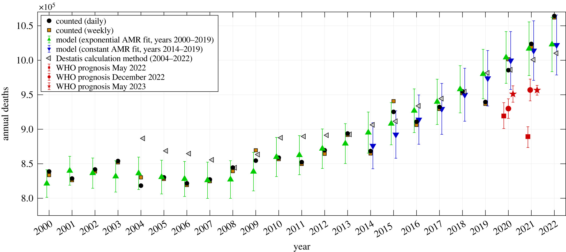

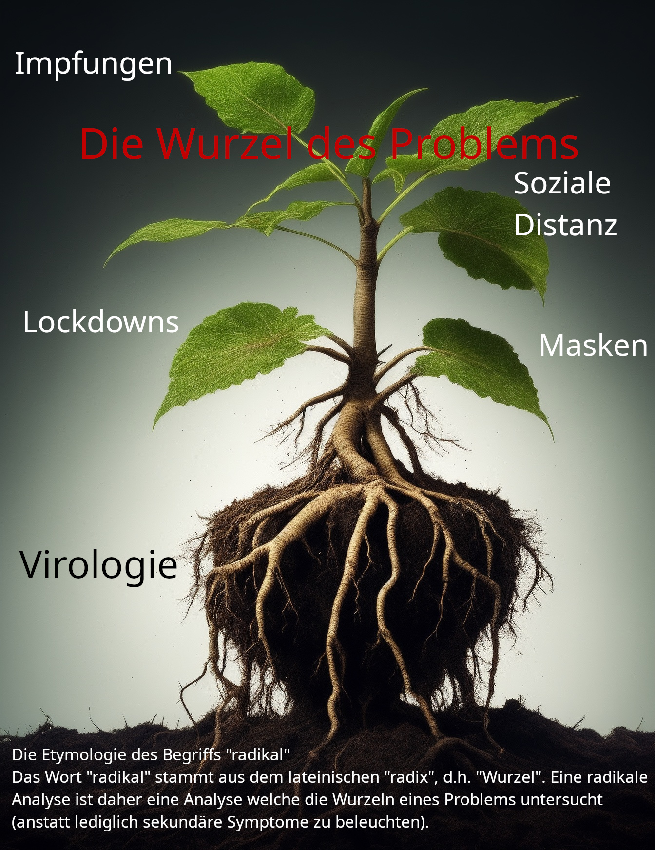
 Thomas Feeley, Ashley E. Fico, Allison Zorzie Shaw, Seyoung Lee, Darrin J. Griffin. (2017) Is the Door-in-the-Face a Concession?. Communication Quarterly 65:1, pages 97-123.

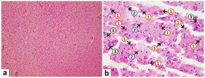

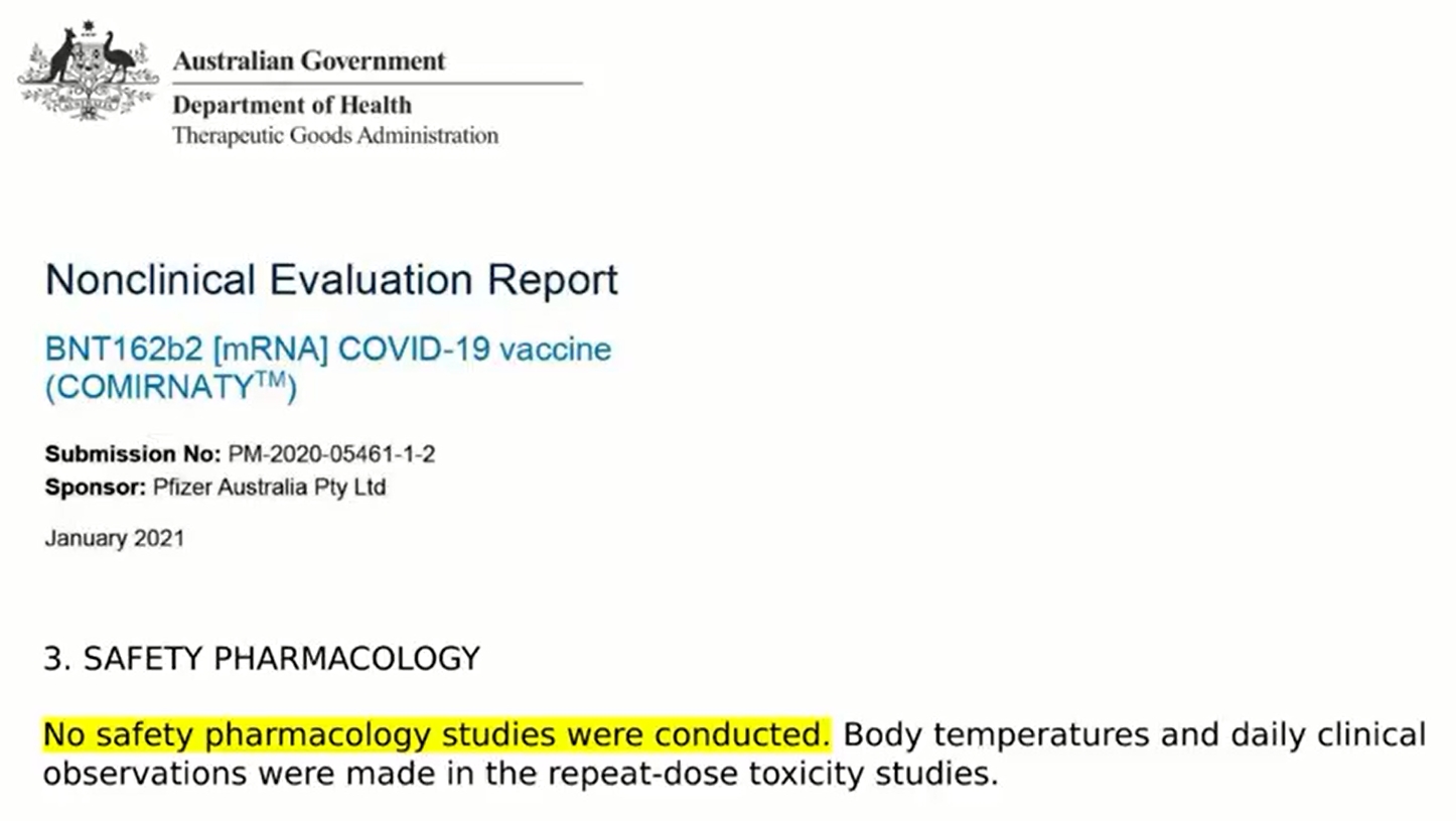
 Wenn man eine Bevölkerung über einen längeren Zeitraum kontrollieren will, muss man ein gewisses Maß an Zustimmung haben. Es ist äußerst schwierig zu erkennen, wie reiner Terrorismus unbegrenzt funktionieren kann. Er kann für eine ziemlich lange Zeit funktionieren, aber ich denke, früher oder später muss man ein Element der Überzeugung einbringen, ein Element, das die Menschen dazu bringt, dem zuzustimmen, was mit ihnen geschieht.
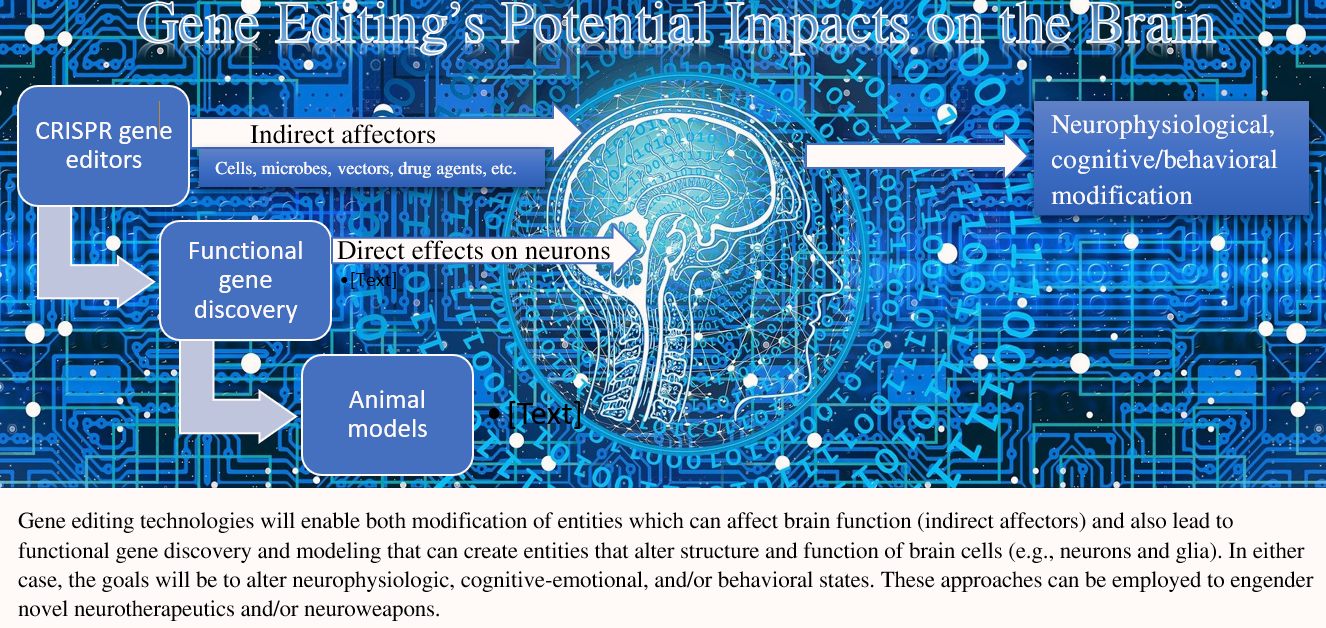


 Die Etymologie der Kybernetik


 Mooresches Gesetz (https://de.wikipedia.org/wiki/Mooresches_Gesetz)